ScholarWorks @ Georgia State University
ScholarWorks @ Georgia State University
Biology Dissertations Department of Biology
4-16-2010
Mechanism (S) of Metal-Induced Apoptosis in Saccharomyces
Mechanism (S) of Metal-Induced Apoptosis in Saccharomyces
Cerevisiae
Cerevisiae
Amrita Mohan Nargund
Georgia State University
Follow this and additional works at: https://scholarworks.gsu.edu/biology_diss
Part of the Biology Commons
Recommended Citation Recommended Citation
Nargund, Amrita Mohan, "Mechanism (S) of Metal-Induced Apoptosis in Saccharomyces Cerevisiae." Dissertation, Georgia State University, 2010.
https://scholarworks.gsu.edu/biology_diss/80
MECHANISM (S) OF METAL-INDUCED APOPTOSIS IN SACCHAROMYCES CEREVISIAE.
by
AMRITA M. NARGUND
Under the direction of Dr. John E. Houghton
ABSTRACT
Heavy metals, such as copper and cadmium have been linked to a number of cellular
dysfunctions in single and multicellular organisms that are associated with apoptosis.
The yeast, Saccharomyces cerevisiae, provides a valuable model for elucidating
apoptosis mechanisms, and this study extends that capability to Cu and Cd-induced
apoptosis. We demonstrate that S. cerevisiae undergoes a glucose-dependent,
programmed cell death in response to low cadmium concentrations, which is initiated
within the first hour of Cd exposure. The response was associated with induction of the
yeast caspase, Yca1p, and was abolished in YCA1∆ mutant. Other apoptotic markers, including sub-G1 DNA fragmentation and hyper-polarization of mitochondrial
membranes, were also evident among Cd-exposed cells. We also show that low levels
exposure. Such cellular responses were verified by analyzing mitochondrial
perturbation, generation of superoxide ions, activation of the yeast caspase1, and the
eventual fragmentation of nuclear DNA (through TUNEL). In analyzing the response of
yeast to the different metals, we also demonstrated that the metal-induced PCD is
instigated through the sequential activity of at least two caspase-like proteins (i.e., Yca1
and Atg4), both of which appear to be in involved in the process of inducing
mitochondrial stress. The additional caspase-like activity is shown to be derived from an
enzyme involved in the latter stages of autophagy (Atg4), and provides an intriguing
association of apoptosis with autophagy. Here we also demonstrate that metals such as
copper and cadmium causes oxidative damage to mitochondrial proteins. Such
oxidative attack is targeted and we show that oxidation of certain crucial proteins is
required for apoptosis upon metal exposure. By showing that such targeted protein
oxidation is dependent on YCA1 and ATG, we also confirm the finding that in yeast that
have been exposed to a heavy metal, YCA1 and ATG are essential for damaging
mitochondria and to initiate apoptosis. These novel findings highlight several new
perspectives about the mechanism of metal-dependent apoptosis, while opening up
future analyses to the power of the yeast model system.
MECHANISM (S) OF METAL-INDUCED APOPTOSIS IN SACCHAROMYCES CEREVISIAE.
by
AMRITA M. NARGUND
A Dissertation Submitted in Partial Fulfillment of the Requirements for the
Degree of
Doctor of Philosophy
in the College of Arts and Sciences
Georgia State University
Copyright by
Amrita M. Nargund
MECHANISM (S) OF METAL-INDUCED APOPTOSIS IN SACCHAROMYCES CEREVISIAE.
by
AMRITA M. NARGUND
Committee Chair: Dr. John E. Houghton
Committee: Dr. Julia K. Hilliard
Dr. Zehava E. Eichenbaum
Electronic Version Approved:
Office of Graduate Studies
College of Arts and Sciences
Georgia State University
DEDICATION
This is to my husband Mandar Mangalvedhekar and to my daughter Gandhali for their
unconditional love and support, which made this accomplishment possible.
To my parents, brothers, sister-in-laws and nieces for their continued support and
encouragement.
ACKNOWLEDGEMENTS
I thank my advisor, Dr. John E. Houghton for his advice and support during my Ph.D.
I also thank my committee member – Dr. Julia Hilliard and Dr. Zehava E. Eichenbaum
for their guidance and support.
Thank you to my colleagues –Pei Ju Chin, Anupama Shanmuganathan, Jiang, Divya
Rao, Prajakta Pradhan, Han, Wen, Ling Wei, Debby Walthall and Sonja Young for their
friendship and technical help.
TABLE OF CONTENTS
ACKNOWLEDGEMENTS ... v
TABLE OF CONTENTS ... vi
LIST OF TABLES ... ix
LIST OF FIGURES ... x
GENERAL INTRODUCTION ... 1
Experimental Question ... 16
References ... 20
CADMIUM INDUCES A HETEROGENEOUS AND CASPASE-DEPENDENT APOPTOTIC RESPONSE IN SACCHAROMYCES CEREVISIAE ... 25
Introduction ... 26
Materials and methods ... 28
Results and discussion ... 32
Conclusions ... 48
Acknowledgements ... 49
References ... 50
METAL TOXICITY RESULTS IN CHANGE IN GENE EXPRESSION PATTERN ... 58
Introduction ... 58
Material and methods ... 61
Discussion ... 82
References ... 90
HEAVY METAL –INDUCED APOPTOSIS IN SACCHAROMYCES CEREVISIAE REQUIRES THE INTERCESSION OF AN AUTOPHAGIC PROTEASE, ATG4 ... 92
Introduction ... 93
Materials and methods ... 96
Results and Discussion ... 99
Acknowledgements ... 113
References ... 114
YEAST CASPASE-1 MEDIATED OXIDATIVE DAMAGE OF MITOCHONDRIAL PROTEINS IN YEAST APOPTOSIS ... 118
Introduction ... 120
Materials and methods ... 124
Results ... 127
Discussion ... 138
Acknowledgements ... 143
References ... 144
GENERAL DISCUSSION ... 150
References ... 161
Appendix A ... 164
Appendix B ... 165
Appendix C ... 166
Appendix D ... 167
Appendix E ... 168
Appendix F ... 169
LIST OF TABLES
TABLE 1: PROTEINS THAT ARE DIFFERENTIALLY EXPRESSED WITHIN 60
MINUTES OF COPPER AND CADMIUM EXPOSURE IN YCA1∆ ... 167
TABLE 2: PROTEINS THAT ARE DIFFERENTIALLY EXPRESSED WITHIN 60
LIST OF FIGURES
FIGURE 1: METAL GENERATE ROS AND CAUSE OXIDATIVE STRESS. ... 3
FIGURE 2: APOPTOSIS PATHWAYS IN YEAST. ... 5
FIGURE 3: INTRINSIC AND EXTRINSIC PATHWAY OF APOPTOSIS. ... 8
FIGURE 4: APOPTOTIC CELL DEATH. ... 9
FIGURE 5: MITOCHONDRIA IN YEAST APOPTOSIS. ... 11
FIGURE 6: MITOCHONDRIA PERMEABILITY TRANSITION DURING APOPTOSIS. 13 FIGURE 7: VIABILITY OF YEAST UPON COPPER AND CADMIUM EXPOSURE. ... 15
FIGURE 8: CADMIUM INDUCES THE YEAST CASPASE, AND A GLUCOSE-DEPENDENT APOPTOSIS ... 33
FIGURE 9: CADMIUM INDUCES MITOCHONDRIAL MEMBRANE HYPERPOLARIZATION AND ENHANCED ROS LEVELS IN A CELL SUBPOPULATION ... 36
FIGURE 10: HYDROGEN PEROXIDE EXPOSURE CAUSES A HOMOGENEOUS INCREASE IN ROS LEVELS IN THE ABSENCE OF APOPTOSIS ... 40
FIGURE 11: CADMIUM-INDUCED APOPTOSIS IN YEAST IS CHARACTERIZED BY PLASMA MEMBRANE DAMAGE ... 42
FIGURE 12: CADMIUM-INDUCED APOPTOSIS IS CASPASE- AND GLUTATHIONE-DEPENDENT ... 45
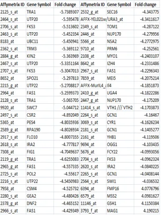
FIGURE 13: HIGHLY DIFFERENTIAL EXPRESSION OF GENES, UPON 1HOUR EXPOSURE OF WILD TYPE YEAST, TO COPPER. ... 64
FIGURE 15: FOLD CHANGE IN EXPRESSION OF APOPTOTIC GENES, UPON
EXPOSURE OF WILD TYPE YEAST TO COPPER. ... 67
FIGURE 16: FOLD CHANGE IN EXPRESSION OF APOPTOTIC GENES, UPON
EXPOSURE OF WILD TYPE YEAST TO CADMIUM. ... 68
FIGURE 17: FOLD CHANGE IN EXPRESSION OF CELL DEATH GENES, UPON
EXPOSURE TO COPPER. ... 69
FIGURE 18: FOLD CHANGE IN EXPRESSION OF CELL DEATH GENES, UPON
EXPOSURE TO CD. ... 70
FIGURE 19: FOLD CHANGE IN EXPRESSION OF AUTOPHAGY GENES, UPON
EXPOSURE TO CU ... 71
FIGURE 20: FOLD CHANGE IN EXPRESSION OF AUTOPHAGY GENES, UPON
EXPOSURE TO CD ... 72
FIGURE 21: FOLD CHANGE IN EXPRESSION OF GLYCOLYTIC GENES, UPON
EXPOSURE TO COPPER. ... 74
FIGURE 22: FOLD CHANGE IN EXPRESSION OF GLYCOLYTIC GENES, UPON
EXPOSURE TO CADMIUM. ... 75
FIGURE 23: FOLD CHANGE IN EXPRESSION OF PENTOSE PHOSPHATE
PATHWAY GENES, UPON EXPOSURE TO COPPER. ... 76
FIGURE 24: FOLD CHANGE IN EXPRESSION OF PENTOSE PHOSPHATE
PATHWAY GENES UPON CADMIUM EXPOSURE. ... 77
FIGURE 25: FOLD CHANGE IN EXPRESSION OF TCA PATHWAY GENES, UPON
FIGURE 26: FOLD CHANGE IN EXPRESSION OF TCA PATHWAY GENES, UPON
CADMIUM EXPOSURE. ... 79
FIGURE 27: FOLD CHANGE IN EXPRESSION OF HISTONE MODIFIERS, UPON CU
EXPOSURE. ... 83
FIGURE 28: FOLD CHANGE IN EXPRESSION OF HISTONE MODIFIERS, UPON CD
EXPOSURE. ... 84
FIGURE 29: FOLD CHANGE IN EXPRESSION OF GLYCOLYTIC GENES, UPON
METAL EXPOSURE AS MEASURED BY REAL TIME RT-PCR. ... 85
FIGURE 30: FOLD CHANGE IN EXPRESSION OF PENTOSE PHOSPHATE
PATHWAY GENES, UPON METAL EXPOSURE AS MEASURED BY REAL TIME
RT-PCR. ... 86
FIGURE 31: FOLD CHANGE IN EXPRESSION OF TRANSLATION ELONGATION
FACTOR, UPON METAL EXPOSURE AS MEASURED BY REAL TIME RT-PCR.
... 87
FIGURE 32: COPPER INDUCES CASPASE-DEPENDENT APOPTOSIS IN
SACCHAROMYCES CEREVISIAE ... 100
FIGURE 33: YCA1 IS NOT THE ONLY ROUTE FOR COPPER MEDIATED
MITOCHONDRIAL MEMBRANE HYPERPOLARIZATION ... 103
FIGURE 34: CASPASE LIKE PROTEIN MAY BE RESPONSIBLE FOR CU INDUCED
APOPTOSIS ... 107
FIGURE 35: ATG4 IS INVOLVED IN PROCESSING OF YCA1P ... 109
FIGURE 37: WILD TYPE MITOCHONDRIAL PROTEINS ARE OXIDIZED ON METAL
TREATMENT. ... 130
FIGURE 38: OXIDATION LEVELS OF INDIVIDUAL PROTEINS THAT ARE
AFFECTED DURING THE APOPTOSIS UPON METAL EXPOSURE. ... 131
FIGURE 39: YCA1 IS REQUIRED FOR MITOCHONDRIAL PROTEIN OXIDATION
DURING APOPTOSIS UPON EXPOSURE TO CD. ... 133
FIGURE 40: ATG4 IS REQUIRED FOR MITOCHONDRIAL PROTEIN OXIDATION
DURING APOPTOSIS UPON EXPOSURE TO CD. ... 135
FIGURE 41: UNLIKE CD, CU CAN OXIDIZE SOME MITOCHONDRIAL PROTEINS
EVEN IN THE ABSENCE OF YCA1 AND ATG4. ... 136
FIGURE 42: VIABILITY CURVE FOR WILD TYPE AND YCA1∆ YEAST CELLS
EXPOSED TO DIFFERENT CONCENTRATIONS OF COPPER ... 152
FIGURE 43: VIABILITY CURVE FOR YEAST CELLS EXPOSED TO DIFFERENT
CONCENTRATIONS OF COPPER AND CADMIUM ... 153
FIGURE 44: AIF1 IS INVOLVED IN METAL INDUCED APOPTOSIS ... 157
FIGURE 45: RELATIVE OXIDATION OF SSC1 ... 164
FIGURE 46: AIF1∆ IS NON APOPTOTIC UPON COPPER AND CADMIUM
EXPOSURE ... 165
FIGURE 47: MITOCHONDRIAL MEMBRANE DEPOLARIZATION STUDY UPON
CADMIUM EXPOSURE. ... 166
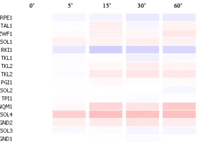
FIGURE 48: GENE EXPRESSION AT VARIOUS TIME POINTS UPON COPPER
FIGURE 49: GENE EXPRESSION AT VARIOUS TIME POINTS UPON CADMIUM
GENERAL INTRODUCTION
Why study metal induced oxidative stress?
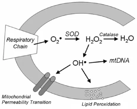
Metals generate reactive oxygen species (ROS) and hence cause oxidative stress in
the cell (Fig 1) 1,51. Metal induced oxidative stress has been implicated in the
pathogenesis of Alzheimer’s disease, Parkinson’s disease, spongiform encephalopathy,
and familial amyotrophic lateral sclerosis 2-3. The imbalance in copper homeostasis is
observed in hereditary neurodegenerative disorders (ND) such as Menke’s and Wilson’s
disease. The impairment of copper transporting gene leads to fatal neurodegeneration
in Menke’s syndrome 4. Moreover, the excessive accumulation of copper in liver, kidney
and brain leads to hepatic, renal and neurological abnormalities in case of Wilson’s
disease 5. Cadmium (Cd) is known to be carcinogenic and known to be associated with
renal damage.
In the above mentioned disorders, metal alters mitochondria and decreases
cytochrome c oxidase activity giving rise to burst of ROS. These ROS that can cause
oxidative stress are also neurotoxic, because they trigger neuronal death due to
apoptosis (programmed cell death) 3,6,7. Loss of neurons because of apoptosis in ND is
Why study metal toxicity, apoptosis and mitochondria dysfunction in
Saccharomyces cerevisiae?
Saccharomyces cerevisiae is an excellent model system for the molecular study of
metal toxicity, because its genome and proteome are well characterized. Yeast is easier
to manipulate experimentally than higher eukaryotes. The effects of metal toxicity and
the response to oxidative stress, which can cause apoptosis, are similar to those seen
in higher eukaryotes. Cellular mechanisms are conserved between higher eukaryotes
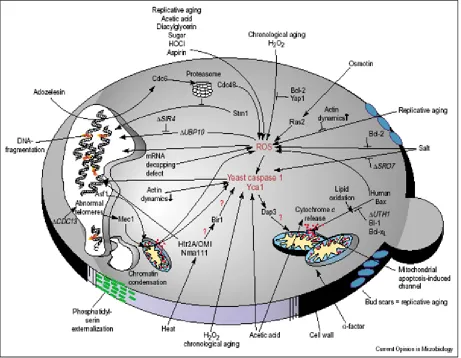
and yeast (Fig 2)32.
Recently, unicellular yeast have been seen to undergo apoptosis under different
stress conditions. As seen in case of higher eukaryotes, ROS are found to be the major
cause of apoptosis in yeast 8. Orthologs of genes such as caspase, AIF1 and BI-1 that
are important for apoptosis, have been characterized in yeast 8-9,10. Yeast has been
found to be useful in elucidating some of the problems related to aging and
neurodegeneration. A mutation in CDC48, a homologue of VCP in mammalian cells,
was first shown to result in apoptosis in yeast and then later was found to be involved in
the formation of inclusion bodies in Paget’s disease and in neurodegeneration caused
by polyglutamine 11-14. Likewise yeast has been proven to be a useful system in the
studies related to Alzheimer’s, Parkinson’s and Huntington’s disease 14-15. Hence, yeast
is a good tool for studying apoptosis related to ageing and neurodegeneration.
In the past, yeast have been studied to elucidate mitochondrial function in cells.
Figure 1: Metal generate ROS and cause oxidative stress.
pathway, as yeast have been shown to undergo mitochondrial-mediated apoptosis
under various stress conditions 16. Although mitochondrial dysfunction has also been
associated with oxidative stress and apoptosis in cases of neurodegenerative diseases
(ND), it is difficult to elucidate even this form of cell death in higher eukaryotes. In yeast,
it is simple to perform mitochondrial manipulations because it is easier to create specific
mutations in mitochondrial DNA. In addition, cells can be grown on a variety of different
carbon sources (fermentative and non-fermentative) to control the mode of respiration,
i.e., aerobic and anaerobic, and thus the different involvement of mitochondria in cellular
activities. As a result, yeast is an excellent model organism to elucidate the events of
mitochondrial-mediated cell death. Even so, the exact path of mitochondria-mediated
apoptosis in yeast is still unclear. The greater the understanding of yeast mitochondria
mediated apoptosis, the easier it will be to solve some important issues regarding these
above mentioned ND 14.
Metal generates Reactive oxygen species (ROS) and cause oxidative stress
In the cell, ROS originates in mitochondria. Mitochondria, a powerhouse of a cell,
can generate energy through the electron transport chain by producing ATP. In doing
so, mitochondria also generate ROS, such as peroxide and superoxide anions but to a
lesser extent. Mitochondria are equipped with antioxidant defenses such as superoxide
dismutase (SOD) and glutathione (GSH imported from cytosol); these convert
superoxide and peroxide to other harmless products before these species can become
a substrate for Haber-Weiss or Fenton's reaction, which generates more potent ROS
Figure 2: Apoptosis pathways in Yeast.
Copper is a transition metal, because it is able to cycle between Cu (II) and Cu (I) as
shown in the reaction below. In doing so, copper can generate superoxides, a reactive
oxygen species (ROS). Copper also qualifies as a redox active metal because by using
Fenton’s reaction it is able to generate more potent ROS such as hydroxyl radical (OH°)
from the less reactive species such as peroxide (H2O2) and superoxide (O2-) that are
generated due to cellular respiration in the mitochondria 3.
Copper is also able to catalyze Haber-Weiss reaction to give OH°18. Hence, in
the presence of excess of copper, cells will be overloaded with potent ROS. Such
humungous amounts of ROS are difficult to be disarmed by antioxidants. Excess ROS
damages all of the cellular macromolecules, such as proteins, lipids and DNA, and
hence a condition called oxidative stress develops inside the cell 19 (Fig 2) 32.
Transition metal Cu:
Cu (II) + e- Æ Cu (I)
Cu (I) + O2 Æ Cu (II) + O2-
Fenton’s reaction:
Cu (I) + H2O2Æ Cu (II) + OH- + OH°
Haber-Weiss cycle:
O2-+ H2O2 Æ O2+ OH- + OH°
2O2- + 2H+ Æ H2O2 + O2
Cadmium by nature is a redox-inactive metal. Cadmium hence cannot catalyze
anti-oxidant defenses or by displacing redox-active metals from proteins 20. Therefore,
cadmium is able to still cause oxidative stress in the cells.
Even though there is a clear difference in ROS species formed by copper and
cadmium, there is no overt difference in cellular oxidation levels between both metals 47
(Shanmuganathan, A., personal communication).
Apoptosis and the role of oxidative stress or ROS in the process
Apoptosis is a programmed cell death i.e. a cell commits suicide. Apoptosis is not a
random process; rather it happens in a very controlled and coordinated manner.
Apoptosis involves a series of events that ultimately lead to the removal of cells from a
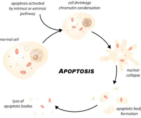
population with no harmful substances released in the environment. An apoptotic cell
shows a typical morphology (e.g., shrinkage of a cell, condensed chromatin, fragmented
DNA), and finally a cell is broken into numerous small apoptotic bodies that are
phagocytosed rapidly. Apoptosis involves a complex signaling network and is an active
process that requires energy. The two pathways by which apoptosis can occur are the
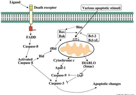
extrinsic and intrinsic pathways (Fig 3,4) 52,53. In the extrinsic pathway, ligands serve as
stimuli when they bind to the receptors on a target cell and trigger activation of
caspases. Caspases are able to fragment DNA and, hence, cause apoptosis. The other
apoptotic path is the intrinsic pathway or mitochondria mediated pathway, in which the
signals for the cell death are internal (e.g., DNA damage, oxidative stress, mutations in
Figure 3: Intrinsic and extrinsic pathway of apoptosis.
Figure 4: Apoptotic cell death.
cell death scenarios indicating that ROS is an important stimulus for the intrinsic
pathway of apoptosis. Numerous studies with antioxidants have shown that oxidative
stress when prevented by scavenging ROS, there is no apoptosis 21. This indicates that
ROS can trigger apoptosis. However, the steps involved in ROS triggered mitochondria
mediated apoptosis are unclear.
ROS is generated and released from mitochondria. Studies have also shown that
once released from mitochondria, ROS can oxidize mitochondrial pore proteins. Such
an oxidation can lead to mitochondrial fission or dysfunction. Mitochondria fission is
associated with apoptosis. Mitochondria are, therefore, not only a source but also the
target of ROS during apoptosis 22.
Mitochondria in apoptosis
In ND, mitochondria dysfunction and apoptosis are commonly observed toxic effects.
Mitochondria are key players in the intrinsic pathway of apoptosis (Fig 5) 14.
Mitochondria dysfunction is a cause of apoptosis in intrinsic pathway. In some cell
types, ROS released from mitochondria causes transient hyperpolarization of the
mitochondrial inner membrane leading to mitochondrial inner membrane
permeabilization (MMP). Because of MMP, solutes less than 1.5KDa can enter the
mitochondrial matrix along with an influx of water leading to mitochondrial swelling and
rupture of an outer mitochondrial membrane. This phenomenon causes a drop in the
Figure 5: Mitochondria in yeast apoptosis.
The pro-apoptotic factors(e.g., Apoptosis inducing factor(AIF) and cytochrome c) are
then released from the inner-mitochondrial space into the cytosol 23-25. Both AIF and
cytochrome c are able to trigger apoptosis. Not only AIF and cytochrome c, but several
other mitochondrial proteins can participate in apoptosis.
Mitochondrial proteins and apoptosis
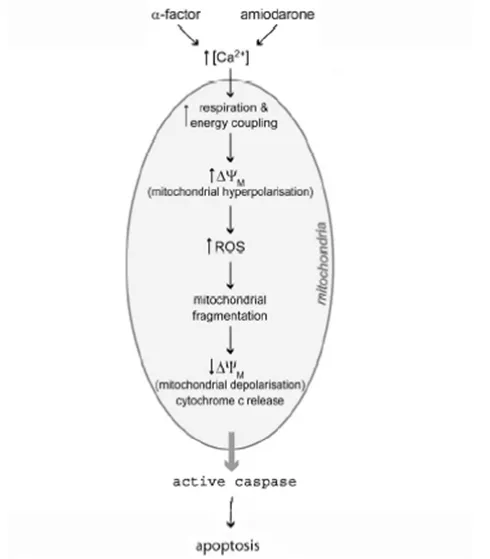
Mitochondria are not only the source but also the target of ROS (Fig 6) 54.
Mitochondrial proteins are oxidized upon exposure to certain oxidants 18. In some
mammalian cell types, oxidation of some of the mitochondrial proteins leads to
mitochondrial fission 29-31.
Isolated yeast mitochondria lacking Voltage dependent anionic channel (a
mitochondrial protein), are not sensitive to human Bax mediated apoptosis as
suggested in Fig 3. 32. Yeast strains lacking mitochondrial pore genes(e.g., POR1 and
ANT1) do not undergo HIV-1 viral protein R (VPR) induced apoptosis 33. On exposure to
oxidizing agent such as paraquat, mitochondrial proteins are carbonylated (oxidized) in
yeast 18. Both copper and cadmium cause oxidative stress by oxidizing the cellular
proteins in yeast (Shanmuganathan A, personal communication) 47. High concentration
of cadmium causes mitochondria mediated pathway in higher eukaryotes 27. To
understand the toxicity associated with relatively low concentrations of copper and
Figure 6: Mitochondria permeability transition during apoptosis.
(e.g., POR1 and ANT1) and mitochondrial collapse in apoptosis.
Mitochondrial flavoprotein, AIF, is present in the inter-mitochondrial space and
has oxidoreductase activity 34. It is an important regulator of cell death in mammals,
because it is involved in mitochondria mediated apoptosis 34. In higher eukaryotes, on
induction of apoptosis, AIF translocates from mitochondria to the nucleus 10,35. In the
nucleus, it fragments DNA itself or with the help of endonuclease G 26,36. AIF has a DNA
binding domain, which is essential for its apoptogenic activity 37. AIF can also contribute
to the release cytochrome c from inter-mitochondrial space with the mechanism still
unclear. Cytochrome c can lead to activation of caspases. Hence, AIF and caspase can
cooperate and cause apoptosis 26,36. The structure and function of AIF are conserved in
Saccharomyces cerevisiae 10. In yeast, AIF essentially performs the same role as in
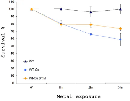
Figure 7: Viability of yeast upon copper and cadmium exposure.
S. cerevisiae YCA1∆ was incubated in the presence of 8mM Cu(NO3)2 and
30µMCd(NO3)2 for up to 3 h. Viability was determined at intervals after the addition of
metals by spreading aliquots from dilutions (OD600 = 1.0) cultures onto YEPD agar and
Experimental Question
Copper (Cu) and cadmium (Cd) are implicated in many neurodegenerative disorders.
5,40. Cadmium is a redox-inactive metal, whereas copper is a redox-active metal and
therefore is able to generate potent reactive oxygen species (ROS) directly as opposed
to cadmium, which is able to do so indirectly. Both the metals cause oxidative stress in
the cell 41. Metal induced oxidative stress is associated with mitochondria dysfunction
and apoptosis (programmed cell death) in cases of neuronal death 39,42-44. In
neurodegenerative disorders, neuronal death due to apoptosis is the major concern 1.
To prevent apoptotic cell death, it is crucial to know the cascade of events leading to
apoptosis and involvement of mitochondria in the process.
In Saccharomyces cerevisiae (yeast), metals such as copper and cadmium
generate ROS and cause cell death with the induction of cell death genes. Yeast, being
a simple unicellular eukaryote, serves as an excellent model to elucidate the
mechanism of metal toxicity. The hypothesis of this dissertation study is that heavy
metals induce apoptosis in yeast in a mitochondria-dependent manner that
SA1: In yeast, what is the cellular response on exposure to copper and cadmium?
Copper has been associated with apoptosis in human hepatoma cells and breast
cancer cell line40-41. Cadmium also has been associated with apoptosis in human
hepatoma cells and cultured renal tissues 3,26. However, cadmium has not been shown
to cause apoptosis in yeast. In addition, low doses of copper have not yet been
associated with apoptosis in yeast. When treated with very low doses of metals, such as
copper and cadmium, yeast cells do show, however, a decrease in viability (Fig 7).
Moreover, upon exposure to either copper or cadmium, proteins are damaged through
metal-induced oxidative stress (Shanmuganathan, personal communication) 47, and
oxidative stress (when induced by hydrogen peroxide) is a known trigger for apoptosis
in yeast (32, 33). To examine the relationship between cadmium or copper and reduced
yeast viability, the following questions were asked:
Specific Aim 1.1: Does exposure to very low doses of cadmium (~30µM) cause
apoptosis (programmed cell death)?
SA2: What is the role of mitochondria in copper and cadmium induced apoptosis,
and how is Yeast caspase-1 involved in the process?
Copper and cadmium toxicity at low concentration has not been studied
extensively in yeast or higher eukaryotes. In higher eukaryotes, Cd-induced apoptosis
appears to involve mitochondria although not much is known about the precise
mechanism. In mitochondria-mediated pathways, mitochondrial proteins such as VDAC
and ANT1 are oxidized in higher eukaryotes 28. Thereafter, this oxidation of
mitochondrial proteins is thought to lead to mitochondrial dysfunction and collapse,
which is believed to trigger apoptosis by releasing pro-apoptotic factors such as AIF and
cytochrome c 32,18,29-30. In yeast, some of the events associated with mitochondrial
perturbation have been observed in case of pheromone induced apoptosis 14. However,
the cascade of such mitochondrial events related to oxidative stress is poorly
understood 14. Exposure of yeast to copper and cadmium does generate oxidative
stress (Shanmuganathan A, personal communication) 47. Therefore, it is important to
study mitochondrial events, such as mitochondrial protein oxidation and involvement of
AIF, in response to the presence of cadmium.
Importantly, cadmium induced apoptosis in higher eukaryotes is thought to be
independent of caspase 49-50. However, preliminary data in our laboratory does suggest
that in yeast, cadmium-induced apoptosis involves both mitochondria and Yeast
caspase-1. Intriguingly, YCA1∆ mutant do not show any mitochondrial perturbation in
response to cadmium suggesting that Yca1 plays a significant role in mitochondrial
perturbations. It is considered important, therefore, in any determination of metal
of the mitochondrial events and/or subsequent stages of the apoptotic process. We
have designed experiments to address the following questions:
Specific Aim 2.1: Does metal-induced oxidative stress include increase oxidation of
mitochondrial proteins? Are proteins such as VDAC and ANT1 targeted?
Specific Aim 2.2: Are proteins such as AIF (Apoptosis Inducing Factor) involved in
apoptosis? If so, do they translocate to the nucleus.
References
1. http://www.iupac.org/goldbook/HT06787.pdf.
2. Avery, S. V. 2001. Metal toxicity in yeasts and the role of oxidative stress. Adv Appl Microbiol 49:111-42.
3. Aydin, H. H., H. A. Celik, R. Deveci, E. Terzioglu, S. Karacali, N. Mete, U. Akarca, and Y. Batur. 2003. Characterization of the cellular response during apoptosis induction in cadmium-treated Hep G2 human hepatoma cells. Biol Trace Elem Res 95:139-53.
4. Bauer, M. K., A. Schubert, O. Rocks, and S. Grimm. 1999. Adenine nucleotide translocase-1, a component of the permeability transition pore, can dominantly induce apoptosis. J Cell Biol 147:1493-502.
5. Bayer, T. A., S. Schafer, H. Breyhan, O. Wirths, C. Treiber, and G. Multhaup. 2006. A vicious circle: role of oxidative stress, intraneuronal Abeta and Cu in Alzheimer's disease. Clin Neuropathol 25:163-71.
6. Cahuana, G. M., J. R. Tejedo, J. Jimenez, R. Ramirez, F. Sobrino, and F. J. Bedoya. 2004. Nitric oxide-induced carbonylation of Bcl-2, GAPDH and ANT precedes apoptotic events in insulin-secreting RINm5F cells. Exp Cell Res 293:22-30.
7. Cande, C., F. Cecconi, P. Dessen, and G. Kroemer. 2002. Apoptosis-inducing factor (AIF): key to the conserved caspase-independent pathways of cell death? J Cell Sci 115:4727-34.
8. Chae, H. J., N. Ke, H. R. Kim, S. Chen, A. Godzik, M. Dickman, and J. C. Reed. 2003. Evolutionarily conserved cytoprotection provided by Bax Inhibitor-1 homologs from animals, plants, and yeast. Gene 323:101-13.
9. Chen, Q., Y. C. Chai, S. Mazumder, C. Jiang, R. M. Macklis, G. M. Chisolm, and A. Almasan. 2003. The late increase in intracellular free radical oxygen species during apoptosis is associated with cytochrome c release, caspase activation, and mitochondrial dysfunction. Cell Death Differ 10:323-34.
10. Costantini, P., A. S. Belzacq, H. L. Vieira, N. Larochette, M. A. de Pablo, N. Zamzami, S. A. Susin, C. Brenner, and G. Kroemer. 2000. Oxidation of a critical thiol residue of the adenine nucleotide translocator enforces Bcl-2-independent permeability transition pore opening and apoptosis. Oncogene 19:307-14.
12. Eisenberg, T., S. Buttner, G. Kroemer, and F. Madeo. 2007. The mitochondrial pathway in yeast apoptosis. Apoptosis 12:1011-23.
13. England, K., and T. G. Cotter. 2005. Direct oxidative modifications of signalling proteins in mammalian cells and their effects on apoptosis. Redox Rep 10:237-45.
14. Fernandez-Checa, J. C., C. Garcia-Ruiz, A. Colell, A. Morales, M. Mari, M. Miranda, and E. Ardite. 1998. Oxidative stress: role of mitochondria and protection by glutathione. Biofactors 8:7-11.
15. Grabarek, J., P. Amstad, and Z. Darzynkiewicz. 2002. Use of fluorescently labeled caspase inhibitors as affinity labels to detect activated caspases. Hum Cell 15:1-12.
16. Gulbins, E., S. Dreschers, and J. Bock. 2003. Role of mitochondria in apoptosis. Exp Physiol 88:85-90.
17. Hayashi, M., S. Fuse, D. Endoh, N. Horiguchi, K. Nakayama, Y. Kon, and T. Okui. 2006. Accumulation of Copper Induces DNA Strand Breaks in Brain Cells of Long-Evans Cinnamon (LEC) Rats, An Animal Model for Human Wilson Disease. Exp Anim 55:419-26.
18. Haynes, C. M., E. A. Titus, and A. A. Cooper. 2004. Degradation of misfolded proteins prevents ER-derived oxidative stress and cell death. Mol Cell 15:767-76.
19. Hirabayashi, M., K. Inoue, K. Tanaka, K. Nakadate, Y. Ohsawa, Y. Kamei, A. H. Popiel, A. Sinohara, A. Iwamatsu, Y. Kimura, Y. Uchiyama, S. Hori, and A. Kakizuka. 2001. VCP/p97 in abnormal protein aggregates, cytoplasmic vacuoles, and cell death, phenotypes relevant to neurodegeneration. Cell Death Differ 8:977-84.
20. Jacotot, E., L. Ravagnan, M. Loeffler, K. F. Ferri, H. L. Vieira, N. Zamzami, P. Costantini, S. Druillennec, J. Hoebeke, J. P. Briand, T. Irinopoulou, E. Daugas, S. A. Susin, D. Cointe, Z. H. Xie, J. C. Reed, B. P. Roques, and G. Kroemer. 2000. The HIV-1 viral protein R induces apoptosis via a direct effect on the mitochondrial permeability transition pore. J Exp Med 191:33-46.
21. Jamers, A., K. Van der Ven, L. Moens, J. Robbens, G. Potters, Y. Guisez, R. Blust, and W. De Coen. 2006. Effect of copper exposure on gene expression profiles in Chlamydomonas reinhardtii based on microarray analysis. Aquat Toxicol 80:249-60.
versatile toolbox for PCR-based tagging of yeast genes: new fluorescent proteins, more markers and promoter substitution cassettes. Yeast 21:947-62.
23. Kehrer, J. P. 2000. Cause-effect of oxidative stress and apoptosis. Teratology 62:235-6.
24. Khan, M. A., P. B. Chock, and E. R. Stadtman. 2005. Knockout of caspase-like gene, YCA1, abrogates apoptosis and elevates oxidized proteins in Saccharomyces cerevisiae. Proc Natl Acad Sci U S A 102:17326-31.
25. Krumschnabel, G., C. Manzl, C. Berger, and B. Hofer. 2005. Oxidative stress, mitochondrial permeability transition, and cell death in Cu-exposed trout hepatocytes. Toxicol Appl Pharmacol 209:62-73.
26. Lee, W. K., M. Abouhamed, and F. Thevenod. 2006. Caspasedependent and -independent pathways for cadmium-induced apoptosis in cultured kidney proximal tubule cells. Am J Physiol Renal Physiol 291:F823-32.
27. Lemarie, A., D. Lagadic-Gossmann, C. Morzadec, N. Allain, O. Fardel, and L. Vernhet. 2004. Cadmium induces caspase-independent apoptosis in liver Hep3B cells: role for calcium in signaling oxidative stress-related impairment of mitochondria and relocation of endonuclease G and apoptosis-inducing factor. Free Radic Biol Med 36:1517-31.
28. Lu, C. X., T. J. Fan, G. B. Hu, and R. S. Cong. 2003. Apoptosis-inducing factor and apoptosis. Sheng Wu Hua Xue Yu Sheng Wu Wu Li Xue Bao (Shanghai) 35:881-5.
29. Lu, J., Y. L. Zheng, D. M. Wu, D. X. Sun, Q. Shan, and S. H. Fan. 2006. Trace amounts of copper induce neurotoxicity in the cholesterol-fed mice through apoptosis. FEBS Lett 580:6730-40.
30. Ludovico, P., F. Rodrigues, A. Almeida, M. T. Silva, A. Barrientos, and M. Corte-Real. 2002. Cytochrome c release and mitochondria involvement in programmed cell death induced by acetic acid in Saccharomyces cerevisiae. Mol Biol Cell 13:2598-606.
31. Lutsenko, S., R. Tsivkovskii, and J. M. Walker. 2003. Functional properties of the human copper-transporting ATPase ATP7B (the Wilson's disease protein) and regulation by metallochaperone Atox1. Ann N Y Acad Sci 986:204-11.
33. Madeo, F., E. Frohlich, M. Ligr, M. Grey, S. J. Sigrist, D. H. Wolf, and K. U. Frohlich. 1999. Oxygen stress: a regulator of apoptosis in yeast. J Cell Biol 145:757-67.
34. Madeo, F., E. Herker, C. Maldener, S. Wissing, S. Lachelt, M. Herlan, M. Fehr, K. Lauber, S. J. Sigrist, S. Wesselborg, and K. U. Frohlich. 2002. A caspase-related protease regulates apoptosis in yeast. Mol Cell 9:911-7.
35. Maestre, I., J. Jordan, S. Calvo, J. A. Reig, V. Cena, B. Soria, M. Prentki, and E. Roche. 2003. Mitochondrial dysfunction is involved in apoptosis induced by serum withdrawal and fatty acids in the beta-cell line INS-1. Endocrinology 144:335-45.
36. Marchetti, P., M. Castedo, S. A. Susin, N. Zamzami, T. Hirsch, A. Macho, A. Haeffner, F. Hirsch, M. Geuskens, and G. Kroemer. 1996. Mitochondrial permeability transition is a central coordinating event of apoptosis. J Exp Med 184:1155-60.
37. Martinou, I., S. Desagher, R. Eskes, B. Antonsson, E. Andre, S. Fakan, and J. C. Martinou. 1999. The release of cytochrome c from mitochondria during apoptosis of NGF-deprived sympathetic neurons is a reversible event. J Cell Biol 144:883-9.
38. O'Brien, K. M., R. Dirmeier, M. Engle, and R. O. Poyton. 2004. Mitochondrial protein oxidation in yeast mutants lacking manganese-(MnSOD) or copper- and zinc-containing superoxide dismutase (CuZnSOD): evidence that MnSOD and CuZnSOD have both unique and overlapping functions in protecting mitochondrial proteins from oxidative damage. J Biol Chem 279:51817-27.
39. Oh, Y. K., K. S. Shin, and S. J. Kang. 2006. AIF translocates to the nucleus in the spinal motor neurons in a mouse model of ALS. Neurosci Lett 406:205-10.
40. Ostrakhovitch, E. A., and M. G. Cherian. 2005. Inhibition of extracellular signal regulated kinase (ERK) leads to apoptosis inducing factor (AIF) mediated apoptosis in epithelial breast cancer cells: the lack of effect of ERK in p53 mediated copper induced apoptosis. J Cell Biochem 95:1120-34.
41. Piret, J. P., T. Arnould, B. Fuks, P. Chatelain, J. Remacle, and C. Michiels. 2004. Mitochondria permeability transition-dependent tert-butyl hydroperoxide-induced apoptosis in hepatoma HepG2 cells. Biochem Pharmacol 67:611-20.
42. protocol, H. L.
43. Rego, A. C., and C. R. Oliveira. 2003. Mitochondrial dysfunction and reactive oxygen species in excitotoxicity and apoptosis: implications for the pathogenesis of neurodegenerative diseases. Neurochem Res 28:1563-74.
44. Rossi, L., M. F. Lombardo, M. R. Ciriolo, and G. Rotilio. 2004. Mitochondrial dysfunction in neurodegenerative diseases associated with copper imbalance. Neurochem Res 29:493-504.
45. Schapira, A. H. 1998. Mitochondrial dysfunction in neurodegenerative disorders. Biochim Biophys Acta 1366:225-33.
46. Schroder, R., G. D. Watts, S. G. Mehta, B. O. Evert, P. Broich, K. Fliessbach, K. Pauls, V. H. Hans, V. Kimonis, and D. R. Thal. 2005. Mutant valosin-containing protein causes a novel type of frontotemporal dementia. Ann Neurol 57:457-61.
47. Shanmuganathan, A., S. V. Avery, S. A. Willetts, and J. E. Houghton. 2004. Copper-induced oxidative stress in Saccharomyces cerevisiae targets enzymes of the glycolytic pathway. FEBS Lett 556:253-9.
48. Shih, C. M., J. S. Wu, W. C. Ko, L. F. Wang, Y. H. Wei, H. F. Liang, Y. C. Chen, and C. T. Chen. 2003. Mitochondria-mediated caspase-independent apoptosis induced by cadmium in normal human lung cells. J Cell Biochem 89:335-47.
49. Shimizu, S., Y. Shinohara, and Y. Tsujimoto. 2000. Bax and Bcl-xL independently regulate apoptotic changes of yeast mitochondria that require VDAC but not adenine nucleotide translocator. Oncogene 19:4309-18.
50. Simon, H. U., A. Haj-Yehia, and F. Levi-Schaffer. 2000. Role of reactive oxygen species (ROS) in apoptosis induction. Apoptosis 5:415-8.
51. Figure made by Dr. houghton
52. http://www.imgenex.com/emarketing/081606_LivinorSurvivin/apoptosis-pathway6.jpg
53. http://www.scq.ubc.ca/wp-content/uploads/2006/07/Apoptosis.gif
CHAPTER 1
CADMIUM INDUCES A HETEROGENEOUS AND CASPASE-DEPENDENT APOPTOTIC RESPONSE IN SACCHAROMYCES CEREVISIAE
Amrita Nargund, Simon V. Avery1 and John E. Houghton*
The toxic metal cadmium is linked to a series of degenerative disorders in humans, in
which Cd-induced programmed cell death (apoptosis) may play a role. The yeast,
Saccharomyces cerevisiae, provides a valuable model for elucidating apoptosis
mechanisms, and this study extends that capability to Cd-induced apoptosis. We
demonstrate that S. cerevisiae undergoes a glucose-dependent, programmed cell death
in response to low cadmium concentrations, which is initiated within the first hour of Cd
exposure. The response was associated with induction of the yeast caspase, Yca1p,
and was abolished in YCA1∆ mutant. Cadmium-dependent apoptosis was also suppressed in a GSH1∆ mutant, indicating a requirement for glutathione. Other apoptotic markers, including sub-G1 DNA fragmentation and hyper-polarization of
mitochondrial membranes, were also evident among Cd-exposed cells. These
responses were not distributed uniformly throughout the cell population, but were
restricted to a subset of cells. This apoptotic subpopulation also exhibited markedly
elevated levels of intracellular reactive oxygen species (ROS) and unusually,
permeabilized plasma membranes. The heightened ROS levels alone were not
sufficient to induce apoptosis. These findings highlight several new perspectives to the
mechanism of Cd-dependent apoptosis and its phenotypic heterogeneity, while opening
Introduction
As a form of programmed cell death (PCD), apoptosis has been shown to provide a
critical contribution to metazoan development, homeostasis and cellular differentiation
1-2. It has also been implicated as a major element in the response network of cells to
external and internal stressors 3, aging 4-5 and disease control; being involved (for
example) in tumor suppression 6-7. Unicellular organisms, such as Saccharomyces
cerevisiae also undergo programmed cell death 8. While this finding was initially
surprising, some of the potential benefits of selective apoptosis, of a few for the sake of
the population as a whole, have now been rationalized 9-10. Moreover, the discovery of
apoptosis in yeast has proven to be invaluable for the elucidation of several key
apoptotic regulators in a variety of other organisms 11-15. Despite the conservation of
only a single caspase-like protein, Yca1p, which regulates caspase-dependent
apoptosis in yeast 16, the retention of specific cell death pathways in common with
humans 11 has made S. cerevisiae a key model system in which to study the
mechanisms that may underlie these shared apoptotic pathways.
The toxic effects of heavy metals, such as cadmium, have been known for a long time.
Cadmium itself is a carcinogen, and elicits a number of toxic effects within cells. These
range from the more chronic genotoxic effects, such as altering the capacity of cells to
mismatch repair DNA 17, to more acute responses involving protein damage and/or lipid
peroxidation 18. The cellular responses to Cd overlap markedly with those attributed to
of ROS directly, it has been shown to promote oxidative stress by depleting the cell’s
anti-oxidant defenses 23-24, or by displacing redox-active metals from proteins 25.
Similarly, the sensitivities of individual yeast cells to oxidative stressors such as
peroxides, superoxide and cadmium are heterogeneous 26, and have been shown to
fluctuate (under varying concentrations of oxidative stressors) during yeast metabolic
oscillations (ultradian rhythms; period <24 h) 27-29, which are associated with concerted
transcriptomic and metabolomic changes 15,30-31.
A major consequence of ROS induced oxidative stress in yeast is apoptosis 16, although
this has not previously been tested in the case of cadmium-induced ROS. Cadmium,
however, has been associated with apoptosis in rat testes 32, human hepatoma cells 33
and cultured renal tissue 34-35. Even so, the precise role(s) that cadmium plays in these
processes is only now starting to become apparent 34,36-37. In particular, the
caspase-dependency of Cd-induced apoptosis remains uncertain 38-39. Here, we demonstrate
that low levels of cadmium induce apoptotic cell death in yeast, and that this induction is
heterogeneous among individual cells within a population. Furthermore, the
programmed cellular response within the affected cells requires the resident caspase,
Yca1p, and, perhaps more intriguingly, glutathione; a critical component in the cellular
Materials and methods
Strains and growth conditions
Saccharomyces cerevisiae BY4741 (MATα his3∆1 leu2Δ0 met15Δ0 ura3∆0) and the
isogenic mutants YCA1∆ and GSH1∆ were obtained from Euroscarf (Frankfurt,
Germany). Experimental cultures were inoculated from 24 h starter cultures derived
from single colonies and grown overnight to exponential phase (OD600~2.0) at 30˚C with
orbital shaking (120 rev min1) in YEPD broth 42. When necessary for experiments,
cadmium nitrate [Cd(NO3)2] was added to growing cultures to a final concentration of 30
µM. Also, when necessary, to test for the requirement for glucose, additional glucose
was not added to the medium.
RNA isolation, microarray analysis and realtime RT-PCR
Total RNA was isolated using Qaigen RNeasy kit. RNA samples were prepared from
BY4741 untreated and samples treated with Cd(NO3)2 for 5, 15, 30 and 60 mins.
Relative quantification RT-PCR was used to confirm the expression levels of YCA1 with
a One Step RT-PCR kit (Qiagen) and the 7500 SDS system (PE Biosystems). Gene
specific probes and primers were as follows: YCA1 (forward primer
5'GGATGCGCAACCCAATGA3'; reverse primer 5'AAATCTTCAGTTTGGC -CACCAT3' ;
probe FAM-TCTTTGTTCCTTCATTATT-CTGGA-TAMRA), ACT1 -endogenous control:
(forward primer 5'ATGCAAACCGCTGCT-CAA3'; reverse primer
-AGATTCAGAGCCC–TAMRA). Fold changes in gene expression were calculated using
the comparative Ct∆∆Ct method 43-44.
Cell viability and tests for apoptotic markers
The viability of Cd-treated and untreated cells was determined according to colony
forming ability. Cultures were diluted and spread plated onto YEPD plates. Colonies
were enumerated after incubation for 3 d at 30°C.
For determination of apoptosis, cell cultures were grown to an OD600 ~ 2.0 and induced
by exposure to 30 μM Cd(NO3)2 for 1 h (unless otherwise specified). Cells were
harvested by centrifugation. To test for DNA fragmentation, the cell pellet was
resuspended in 300 μl of ddH2O, and 700 μl of 95% ethanol was added before
incubation for 3 d at 4°C. Cells were harvested by centrifugation and the pellet
resuspended in 1 ml of 50 mM citrate buffer, pH 7.4. Cells were then re-pelleted and
resuspended in citrate buffer with 0.25 mg ml-1 RNase, followed by incubation for 2 h at
50°C. Finally, cells were pelleted and resuspended in 1 ml of 16 μg ml-1 propidium
iodide (PI) in 50 mM citrate buffer. After incubation for 30 min at room temperature, the
PI-stained cells were analyzed (613 nm) by flow cytometry with a FACSCanto (BD
Biosciences) 20.
The presence of yeast caspase was detected using an SR_FLICA activated-caspase
detection kit (Immunochemistry Technologies, LLC) 45. Cell pellets, obtained as
described above, were resuspended to OD600 ~1.0 in YEPD broth and incubated with
530nm/590nm emission using a FACSCanto (BD Biosciences). Images were captured
using a Zeiss Axioimager fluorescent microscope equipped with Zeiss CP-ACHROMAT
100X/1.25 oil objective, a rhodamine filter and a Zeiss AxioCam MRc5.
Intracellular ROS were detected using the oxidant-sensitive dye dihydro-rhodamine-123
(DHR123, Sigma Aldrich) 46. DHR123 was added at 5 µg per ml of cell culture, from a
2.5 mg ml-1 stock solution in ethanol, and cells were incubated at 30˚C for 3 h. Cells
were observed by fluorescence microscopy as described above. Fluorescence was also
determined with the FACSCanto at 525 nm to measure the ROS content. To determine
whether the same cells that have high intracellular ROS are also caspase-positive, cells
were incubated with both DHR123 and SR_FLICA reagent for 3 h. Spectral overlap of
the dye emissions at 535 nm and 590 nm was compensated using FACS Diva software
(v 5.0.1), with reference to control cell samples.
To evaluate mitochondrial membrane potential, cells (obtained as described above)
were incubated with a final concentration of 2 µM rhodamine-123 and analyzed with the
FACSCanto at an emission wavelength of 535 nm
PI assay of plasma membrane permeabilization
Cells were grown to an OD600 ~ 2.0 and treated with Cd as described above. Cells were
harvested by centrifugation and the pellet was resuspended in YEPD and diluted to
OD600 ~1.0. PI was added to 1 ml of the Cd treated and control (untreated) cells to a
30˚C. Cells were examined by fluorescence microscopy, and by flow cytometry (emission 613 nm) using a FACSCanto. To determine whether the same cells had high
intracellular ROS and high PI, cells were incubated with both DHR123 and PI for 3 h.
Compensation for spectral overlap between the two fluorochromes at emission
Results and discussion
Cadmium induces the yeast caspase, and a glucose-dependent apoptosis
Previously it was established that 30-50 μM Cd(NO3)2 was sufficient to initiate
discernible oxidative stress in yeast, but insufficient to cause acute cellular damage and
extensive loss of viability 22,47. In preliminary microarray analyses of the transcriptional
response of S. cerevisiae to 30 µM Cd (Fig 16), we noted that a principal upregulated
gene in Cd-treated cultures was YCA1. YCA1 encodes the pivotal apoptotic marker
protein, yeast caspase 16,48. Consequently, RT-PCR measurements were used to
investigate this upregulation of YCA1 more rigorously (Fig. 8A). These analyses
revealed a 2-log increase in the YCA1 mRNA levels of Cd-treated versus control cells,
an increase that was detectable within 5 min and sustained over a 1 h time course of
cadmium exposure.
YCA1 induction has previously been observed in yeast cells that have been shown to
undergo apoptosis 16,46. We hypothesized, therefore, that 30 μM Cd(NO3)2 provokes a
similar caspase-mediated apoptotic response in S. cerevisiae. In support of this
hypothesis, we initially made observations of DNA fragmentation in Cd-exposed cells
using the fluorescent DNA stain propidium iodide (PI) 20,49. Preliminary experiments
indicated that after 3 h Cd exposure, ∼10% of cells had a well-defined, weakly staining
“sub-G1” DNA content, indicative of apoptosis. Moreover, given that significant YCA1
induction occurred between 5 and 60 min of Cd exposure (Fig. 8A), the possibility that 1
Figure 8: Cadmium induces the yeast caspase, and a glucose-dependent apoptosis
(A) Transcriptional induction of the yeast caspase gene (YCA1) was determined using realtime RT-PCR at various intervals after the exposure of S. cerevisiae (BY4741) to 30 µM Cd(NO3)2 in YEPD medium. Fold changes in YCA1 mRNA were calculated using
SDS software through the ∆∆Cτ method, with ACT1 mRNA as an internal control. The data represent fold-changes (log2) in YCA1 mRNA levels. (B) Cells were exposed to 30
µM Cd(NO3)2 for 1 h then ‘recovered’ in fresh YEPD medium for 3 h. Cellular DNA was
Accordingly, cells were incubated with 30 μM Cd(NO3)2 for 1 h, followed by a 3 h
‘recovery’ period in fresh YEPD growth medium to allow common apoptotic indicators,
such as DNA fragmentation, to become apparent. The presence of a tell-tale, distinctive
sub-G1 peak (Fig. 8C), representative of a subpopulation of cells within the cultures,
indicated that 1 h exposure to Cd was indeed sufficient to promote an apoptosis-like
response (Fig. 8C). This was despite the fact that overt cellular indicators of apoptosis
(e.g., enhanced cellular complexity associated with cellular “blebbing”) did not become
apparent until much later (Fig 32B).
As increased expression of YCA1 was observed by both microarray and real time
RT-PCR analyses (Fig. 8A), we ascertained whether activated caspase could be used
to monitor apoptotic responses of individual cells to cadmium. We employed a
fluorescent dye with the caspase inhibitor SR_VAD_FMK (SR_FLICA), which is a strong
and selective indicator of the presence of “activated” caspase 45,50. Staining of cells that
had been exposed to Cd revealed that some cells had markedly increased levels of
activated caspase (Fig 8C, D). Flow cytometric analysis further indicated that this
caspase-positive subpopulation comprised approximately 10-15% of the total cell
population. Specificity of the fluorescent marker for caspase was confirmed by showing
that the SR-FLICA-positive subpopulation was completely abolished in a (Cd-exposed)
YCA1∆ mutant (Fig 8E).
Unlike necrotic cell death scenarios, programmed apoptotic responses to cellular stress
applied SR_FLICA to determine the requirement for glucose in the response of S.
cerevisiae to 30 μM Cd. Cells that were exposed to cadmium (as above) failed to
exhibit any signs of caspase-specific apoptosis in the absence of glucose (Fig 8D).
Similarly, cells that were exposed to cadmium in the presence of glucose, but which
were subsequently allowed to recover from Cd exposure in its absence, also failed to
exhibit any caspase-specific apoptotic response (Fig. 8D). These results indicated that
the apoptotic response to cadmium is dependent on the continued metabolism of
glucose, consistent with other types of stress–induced apoptotic responses observed in
yeast 46,49.
Cadmium induces mitochondrial membrane hyper-polarization and enhanced
ROS levels in a cell subpopulation
To substantiate an apoptotic response to 30 μM Cd and to examine a potential role for
mitochondria in the apoptotic process, we tested additional markers of programmed cell
death. Mitochondrial dysfunction in yeast has been discovered in a number of apoptotic
scenarios, the effects including mitochondrial membrane hyper-polarization, oxidative
bursts and, ultimately, breakdown of membrane potential and mitochondrial
fragmentation 52-54. Alterations in the mitochondrial membrane potential (Δψm; an early
indicator of apoptosis) were assessed in Cd-treated cells, according to uptake of the
Figure 9: Cadmium induces mitochondrial membrane hyperpolarization and enhanced ROS levels in a cell subpopulation
Cells were incubated in the absence or presence of 30 µM Cd(NO3)2 for 1 h then
recovered in fresh YEPD medium for 3 h. (A) Changes in the mitochondrial membrane potential as a consequence of Cd treatment were determined following staining with RH123. Fluorescence from RH123 was analyzed with flow cytometry. Control cells were treated in exactly the same way as the +Cd cells (except for the exclusion of Cd from incubations). (B) Heterogeneous production of intracellular ROS was detected using the oxidant-sensitive fluorescent dye DHR123. Cells were stained with DHR123 after incubation in the absence or presence of Cd as above. The left-most panel is a representative phase contrast image, and the images to its right are fluorescent micrographs of the same cells stained with Hoechst (middle) and DHR123 (right). (C) The dot plots show cells treated in the same way as in (B) and analyzed with flow cytometry. (D) Cells were dual-stained with DHR123 (for ROS levels) and SR_FLICA (for caspase activation), before flow cytometric analysis. The sector labeled ‘Q2’ contains cells that are both ROS- and activated caspase- positive.
In contrast to control (untreated) cells, a sub-population (comprising ∼5-10% of the total
population of Cd-exposed cells) exhibited a distinct hyper-polarization of the
mitochondrial membrane (Fig. 9A). This fraction of cells corresponded well with that
exhibiting high levels of bound SR_FLICA (Fig. 8D), suggesting that these same cells
induce a caspase-related, hyper-polarization of their mitochondrial membranes.
Elevated cellular ROS levels are also a characteristic of apoptotic yeast cells 16,46.
Furthermore, even though cadmium itself cannot induce ROS directly, it is a potent
pro-oxidant and can promote oxidative stress even at the relatively low concentrations used
here 22. Considering also that mitochondria are principal sources of cellular ROS and
that Cd-treatment is associated with mitochondrial membrane hyper-polarization (Fig.
9A), we tested for ROS over-production in cells exposed to 30 μM Cd.
Dihydrorhodamine123 (DHR123) was used to monitor the presence of ROS in cells, as
this dye readily permeates cells and is quantitatively oxidized to its fluorescent product
in the presence of ROS 10. Analysis of ROS levels in this way showed that cadmium
treatment did indeed elicit a marked increase in ROS production, and in a similar
manner to that observed for caspase and mitochondrial membrane hyper-polarization
(Fig. 9B,C). It should be noted that there is a potential for dead cells to bind fluorescent
dyes, such as fcDHR123 55-56, nonspecifically, giving rise to the artifactual labeling of
dead cells and cellular debris. Such concerns were alleviated by confirming that the
stained cells were still living, by their location in forward scatter plots, and by the fact
that the cells remained responsive to antioxidants, such as N-Acetylcysteine (NAC: Fig.
To determine whether the cells generating increased ROS, following Cd treatment (Fig.
9B, C), constitute the same apoptotic sub-population of cells as those showing
increased activated caspase (Fig. 8D), cells were simultaneously labeled with caspase
inhibitor SR_VAD_FMK and DHR123. These analyses required compensation for the
spectral overlap of the fluorochromes (see Materials and Methods section). The
resultant dot-plots showed that the cells, which had accumulated high ROS were the
same cells that exhibited high levels of caspase-induced apoptosis (Fig. 9D).
As ROS have been shown to be key regulators of apoptosis in yeast 46, and knowing
that there are marked overlaps in the responses of cells to cadmium and hydrogen
peroxide 21,57, it could be argued that it is the heightened production of ROS that is
responsible for the caspase-dependent apoptotic response of yeast to cadmium.
To test this idea, various concentrations of the antioxidant, N-Acetylcysteine (NAC)
were added to the cells to determine whether the removal of ROS was involved in the
apoptotic response. N-Acetylcysteine is a thiol containing antioxidant that is commonly
used as an ROS scavenger. At low concentrations (4mM) addition of NAC had little, if
any affect, upon either the levels of ROS in the cells, or their apoptotic response. At
higher levels (10 mM, 20 mM and 40mM), however, NAC was seen to reduce the
proportion of cells that exhibited exaggerated levels of ROS (above threshold) by ~36%,
96% and >98%, respectively. This effect was also reflected in a reduced apoptotic
response, which was completely abrogated at the higher concentrations (Fig. 8E).
Unfortunately, especially at the higher concentrations, it is feasible that NAC may also
Consequently, to test the role of heightened ROS in the Cd-induced apoptosis,
apoptotic indicators of cells treated with cadmium (namely caspase activation or
mitochondrial membrane hyper-polarization, described above [Fig. 9]), were compared
with those of cells exposed to a range of H2O2 concentrations, under otherwise similar
conditions. Even at the highest concentration of H2O2 tested (5 mM), the presence of
H2O2 in the growth medium for 1 h failed to cause any perceptible apoptotic response
within the exposed cells, according to an absence of caspase activation or
mitochondrial membrane hyper-polarization (Fig. 10A). H2O2-induced caspase
activation and apoptosis evidently requires prolonged exposures such as those used
previously 16. Nonetheless, relatively high concentrations of H2O2 (1 - 5 mM) did cause
elevated levels of ROS in cells after a 1 h exposure (Fig. 10B). These levels were at
least as great as those observed with 30 μM Cd(NO3)2 (Fig. 9B) without, however,
eliciting any concomitant apoptotic cell death. Consequently, the levels of ROS in
Cd-exposed cells, alone, are not sufficient to explain the apoptotic response to Cd seen
after 1 h exposure.
An additional observation made during the above series of experiments was that cellular
ROS levels increased gradually with increasing H2O2 concentration (Fig. 10B), and in a
much more homogeneous fashion than those seen with Cd (Figs. 9B). Thus, Cd
Figure 10: Hydrogen peroxide exposure causes a homogeneous increase in ROS levels in the absence of apoptosis
Cells were incubated in the absence or presence of H2O2 for 1 h then allowed to recover
in fresh YEPD medium for 3 h. (A) The cells were stained for caspase activation with SR_FLICA (upper panels) or for mitochondrial membrane hyperpolarization with RH123 (lower panels) and analyzed by flow cytometry. Control cells (left panels) were not incubated with H2O2, whereas the test cells (right panels) were treated with 5 mM H2O2.
Similar data as for 5 mM H2O2 were obtained for cells treated with 0.5 mM or 1 mM
H2O2 (not shown). (B) Cells treated with the indicated concentrations of H2O2 were
individual cells had either low or high ROS), whereas ROS levels were relatively uniform
across the population after 1 h exposure to any concentration of H2O2. Binary
phenotypes commonly stem from bistable gene expression patterns upstream. In turn,
these can arise from positive auto-regulatory feedback and similar regulatory
mechanisms, increased input of noise in regulatory cascades, or changes in the rates of
promoter state transitions 26. The binary nature of ROS generation among Cd-exposed
cells sets this apoptotic response apart from that of cells exposed to H2O2, and
reinforces the value of Cd-induced stress as a model heterogeneous phenotype 27.
Cadmium causes membrane damage
In an attempt to obtain additional labels for differentiating apoptotic and non-apoptotic
cells within the Cd-exposed population, propidium iodide (PI) staining was employed.
The application of PI as a DNA stain (Fig. 8B) relies on a membrane-permeabilization
step to allow entry of the dye. The PI-impermeability of live (or apoptotic) yeast cells is
commonly exploited to distinguish these from necrotic cells, in which the cellular entry of
PI indicates that the membranes have been severely compromised 58. As even low Cd
concentrations cause peroxidation of membrane lipids in yeast 20, we speculated that
cadmium could be causing some cellular necrosis, in addition to the observed
apoptosis; a possibility that we sought to test with PI (Fig. 11). Unexpectedly, the
proportion of Cd-treated cells that we found to be PI-positive (∼10%) approximated the
Figure 11: Cadmium-induced apoptosis in yeast is characterized by plasma membrane damage
Cells were incubated in the absence or presence of 30 µM Cd(NO3)2 for 1 h then
programmed cell death. To ascertain whether the cells that were undergoing apoptosis
were the same cells that had become PI-permeant, Cd-exposed cells were
dual-labeled. Owing to a significant overlap in the fluorescence properties of PI and most of
the other dyes used in this study, only DHR123 could be used in combination with PI
here (with appropriate compensation). Dual-labeling with PI and DHR123 demonstrated
that the cells which accumulated high ROS (which were also those with hyper-polarized
mitochondrial membranes; Fig. 9C) were the same cells that had become sufficiently
permeabilized for PI to gain entry (Fig 11B). While permeabilization of plasma
membranes is normally associated with late apoptosis, it is unusual for it to be evident
so early after the onset of apoptosis.. Moreover, permeabilization of the plasma
membrane does not occur in the YCA1∆ mutant (Fig. 11C), indicating that this,
seemingly Cd-specific, membrane damage requires an active caspase. In this regard it
is interesting to note that, even after prolonged exposure to H2O2, which does initiate a
caspase dependent apoptosis 16, there is no associated permeability to PI (data not
shown). One common cause of membrane permeabilization in cells is lipid peroxidation,
which is known to be a principal mechanism of Cd toxicity 18,20. Lipid peroxidation has
previously been implicated in the apoptotic cell death of yeast, mediated by the
heterologous action of human Bax 59. Furthermore, fatty acid induced lipoapoptosis in
the fission yeast, Schizosaccharomyces pombe, has also been shown to have an
essential ROS component 60. Thus, it is possible that Cd-induced lipid peroxidation (and
associated plasma membrane permeabilization) provides additional cellular cues that
The apoptotic response to cadmium is abrogated in caspase- and
glutathione-deficient mutants
Having demonstrated the apparent involvement of caspase in Cd-induced membrane
damage and apoptosis (Fig. 8, 2D), YCA1∆ mutant was further used to investigate
whether the apoptotic response, itself, was dependent on this activated caspase. Cell
viability was determined according to colony forming ability on YEPD agar after
incubation in broth with 30 μM Cd(NO3)2. These analyses showed that there was a
progressive loss in viability (from ~20–40% cell death) of wild type cells with time of
incubation in the presence of Cd (Fig. 12A). This progression was mirrored by the
proportions of cells undergoing caspase-induced apoptosis under similar conditions of
prolonged exposure to cadmium (Fig. 12A). In corroboration of the suggestion that the
viability loss was due to a caspase-mediated response, ∼100% of YCA1∆ cells retained
their viability under identical conditions of Cd exposure (Fig. 12A). Not unexpectedly,
the YCA1∆ mutant did not exhibit any upregulation of activated caspase within the first
hour of Cd exposure (Fig 8E). Moreover, these cells also failed to show any
overproduction of intracellular ROS (Fig. 12B left panels) or mitochondrial membrane
hyper-polarization (Fig. 12B, right panels), which all occur in wild type cells (Fig. 9). This
indicates not only that caspase production is required for Cd-induced apoptosis, but
also that the activation of caspase precedes (and is potentially a prerequisite for) the
stages of the programmed cascade in this cell death response that involve
Figure 12: Cadmium-induced apoptosis is caspase- and glutathione-dependent
S. cerevisiae BY4741 (wild type) or the isogenic mutants YCA1∆ and GSH1∆ were incubated in the presence of 30 µM Cd(NO3)2 for up to 3 h. (A) Viability was determined
at intervals after the addition of Cd by spread plating aliquots from dilutions (OD600 =
1.0) of the wild type ({), YCA1∆ (z) and GSH1∆ ( ) cultures onto YEPD agar. Colonies were enumerated after incubation for 3 d at 30°C. Percentage viability was calculated with reference to the number of colonies formed by untreated cells (zero time). The histogram shows the proportion of apoptotic cells within wild type cultures at intervals after Cd addition, determined with SR_FLICA staining. No SR_FLICA-positive cells were detected in YCA1∆ cultures. (B) Cells of the YCA1∆ mutant were tested for ROS levels (DHR123 fluorescence) and mitochondrial membrane hyper-polarization (RH123 fluorescence), after incubation for 1 h in the absence (top panels) or presence of 30 µM Cd(NO3)2 (lower panels), followed by 3 h recovery in fresh YEPD. The