© 2015, IRJET ISO 9001:2008 Certified Journal
Page 236
An Impression of Cancers and Survey of Techniques in Image
Processing for Detecting Various Cancers: A Review
Ms.P.Geetha
1, Dr.V.Selvi
21
M.Phil Research Scholar of Computer Science, Thanthai Hans Roever College, Perambalur, Tamilnadu, India.
2
Assistant professor Computer Science,Mother Teresa Women's University,Kodaikanal ,Tamilnadu, India.
---***---Abstract -
A cancer is an abnormal growth of cells typically derived from a single abnormal cell. The cells have omitted normal run mechanisms and thus are capable to expand constantly, attack adjacent tissues, travel to distant parts of the body and support the growth of new blood vessels from which the cells derive nutrients. Cancerous cells can increase from any tissue within the body. As cancerous cells cultivate and multiply, they form a mass of cancerous tissue called a tumor that attacks and destroys normal adjacent tissues. The term tumor refers to an abnormal growth. Tumors can be cancerous or noncancerous. Cancerous cells from the initial site can spread throughout the body. In recent years the image processing mechanisms are used widely in several medical areas for improving earlier detection and treatment stages, especially in various cancer tumors.Key Words:
Benign tumors, Computer Aided Detection
and Classification, Image Processing, Impression of
Cancers, Malignant tumors.
1. INTRODUCTION
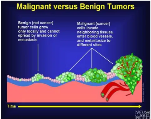
Cancer cells may be able to influence the normal cells, molecules, and blood vessels that surround and feed a tumor an area known as the microenvironment. In all types of cancer, some of the body’s cells begin to divide without stopping and spread into surrounding tissues. The cells divide uncontrollably and may grow into adjacent tissue or spread to distant parts of the body [1]. The mass of cancer cells eventually become large enough to produce lumps, masses, or tumors that can be detected. Tumors can be benign or malignant. Fig1 shows the variation between malignant and benign tumors,
Benign tumors are not cancer. It do not spread into, or invade, nearby tissues. Benign tumors can sometimes be quite large, however. When
removed, they usually don’t grow back, whereas malignant tumors sometimes do. Unlike most benign tumors elsewhere in the body, benign brain tumors can be life threatening [1].
[image:1.595.307.550.378.570.2] Malignant tumors are cancer, which means they can spread into, or invade, nearby tissues. In addition, as these tumors grow, some cancer cells can break off and travel to distant places in the body through the blood or the lymph system and form new tumors far from the original tumor [1].
Fig -1: Malignant versus Benign Tumors
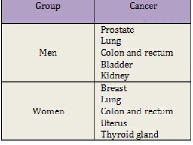
The most common cancers are at the top of the list, and go down in decreasing frequency. The order is based on estimates for 2013 from the American Cancer Society. Skin cancer is probably the most common cancer in both men and women. [2].
© 2015, IRJET ISO 9001:2008 Certified Journal
Page 237
Table -1:Most Common Cancers in Men and Women2. CANCER CELL ORIGINATION
The first step in cancer development is initiation, in which a change in the cell’s genetic material primes the cell to become cancerous. The change in the cell’s genetic material may occur spontaneously or be brought on by an agent that causes cancer (a carcinogen). Carcinogens include many chemicals, tobacco, viruses, radiation, and sunlight. However, not all cells are equally susceptible to carcinogens. A genetic flaw in a cell may make it more susceptible. Even chronic physical irritation may make a cell more susceptible to carcinogens [3].
The genetic changes that contribute to cancer tend to affect three main types of genes proto-oncogenes, tumor suppressor genes, and DNA repair genes. These changes are sometimes called “drivers” of cancer.
Proto-oncogenes are involved in normal cell growth and division. However, when these genes are altered in certain ways or are more active than normal, they may become cancer-causing genes allowing cells to grow and survive when they should not.
Tumor suppressor genes are also involved in controlling cell growth and division. Cells with certain alterations in tumor suppressor genes may divide in an uncontrolled manner.
[image:2.595.64.259.128.274.2] DNA repair genes are involved in fixing damaged DNA. Cells with mutations in these genes tend to develop additional mutations in other genes. Together, these mutations may cause the cells to become cancerous [3].
Fig -2: Name of the chart
Paragraph comes content here. Paragraph comes content here. Paragraph comes content here. Paragraph comes content here. Paragraph comes content here. Paragraph comes content here. Paragraph comes content here. Paragraph comes content here.
In Fig.2, cancer cells break away from where they first formed (primary cancer), travel through the blood or lymph system, and form new tumors (metastatic tumors) in other parts of the body. The metastatic tumor is the same type of cancer as the primary tumor
Cancerous cells develop from healthy cells in a complex process called malignant transformation. Cancer can grow directly into surrounding tissue or “spread” to tissues or organs, nearby or distant. Cancer can spread through the lymphatic system. This type of spread is typical of carcinomas. For example, breast cancer usually spreads first to the nearby lymph nodes in the armpit, and only later does it spread to distant sites. Cancer can also spread via the bloodstream [3]. Fig3 shows the factors believed to contribute to global causes of cancer,
[image:2.595.310.517.536.719.2]
© 2015, IRJET ISO 9001:2008 Certified Journal
Page 238
This type of spread is typical of sarcomas. SomeEnvironmental factors of cancer occurrences are described in the following table.
Table -2: Environmental factors of cancer
2.1 Cancer stage grouping
[image:3.595.314.555.111.249.2]Doctors determine the stage of a cancer by combining the T, N, and M classifications. Most cancers have four stages, stages I to IV. Some cancers also have a stage 0 [4].
Table -3: Stages of cancer
2.2 Types of Cancer
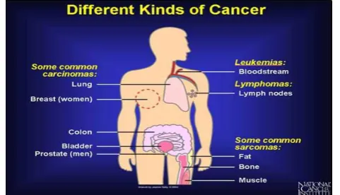
There are more than 100 types of cancer. Types of cancer are usually named for the organs or tissues where the cancers form. For example, lung cancer starts in cells of the lung, and brain cancer starts in cells of the brain. Cancers also may be described by the type of cell that formed them, such as an epithelial cell or a squamous cell [5]. Fig4 shows the different kinds of cancers for human,
Fig -4: Types of cancer
Leukemias and lymphomas are cancers of the blood and blood-forming tissues and cells of the immune system. It arises from blood-forming cells and crowd out normal blood cells in the bone marrow and bloodstream. Cancer cells from lymphomas expand lymph nodes, producing large masses in the armpit, groin, abdomen, or chest.
Carcinomas are cancers of cells that line the skin, lungs, digestive tract, and internal organs. Examples of carcinomas are cancer of the skin, lungs, colon, stomach, breasts, prostate, and thyroid gland. Typically, carcinomas occur more often in older than in younger people [5].
Sarcomas are cancers of mesodermal cells. Mesodermal cells normally form muscles, blood vessels, bone, and connective tissue. Examples of sarcomas are leiomyosarcoma (cancer of smooth muscle that is found in the wall of digestive organs) and osteosarcoma (bone cancer). Typically, sarcomas occur more often in younger than in older people.
Melanoma is cancer that begins in cells that become melanocytes, which are specialized cells that make melanin (the pigment that gives skin its color). Most melanomas form on the skin, but melanomas can also form in other pigmented tissues, such as the eye [5].
[image:3.595.45.274.465.631.2]© 2015, IRJET ISO 9001:2008 Certified Journal
Page 239
2.3 Cancer control
It consists of prevention, detection, diagnosis, treatment, reducing incidence and prevalence. In primary control, reducing the exposure to the risk factors, control of tobacco and alcohol consumption. Control of these two will reduce the total burden of cancer by 1 million cases per year. The following are some common diagnostic tests:
Diagnostic testing involves tests and procedures to confirm the presence of disease and identify the correct tumor type, location, extent and stage [6].
2.4 Types of Treatment
[image:4.595.306.557.60.262.2]There are many types of cancer treatment. The types of treatment that you receive will depend on the type of cancer you have and how advanced it is. The following table includes main types of cancer treatment,
Table -4: Treatments of cancer
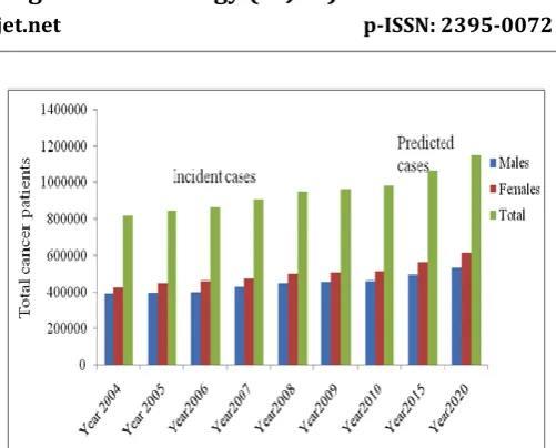
Some people with cancer will have only one treatment. But most people have a combination of treatments, such as surgery with chemotherapy and/or radiation therapy [3]. The following chart (Chart -1) illustrates the cancer scenario in India,
Chart -1: Year wise total cancer prevalence in India
3.
COMPUTER
AIDED
DETECTION
AND
CLASSIFICATION
Cancer cells are defined with wide range of features and may be easily misinterpreted by radiologists while reading large amount of results provided in screening programs. To help radiologists provide an accurate recognition, a computer-aided detection (CAD) and computer-aided classification (CAC) algorithms are being developed. These algorithms can help doctors and radiologists for getting a more reliable and effective diagnoses and help reducing the number of false positives. Fig6 represents the cycle of image processing techniques involved in clarifying the cancer cells [7].
Fig -5: Digital image and Principle phases of cancer cell detection
[image:4.595.324.526.459.620.2]© 2015, IRJET ISO 9001:2008 Certified Journal
Page 240
In feature extraction, when the input data given to analgorithm is too large to be processed and it is suspected to be notoriously redundant (much data, but not much information) then the input data will be transformed into a reduced representation set of features. Transforming the input data into the set of features is called feature extraction [8].
Computer aided detection (CAD) techniques may be useful to detect Benign and Malignant in input image. Confirmation of whether the tumor is actually a malignant and its specific type are left to the human operator or subsequent processing methods. Computer aided classification (CAC) techniques may be able to positively identify a tumor as a malignant and determine the type and orientation of the malignant involved.
4. LITERATURE SURVEY
The CAD system proposed by Gletsos Miltiades, et al. [16] consists of two basic modules: the feature extraction and the classifier modules. In their work, region of interest (liver tumor) were identified manually from the CT liver images and then fed to the feature extraction module. The total performance of the system was 97% for validation set and100% for testing set.
B.Monica Jenefer and V.Cyrilraj used the image processing techniques with MRI images to detecting Breast cancer, for Image Enhancement Speckle Noise Removal and EM algorithm, Modified Watershed Segmentation, GLCM Feature Extraction technique and SVM classification proved its performance via performance metrics such as Sensitivity is 97.5%, Specificity is 100% and its Accuracy in classification is 98% [17].
Mahmoud Elgamal, Saudii ArabiaThis presents two hybrid techniques for the classification of the skin images to predict it if exists. The proposed hybrid techniques consist of three stages, namely, feature extraction, dimensionality reduction, and classification. In the first stage, we have obtained the features related with images using discrete wavelet transformation. In the second stage, the features of skin images have been reduced using principle component analysis to the more essential features. In the classification stage, two classifiers based on supervised machine learning have been developed. The first classifier based on feed forward back-propagation artificial neural network and the second classifier based on k-nearest neighbor. The classifiers have been used to classify
subjects as normal or abnormal skin cancer images. A classification with a success of 95% and 97.5% has been obtained by the two proposed classifiers and respectively. This result shows that the proposed hybrid techniques are robust and effective [18].
Disha Sharma, Gagandeep Jindal using the approach that starts by extracting the lung regions from the CT image using several image processing techniques, including bit image slicing, erosion, weiner filter. They introduced the using of bit plane slicing technique that is used in the first step in the extraction process to convert the CT image into a binary image. Bit-plane slicing technique is both faster and data- and user-independent compared to the thresholding technique. After the extraction step, the extracted lung regions are segmented using egion growing segmentation algorithm. Then, the initial lung candidate nodules resulting from the Region growing segmentation are analyzed to extract a set of features to be used in the diagnostic rules. After that to segment the lung region. In this region to detect cancerous region and to get the accurate result. The accuracy of 80% approximate the accuracy indicated by surgeons and radiologists for locating cancerous nodules during reading clinical CT images in 2.5–7.0 mm [19].
Shrikant D. Kale, Krushil M. Punwatkar used the gray level co-occurrence matrix (GLCM) texture characterization techniques are used for feature extraction & extracted features are classified using scaled conjugate gradient backpropagation training neural network (SCGBNN) for diagnosis of thyroid cancer in ultrasound images are described. The experimental results show the performance measure of SCG backpropagation training neural network in terms classification accuracy [20].
© 2015, IRJET ISO 9001:2008 Certified Journal
Page 241
5. CONCLUSION
Images are essential components in many fields of medicine, especially for the diagnosis process of tumors, technology based approaches such as digital image processing help physicians to have better interpretation, faster detection and increases accuracy and objectivity of diagnosis. In cancer diagnosis and treatment field the goal of digital image processing is to locate and extract meaningful items from images and classify the type and
stage of cancer. Looking at the experiences gained will help specialists to choose the appropriate technique for optimization of diagnosis through medical imaging. This paper presents an impression of cancers and literature survey on detection of cancers by previous researchers. A detailed comparative analysis between the various image processing techniques are to be presented in this work. From the literature review, it indicates that developments of new methods are required to detect cancer cells more efficiently.
Filter (architectura
l and textural features)
back propagation
Neural network
Aparna Kanakatte[8]
2008 PET LUNG _
Standard Uptake Values(SUV) Discrete Cosine Transform, Wavelets
k-NN, SVM 97%
Disha Sharma [9] 2011 CT LUNG Wiener Filter Edge detection
(Sobel) Texture Features Diagnostic Indicators 80% Seena Thomas, Anjali Vijayan [10]
2015 Biopsy Colon Wiener Filter, Adaptive Histogram Equalization
Graph Cut GLCM Kernel Sparse Representation
Classifier
94%
Eggi Intan Putri,, Rita Magdalena
[11]
2015 Ultrasound Cervical Bwareaopen Adaptive Thresholding
_ K-Nearest
Neighbor
100%
D.-Y. KIM & J.-W. PARK [12]
2015 CT Kidney Histogram
Equalization Thresholding and region-growing Texture Analysis _ 85% Shijun Wang,Karen
Burtt, 2014 MP-MRI
Prostate ADC Map Graph-cut
algorithm
Statistical Features
SVM
99.6%
Shrikant D. Kale
, Krushil M. Punwatkar [13]
2013 Ultrasound Thyroid LP and DFB
Filters
Region based
Active contour
GLCM Scaled conjugate
Gradient back propagation training neural network 94.11% Mahmoud Elgamal[14]
2013 CT Skin Anisotropic Diffusion Filters(ADF)
Superficial Spreading
Melanoma (SSM)
DWT K nearest
neighbor
97.5%
Abdulmuhssin Binhssan[15]
2015 MRI Bone average & bilateral filter Thresholding segmentation Morphologic al Operation _ 91.3%
© 2015, IRJET ISO 9001:2008 Certified Journal
Page 242
REFERENCES
[1] https://www.urmc.rochester.edu/Encyclopedia
[2] http://www.msdmanuals.com/home/cancer
[3] http://www.cancer.gov/about-cancer
[4] http://www.cancer.net
[5] http://patient.info/health/cancer-a-general-overview
[6] http://www.cancercenter.com/
[7] Shekhar Singh "Cancer Cells Detection and Classification in Biopsy Image" International Journal of Computer Applications (0975 – 8887) Volume 38– No.3, January 2012.
[8] Aparna Kanakatte, Nallasamy Mani, Bala Srinivasan, Jayavardhana Gubbi, “Pulmonary Tumor Volume Detection from Positron Emission Tomography Images”, International Conference on Biomedical Engineering and Informatics, pp: 213- 217, 2008.
[9] Disha Sharma, Gagandeep Jindal, “Identifying Lung Cancer Using Image Processing Techniques”, International Conference on Computational Techniques and Artificial Intelligence (ICCTAI), pp: 115-120, 2011.
[10] Seena Thomas, Anjali Vijayan"Automated Colon Cancer Detection Using Kernel Sparse Representation Based Classifier International Journal of Engineering and Advanced Technology (IJEAT) ISSN: 2249 – 8958, Volume-4 Issue-6, August 2015
[11] Eggi Intan Putri, Rita Magdalena & Ledya Novamizanti "Detection of Cervical Cancer Disease using Adaptive Thresholding Method by Image Processing" GTAR-2015, Vol. 2, 477-486 ISBN: 978-969-9948-30-5
[12] D.-Y. KIM & J.-W. PARK "Computer-Aided Detection of Kidney Tumor on Abdominal Computed Tomography Scans” ISSN: 1600-0455
(Online) Journal homepage:
http://www.tandfonline.com/loi/iard20, 08 October 2015.
[13] Shrikant D. Kale1, Krushil M. Punwatkar2 "Texture Analysis of Thyroid Ultrasound Images for Diagnosis of Benign and Malignant Nodule
using Scaled Conjugate Gradient Backpropagation Training Neural Network " IJCEM International Journal of Computational Engineering & Management, Vol. 16 Issue 6, November 2013 ,ISSN (Online): 2230-7893
[14] Mahmoud Elgamal"AUTOMATIC SKIN CANCER IMAGES CLASSIFICATION “(IJACSA) International Journal of Advanced Computer Science and Applications,Vol. 4, No. 3, 2013.
[15] Abdulmuhssin Binhssan "Enchondroma tumor Detection International Journal of Advanced Research in Computer and Communication Engineering,Vol. 4, Issue 6, June 2015.ISSN (Online) 2278-1021
[16] Seo, K.S. “Improved fully Automatic Liver Segmentation using Histogram tail threshold Algorithms” Computational Science ICCS, vol. 35, pp. 822 -825, May 2005.
[17] B.Monica Jenefer, V.Cyrilraj "An Efficient Image Processing Methods for Mammogram Breast Cancer Detection" Journal of Theoretical and Applied Information Technology 10th November 2014. Vol. 69 No.1.
[18] Mahmoud Elgamal,Saudii "Automatic Skin Cancer Images Classification" (IJACSA) International Journal of Advanced Computer Science and Applications, Vol. 4, No. 3, 2013.
[19] Disha Sharma, Gagandeep Jindal " Identifying Lung Cancer Using Image Processing Techniques " International Conference on Computational Techniques and Artificial Intelligence (ICCTAI'2011)