HDL action on the vascular wall: is the answer
NO?
Philip W. Shaul, Chieko Mineo
J Clin Invest. 2004;
113(4)
:509-513.
https://doi.org/10.1172/JCI21072
.
Circulating levels of HDL cholesterol are inversely related to the risk of atherosclerosis, and
therapeutic increases in HDL reduce the incidence of cardiovascular events. A new study
shows that HDL-associated lysophospholipids stimulate the production of the potent
antiatherogenic signaling molecule NO by the vascular endothelium.
Commentary
Find the latest version:
1. Foster, P.S., Hogan, S.P., Ramsay, A.J., Matthaei, K.I., and Young, I.G. 1996. Interleukin 5 deficien-cy abolishes eosinophilia, airways hyperreactivi-ty, and lung damage in a mouse asthma model. J. Exp. Med.183:195–201.
2. Hamelmann, E., et al. 1997. Antiinterleukin-5 antibody prevents airway hyperresponsiveness in a murine model of airway sensitization. Am. J. Respir. Crit. Care Med.155:819–825.
3. Corry, D.B., et al. 1996. Interleukin 4 but not interleukin 5 or eosinophils, is required in a murine model of acute airway hyperresponsive-ness. J. Exp. Med.183:109–117.
4. Mattes, J., et al. 2002. Intrinsic defect in T cell pro-duction of interleukin (IL)-13 in the absence of both IL-5 and eotaxin precludes the development of eosinophilia and airways hyperreactivity in experimental asthma. J. Exp. Med. 195:1433–1444. 5. Mauser, P.J., et al. 1995. Effects of an antibody to IL-5 in a monkey model of asthma. Am. Rev. Respir. Dis.152:467–472.
6. Leckie, M.J., et al. 2000. Effects of an interleukin-5 blocking monoclonal antibody on eosinophils, airway hyper-responsiveness, and the late asth-matic response. Lancet.356:2144–2148.
7. Kips, J.C., et al. 2003. Effect of SCH55700, a humanized anti-human interleukin-5 antibody, in severe persistent asthma: a pilot study. Am. J. Respir. Crit. Care Med.167:1655–1659. 8. O’Byrne, P.M., Inman, M.D., and Parameswaran,
K. 2001. The trials and tribulations of IL-5, eosinophils, and allergic asthma. J. Allergy Clin. Immunol. 108:503–508.
9. Kay, A.B., and Menzies-Gow, A. 2003. Eosinophils and interleukin-5: the debate continues. Am. J. Respir. Crit. Care Med.167:1586–1587.
10. Flood-Page, P.T., Menzies-Gow, A.N., Kay, A.B., and Robinson, D.S. 2003. Eosinophil’s role remains uncertain as anti-interleukin-5 only par-tially depletes numbers in asthmatic airway. Am. J. Respir. Crit. Care Med.167:199–204.
11. Blyth, D.I., Wharton, T.F., Pedrick, M.S., Savage, T.J., and Sanjar, S. 2000. Airway subepithelial fibrosis in a murine model of atopic asthma: suppression by dexamethasone or anti-inter-leukin-5 antibody. Am. J. Respir. Cell Mol. Biol.
23:241–246.
12. Trifilieff, A., Fujitani, Y., Coyle, A.J., Kopf, M., and Bertrand, C. 2001. IL-5 deficiency abolishes aspects of airway remodelling in a murine
model of lung inflammation. Clin. Exp. Allergy.
31:934–942.
13. Cho, J.Y., et al. 2004. Inhibition of airway remod-eling in IL-5–deficient mice. J. Clin. Invest.
113:551–560. doi:10.1172/JCI200419133. 14. Huang, X.Z., et al. 1996. Inactivation of the
inte-grin β6subunit gene reveals a role of epithelial integrins in regulating inflammation in the lung and skin. J. Cell Biol.133:921–928.
15. Munger, J.S., et al. 1999. The integrin αvβ6binds and activates latent TGF β1: a mechanism for reg-ulating pulmonary inflammation and fibrosis. Cell.96:319–328.
16. Phipps, S., et al. 2002. The relationship between allergen-induced tissue eosinophilia and markers of repair and remodeling in human atopic skin. J. Immunol. 169:4604–4612.
17. Flood-Page, P., et al. 2003. Anti-IL-5 treatment reduces deposition of ECM proteins in the bronchial subepithelial basement membrane of mild atopic asthmatics. J. Clin. Invest.
112:1029–1036. doi:10.1172/JCI200317974. 18. Mauser, P.J., et al. 1993. Inhibitory effect of the
TRFK-5 anti-IL-5 antibody in a guinea pig model of asthma. Am. Rev. Respir. Dis.148:1623–1627.
HDL action on the vascular wall:
is the answer NO?
Philip W. Shaul and Chieko Mineo
Department of Pediatrics, University of Texas Southwestern Medical Center, Dallas, Texas, USA
Circulating levels of HDL cholesterol are inversely related to the risk of atherosclerosis, and therapeutic increases in HDL reduce the incidence of cardiovascular events. A new study (see the related article beginning on page 569) shows that HDL-associated lysophospholipids stimulate the production of the potent antiatherogenic signaling molecule NO by the vascular endothelium.
J. Clin. Invest.113:509–513 (2004). doi:10.1172/JCI200421072.
The risk of cardiovascular disease from atherosclerosis is inversely pro-portional to serum levels of HDL and the major HDL apolipoprotein apoAI (1). In fact, low HDL levels predict an increased risk of coronary artery dis-ease independently of LDL levels, and 60–70% of major cardiovascular
events cannot be prevented with cur-rent approaches focused on LDL, such as statin therapy (2). In addition, low HDL levels are particularly com-mon in males with early-onset ather-osclerosis (3). Based on these obser-vations, prevention trials have been performed with agents such as niacin and fibrates, which raise HDL, and they indicate that modest increases in HDL independently yield a signifi-cant reduction in cardiovascular events (4–6). Thus, there is com-pelling evidence that HDL is not sole-ly a marker of lower risk of cardiovas-cular disease but instead is a mediator of vascular health.
Up until recently the protective fea-tures of HDL had been attributed pri-marily to its classical function of removing cholesterol from peripheral
tissues and transferring it to the liver in a process known as reverse cholesterol transport (RCT). The delivery of cho-lesteryl ester from HDL to cells such as hepatocytes entails apoAI–mediated HDL cell surface interaction with scavenger receptor class B, member I (SR-BI), to which HDL binds with high affinity (7). Despite detailed under-standing of HDL and RCT, the mecha-nisms by which HDL and apoAI are atheroprotective remain complex and not fully understood (8). This is partic-ularly apparent when one considers evidence that circulating levels of HDL and apoAI do not regulate RCT (9, 10), and that individuals with homozygous deficiency in the plasma cholesteryl ester transferase protein, which en-hances HDL cholesterol delivery to the liver, have decreased risk of coronary artery disease (11).
Our basic understanding of the role of HDL in vascular biology has entered a new era in the past few years as direct modes of action of HDL on vascular cells have been elucidated. In particular, it has been demonstrated that HDL causes potent stimulation of eNOS activity through binding to SR-BI, which is expressed in endothelium (12, 13). Similarly, HDL enhances endothe-lium- and NO-dependent relaxation in aortas from wild-type but not SR-BI– knockout mice (12). The HDL-induced increase in NO production may be crit-ical to the atheroprotective features of HDL, as diminished bioavailablity of NO has a key role in the early
pathogen-Address correspondence to: Philip W. Shaul, Department of Pediatrics, University of Texas Southwestern Medical Center at Dallas, 5323 Harry Hines Boulevard, Dallas, Texas 75390, USA. Phone: (214) 648-2015; Fax: (214) 648-2481;
E-mail: [email protected].
Conflict of interest: The authors have declared that no conflict of interest exists.
esis of hypercholesterolemia-induced vascular disease and atherosclerosis (14). However, the mechanisms by which HDL activates eNOS and the physiological and pathological implica-tions are yet to be fully clarified.
In the current issue of the JCI, Nofer and colleagues provide intrigu-ing evidence that HDL-induced NO production may be mediated by the HDL-associated lysophospholipids sphingosylphosphorylcholine (SPC), sphingosine-1-phosphate (S1P), and lysosulfatide (LSF) (15). They de-monstrate that SPC, S1P, and LSF each cause eNOS-dependent relax-ation of precontracted aortic rings from mice, and that blood pressure falls following intra-arterial infusion of HDL, SPC, S1P, or LSF in rats pre-treated with endothelin. The lyso-phospholipid receptor S1P3 was
implicated in further studies of aor-tic rings from receptor-null mice (15). Collectively these observations suggest that lysophospholipids un-derlie an important component of HDL action on the vascular wall.
Features of HDL that mediate signaling to eNOS
Both human and animal studies indi-cate that apoAI is the apolipoprotein principally responsible for the athero-protective features of HDL (16). In ear-lier studies of eNOS activation, lipid-free apoAI did not stimulate the enzyme in cultured endothelial cells, yet antibody to apoAI blocked eNOS activation by HDL in isolated endothe-lial plasma membranes, indicating that apoAI is necessary but not suffi-cient for eNOS stimulation (12). The work of Nofer and colleagues suggests that cargoes of the HDL particle, namely SPC, S1P, and LSF, mediate the signaling to eNOS (15). In their studies of aortic ring relaxation in response to HDL, SPC, S1P, and LSF, the relative contribution of the lysophospholipids to signal activation by HDL is uncertain because the con-centrations provided by HDL were 20-fold lower than those for the individu-ally tested lysophospholipids. The experiments with aortas from S1P3
-knockout mice more clearly suggest that 50–60% of the response to HDL is mediated by lysophospholipids (15).
However, as the process is SR-BI de-pendent and recent work on the SR-BI adaptor molecule PDZK1 indicates that the mechanisms determining SR-BI abundance may be complex (17), poten-tial changes in SR-BI expression in S1P3receptor–null mice should be
eval-uated. Interestingly, Kimura and coworkers recently observed that the migration and survival of cultured endothelial cells is also enhanced by lysophospholipids (18), but it has not yet been determined whether eNOS mediates these processes.
Previous work by Gong and cowork-ers suggests that another potential cargo of HDL, namely estradiol, plays a role in eNOS activation by the lipoprotein (19). They reported that HDL isolated from female humans or mice had greater capacity to stimu-late the enzyme than did HDL from male humans or mice. These findings are quite exciting, but it should be noted that the experiments implicat-ing a direct role of estradiol via estro-gen receptor activation were limited. In addition, as pointed out by Nofer et al. (15), HDL-mediated responses were apparent at lipoprotein concen-trations that would provide estradiol at levels as low as 5 × 10–16M (19),
which is six orders of magnitude below the concentrations shown to activate eNOS in prior reports (20, 21). Furthermore, human studies demonstrating that brief elevations in HDL enhance endothelial function have been performed mainly in males (see below), suggesting a lack of dependence on estradiol. Nofer and colleagues also compared responses to male and female HDL and found no difference. As is the case for the new information gained regarding lysophospholipids, the findings relat-ed to estradiol are of considerable potential importance, but interpreta-tion and extrapolainterpreta-tion at this point should be done with caution.
Relevant signaling events proximal to eNOS
Over the past decade it has been appre-ciated that multiple signal transduc-tion pathways converge to regulate the activity of eNOS. Stimulation often occurs in association with elevations in cytosolic Ca2+concentrations and
Ca2+-calmodulin binding, and tyrosine
kinase-MAPK signaling is also fre-quently involved. In addition, protein kinases including Akt kinase, which is downstream of PI3K, enhance eNOS activity through serine phosphoryla-tion at posiphosphoryla-tion 1,177 of human eNOS. In contrast, phosphorylation of Thr 497 yields attenuated eNOS activity (22). Regarding HDL regulation of eNOS, Mineo and colleagues deter-mined that HDL causes an increase in phosphorylation at Ser 1,179 of bovine eNOS (equivalent to Ser 1,177 in hu-man eNOS) and no change in Thr 497 phosphorylation. Further experiments indicated that HDL stimulation of eNOS also requires MAPK activation and that the nonreceptor tyrosine kinase src and PI3K reside upstream in the signaling pathway and cause par-allel activation of Akt and MAPK and their resultant independent modula-tion of the enzyme (23).
The current report by Nofer and coworkers provides additional in-sights into HDL signaling to eNOS (15). Along with data confirming HDL-induced phosphorylation of Akt and eNOS, they show that HDL caus-es an increase in intracellular Ca2+,
and that Ca2+ is required for
HDL-induced NO formation. In parallel, SPC, LSF, and S1P caused eNOS phosphorylation at Ser 1,177 and in-creases in intracellular Ca2+. However,
infor-mation indicates that HDL activation of eNOS entails the stimulation of multiple kinase cascades and calcium mobilization.
Evidence of HDL modulation of eNOS in humans
Whereas the functional link between HDL and eNOS has been appreciated
only recently, the relationship be-tween HDL and endothelium-de-pendent vasodilation has been known for some time. In studies of coronary vasomotor responses to acetylcholine, it was noted in 1994 that patients with elevated HDL have greater vasodilator and attenuated vasocon-strictor responses (24). Studies of flow-mediated vasodilation of the brachial artery have also shown that HDL cholesterol is an independent predictor of endothelial function (25). The direct, short-term impact of HDL on endothelial function also has recently been investigated in humans. One particularly elegant study recent-ly evaluated forearm blood flow responses in individuals who are het-erozygous for a loss-of-function mutation in the ATP-binding cassette transporter 1 (ABCA1) gene. Com-pared with controls, ABCA1 het-erozygotes (six men and three women) had HDL levels that were decreased by 60%, their blood flow responses to endothelium-dependent vasodilators were blunted, and endothelium-inde-pendent responses were unaltered. After a 4-hour infusion of apoAI/ phosphatidylcholine disks, their HDL level increased threefold and endothe-lium-dependent vasomotor respons-es were fully rrespons-estored (26). It has also been observed that endothelial func-tion is normalized in hypercholes-terolemic men with normal HDL lev-els shortly following the administration of apoAI/phosphatidylcholine parti-cles (27). Thus, evidence is now accu-mulating that HDL is a robust posi-tive modulator of endothelial NO production in humans.
More than an eNOS agonist
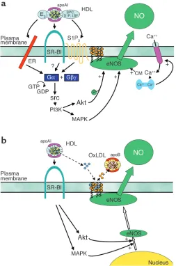
[image:4.585.77.330.54.436.2]In addition to the modulation of NO production by signaling events that rapidly dictate the level of enzymatic activity, important control of eNOS involves changes in the abundance of the enzyme. In a clinical trial by the Karas laboratory of niacin therapy in patients with low HDL levels (nine males and two females), flow-mediat-ed dilation of the brachial artery was improved in association with a rise in HDL of 33% over 3 months (28). They also demonstrated that eNOS expres-sion in cultured human endothelial Figure 1
HDL enhances NO production by eNOS in vascular endothelium. (a) HDL causes membrane-initiated signaling, which stimulates eNOS activity. The eNOS protein is localized in cholesterol-enriched (orange circles) plasma membrane caveolae as a result of the myristoylation and palmitoylation of the protein. Binding of HDL to SR-BI via apoAI causes rapid activation of the nonreceptor tyrosine kinase src, leading to PI3K activation and downstream activation of Akt kinase and MAPK. Akt enhances eNOS activity by phosphorylation, and independent MAPK-mediated processes are additionally required. HDL also causes an increase in intracellular Ca2+
concentration (intracellular Ca2+store shown in blue; Ca2+channel shown in pink), which
enhances binding of calmodulin (CM) to eNOS. HDL-induced signaling is mediated at least partially by the HDL-associated lysophospholipids SPC, S1P, and LSF acting through the G pro-tein–coupled lysophospholipid receptor S1P3. HDL-associated estradiol (E2) may also activate
cells is increased by HDL exposure for 24 hours. They further showed that the increase in eNOS is related to an increase in the half-life of the protein, and that this is mediated by PI3K–Akt kinase and MAPK (29). Thus, the same mechanisms that underlie the acute activation of eNOS by HDL appear to be operative in upregulating the expression of the enzyme.
Another important feature of eNOS regulation involves the myristoylation and palmitoylation of the protein, resulting in trafficking to caveolae, which are a subclass of liquid-ordered, cholesterol-rich domains (lipid rafts). The eNOS protein resides within a signaling module in caveolae that contains all of the molecular machin-ery necessary for its regulation by multiple ligands (22). The impact of lipoproteins on the lipid environment in caveolae and on eNOS localization was assessed by the Smart laboratory. Oxidized LDL was found to cause 90% of eNOS to leave caveolae, and acetyl-choline-induced activation was atten-uated. Further studies showed that this was due to oxidized LDL–induced depletion of caveolae cholesterol mediated by CD36 (30). However, coincubation of cells with HDL pre-vented oxidized LDL–induced translo-cation of eNOS from caveolae and restored acetylcholine activation. Additional work showed that this process is mediated by SR-BI and that it is related to the maintenance of caveolae cholesterol content due to cholesteryl ester uptake from HDL (31). Thus, along with its capacity to serve as an agonist for eNOS and to upregulate its expression, HDL may preserve the lipid environment in caveolae in the face of oxidized LDL, thereby maintaining the signaling module required for effective endothelial NO production and the atheroprotection that NO affords.
Unanswered questions
Our current understanding of the mechanisms by which HDL enhances endothelial NO production is summa-rized in Figure 1. The work by Nofer and colleagues on lysophospholipids (15) and the prior studies of HDL-cou-pled estradiol have provided valuable information about the potential
com-ponents of HDL that mediate eNOS activity (19). However, perhaps related solely to the complexity of the lipopro-tein, it is likely that additional charac-teristics are also important to induc-ing signalinduc-ing in the endothelium. In particular, the role of the best described function of HDL in mediat-ing cholesterol flux has yet to be deter-mined. In addition, the impact of apolipoproteins besides apoAI should be considered. For example, a potential role for apoAII was revealed in studies in which eNOS activation by HDL in isolated endothelial plasma mem-branes was augmented by antibody to apoAII (12). The features of SR-BI req-uisite to eNOS activation are also unknown. The work related to lyso-phospholipids and estradiol suggests that SR-BI serves to tether HDL to the extracellular surface for the delivery of these ligands (15, 19). However, domains of SR-BI other than the extra-cellular loop may be necessary, as sug-gested by experiments in the endothe-lial cell plasma membrane paradigm in which antibody to the SR-BI cytoplas-mic C-terminal tail prevented eNOS activation (12). In addition, the most proximal events occurring upon bind-ing of HDL to SR-BI are unclear at present, and they represent the poten-tially most novel aspects of HDL action in endothelial cells. Perhaps most importantly, now that initial mechanistic insights have been gained in vitro, discrete manipula-tions of the HDL–SR-BI–eNOS path-way will be required in intact animal models to delineate if these process-es mediate endothelial cell function in a physiological or pathophysiolog-ical context. With greater under-standing in hand regarding both the underlying mechanisms and their implications, it will then be possible to consider therapeutic manipula-tion of direct HDL acmanipula-tion on the endothelium to optimize cardiovas-cular health.
Acknowledgments
We are indebted to Marilyn Dixon for preparing this manuscript. This work was supported by National Institutes of Health grants HD30276, HL58888, and HL53546 (P.W. Shaul), the Crystal Charity Ball Center for Pediatric
Criti-cal Care Research, and the Lowe Foun-dation. C. Mineo is also supported by the Scientist Development Program of the American Heart Association.
1. Gordon, D.J., and Rifkind, B.M. 1989. High-den-sity lipoprotein—the clinical implications of recent studies. N. Engl. J. Med.321:1311–1316. 2. Foody, J.M., et al. 2000. HDL cholesterol level predicts survival in men after coronary artery bypass graft surgery: 20-year experience from The Cleveland Clinic Foundation. Circulation.
102:III90–III94.
3. Wilson, P.W., Abbott, R.D., and Castelli, W.P. 1988. High density lipoprotein cholesterol and mortali-ty. The Framingham Heart Study. Arteriosclerosis.
8:737–741.
4. Rubins, H.B., et al. 1999. Gemfibrozil for the sec-ondary prevention of coronary heart disease in men with low levels of high-density lipoprotein cholesterol. Veterans Affairs High-Density Lipoprotein Cholesterol Intervention Trial Study Group. N. Engl. J. Med.341:410–418.
5. Brown, B.G., et al. 2001. Simvastatin and niacin, antioxidant vitamins, or the combination for the prevention of coronary disease. N. Engl. J. Med.
345:1583–1592.
6. Boden, W.E. 2000. High-density lipoprotein cho-lesterol as an independent risk factor in cardio-vascular disease: assessing the data from Fram-ingham to the Veterans Affairs High-Density Lipoprotein Intervention Trial. Am. J. Cardiol.
86:19L–22L.
7. Krieger, M. 1999. Charting the fate of the “good cholesterol”: identification and characterization of the high-density lipoprotein receptor SR-BI. Annu. Rev. Biochem.68:523–558.
8. Krieger, M. 1998. The “best” of cholesterols, the “worst” of cholesterols: a tale of two receptors. Proc. Natl. Acad. Sci. U. S. A.95:4077–4080. 9. Jolley, C.D., Woollett, L.A., Turley, S.D., and
Dietschy, J.M. 1998. Centripetal cholesterol flux to the liver is dictated by events in the peripheral organs and not by the plasma high density lipoprotein or apolipoprotein A-I concentration. J. Lipid Res.39:2143–2149.
10. Brousseau, M.E., et al. 2000. Cellular cholesterol efflux in heterozygotes for tangier disease is markedly reduced and correlates with high den-sity lipoprotein cholesterol concentration and particle size. J. Lipid Res.41:1125–1135. 11. Zhong, S., et al. 1996. Increased coronary heart
disease in Japanese-American men with mutation in the cholesteryl ester transfer protein gene despite increased HDL levels. J. Clin. Invest.
97:2917–2923.
12. Yuhanna, I.S., et al. 2001. High-density lipopro-tein binding to scavenger receptor-BI activates endothelial nitric oxide synthase. Nat. Med.
7:853–857.
13. Li, X.A., et al. 2002. High density lipoprotein binding to scavenger receptor, class B, type I activates endothelial nitric-oxide synthase in a ceramide-dependent manner. J. Biol. Chem.
277:11058–11063.
14. Cohen, R.A. 1995. The role of nitric oxide and other endothelium-derived vasoactive sub-stances in vascular disease. Prog. Cardiovasc. Dis.
38:105–128.
15. Nofer, J.-R., et al. 2004. HDL induces NO-dependent vasorelaxation via the lysophospho-lipid receptor S1P3. J. Clin. Invest.113:569–581.
doi:10.1172/JCI200418004.
16. Rader, D.J. 2003. Regulation of reverse choles-terol transport and clinical implications. Am. J. Cardiol.92:42J–49J.
18. Kimura, T., et al. 2003. High-density lipoprotein stimulates endothelial cell migration and survival through sphingosine 1-phosphate and its recep-tors. Arterioscler. Thromb. Vasc. Biol.23:1283–1288. 19. Gong, M., et al. 2003. HDL-associated estradiol stimulates endothelial NO synthase and vasodila-tion in an SR-BI-dependent manner. J. Clin. Invest.
111:1579–1587. doi:10.1172/JCI200316777. 20. Lantin-Hermoso, R.L., et al. 1997. Estrogen
acute-ly stimulates nitric oxide synthase activity in fetal pulmonary artery endothelium. Am. J. Physiol.
273:L119–L126.
21. Caulin-Glaser, T., Garcia-Cardena, G., Sarrel, P., Sessa, W.C., and Bender, J.R. 1997. 17 beta-estra-diol regulation of human endothelial cell basal nitric oxide release, independent of cytosolic Ca2+ mobilization. Circ. Res.81:885–892. 22. Shaul, P.W. 2002. Regulation of endothelial nitric
oxide synthase: location, location, location. Annu. Rev. Physiol.64:749–774.
23. Mineo, C., Yuhanna, I.S., Quon, M.J., and Shaul, P.W. 2003. HDL-induced eNOS activation is mediated by Akt and MAP kinases. J. Biol. Chem.
278:9142–9149.
24. Zeiher, A.M., Schachlinger, V., Hohnloser, S.H., Saurbier, B., and Just, H. 1994. Coronary athero-sclerotic wall thickening and vascular reactivity in humans. Elevated high-density lipoprotein lev-els ameliorate abnormal vasoconstriction in early atherosclerosis. Circulation.89:2525–2532. 25. Li, X.P., et al. 2000. Protective effect of high
den-sity lipoprotein on endothelium-dependent vasodilatation. Int. J. Cardiol.73:231–236. 26. Bisoendial, R.J., et al. 2003. Restoration of
endothelial function by increasing high-density lipoprotein in subjects with isolated low high-density lipoprotein. Circulation.107:2944–2948. 27. Spieker, L.E., et al. 2002. High-density lipoprotein restores endothelial function in hypercholes-terolemic men. Circulation.105:1399–1402.
28. Kuvin, J.T., et al. 2002. A novel mechanism for the beneficial vascular effects of high-density lipoprotein cholesterol: enhanced vasorelaxation and increased endothelial nitric oxide synthase expression. Am. Heart J.144:165–172. 29. Ramet, M.E., et al. 2003. High-density lipoprotein
increases the abundance of eNOS protein in human vascular endothelial cells by increasing its half-life. J. Am. Coll. Cardiol.41:2288–2297. 30. Blair, A., Shaul, P.W., Yuhanna, I.S., Conrad, P.A.,
and Smart, E.J. 1999. Oxidized low density lipopro-tein displaces endothelial nitric-oxide synthase (eNOS) from plasmalemmal caveolae and impairs eNOS activation. J. Biol. Chem.274:32512–32519. 31. Uittenbogaard, A., Shaul, P.W., Yuhanna, I.S., Blair, A., and Smart, E.J. 2000. High density lipoprotein prevents oxidized low density lipoprotein-induced inhibition of endothelial nitric-oxide synthase localization and activation in caveolae. J. Biol. Chem.275:11278–11283.
Fighting cancer by disrupting C-terminal
methylation of signaling proteins
Steven Clarke
1,2and Fuyuhiko Tamanoi
2,31Department of Chemistry and Biochemistry, 2Molecular Biology Institute, and
3Department of Microbiology, Immunology, and Molecular Genetics, Jonsson
Comprehensive Cancer Center, University of California, Los Angeles, California, USA
Protein methylation at the C-terminus of mammalian isoprenylated pro-teins has been implicated in membrane attachment, protein-protein inter-actions, and protein stability. A new paper describes surprising results: in the absence of methylation some target proteins have increased stability, whereas others have decreased stability. The decreased stability of the RhoA protein is correlated with an increased resistance to Ras-dependent trans-formation and suggests the basis for the development of a new approach to antitumor therapy (see the related article beginning on page 539).
J. Clin. Invest.113:513–515 (2004). doi:10.1172/JCI200421059.
Covalent modification of proteins facilitates a tremendous expansion of their functional potential. Some mod-ifications serve to enlarge the chemical diversity of proteins beyond that pro-vided by the 20 standard amino acids utilized in protein synthesis, providing the new shapes and reactive groups that allow new types of binding and catalytic interactions. Other modifica-tions, often reversible, serve to modu-late protein function. Among the large
number of reactions that modify intra-cellular eucaryotic proteins are three sequential enzymatic steps that recog-nize proteins synthesized with a C-ter-minal CAAX tetrapeptide motif, where C is a cysteine residue, A is generally an aliphatic residue, and X can be a vari-ety of residues (1–3). Such proteins are initially lipidated in a reaction that adds either a 15-carbon farnesyl or a 20-carbon geranylgeranyl group to the sulfur atom of the cysteine residue. This step is followed by the removal of the three AAX amino acids generating a C-terminal isoprenylcysteine residue. The third step is a potentially re-versible methyl esterification reaction of the newly exposed C-terminal cys-teinyl carboxyl group by a membrane-bound enzyme of the endoplasmic reticulum (Figure 1). Many proteins
are modified in this way, including some nuclear lamins, a cGMP phos-phodiesterase, and several small G-proteins involved in cell signaling, such as the Ras and Rho proteins. The importance of Ras, especially the acti-vated oncogenic forms, has led to a large interest in this pathway in terms of the possibility of controlling human cancers.
These reactions can provide the modified protein increased membrane attachment with the newly formed hydrophobic C-terminus (4), directed binding to signaling partners via the isoprenyl and methyl groups (5, 6), and protection against proteolytic degradation (7, 8). In this issue of the JCI, Bergo et al. provide new insight into these functions and have identi-fied new targets for the development of anticancer drugs (9). In this study, they developed transgenic mice (and fibroblast cell lines derived from these mice) where the expression of the Icmt gene encoding isoprenylcysteine car-boxyl methyltransferase (Icmt) can be controlled by the Cre-loxP system. There are two striking results from this study. First, the authors show that loss of the methyltransferase can have different effects in different methyl-accepting proteins. For example, they show that Ras proteins are stabilized but the RhoA protein is destabilized. Second, they show that the decreased levels of RhoA in the absence of methy-lation results in increased levels of p21Cip1and the inhibition of the cell
cycle. The authors thus present a case for anticancer therapeutic approaches based on inhibiting Icmt.
Address correspondence to: Steven Clarke, 640 Boyer Hall, University of California, Los Angeles, California 90095-1569, USA. Phone: (310) 825-8754; Fax: (310) 825-1968; E-mail: [email protected].
Conflict of interest: The authors have declared that no conflict of interest exists.
Nonstandard abbreviations used: