Angiotensin II stimulates expression of the
chemokine RANTES in rat glomerular
endothelial cells. Role of the angiotensin type 2
receptor.
G Wolf, … , U Helmchen, R A Stahl
J Clin Invest.
1997;
100(5)
:1047-1058.
https://doi.org/10.1172/JCI119615
.
Glomerular influx of monocytes/macrophages (M/M) occurs in many immune- and
non-immune-mediated renal diseases. The mechanisms targeting M/M into the glomerulus are
incompletely understood, but may involve stimulated expression of chemokines. We
investigated whether angiotensin II (ANG II) induces the chemokine RANTES in cultured
glomerular endothelial cells of the rat and in vivo. ANG II stimulated mRNA and protein
expression of RANTES in cultured glomerular endothelial cells. The ANG II-induced
RANTES protein was chemotactic for human monocytes. Surprisingly, the ANG
II-stimulated RANTES expression was transduced by AT2 receptors because the AT2
receptor antagonists PD 123177 and CGP-42112A, but not an AT1 receptor blocker,
abolished the induced RANTES synthesis. Intraperitoneal infusion of ANG II (500 ng/h) into
naive rats for 4 d significantly stimulated glomerular RANTES mRNA and protein
expression compared with solvent-infused controls. Immunohistochemistry revealed
induction of RANTES protein mainly in glomerular endothelial cells and small capillaries.
Moreover, ANG II- infused animals exhibited an increase in glomerular ED-1- positive cells
compared with controls. Oral treatment with PD 123177 (50 mg/liter drinking water)
attenuated the glomerular M/M influx without normalizing the slightly elevated systolic blood
pressure caused by ANG II infusion, suggesting that the effects on blood pressure and
RANTES induction can be separated. We conclude that the vasoactive peptide ANG II may
play an important role in glomerular chemotaxis of M/M […]
Research Article
Find the latest version:
J. Clin. Invest.
© The American Society for Clinical Investigation, Inc. 0021-9738/97/09/1047/12 $2.00
Volume 100, Number 5, September 1997, 1047–1058 http://www.jci.org
Angiotensin II Stimulates Expression of the Chemokine RANTES in Rat Glomerular
Endothelial Cells
Role of the Angiotensin Type 2 Receptor
Gunter Wolf,* Fuad N. Ziyadeh,§ Friedrich Thaiss,* John Tomaszewski,i Robert J. Caron,i Ulrich Wenzel,* Gunther Zahner,*
Udo Helmchen,‡ and Rolf A.K. Stahl*
*Department of Medicine, Division of Nephrology and Osteology; ‡Department of Pathology, University of Hamburg, D-20246 Hamburg, Germany; the §Renal-Electrolyte and Hypertension Division and the Penn Center for Molecular Studies of Kidney Diseases, and iDepartment of Pathology, University of Pennsylvania School of Medicine, Philadelphia, Pennsylvania 19104-6144
Abstract
Glomerular influx of monocytes/macrophages (M/M) occurs
in many immune- and non-immune-mediated renal
dis-eases. The mechanisms targeting M/M into the glomerulus
are incompletely understood, but may involve stimulated
expression of chemokines. We investigated whether
angio-tensin II (ANG II) induces the chemokine RANTES in
cul-tured glomerular endothelial cells of the rat and in vivo.
ANG II stimulated mRNA and protein expression of
RANTES in cultured glomerular endothelial cells. The
ANG II–induced RANTES protein was chemotactic for
human monocytes. Surprisingly, the ANG II–stimulated
RANTES expression was transduced by AT
2receptors
because the AT
2receptor antagonists PD 123177 and
CGP-42112A, but not an AT
1receptor blocker, abolished the
in-duced RANTES synthesis. Intraperitoneal infusion of ANG II
(500 ng/h) into naive rats for 4 d significantly stimulated
glomerular RANTES mRNA and protein expression
com-pared with solvent-infused controls. Immunohistochemistry
revealed induction of RANTES protein mainly in
glomeru-lar endothelial cells and small capilglomeru-laries. Moreover, ANG II–
infused animals exhibited an increase in glomerular ED-1–
positive cells compared with controls. Oral treatment with
PD 123177 (50 mg/liter drinking water) attenuated the
glomerular M/M influx without normalizing the slightly
ele-vated systolic blood pressure caused by ANG II infusion,
suggesting that the effects on blood pressure and RANTES
induction can be separated. We conclude that the
vasoac-tive peptide ANG II may play an important role in
glomer-ular chemotaxis of M/M through local induction of the
chemokine RANTES. The observation that the ANG II–
mediated induction of RANTES is transduced by AT
2re-ceptors may influence the decision as to which substances
might be used for the therapeutic interference with the
ac-tivity of the renin–angiotensin system. (
J. Clin. Invest.
1997.
100:1047–1058.) Key words: angiotensin II
•RANTES
•glomerular endothelial cells
•macrophages/monocytes
Introduction
Glomerular infiltration with macrophages/monocytes (M/M)1 is a common feature in many immune- and non-immune-medi-ated glomerular diseases of the kidney (1–5). It is believed that the glomerular influx of M/M is crucial in the progression of renal disease towards the irreversible structural changes of glomerulosclerosis (3, 6). Glomerular M/M are locally acti-vated mononuclear cells that produce an array of cytokines, growth factors, reactive oxygen species, proteases, eicosanoids, coagulation products, and nitric oxide, which may all induce tissue injury and stimulate resident glomerular cells to synthe-size extracellular matrix proteins (6, 7). Thus, it is of consider-able interest to identify the mechanism of glomerular M/M re-cruitment. Although several factors, including interleukin-1, TNF-a, platelet activating factor, leukotrienes, and comple-ment components are all chemotactic for M/M, a novel class of proinflammatory chemoattractant cytokines called chemo-kines, has been identified and characterized in the past few years (8–10). Interleukin-8 and monocyte chemoattractant protein-1 (MCP-1) are probably the most thoroughly studied members of this superfamily (11, 12). RANTES is a member of the C-C-chemokine subfamily with chemoattractant proper-ties for M/M, eosinophil and basophil granulocytes, and for T lymphocytes (13, 14). TNF-a and LPS stimulate RANTES expression in the kidney as well as in cultured renal proximal tubular and mesangial cells (15, 16). It remains unclear, how-ever, whether other cell types in the kidney may express RANTES.
We have had a long interest in the nonhemodynamic mech-anisms of how angiotensin II (ANG II) may be involved in the progression of chronic renal disease (17). The observation that angiotensin-converting enzyme (ACE) inhibitors prevent the renal influx of M/M in some models of chronic renal disease prompted us to study potential effects of ANG II on RANTES expression in cultured glomerular endothelial cells (18–20).
Part of this study was presented at the 29th meeting of the American Society of Nephrology (November 3–6, 1996), and was published in abstract form (J. Am. Soc. Nephrol. 7:1725, 1996).
Address correspondence to Gunter Wolf, M.D., University of Hamburg, University Hospital Eppendorf, Department of Medicine, Division of Nephrology and Osteology, Pavillion 61, Martinistrabe 52, D-20246 Hamburg, Germany. Phone: 40-4717-3667; FAX: 49-40-4717-5186.
Received for publication 20 March 1997 and accepted in revised form 20 May 1997.
This cell type was selected since a transendothelial gradient of soluble chemokines is most likely responsible for the glomeru-lar influx of M/M (21). Moreover, haptotaxis in response to immobilized chemoattractant molecules on the endothelial surface may be important in this process (21). Our studies demonstrate that ANG II stimulates RANTES protein and mRNA expression in cultured glomerular endothelial cells iso-lated from rats (GER). Furthermore, the secreted RANTES is chemotactic for M/M as assessed in a Boyden chamber. The in-duction of RANTES by ANG II is surprisingly mediated by AT2 receptors. Short-term ANG II infusion for 4 d in rats with osmotic minipumps stimulates RANTES protein and mRNA expression in isolated glomeruli, but not in other renal struc-tures. These data demonstrate for the first time that the vaso-active peptide ANG II may have immunomodulatory proper-ties in the kidney through the induction of the chemokine RANTES.
Methods
Cell culture.GER are a nontransformed glomerular endothelial cell line isolated from adult Sprague-Dawley rats. These cells have been previously characterized in detail, and exhibit positive staining for the endothelial markers Factor VIII, CD 31, endothelial leukocyte adhe-sion molecule-1, and the lectin BS1 (22). Cells were routinely carried in Dulbecco’s modified Eagle’s medium (DMEM with 450 mg/dl glu-cose; GIBCO BRL, Gaithersburg, MD) supplemented with 100 U/ml penicillin, 100 mg/ml streptomycin, 2 mM glutamine, and 10% heat-inactivated FCS at 378C in 5% CO2. GER were passaged twice a
week by light trypsinization. All experiments were performed at pas-sage 15–25. For some experiments, primary culture of rat mesangial cells (MC) were used. MC were isolated from outgrowth of isolated glomeruli obtained from Sprague-Dawley rats by differential sieving as previously described (23). MC stained positive for the Thy-1 anti-gen, but failed to bind the anti-Factor VIII antibody. MC showed contractions after treatment with ANG II, indicating expression of functional ANG II receptors. MC were used at passage 5–8.
ANG II receptor expression. We have previously shown that GER express high-affinity receptors for ANG II (22). The present receptor binding studies were performed to characterize the expression of ANG II-receptor subtypes. Binding studies were performed on 5 3 104
cells grown to subconfluence in 24-well culture plates (Nunc Inc., Na-perville, IL) in assay buffer consisting of 150 mM NaCl, 5 mM MgCl2,
50 mM Tris-HCl (pH 7.1), 5 mM EDTA, 0.7% bovine serum albu-min, 0.5% aprotinin, and 1 mM phenylmethylsulfonyl fluoride on a shaking platform at 228C. For displacement experiments, 0.5 pM [125I][Sar1, Ile8]ANG II (2,000 Ci/mmol, Amersham Buchler GmbH,
Braunschweig, Germany) was incubated in the presence or absence of various concentrations of the AT1 receptor antagonist losartan
(gift of Merck, Sharp & Dohme, Munich, Germany) and the AT2
blocker PD123177 (gift of Parke-Davis, Warner Lambert Co., Ann Arbor, MI). After an incubation period of 2 h, cells were washed three times with ice-cold PBS to remove unbound [125I][Sar1, Ile8
]-ANG II. The cells were dissolved in 1 N NaOH, and the amount of radioactivity was counted in a gamma scintillation counter. Nonspe-cific binding was determined in the presence of 1023 M
nonradioac-tive [Sar1, Ile 8]ANG II (Sigma Chemical Co., St. Louis, MO) and was
less than 10% of total binding. Specific binding of [125I][Sar1, Ile8
]-ANG II in the absence of competitors was considered 100%. Experi-ments were repeated four times with duplicate measureExperi-ments for each experiment.
To gain insight into whether GER express mRNA for AT2
recep-tors, cDNA amplification after reverse transcription was performed because abundance of transcripts was too low by Northern hybridiza-tion (data not shown). Total RNA was isolated by repeated phenol– chloroform extractions and isopropanol precipitation as described by
Chomczynski and Sacchi (24). A total of 10 mg RNA was reverse transcribed using 0.7 mg of poly-d(T)primer (Pharmacia Diagnostics AB, Uppsala, Sweden) in the presence of 500 U of Maloney murine leukemia virus reverse transcriptase diluted in 50 ml of a buffer con-taining 50 mM Tris-HCl (pH 8.3), 75 mM KCl, 3 mM MgCl2, 10 mM
DTT, and 500 mM dNTP (25). After incubation for 90 min at 378C, the reaction was precipitated with 25 ml 7.5 M ammonium acetate and 50 ml isopropanol, and pellets were recovered by centrifugation. Af-ter washing in 70% ethanol, pellets were resuspended in 50 ml dis-tilled water. For the polymerase chain reaction, 5 ml of cDNA was combined with a total of 0.15 mg of each of the following primers spe-cific for AT2 receptor: sense 59
GGGGATGGAGCGAGCACA-GAATTG39, antisense 59AGTCTATCTATAAGAGTAATAGG39 (26). Amplification reactions were performed with the GeneAmp™ kit (Perkin Elmer Cetus, Ueberlingen, Germany) using 200 mM of each dNTP, 2.5 U of Amplitaq polymerase in 100 ml PCR buffer (20 mM Tris–HCl, pH 8.3; 25 mM KCl, 2.0 mM MgCl2, and 0.05%
Tween 20). A total of 40 cycles was performed with an annealing tem-perature of 608C for 1.5 min, an extension step at 728C for 1.5 min, and a denaturation step at 928C for 1 min (25, 27). 15 ml of the reac-tion product was separated in a 1.9% agarose gel containing 0.5 mg/ml ethidium bromide. The correct identity of the amplification product was tested by sequencing after ligation into the TA II vector (Invitro-gen Corp., San Diego, CA) and comparison with the published se-quence. As an additional control, cDNA amplification was per-formed after reverse transcription of total RNA from isolated glomeruli of Sprague-Dawley rats.
RANTES mRNA expression. A total of 107 GER or MC were
made quiescent in DMEM without serum for 24 h, and were subse-quently incubated for another 24 h with 1028–1026 M ANG II (Sigma
Chemical Co.). For time-course experiments, 1027 M ANG II was
given for 6–24 h. ANG II was free of measurable endotoxin concen-trations (endotoxin , 0.001 ng/ml, endotoxin kit from Sigma Chemi-cal Co.). Additional cells were treated with ANG II and either 1026 M
losartan or PD 123177. Some cells were also incubated with 1 mg/ml LPS from Escherichia coli, serotype 02:B6 (endotoxin . 10,000 U/mg LPS; Sigma Chemical Co.). At the end of the stimulation period, cell layers were washed twice with RNAse-free PBS, and total RNA was isolated as described above. The quantity and purity of the prepara-tions were assessed by measuring absorption at 260 and 280 nm. Total cellular RNA (20 mg/lane) was denatured in formamide-formalde-hyde, and was electrophoresed through a 1.2% agarose gel containing 2.2 M formaldehyde (27). After completion of electrophoresis, RNA was vacuum-blotted onto a nylon membrane (Zetabind; Cuno Inc., Meriden, CT), and short-wave UV cross-linked. All hybridization steps were carried out in a rotating drum in a temperature-controlled oven. The Rapidhyb™ system (Amersham Buchler GmbH) was used as recommended by the manufacturer. An EcoRI-XhoI insert from the murine RANTES clone pMuR3 (16) and a 2.0-kb cDNA insert of the plasmid pMC1 encoding the murine 18S ribosomal RNA band were radioactively labeled using hexamer primers with 5 mCi [32P]deoxyadenosine 59-triphosphate (3,000 Ci/mmol; New England
Gaussian integration with the computer program GS 365W (Hoe-fer). Relative changes in RNA were calculated after assigning hy-bridization in control lanes a relative value of one (27). Samples were normalized for the signal intensity of the 18S hybridizations. All Northern blot experiments were repeated three times with simi-lar results.
RANTES ELISA. RANTES protein production was measured in cell culture supernatants with a commercial solid-phase ELISA as-say that uses two antibodies specific for RANTES (R&D Systems, Minneapolis, MN). For this assay, 106 GER or MC were made
quies-cent in serum-free DMEM, and were subsequently stimulated with different concentrations of ANG II (1028–1026 M) in the presence or
absence of 1026 M losartan, 1026 M PD 123177, or 1027 M
CGP-42112A (Neosystem, Strasbourg, France) for 24 h. Additional cells were treated with 1 mg/ml LPS. At the end of the experiment, super-natants were harvested, and RANTES protein concentrations were directly measured in culture supernatants with the ELISA as recom-mended by the manufacturer. Cells were released from the plates with trypsin/EDTA, and were counted in a hemocytometer. Concen-trations were expressed as fg RANTES/104 cells. Experiments were
independently repeated six times with duplicate measurements. Monocyte chemotactic assay. Monocyte chemotactic activity was determined in modified Boyden chambers (NeuroProbe, Cabin John, MD) by using freshly prepared human peripheral blood mononuclear cells exactly as previously described (28–30). In brief, 104 GER were
plated in 24-well plates, stimulated as indicated, and medium was col-lected after 24 h, centrifuged at 10,000 g for 5 min, and stored at 708C until assay. After a 30-min incubation at 378C with or without 30 mg/ ml neutralizing polyclonal goat anti-RANTES antibody (R&D Sys-tems), different dilutions in serum-free DMEM were assayed for monocyte migration using 3.5 3 106 monocytes/ml, freshly prepared
from human blood by ficoll gradient centrifugation (28). As addi-tional control, 1026 M ANG II was directly added to nonconditioned
DMEM, and the chemotaxis assay was performed. Chemotactic ac-tivity was expressed as the mean number of monocytes migrating per field in 10 high-power fields (29). Background migration in response to nonconditioned medium (only serum-free DMEM) was 1.0660.35 (n5 10) and was subtracted from all values. Chemotactic assays were independently performed six times with duplicates for each experi-ment.
Animal experiments. To test the effect of ANG II delivery in vivo, male Sprague-Dawley rats (SAVO-Ivanovas, Kissleg, Germany) with a body weight of 150 g were anaesthetized by ether, and osmotic minipumps (Model 2002; Alzet, Palo Alto, CA) were intraperito-neally placed under sterile conditions. These pumps delivered 500 ng ANG II dissolved in 0.9% NaCl per h with a pumping rate of 0.5 ml/h. Control animals received only 0.9% NaCl. As an additional positive control, a limited number of rats (n5 3) were infused with 500 ng of LPS from E. coli, serotype 02:B6 (endotoxin . 10,000 U/mg LPS; Sigma Chemical Co.) per h. Some animals were directly treated after pump implantation with the AT2 receptor antagonist PD 123177 in
the drinking water (50 mg/liter) ad libitum. After 4 d, systolic blood pressures were measured by tail plethysmography under light ether anesthesia, blood was collected in EDTA tubes on ice by puncturing the aorta, and samples were stored at 2708C after plasma separation. Plasma renin activity (PRA) was measured as the generation of ANG I/ml/h with RIA using commercially available reagents (Sorin Bio-medica, Saluggia, Italy). Kidneys were removed, and glomeruli were isolated by differential sieving with ice-cold Krebs-Ringer buffer as previously described (23). Purity of the preparations was assessed by light microscopy. Part of the glomeruli was directly lysed in 10 ml of a buffer containing 4 M guanidine thiocyanate, 25 mM sodium citrate (pH 7.0), 0.5% sodium lauroyl sarcosinate, and 0.7% b -mercaptoeth-anol. Total RNA was prepared, and Northern blots for RANTES hy-bridization were performed as described above. In addition to glom-eruli, the remnant renal tissue was also collected for RNA isolation. Some glomeruli were not lysed, but incubated in serum-free DMEM at 378C for 2 h in a shaker. After centrifugation, the protein content
of the glomeruli was determined by the Lowry method (31), and chemotactic activity of the supernatants was measured in various di-lutions as described above. As an additional control, 30 mg/ml neu-tralizing polyclonal goat anti-RANTES antibody or control IgG were added to some supernatants before the chemotactic assay was per-formed. Isolated glomeruli from control animals and ANG II–infused rats were also lysed in disruption buffer (60 mM Tris-HCl, pH 6.8, 2% SDS, and 100 mM dithiothreitol) and the protein content was mea-sured by a modification of the Lowry method, which is insensitive to the used concentrations of SDS and dithiothreitol (31). Equal amounts of protein (100 mg/lane) were loaded onto a denaturing 15% SDS-polyacrylamide gel. Low molecular weight Rainbow™ markers (2,350–46,000 kD; Amersham Buchler GmbH) were used as stan-dards. Proteins were electroblotted onto nitrocellulose (Hybond-N; Amersham Buchler GmbH), and membranes were stained with 0.2% Ponceaus S (Sigma Chemical Co.) to test for complete protein transfer. Detection of RANTES was performed exactly as previously de-scribed (16) using a 1:500 dilution of a mouse monoclonal anti– human RANTES antibody (R & D Systems). The secondary anti-body was a rabbit horseradish peroxidase-conjugated anti–mouse IgG in a 1:1000 dilution (Transduction Laboratories, Lexington, KY). Peroxidase labeling was detected with luminescence immunodetec-tion (ECL; Amersham Buchler GmbH) according to the manufac-turer’s recommendations.
Immunohistochemistry for M/M. To analyze the infiltration of M/M into glomeruli, a separate series of animals consisting of five rats per group were treated as described above. Kidneys were perfused in situ with ice-cold 0.9% NaCl, and tissues were fixed in Carnoy’s solution. Kidney tissue was cut and stained with a monoclonal antibody di-rected against the rat monocyte-specific marker ED-1 (Chemicon In-ternational, Inc., Temecula, CA). The stainings were developed using the alkaline phosphatase anti-alkaline phosphatase technique. All quantitative morphologic analyses were performed in a blinded fash-ion. Evaluation of ED-1–positive cells was performed by counting positive cells in 30 glomeruli in each section from five (three LPS-infused rats) individual animals.
Statistical analysis. All values are presented as means6SEM. Sta-tistical significance among multiple groups was tested with nonparamet-ric Kruskal-Wallis test. A P value of , 0.05 was considered significant.
Results
ANG II receptor subtypes. We have previously shown that GER expressed high affinity receptors for ANG II (22). The present studies were performed to characterize possible ANG II receptor subtypes. Fig. 1 A demonstrates that competition experiments with the AT1 antagonist losartan replaces z 80%
of the tracer [125I][Sar1, Ile8] ANG II. The AT
2 receptor blocker PD 123177 appears to have at lower concentrations a slightly higher affinity to the ANG II receptor compared with losartan, and replaces up to 30% of the radioactive tracer (Fig. 1 A). The complex competition characteristics are compatible with expression of both AT1 and AT2 receptors on GER. The presence of specific transcripts for the AT2 receptor was de-tected by cDNA amplification after reverse transcription (Fig. 1 B). In addition to the predicted size and selecting primers that span introns, the identity of the band was confirmed by dideoxynucleotide chain termination sequencing after cloning in the TA II vector, and comparison with the published se-quence (26). As an additional control, cDNA amplification for AT2 receptor mRNA was performed with RNA from isolated glomeruli obtained from normal male Sprague-Dawley rats (Fig. 1 B). Northern blots with 20 mg total RNA, however, failed to detect a discrete signal, probably due to the low abun-dance of AT2 receptor transcripts.
RANTES production in GER. We first used a sandwich ELISA to quantify RANTES protein in cell culture
superna-tants of GER after stimulation with ANG II. To control for any growth stimulatory effects of ANG II on GER, RANTES protein secretion was normalized to total cell counts. As shown in Fig. 2, a single dose of 1028–1026 M ANG II for 24 h significantly stimulated secretion of RANTES from GER. This effect was specific for these cells because syngeneic mes-angial cells failed to secrete RANTES after ANG II treatment (Fig. 2). No endotoxin could be detected in the used ANG II preparation, indicating that the induced RANTES secretion is not due to contamination with endotoxins. 1 mg/ml LPS for 24 h, however, strongly stimulates RANTES secretion in both glomerular cell types in accordance with previous observations (15, 32). This response appears to be specific for RANTES be-cause ANG II failed to stimulate the release of MCP-1, an-other chemokine of the C-C family (data not shown). To char-acterize the subtype of the ANG II receptor which is responsible for signal transduction of this response, GER were stimulated in the presence of 1026 M of the AT
1 antagonist losartan, or 1026 M of the AT
2 blocker PD 123177. In addition, 1026 M of the peptide AT
2 antagonist CGP-42112A with some agonistic properties was also used. ANG II–stimulated RANTES secretion was almost completely abolished in the presence of the AT2 antagonists PD 123177 or CGP-42112A, whereas the AT1 blocker losartan had no significant effect (Fig. 3).
We next tested whether secreted RANTES induced by ANG II treatment is chemotactic for human monocytes. Hu-man monocytes were used because they can be easily isolated in large quantities, and samples from the same donor are a reli-able system for multiple analysis (29). As shown in Fig. 4 A, conditioned supernatant from ANG II–treated GER in
vari-Figure 1. ANG II-receptor expression in GER. (A) Competition of the AT1 receptor antagonist losartan and the AT2 blocker PD 123177 for
[125I][Sar1-Ile8] ANG II binding sites on GER. GER express AT
1 and AT2 receptors with complex binding characteristics. Mean values of four
independent binding experiments with duplicates for each measurement. (B) cDNA amplification of reverse-transcribed RNA isolated from GER and normal rat glomeruli using specific primers for the rat AT2 receptor. The predicted band at 476 bp indicates the presence of AT2
[image:5.612.71.531.422.660.2]ous dilutions has significantly more chemotactic activities for human monocytes than culture supernatant from control GER that were not treated with ANG II. A maximal monocyte mi-gration was observed at a 1:2 dilution of the conditioned super-natant. A bell-shape activity curve with an optimal monocyte migration at a distinct dilution is typical for chemotactic as-says, and represents a genuine chemotactic response (33). A
Figure 2. RANTES secretion in culture supernatants. A single dose of 1028–1026 M ANG II for 24 h significantly stimulated RANTES
[image:6.612.319.558.61.457.2]protein secretion in GER, but not in syngeneic MC as measured by a sandwich ELISA. LPS stimulated RANTES secretion in both cell cultures. n5 6 independent stimulation experiments with duplicate measurements; *P , 0.01 versus control; **P , 0.001 versus controls. White bars, MC; striped bars, GER.
Figure 3. Effect of ANG II receptor blocker on RANTES secretion. Quiescent GER were stimulated for 24 h with 1026 M ANG II in the
presence or absence of 1026 M of the receptor antagonists. The ANG
II–stimulated RANTES secretion was attenuated by the AT2
recep-tor antagonists PD 1231777 (PD) or CGP-42112A (CGP), but not by the AT1 antagonist losartan (los). n5 6 independent stimulation
ex-periments with duplicate measurements; **P , 0.01 versus unstimu-lated controls; *P , 0.05 versus cells treated with ANG II only.
Figure 4. Chemotactic assay for human monocytes. Conditioned me-dium from GER treated for 24 h with a single dose of 1026 M ANG II
or supernatant from GER incubated with solvent (0.9% NaCl) were diluted with endotoxin-free sterile PBS and used for the chemotactic assay in a modified Boyden chamber. Migrated monocytes were quantitated by counting after staining of filter. (A) 1:2 dilution of con-ditioned medium from ANG II–treated cells showed the maximal chemotactic activity. All other dilutions of conditioned supernatants from ANG II–incubated GER, however, exhibited more chemotactic activity than control supernatants. Such a bell-shape dilution curve is typical for chemotactic assay. n5 6 separate GER stimulation exper-iments with subsequent chemotactic assays in duplicate. *P , 0.05 versus control medium; **P , 0.001 versus control medium. White squares, control medium; black circles, ANG II medium. (B) 1:2 dilu-tion of condidilu-tioned supernatant from ANG II–treated GER was supplemented with 30 mg/ml of a neutralizing goat anti-RANTES antibody (anti-RANTES Ab) or normal goat IgG. The neutralizing anti-RANTES antibody almost completely abolished the chemotactic activity in conditioned supernatant from ANG II–treated cells, indi-cating that the chemotactic factor for human monocytes is RANTES. Control goat IgG did not influence the chemotactic activity of condi-tioned supernatant obtained from ANG II–treated GER. Direct addi-tion of 1026 M ANG II in serum-free DMEM into the Boyden
[image:6.612.58.294.455.651.2]neutralizing goat anti-RANTES antibody, but not normal goat IgG, blocked the chemotactic activity in culture supernatant from ANG II–treated GER, indicating that the response is due to the secretion of RANTES and not of other chemokines (Fig. 4 B). Direct addition of 1026 M ANG II to serum-free DMEM failed to induce any significant chemotaxis (Fig. 4 B). This observation strongly suggests that the vasoactive peptide per se is not chemotactic for monocytes.
As demonstrated in Fig. 5, treatment of quiescent GER with 1028–1026 M ANG II stimulated the expression of RANTES mRNA (control: 1.00; 1028 M ANG II: 3.15; 1027 M ANG II: 3.22; 1026 M ANG II: 3.65 relative changes in mRNA expression). ANG II, however, failed to induce RANTES transcripts in syngeneic MC. As predicted, RANTES mRNA can be induced in MC by LPS (Fig. 5). Time-course experi-ments revealed maximal induction of RANTES mRNA 24 h after stimulation with 1027 M ANG II, although expression was already detectable after 6 h of incubation (Fig. 6).
In vivo induction of RANTES. To confirm our cell culture observations in vivo, rats were intraperitoneally infused for 4 d with ANG II. This time course was selected to investigate early potential changes in the expression of RANTES, because longer infusion of ANG II induces irreversible structural changes in the kidney (34). ANG II–infused rats had slightly but significantly elevated systolic blood pressure (control-infused: 7361.6; ANG II-infused: 9763.3 mmHg, P , 0.001,
n5 6). Treatment with the AT2 antagonist PD 123177 in the drinking water did not significantly lower systolic blood
pres-sure (ANG II-infused 1 oral PD 123177: 10063.4 mmHg, not significant versus ANG II–infused only, n5 6). ANG II–infused rats had a significantly lower PRA compared with controls (control-infused: 13.462.7; ANG II–infused: 6.861.2 ANG I/ml/h,
P , 0.05, n5 6).
Isolated glomeruli from ANG II–infused animals and con-trols were incubated for 2 h on a shaker at 37°C with serum-free DMEM. After incubation, the medium was assayed for monocyte chemotaxis, and the protein content of the glomer-uli was measured. As shown in Fig. 7 A, DMEM incubated with glomeruli from ANG II–infused animals exhibited signifi-cantly more chemotactic activity than medium which was con-ditioned by incubation with glomeruli from control-infused rats. Addition of 30 mg/ml of the neutralizing anti-RANTES antibody significantly inhibited this activity (conditioned me-dium from ANG II-infused rats 1 anti-RANTES antibody: 6.260.5 migrated monocytes, P , 0.01 versus conditioned me-dium without anti-RANTES antibody, n5 8). Electrophoresis of 100 mg glomerular protein with subsequent Western blot-ting applying an anti-RANTES antibody revealed an 8-kD band only in the glomeruli obtained from ANG II–infused, but not from control-infused rats (Fig. 7 B).
Isolated glomeruli from ANG II–infused animals ex-pressed more RANTES mRNA than control rats infused with 0.9% NaCl only (control-infused: 1.00; ANG II-infused: 1.97 relative changes in mRNA expression [see Fig. 8]). Although there was some variation in the expression of RANTES tran-scripts in controls that may be due to some stress response
as-Figure 5. RANTES mRNA expression. GER or MC were stimulated for 24 h with a single dose of 1028–1026 M ANG II. A total of 20 mg total
[image:7.612.57.537.57.358.2]sociated with peritoneal surgery and wound healing, there was in all three series (with four rats in each series) always a higher RANTES mRNA expression in isolated glomeruli from ANG II rats compared with control-infused animals. No significant RANTES mRNA expression, however, was detected in the re-maining renal tissue after removal of glomeruli (Fig. 8). Oral treatment with the AT2 receptor antagonist PD 123177 (50 mg/ liter drinking water ad libitum) attenuated the increase in RANTES transcripts in ANG II–infused rats (control: 1.00; ANG II–infused: 1.70; ANG II–infused 1 PD 123177: 0.98 rel-ative changes in mRNA expression (Fig. 9).
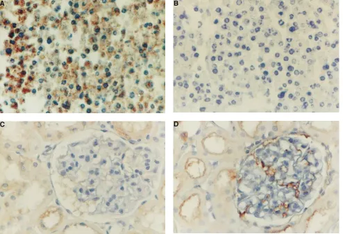
We could not detect RANTES protein expression using a polyclonal or a monoclonal antibody in Carnoy’s solution or buffered formaldehyde, suggesting that the specific epitopes are destroyed by the fixation process (data not shown). Frozen sections exhibited unsatisfactory preservation of morphology. Thus, we had to rely on glutaraldehyde perfusion-fixed renal tissue which, in combination with biotin–tyramide staining en-hancement, offers an excellent sensitivity and exact morpho-logical localization of staining. To test further the specificity of the used monoclonal anti-RANTES antibody, we used fibro-blasts challenged with TNF-a as a control system. As shown in Fig. 10 A, treatment of NIH-3T3 fibroblasts with TNF-a for 24 h induced a strong cytoplasmic staining for RANTES protein. In contrast, no specific staining was observed in TNF-a–treated cells when the used antibody was preabsorbed with recombi-nant RANTES, indicating the specificity of this antibody (Fig. 10 B). Although there was some limited tubular staining, glomeruli were only occasionally positive for RANTES pro-tein in animals infused with vehicle (Fig. 10 C). A strong
[image:8.612.67.467.56.334.2]spe-cific staining for RANTES protein, however, was observed in ANG II–infused rats (Fig. 10 D). This staining was principally localized to small capillaries and glomerular endothelium with some glomerular epithelial cells showing also a positive reac-tion (Fig. 10 D). Control sections that were stained only with the secondary antibody or irrelevant control sera showed no staining indicating the specificity of the response (data not shown). Quantification of positive glomerular endothelium in 100 glomeruli from a total of three rats in each group revealed
Figure 6. Time course of RANTES mRNA expression. Stimulation of GER with a single dose of 1027 M ANG II induced RANTES
tran-scripts after 6 h, although maximal expression was observed after 24 h of incubation. This blot is representative of three independent exper-iments with similar results.
Figure 7. RANTES protein expression in isolated glomeruli from rats infused with ANG II. (A) Isolated glomeruli (200 mg) from ANG II-infused animals or solvent-infused animals (pooled from four animals in each group) were incubated for 2 h in serum-free DMEM at 378C. After incubation, glomeruli were removed by cen-trifugation, and the supernatants were assayed for chemotactic activ-ity in a modified Boyden chamber. The conditioned medium from ANG II-treated animals revealed a significantly higher chemotactic activity for monocytes compared with the medium that was incubated with glomeruli from control-infused animals. The figure shows pooled data from two series of animal experiments with four rats in each group. Glomeruli from each series were independently incu-bated four times with subsequent chemotactic assay. **P , 0.001 ver-sus control-medium; *P , 0.05 versus control-medium. White bars, control medium; gray bars, ANG II medium. (B) Western blot of 100 mg glomerular protein obtained from ANG II–infused or control-infused rats. The blot was incubated with an anti-RANTES antibody. Molecular weight markers are shown on the right side. A single band of z 8 kD is detectable in the glomerular lysates isolated from
[image:8.612.252.547.58.383.2]an increase from 1% in control-infused rats to 26% in ANG II–treated animals. There was no change in the slight staining of tubular structures by ANG II infusion. Moreover, ANG II– infused animals exhibited significantly more glomerular ED-1– positive cells compared with control-infused rats (Table I). ANG II–infused animals had almost as many ED-1–positive
cells as LPS-infused rats, which served as a positive control (Table I). Oral treatment with the AT2 receptor antagonist PD 123177 reduced this glomerular influx of ED-1–positive cells (Table I).
Discussion
Glomerular influx of M/M is a common feature of immune-mediated glomerular injury (1–3). In addition, glomerular in-filtration with M/M occurs early even in renal diseases that are traditionally considered to be of nonimmune origin, such as di-abetic nephropathy and glomerulosclerosis subsequent to re-nal ablation (4, 5). Release of various cytokines, proteinases, and toxic oxygen species by M/M clearly contributes to the ini-tiation and progression of renal failure in such pathophysiolog-ical situations (6). ACE inhibitors and to some extent ANG II receptor blockers reduce glomerular injury in various models of chronic renal failure (35–38). There exists suggestive evi-dence that these effects are independent of the reduction in systemic blood pressure, and they may not even be associated with a decrease in glomerular capillary pressure (17, 19, 39). Treatment with ACE inhibitors also reduces glomerular and tubulointerstitial infiltration with M/M in different immune and nonimmune models of renal disease (18, 19, 36, 39).
[image:9.612.57.241.58.321.2]Although there is some evidence that other chemotactic factors such as osteopontin or lipid factors may be involved in ANG II–mediated invagination of M/M into the tubulointer-stitium (40), the role of glomerular chemokine expression in the recruitment of M/M is only incompletely understood. We have, for example, recently shown that glomerular MCP-1 ex-pression is enhanced in a model of proliferative glomerulone-phritis (41). This increased MCP-1 expression was associated with an increase in glomerular influx of M/M. In contrast, ex-pression of RANTES occurs later in this model and may not
[image:9.612.55.300.443.671.2]Figure 8. Northern blot analysis of RNA from rats infused for 4 d with ANG II (ANG II) or control-sol-vent (Control). The expression of RANTES mRNA was significantly higher in ANG II–infused animals compared to control-infused rats. No RANTES transcripts were de-tectable in RNA isolated from the rest tissue after glomeruli were re-moved by sieving. Although there was some variation in the intensity of the hybridization signals that may be due to RANTES induction by operative stress, RANTES tran-scripts were always more abundant in the ANG II–infused animals in all three series consisting of four an-imals in each group. This blot is rep-resentative for three independent experiments.
Figure 9. Northern blot analysis of isolated glomeruli from ANG II or control-infused rats treated with PD 123177 in the drinking water. ANG II infusion stimulated expression of RANTES mRNA in iso-lated glomeruli. Oral treatment with the AT2 receptor blocker PD
play a significant role in the early wave of glomerular M/M in-vagination (Wolf et al., unpublished data.)
The present study was undertaken to test the hypothesis that ANG II may influence the expression of the chemokine RANTES in GER. Our data collectively indicate that ANG II induces mRNA and protein expression of RANTES in cul-tured GER. In addition, the induced RANTES is chemotactic for monocytes as measured in a chemotactic assay. The obser-vation is surprising, however, that the signal transduction of ANG II occurs through the AT2 receptors because two differ-ent AT2 receptor antagonists, but not the AT1 receptor blocker losartan, inhibit the ANG II-induced RANTES ex-pression. Almost all effects of ANG II in the kidney are trans-duced by the seven-membrane–spanning, G-protein–coupled AT1 receptors (42, 43). The mechanism of signal transduction through the AT1 receptor is a topic of current research; activa-tion of the AT1 receptor causes phosphorylation of various cel-lular proteins, although the receptor itself has no kinase activ-ity (44). Furthermore, the AT1 receptor couples to multiple other effector systems such as phospholipase C, D, and A2, adenylate cyclase, and ion channels in distinct cells (44). Much less is known, however, about the AT2 receptor and its signal
[image:10.612.58.552.60.397.2]transduction pathways. The recently cloned AT2 receptor also has the putative seven-transmembrane domain structure, but with very little homology with the AT1 receptor (45, 46). The AT2 receptor may be involved in the regulation of intracellular phosphotyrosine phosphatases (47, 48). Although the AT2 re-ceptor is abundantly expressed in the fetal kidney, only 5–10% of ANG II receptors in the adult kidney are of the AT2 sub-Figure 10. Immunohistochemistry for RANTES protein. (A) NIH-3T3 fibroblasts stimulated for 24 h with TNF-a revealed positive cytoplasmic staining for RANTES protein using a monoclonal antihuman RANTES antibody. (B) Preabsorption of the antibody with recombinant RANTES, and subsequent staining of TNF-a–challenged 3T3 cells revealed no positive staining indicating the specificity of this antibody. (C) Kidney section of animals infused with solvent for 4 d revealed only a slight staining of tubular cells with no detection of RANTES protein in the glomerular tuft. (D) Infusion of ANG II for 4 d, however, induced RANTES staining which appeared to be principally localized in glomerular endothelial cells and small capillaries. See Methods for details. 3200.
Table I. ED-1–Positive Cells per Glomerular Cross-section 4 d After Infusion
Infusion No. of cells
Control (solvent) 0.2560.04
Control (solvent) and oral treatment with PD 123177 0.1860.04
ANG II 0.4660.03*
ANG II and oral treatment with PD 123177 0.2460.02‡
LPS 0.4860.02*
A total of 20 glomeruli were counted from each individual animal (n 5 3
for LPS infusion, n 5 5 for all other groups). *P , 0.01 versus control
[image:10.612.315.557.617.703.2]type (49). Autoradiography studies demonstrate expression mainly in preglomerular vessels, the renal capsule, and only lit-tle in the glomerular tufts (42, 43, 50). Which effects of ANG II in the kidney are actually transduced by AT2 receptors is cur-rently incompletely understood, but it has been recently de-scribed that activation of AT2 receptors blunt pressure-induced natriuresis (49, 51). Our GER express AT1 and AT2 receptors with the AT1 subtype being much more abundant. This is in good agreement with recent studies demonstrating an AT1 to AT2 ratio of 80:20 in cultured rat coronary endothe-lial cells (52). We have previously demonstrated that ANG II stimulates a slight, but significant proliferation of GER, a re-sponse that is solely mediated by AT1 receptors, and is associ-ated with stimulation of mitogen-activassoci-ated protein kinases (22). In contrast to observations in cultured microvascular cells from coronary arteries (52), we do not observe an inhibition of proliferation associated with activation of the AT2 receptor. So far, the only known effect that is transduced through AT2 re-ceptors in GER is the stimulation of RANTES expression. The ANG II–stimulated increase in RANTES mRNA and protein is specific for GER because syngeneic mesangial cells showed no response after treatment with ANG II. Whether this failure of RANTES induction in mesangial cells is due to the absence of AT2 receptors in these cells remains to be inves-tigated.
There exists some evidence that angiotensin III may be chemotactic for polymorphonuclear neutrophils, but chemoat-traction for M/M has not been tested (53). Moreover, it has been reported that ANG II activates M/M under certain cir-cumstances (54, 55). In our experiments, ANG II alone was not chemotactic for human monocytes and the chemotactic activity of conditioned supernatants from ANG II–treated GER was abolished in the presence of the neutralizing anti-RANTES antibody, strongly indicating that induction of RANTES is necessary, and ANG II per se is not chemotactic for monocytes in our experimental setting. Interestingly, a pre-vious study reported release of a neutrophil chemoattractant factor from human and bovine arterial endothelial cells after treatment with ANG II (56). This factor was not an eicosanoid or a phospholipid (56). Although the nature of this factor is not known, it is possible that some of the chemoattractant ac-tivity may be due to chemokine induction by ANG II.
Our data from our in vivo infusion experiments clearly demonstrate that glomeruli from ANG II–infused rats pro-duce more RANTES compared with control-infused rats. We selected a short-time infusion period with lower concentra-tions of ANG II than previously used because we wanted to study early changes before structural alterations such as glom-erulosclerosis or interstitial fibrosis occurred (34). Since we studied isolated glomeruli, we do not currently know whether glomerular endothelial cells are indeed the only source of the ANG II–mediated increase in RANTES mRNA production. We tried very hard to evaluate RANTES protein expression by immunhistochemistry through the application of different approaches. Unfortunately, routinely applied fixation proce-dures such as Carnoy’s solution or formaldehyde fixation also destroyed the specific RANTES epitopes. Thus, we had to rely on glutaraldehyde-perfused kidneys with some particular modifications of the staining procedure to localize RANTES protein expression. This method was highly sensitive and re-producible in our hands. Additional experiments with preab-sorbed antibody on TNF-a–treated fibroblasts confirmed the
specificity of the staining method. Immunohistochemistry ex-periments using a specific anti-RANTES antibody revealed a positive glomerular staining in ANG II–infused rats, whereas no RANTES protein expression could be detected in control-infused animals. The positive staining may be principally local-ized to small capillaries and glomerular endothelium. We can-not, however, rule out that other glomerular structures such as epithelial cells may also show positive staining for RANTES, although quantification revealed that this staining did not in-crease in ANG II–infused rats. Along this line, it may be very difficult to demonstrate convincingly chemokine expression by immunohistochemistry since the small molecules may be cleared rapidly and removed from the tissue. Moreover, only very lim-ited information hitherto exists in the literature demonstrating positive renal immunohistochemistry for RANTES (57, 58). The positive RANTES immunohistochemistry in ANG II– infused animals does not necessarily imply that RANTES pro-tein is produced there, because circulating RANTES may sim-ply adhere specifically or nonspecifically to the endothelium. Since the glomerular localization of M/M was different from RANTES staining, however, it is not likely that a primary in-flux of M/M may subsequently release RANTES protein. Treatment in vivo with the AT2 receptor blocker PD 123177 partly abolished the ANG II–stimulated increase in RANTES expression in isolated glomeruli without influencing the sys-tolic blood pressure. In addition, we have obtained preliminary evidence from pilot experiments that losartan, although lower-ing the ANG II–mediated increase in blood pressure, does not influence glomerular RANTES expression (our unpublished observations).
Glomerular ED-1 positive cells were significantly increased in rats infused for 4 d with ANG II compared with controls. The AT2 receptor blocker PD 123177 abolished this glomeru-lar influx of M/M. Although we cannot totally rule out that ANG II per se in the absence of RANTES induction may stim-ulate glomerular M/M influx, these data combined with the ob-served RANTES expression are highly suggestive that the ANG II–mediated glomerular infiltration of M/M depends on RANTES expression. Interference with RANTES expression using neutralizing antibodies or antisense technology, how-ever, is necessary to demonstrate unequivocally a causal rela-tionship between ANG II, RANTES expression, and glomeru-lar M/M influx. In comparison with our studies, Johnson et al. (34) infused a higher dose ANG II into rats for 14 days and ob-served a higher systolic blood pressure associated with mild tu-bulointerstitial fibrosis and interstitial monocyte infiltration but only little glomerular infiltration of M/M. Since glomerular infiltration of M/M is a dynamic process, it is possible that ear-lier time-points after ANG II infusion such as selected in our study may show a higher degree of glomerular M/M infiltra-tion.
possible that ANG II–stimulated endothelial cells may synthe-size chemoattracting factors such as RANTES, which attract M/M into the glomerular tuft.
Dietary protein restriction reduces the glomerular influx of M/M in several models of immune- and non-immune-medi-ated glomerular disease (60). Since reduction of dietary pro-tein intake also downregulates renin transcription (61), it is tempting to speculate that a decrease in intrarenal ANG II concentration which in turn reduces RANTES expression is responsible for some of the immune-protective effects associ-ated with protein restriction.
Taking into consideration that RANTES induction in GER is mediated by AT2 receptors, ACE inhibitors or a combina-tion of AT1 and AT2 receptor antagonists may be more effec-tive in preventing chemokine induction than an AT1 receptor blocker only.
Acknowledgments
We thank R. Schroeder and U. Kneissler for excellent technical help. This study was supported by grants from the Deutsche For-schungsgemeinschaft (Sta 193/6-3, Wo 460/2-2). Additional support was obtained from National Institutes of Health grants DK-07006, DK-44513, and DK-45191.
References
1. Cattell, V. 1994. Macrophages in acute glomerular inflammation. Kidney Int. 45:945–952.
2. Nikolic-Paterson, D.J., H.Y. Lan, P.A. Hill, and R.C. Atkins. 1994. Mac-rophages in renal injury. Kidney Int. Suppl. 45:S79–S82.
3. Van Goor, H., G. Ding, D. Kees-Folts, J. Grond, G.F. Schreiner, and J.R. Diamond. 1994. Biology of disease. Macrophages and renal disease. Lab. In-vest. 71:456–464.
4. Floege, J., M.W. Burns, C.E. Alpers, A. Yoshimura, P. Pritzl, K. Gordon, R.A. Seifert, D.F. Bowen-Pope, W.G. Couser, and R.J. Johnson. 1992. Glomer-ular cell proliferation and PDGF expression precede glomerulosclerosis in the remnant kidney model. Kidney Int. 41:297–309.
5. Young, B.A., R.J. Johnson, C.E. Alpers, E. Eng, K. Gordon, J. Floege, and W.G. Couser. 1995. Cellular events in the evolution of experimental dia-betic nephropathy. Kidney Int. 47:935–944.
6. Schreiner, G.F. 1991. The role of the macrophages in glomerular injury. Sem. Nephrol. 11:268–275.
7. Nathan, C.F. 1987. Secretory products of macrophages. J. Clin. Invest. 79: 319–326.
8. Oppenheim, J.J., C.O.C. Zachariae, N. Mukaida, and K. Matsushima. 1991. Properties of the novel proinflammatory supergene intercrine cytokine family. Annu. Rev. Immunol. 9:617–648.
9. Schlöndorff, D. 1995. The role of chemokines in the initiation and pro-gression of renal disease. Kidney Int. 47 (Suppl. 49):S44–S47.
10. Wenzel, U.O., and H.E. Abboud. 1995. Chemokines and renal disease. Am. J. Kidney Dis. 26:982–994.
11. Huber, A.R., S.L. Kunkel, R.F. Todd, and S.J. Weiss. 1991. Regulation of transendothelial neutrophil migration by endogenous interleukin-8. Science (Lond.). 4:99–102.
12. Ro, A. 1992. Endothelial cell binding of NAP-1/Il-8: role in neutrophil emigration. Immunol. Today. 13:291–294.
13. Schall, T.J. 1991. Biology of the RANTES/SIS cytokine family. Cyto-kine. 3:165–183.
14. Wolf, G., B. Luckow, and D. Schlöndorff. 1995. Molecular biology and function of the chemoattractant cytokines RANTES and CSF-1. Kidney func-tion in health and disease. In Molecular Nephrology. D. Schlöndorff and J.V. Bonventre, editors. Marcel Dekker, Inc., New York. 673–679.
15. Wolf, G., S. Aberle, F. Thaiss, P.J. Neilson, A.M. Krensky, E.G., Neil-son, and R.A.K. Stahl. 1993. TNFa induces expression of the chemoattractant cytokine RANTES in cultured mouse mesangial cells. Kidney Int. 44:795–804.
16. Heeger, P., G. Wolf, C. Meyers, M.J. Sun, S.C. O’Farrel, A.M. Krensky, and E.G. Neilson. 1992. Isolation and characterization of cDNA from renal tu-bular epithelium encoding murine RANTES. Kidney Int. 41:220–225.
17. Wolf, G., and E.G. Neilson. 1993. Angiotensin II as a renal growth fac-tor. J. Am. Soc. Nephrol. 3:1531–1540.
18. Jardine, A.G. 1995. Angiotensin II and glomerulonephritis. J. Hyper-tens. 13:487–493.
19. Egido, J. 1996. Vasoactive hormones and renal sclerosis. Kidney Int. 49: 578–597.
20. Klahr, S., S. Ishidoya, and J. Morrissey. 1995. Role of angiotensin II in the tubulointerstitial fibrosis of obstructive nephropathy. Am. J. Kidney Dis. 26: 141–146.
21. Takahashi, M., J.I. Masuyama, U. Ikeda, S.I. Kitagawa, T. Kasahara, M. Saito, S. Kano, and K. Shimada. 1995. Suppressive role of endogenous endothe-lial monocyte chemoattractant protein-1 on monocyte transendotheendothe-lial migra-tion in vitro. Arterioscler. Thromb. Vasc. Biol. 15:629–636.
22. Wolf, G., F.N. Ziyadeh, G. Zahner, and R.A.K. Stahl. 1996. Angiotensin II is mitogenic for cultured rat glomerular endothelial cells. Hypertension (Dal-las). 27:897–905.
23. Stahl, R.A.K., M. Paravicini, and P. Schollmeyer. 1984. Angiotensin II stimulates prostaglandin E2 and 6-keto PGF1 formation by isolated human glomeruli. Kidney Int. 26:30–34.
24. Chomczynski, P., and N. Sacchi. 1987. Single-step method of RNA isola-tion by acid guanidinium thiocyanate-phenol-chloroform extracisola-tion. Anal. Bio-chem. 132:6–13.
25. Wolf, G., G. Zahner, R. Schroeder, and R.A.K. Stahl. 1996. Transform-ing growth factor beta mediates the angiotensin II-induced stimulation of col-lagen type IV synthesis in cultured murine proximal tubular cells. Nephrol. Dial. Transplant. 11:263–269.
26. Kobayashi, S., J. Ohnishi, Y. Nibu, S. Nishimatsu, S. Umemura, M. Ishii, K. Murakami, and H. Miyazaki. 1995. Cloning of the rat angiotensin II type 2 receptor gene and identification of its functional promoter region. Biochim. Biophys. Acta. 1262:155–158.
27. Wolf, G., E. Mueller, R.A.K. Stahl, and F.N. Ziyadeh. 1993. Angio-tensin II-induced hypertrophy of cultured murine proximal tubular cells is me-diated by endogenous transforming growth factor-b. J. Clin. Invest. 92:1366– 1372.
28. Vissers, M.C., S.A. Jester, and J.C. Fantome. 1988. Rapid purification of human peripheral blood monocytes by centrifugation through Ficoll-hypaque and Sepracell-MN. J. Immunol. Methods. 110:203–207.
29. Wenzel, U.O., B. Fourqueray, G. Grandaliano, Y.S. Kim, C. Karamit-sos, A.J. Valente, and H.E. Abboud. 1995. Thrombin regulates expression of monocyte chemoattractant protein-1 in vascular smooth muscle cells. Circ. Res. 77:503–509.
30. Leonard, E.J., T. Yoshimura, A. Skeel, and R. Goodwin. 1990. A multi-well cell settling and adherence chamber for morphology and differential counting. Biotechniques. 9:684–689.
31. Stoscheck, C.M. 1990. Quantitation of protein. Methods Enzymol. 182: 50–68.
32. Marfaing-Koka, A., O. Devergne, G. Gorgone, A. Portier, T.J. Schall, P. Galanaud, and D. Emilie. 1995. Regulation of the production of RANTES chemokine by endothelial cells. Synergistic induction by IFN-g plus TNF-a and inhibition by IL-4 and IL-13. J. Immunol. 154:1870–1878.
33. Maderazo, E.G., and P.A. Ward. 1986. Leukocyte chemotaxis. In Man-ual of Clincial Laboratory Immunology. N.R. Rose, H. Friedman, and J.L. Fa-hey, editors. American Society of Microbiology, Washington, DC. pp. 290–294. 34. Johnson, R.J., C.E. Alpers, A. Yoshimura, D. Lombardi, P. Pritzl, J. Floege, and S.M. Schwartz. 1992. Renal injury from angiotensin II-mediated hypertension. Hypertension (Dallas). 19:464–474.
35. Lafayette, R.A., G. Mayer, S.K. Park, and T.W. Meyer. 1992. Angio-tensin II receptor blockade limits glomerular injury in rats with reduced renal mass. J. Clin. Invest. 90:766–771.
36. Diamond, J.R., and S. Anderson. 1990. Irreversible tubulointerstitial damage associated with chronic aminonucleoside nephrosis. Amelioration by angiotensin I converting enzyme inhibition. Am. J. Pathol. 137:1323–1332.
37. Thaiss, F., C. Haas, U. Helmchen, U. Wenzel, and R.A.K. Stahl. 1996. Angiotensin-converting enzyme inhibition in experimental in-situ immune complex glomerulonephritis: influence on renal function, proteinuria, and mor-phology. Nephrol. Dial. Transplant. 11:40–46.
38. Lee, S.K., S.Y. Jin, D.C. Han, S.D. Hwang, and H.B. Lee. 1993. Effects of delayed treatment with enalapril and/or lovastatin on the progression of glomerulosclerosis in 5/6 nephrectomized rats. Nephrol. Dial. Transplant. 8: 1338–1343.
39. Harris, R.C., and M. Martinez-Maldonado. 1995. Angiotensin II-medi-ated renal injury. Miner. Electrolyte Metab. 21:328–335.
40. Giachelli, C.M., R. Pichler, D. Lombardi, D.T. Denhardt, C.E. Alpers, S.M. Schwartz, and R.J. Johnson. 1994. Osteopontin expression in angiotensin II-induced tubulointerstitial nephritis. Kidney Int. 45:515–524.
41. Stahl, R.A.K., F. Thaiss, M. Disser, U. Helmchen, K. Hora, and D. Schlöndorff. 1993. Increased expression of monocyte chemoattractant protein-1 in anti-thymocyte antibody-induced glomerulonephritis. Kidney Int. 44:1036– 1047.
42. Zhuo, J., K. Song, P.J. Harris, and F.A.O. Mendelsohn. 1992. In vitro autoradiography reveals predominantly AT1 angiotensin II receptors in rat kid-ney. Renal Physiol. Biochem. 15:231–239.
43. Edwards, R.M., and N. Aiyar. 1993. Angiotensin II receptor subtypes in the kidney. 1993. J. Am. Soc. Nephrol. 3:1643–1652.
Saavedra, and P.B. Timmermans, editors. Plenum Publishing Corporation, New York. 1–15.
45. Mukoyama, M., M. Nakajama, M. Horiuchi, H. Sasamura, R.E. Pratt, and V. Dzau. 1993. Expression cloning of type 2 angiotensin II receptor reveals a unique class of seven-transmembrane receptors. J. Biol. Chem. 268:24539– 24542.
46. Kambayashi, Y., S. Bardhan, K. Tahahashi, S. Tsuzuki, H. Inui, T. Ha-makubo, and T. Inagami. 1993. Molecular cloning of a novel angiotensin II re-ceptor isoform involved in phosphotyrosine phosphatase inhibition. J. Biol. Chem. 268:24543–24546.
47. Brechler, V., S. Reichlin, M. De Gasparo, and S.P. Bottari. 1994. Angio-tensin II stimulates protein tyrosine phosphatase activity through a G-protein independent mechanism. Recept. Chann. 2:89–97.
48. Takahasi, K., S. Bardhan, Y. Kambayashi, H. Shirai, and T. Inagami. 1994. Protein tyrosine phosphatase inhibition by angiotensin II in rat pheochro-mocytoma cells through type 2 receptor, AT2. Biochem. Biophys. Res. Com-mun. 198:60–66.
49. Ardaillou, R. 1994. Angiotensin II type II receptor. Does it have a role in the normal or diseased kidney? Nephrol. Dial. Transplant. 9:1364–1365.
50. Sechi, L.A., E.F. Grady, C.A. Griffin, J.E. Kalinyak, and M. Scham-belan. 1992. Distribution of angiotensin II receptor subtypes in rat and human kidney. Am. J. Physiol. 262:F236–F240.
51. Lo, M., K.L. Liu, P. Lantelme, and J. Sassard. 1995. Subtype 2 of angio-tensin II receptors controls presure-natriuresis in rats. J. Clin. Invest. 95:1394– 1397.
52. Stoll, M., U.M. Steckelings, M. Paul, S.G. Bottari, R. Metzger, and T. Unger. 1995. The angiotensin AT2-receptor mediates inhibition of cell prolifer-ation in coronary endothelial cells. J. Clin. Invest. 95:651–657.
53. Yamamoto, Y., T. Yamaguchi, M. Shimamura, and T. Hazato. 1993. Angiotensin III is a new chemoattractant for polymorphonuclear cells. Bio-chem. Biophys. Res. Commun. 193:1038–1043.
54. Weinstock, J.V., and J.T. Kassab. 1984. Angiotensin II stimulation of granuloma macrophages phagocytosis and actin polymerization in murine schistosomiasis mansoni. Cell. Immunol. 89:46–54.
55. Hahn, A.W., U. Jonas, F.R. Bühler, and T.J. Resink. 1994. Activation of human peripheral monocytes by angiotensin II. FEBS Lett. 347:178–180.
56. Farber, H.W., D.M. Center, S. Rounds, and S.M. Danilov. 1990. Com-ponents of the angiotensin system cause release of a neutrophil chemoattrac-tant from cultured bovine and human endothelial cells. Eur. Heart J. 11 (Suppl.B):100–107.
57. Pattison, J., P.J. Nelson, P. Huie, I. von Leuttichau, G. Farshid, R.K. Sibley, and A.M. Krensky. 1994. RANTES chemokine expression in cell-medi-ated transplant rejection of the kidney. Lancet. 343:209–211.
58. von Luettichau, I., P.J. Nelson, J.M. Pattison, M. van de Rijn, P. Huie, R. Warnke, C.J. Wiedermann, R.A.K. Stahl, R.K. Sibley, and A.M. Krensky. 1996. RANTES chemokine expression in diseased and normal human tissues. Cytokine. 8:89–98.
59. Lee, L.K., T.W. Meyer, A.S. Pollock, and D.H. Lovett. 1995. Endothe-lial cell injury initiates glomerular sclerosis in the rat remnant kidney. J. Clin. Invest. 96:953–964.
60. Diamond, J.R. 1990. Effects of dietary interventions on glomerular pathophysiology. Am. J. Physiol. 258:F1–F8.