Corrigendum
(October 2005)
TWEAK induces liver progenitor cell
proliferation
Aniela Jakubowski, … , D. Montgomery Bissell, Linda C.
Burkly
J Clin Invest.
2005;
115(9)
:2330-2340.
https://doi.org/10.1172/JCI23486
.
Progenitor (“oval”) cell expansion accompanies many forms of liver injury, including alcohol
toxicity and submassive parenchymal necrosis as well as experimental injury models
featuring blocked hepatocyte replication. Oval cells can potentially become either
hepatocytes or biliary epithelial cells and may be critical to liver regeneration, particularly
when hepatocyte replication is impaired. The regulation of oval cell proliferation is
incompletely understood. Herein we present evidence that a TNF family member called
TWEAK (TNF-like weak inducer of apoptosis) stimulates oval cell proliferation in mouse
liver through its receptor Fn14. TWEAK has no effect on mature hepatocytes and thus
appears to be selective for oval cells. Transgenic mice overexpressing TWEAK in
hepatocytes exhibit periportal oval cell hyperplasia. A similar phenotype was obtained in
adult wild-type mice, but not Fn14-null mice, by administering TWEAK-expressing
adenovirus. Oval cell expansion induced by 3,5-diethoxycarbonyl-1,4-dihydrocollidine
(DDC) was significantly reduced in Fn14-null mice as well as in adult wild-type mice with a
blocking anti-TWEAK mAb. Importantly, TWEAK stimulated the proliferation of an oval cell
culture model. Finally, we show increased Fn14 expression in chronic hepatitis C and other
human liver diseases relative to its expression in normal liver, which […]
Research Article
Hepatology
Find the latest version:
TWEAK induces liver progenitor
cell proliferation
Aniela Jakubowski,1 Christine Ambrose,1 Michael Parr,1 John M. Lincecum,1 Monica Z. Wang,1
Timothy S. Zheng,1 Beth Browning,1 Jennifer S. Michaelson,1 Manfred Baestcher,2
Bruce Wang,3 D. Montgomery Bissell,3 and Linda C. Burkly1
1Departments of Exploratory Science, Discovery Biology, and Validation Biology, Biogen Idec Inc., Cambridge, Massachusetts, USA. 2Department of Molecular and Medical Genetics, Oregon Health Sciences University, Portland, Oregon, USA.
3Division of Gastroenterology and Liver Center, University of California San Francisco, San Francisco, California, USA.
Progenitor (“oval”) cell expansion accompanies many forms of liver injury, including alcohol toxicity and
submassive parenchymal necrosis as well as experimental injury models featuring blocked hepatocyte
repli-cation. Oval cells can potentially become either hepatocytes or biliary epithelial cells and may be critical to
liver regeneration, particularly when hepatocyte replication is impaired. The regulation of oval cell
prolif-eration is incompletely understood. Herein we present evidence that a TNF family member called TWEAK
(TNF-like weak inducer of apoptosis) stimulates oval cell proliferation in mouse liver through its receptor
Fn14. TWEAK has no effect on mature hepatocytes and thus appears to be selective for oval cells. Transgenic
mice overexpressing TWEAK in hepatocytes exhibit periportal oval cell hyperplasia. A similar phenotype was
obtained in adult wild-type mice, but not Fn14-null mice, by administering TWEAK-expressing adenovirus.
Oval cell expansion induced by 3,5-diethoxycarbonyl-1,4-dihydrocollidine (DDC) was significantly reduced
in Fn14-null mice as well as in adult wild-type mice with a blocking anti-TWEAK mAb. Importantly, TWEAK
stimulated the proliferation of an oval cell culture model. Finally, we show increased Fn14 expression in
chronic hepatitis C and other human liver diseases relative to its expression in normal liver, which suggests a
role for the TWEAK/Fn14 pathway in human liver injury. We conclude that TWEAK has a selective mitogenic
effect for liver oval cells that distinguishes it from other previously described growth factors.
Introduction
Compensatory epithelial hyperplasia following liver injury involves 1 of 3 different cellular compartments, depending on the nature of the injury. With simple loss of hepatocyte mass, the paradigm of which is partial hepatic resection, hepatocytes within the remaining lobes proliferate. When the injury consists of biliary obstruction, proliferation of the biliary epithelium is prominent. Injury that both reduces hepatocyte mass and impairs hepatocyte proliferation elicits the appearance of “oval” cells. Oval cells arise from the portal area of the lobule, which is the location of the putative hepatic stem cell. These cells are viewed as a progenitor cell type because of their known ability to differenti-ate into either hepatocytes or biliary epithelial cells (BECs) (1). With the development of improved techniques for detecting oval cells in liver sections, it has become apparent that the oval cell compartment participates in a range of injuries to human liver (2) and may be critical to the survival of patients with acute liver failure (3, 4). There are many rodent models for oval cell prolif-eration. A commonly used model involves treating rats with the carcinogen 2-acetylaminofluorene (2-AAF) followed by a growth stimulus in the form of partial hepatectomy (AAF/PH) or carbon
tetrachloride (AAF/CCl4). This results in impressive growth of a population of small ovoid cells with a large ratio of nucleus to cytoplasm (5–7). These oval cells express phenotypic markers of both immature hepatocytes and BECs, such as α-fetoprotein and cytokeratins 8 and 18 respectively (7–9).
Hepatic regeneration after PH is a multistep process involving numerous cytokines and growth factors. HGF, EGF, and TGF-α
are viewed as primary stimuli but require that hepatocytes be primed by other soluble factors, including TNF and IL-6 (1, 10). Some of the same factors have been implicated in oval cell pro-liferation (1, 9, 11–15). However, their effects are not specific for oval cells, which raises the question of whether they are both necessary and sufficient.
TNF-like weak inducer of apoptosis (TWEAK) (16) is a member of the TNF-α family of growth regulators that has weak apoptotic activity for certain human tumor cell lines (17). Its mRNA is broad-ly expressed in normal tissues, including the liver. Studies using an array of cell types have indicated that its putative actions are broad and include regulation of cell survival, proliferation and migration, and induction of proinflammatory cytokines and chemokines (18). A TWEAK receptor known as Fn14, a member of the TNF receptor family, has been identified (19, 20). Its expression is restricted to epi-thelial and mesenchymal cell types. In the liver of the growing new-born, Fn14 is highly expressed (21). It is minimally expressed in the adult except during injury and repair, such as following PH in mice and hepatocellular carcinoma (HCC) in mice and humans (20). In the course of studies exploring the biological role of TWEAK, we prepared a transgenic mouse expressing the gene under control of the α1 antitrypsin (AAT) promoter, which is primarily expressed in the liver (22). The livers in these mice exhibited a striking periportal
Nonstandard abbreviations used: AAF, acetylaminofluorene; AAT, α1 antitrypsin; ALT, alanine aminotransferase; BEC, biliary epithelial cell; DDC, 3,5-diethoxycar-bonyl-1,4-dihydrocollidine; HCC, hepatocellular carcinoma; NASH, nonalcoholic steatohepatitis; NRC-1, normal rat cholangiocyte–1; Ntg, nontransgenic; PCNA, proliferating cell nuclear antigen; PH, partial hepatectomy; TWEAK, TNF-like weak inducer of apoptosis.
Conflict of interest: The authors have declared that no conflict of interest exists.
outgrowth of oval cells but otherwise appeared healthy. The find-ings were confirmed by transiently overexpressing TWEAK in the adult mouse liver and by chemically inducing oval cell prolifera-tion in the mouse. In the latter model, anti-TWEAK mAb blocked oval cell expansion. TWEAK overexpression in these models had no detectable effect on hepatocyte proliferation. Moreover, the TWEAK receptor, Fn14, was present on ductal structures and oval cells. In addition, TWEAK was shown to be mitogenic in an oval cell culture model. We concluded that TWEAK initiates prolifera-tion of oval cells and is specific to this populaprolifera-tion. The potential relevance to human liver disease is supported by additional data demonstrating increased Fn14 expression on bile ducts and duct-like structures in human cirrhotic liver.
Results
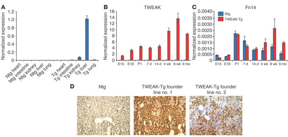
TWEAK transgenic mice. Transgenic mice carrying full-length murine TWEAK cDNA under the murine AAT promoter (TWEAK-Tg) were prepared. Two lines were developed. TWEAK transgene expression was predominantly hepatic, but low levels were also detected in the kidney by quantitative PCR (Figure 1A). The developmental expression of TWEAK and Fn14 was also analyzed by quantitative PCR. In nontransgenic (Ntg) mice, TWEAK expression was very low at all tested times. In contrast, in TWEAK-Tg mice, TWEAK was detectable at low levels as early as E15.5, and its expression continuously increased, reaching high levels in the adult (Figure 1B). In both TWEAK-Tg and Ntg animals, Fn14 expression only reached significant levels begin-ning at birth: it remained relatively high in adult and older trans-genic mice but decreased in the age-matched Ntg mice, which suggests that it is subject to ligand regulation (Figure 1C).
One founder line strongly expressed TWEAK protein in the liver in hepatocytes and focally and weakly in the kidney (Figure 1D); the protein was not detected in other organs. The second found-er line exhibited a weakfound-er expression of TWEAK protein in the liver. Circulating TWEAK protein at levels of 8–25 ng/ml was detected only in the high-expressing line.
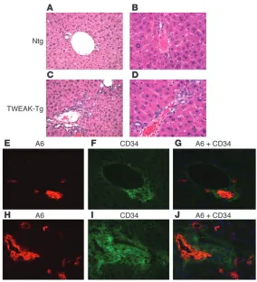
Transgenic TWEAK overexpression results in oval cell proliferation. An increased number of oval cells, defined as small cells with an oval nucleus and scant cytoplasm, spreading from the periportal region was observed in TWEAK-Tg mice as compared with Ntg mice. This phenotype was observed as early as 2 weeks of age (data not shown). By 8 weeks and thereafter to 5–8 months, TWEAK-Tg livers exhib-ited a relatively high number of cells organized either in single file or in duct-like structures in the periportal region and extending into the liver lobule. (Figure 2, C and D). The majority of the duct-like structures were irregularly shaped with a poorly defined lumen. The livers of Ntg littermates were normal, each portal tract having 1 or 2 well-formed bile ducts (Figure 2, A and B).
[image:3.585.45.536.76.310.2]We used immunohistochemical staining of serial sections to fur-ther characterize the proliferating cells. As in previously published work, A6 antigen (23, 24) and the hematopoietic marker CD34 (25) were expressed by both proliferating oval cells and normal BECs (26). In control mice, A6 and CD34 coexpression was detect-able only in bile ducts (Figure 2, E–G), with CD34 being present also in the portal vein endothelium (data not shown). In contrast, TWEAK-Tg livers exhibited an increased number of periportal A6- and CD34-expressing cells (Figure 2, H and I), with some cells dis-playing both markers (Figure 2J). The data indicate that TWEAK-Tg mice have increased numbers of oval cells based on their morphol-ogy, location, and marker expression. The immaturity of the cells
Figure 1
is inferred from the fact that most are not organized as ductular structures; however, the latter were also present, suggesting that a range of maturation states may be present.
TWEAK is proapoptotic in some settings, which raises the pos-sibility that the observed proliferation of oval cells was secondary to hepatocyte death. To test this, we examined proliferation and apoptosis in TWEAK-Tg relative to Ntg livers. Only periportal oval cells and BECs exhibited significantly increased proliferation in TWEAK-Tg relative to that in Ntg mice (Figure 3A) while hepato-cyte proliferation was similar in both (Figure 3B). The increased proliferation of oval cells in the TWEAK-Tg mice was evident as early as 2 weeks of age and persisted thereafter although it decreased somewhat with age. Cell death was examined with TUNEL stain-ing (Figure 3, C and D). At 2 weeks and 8 weeks, when oval cell proliferation was most evident, there was no difference in hepato-cyte apoptosis between the TWEAK-Tg and Ntg mice (Figure 3D). At 7 months, the TWEAK-Tg liver showed an increase in apoptotic hepatocytes relative to a very low rate of apoptosis in age-matched controls. The significance of this finding is uncertain. Interestingly, at early time points, oval cell apoptosis was increased (Figure 3C), suggesting that increased proliferation in TWEAK-Tg mice is accompanied by a detectable increase in cell turnover. Finally,
TWEAK-Tg mice had normal serum transaminases and no detectable inflammation or bile deposits (data not shown). Taken together, the data indicate that the proliferative phenotype is not due to an impaired hepa-tocyte response to injury.
Fn14 expression is associated with BECs and periportal cells. If oval cells respond directly to TWEAK, they pre-sumably express the TWEAK receptor, Fn14. This was examined by in situ hybridization with both embryo and adult livers. At E18.5 both transgenic (Figure 4E) and control mice (Figure 4A) expressed similar levels of Fn14 mRNA in the ductal plate cell layers surround-ing the portal mesenchyme from which bile ductules emerge (27). Low levels of Fn14 were also uniformly expressed in the parenchyma. Fn14 expression peaked in newborn mice, being slightly increased in TWEAK-Tg mice (Figure 4F) relative to Ntg littermates (Figure 4B) and was also expressed in BCEs and surrounding peri-portal cells (Figure 4, B and F), with scattered and gen-erally lower expression in the parenchyma. In the adult livers of both transgenic and control mice, Fn14 expres-sion was sharply diminished. In Ntg adult mice, it was detectable mostly in BECs, in a few periportal cells, and at a very low level in the parenchyma (Figure 4, C and D). In contrast, in adult TWEAK-Tg liver, Fn14 expression was strikingly elevated in the region of oval cell hyper-plasia (Figure 3, G and H).
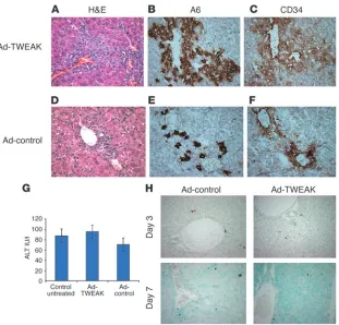
Role of TWEAK in adult mice. As shown in Figure 1B, TWEAK transgene mRNA expression is driven by the AAT promoter, is detectable as early as day 15 of fetal development, and, as previously reported, increases rap-idly through day 18 (28). This raises the possibility that TWEAK exerts its effects mainly in utero rather than in the adult. This was explored in 2 different models. First we transiently overexpressed TWEAK in normal adult mouse liver using an adenoviral vector (Ad-TWEAK). Control mice received an equivalent number of viral particles containing GFP and lacking TWEAK cDNA (Ad-control), and transduction was confirmed by GFP expression in the liver (data not shown). Seven days after Ad-TWEAK adminis-tration, the liver exhibited a remarkable proliferation of oval cells, some of which were organized in duct-like structures (Figure 5A). The identity of the cells was determined both by morphological criteria and by the coexpression of A6 and CD34 markers (Figure 5, B and C). Mice receiving the control adenovirus showed a minimal amount of ductal proliferation (Figure 5, D–F).
Adenoviruses caused a mild inflammatory response and limited hepatocyte damage that peaked by day 7. Since TWEAK was report-ed to induce apoptosis in some systems (29–32), we wishreport-ed to rule out the possibility that TWEAK and an adenoviral vector could act synergistically to enhance hepatocyte damage and thus stimulate oval cell proliferation. Therefore, we assessed serum alanine amino-transferase (ALT) levels and apoptosis in the liver both prior to and at the peak of the inflammatory response. We found normal ALT values and very few apoptotic hepatocytes in both Ad-TWEAK– and Ad-control–treated mice prior to day 7 (Figure 5, G and H).
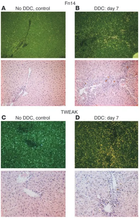
[image:4.585.42.326.80.392.2]The second model we used to test the role of TWEAK in adult mice was a liver injury model in which oval cell proliferation was induced chemically by 3,5-diethoxycarbonyl-1,4-dihydrocollidine (DDC) feeding. First we asked whether the TWEAK pathway is
Figure 2
expressed in this model. Seven days after the start of DDC feeding, we found that Fn14 expression was markedly upregulated in the reactive portal area (Figure 6B) in contrast to Fn14 expression in control mice (Figure 6A), supporting the possibility that TWEAK pathways play a role in the regulation of these cells. The scattered and low expression of Fn14 in the lobule was similar in DDC-treated and control mice. Expression of TWEAK itself in control mice was present across the lobule, possibly in resident stellate
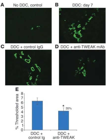
[image:5.585.43.440.78.346.2]and Kupffer cells (Figure 6C). In comparison, in DDC-fed mice, TWEAK was strikingly elevated in reactive portal areas; it appeared also to be slightly higher in the parenchyma (Figure 6D). When DDC-fed mice were treated with a blocking anti-TWEAK mAb, the proliferation of A6-positive oval cells was significantly inhibited (Figure 7, D and E) relative to that in untreated DDC-fed mice and control IgG-treated DDC-fed mice (Figure 7, B and C). Control Ig had no effect on the DDC-induced progenitor cell proliferation
Figure 4
[image:5.585.171.541.472.742.2]Increased Fn14 mRNA expres-sion in the hyperplastic regions of adult TWEAK-Tg relative to Ntg livers. Fn14 mRNA expres-sion was detected by in situ hybridization using a murine Fn14 riboprobe template. (A–D) Ntg liver in (A) E18.5 embryo, (B) newborn, and (C) 8-week-old mouse. Darkfield (top) and brightfield (bottom) views are shown. Magnification, ×250. (D) Ntg 8-week-old liver. Magnifica-tion, ×640. (E–H) TWEAK-Tg liver in (E) E18.5 embryo, (F) newborn, and (G) 8-week-old mouse. Darkfield (top) and brightfield (bottom) views are shown. Magnification, ×250. (H) TWEAK-Tg 8-week-old liver. Magnification, ×640.
Figure 3
compared with the untreated DDC-fed mice (Figure 7, B and C). We conclude from these 2 models that TWEAK causes prolifera-tion with selectivity for oval cells in adult mice.
TWEAK-induced oval cell proliferation is Fn14 dependent. Accumu-lating evidence suggests that Fn14 is the receptor for TWEAK, although some studies have opened the possibility of additional receptors (18). In order to examine the role of Fn14 in mediating the mitogenic effect of TWEAK, we recently generated Fn14-null mice (Fn14-KO), which were fertile and viable with no gross or histologi-cal abnormality. For this purpose, the 2 animal models described above were utilized in the Fn14-KO mice. As described above and shown by A6 staining in Figure 5B, Ad-TWEAK caused a prominent oval cell and ductal hyperplasia in the wild-type mice (Figure 8A) relative to Ad-control–treated wild-type mice, which showed no sig-nificant proliferation (Figure 8C). However, Fn14-KO mice adminis-tered Ad-TWEAK exhibited no hyperplastic phenotype (Figure 8B), and their livers appeared histologically similar to those of Ad-con-trol–treated wild-type or Fn14-KO mice (Figure 8, C–E), indicating that the mitogenic effect of TWEAK on oval cells in the liver is
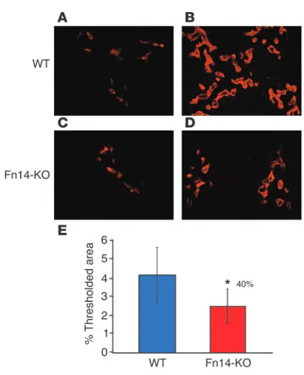
exclu-sively Fn14 dependent. Thus, the numbers of A6-positive cells in the Fn14-KO mice treated with Ad-TWEAK were low and comparable to those in either wild-type or Fn14-KO mice treated with Ad-control, as quantified by MetaMorph software (Figure 9E). We wished to test wheth-er Fn14 also mediates oval cell prolifwheth-eration in the physiological model of DDC feeding. As described above, DDC-fed wild-type mice exhib-ited marked oval cell proliferation (Figure 9B) relative to wild-type mice fed a regular diet (Figure 9A). DDC-fed Fn14-KO mice exhibited 40% less oval cell proliferation than DDC-fed wild-type mice (Figure 9, D and E). This reduc-tion in oval cell proliferareduc-tion was similar to the effect obtained with pharmacological blocking. There was no significant difference between the wild-type and Fn14-KO mice maintained on a regular diet, either histologically or in the amount of A6 expression (Figure 9, A and C). These data provide independent evidence for the role of the TWEAK/Fn14 pathway in the induc-tion of oval cell proliferainduc-tion.
TWEAK is mitogenic for normal rat cholangiocyte–1 cells. As a direct test of the mitogenic effect of TWEAK on bile duct–derived epithelial cells, this factor was added to the medium of normal rat cholangiocyte–1 (NRC-1) cells. The NRC-1 cell line expresses β6 integrin and thus
repre-sents reactive BECs or mature oval cells (data not shown). The cultures were placed in serum-free medium for 24 hours, at which point they were growth-arrested. TWEAK was added and the response assessed by incorporation of BrdU into the nucleus. TWEAK stimulated DNA synthesis in a dose-dependent fashion. At the optimal concentration (100 ng/ml), 40% of the culture was BrdU-positive (Figure 10).
Fn14 is expressed in BECs and duct-like structures in human liver disease. Extending the results in mice to human liver disease, we asked whether Fn14 was expressed in diseases involving progenitor cell prolifera-tion, such as chronic viral hepatitis, alcoholic liver disease, and nonalcoholic fatty liver disease (3, 33). In normal liver, we found that Fn14 is expressed in bile ducts and in some smooth muscle cells surrounding them (Figure 11A). Interestingly, increased Fn14 expression was detected in nonalcoholic steatohepatitis (NASH), alcoholic liver disease, and HCV with HCC (Figure 11, B–D), in which Fn14 was found in the abundant duct-like structures and in bile ducts. In some sections, Fn14 also appeared to be expressed in the fibrotic regions, possibly representing trapped ductal struc-tures and smooth muscle cells.
Discussion
This report shows that TWEAK selectively stimulates prolifera-tion of a hepatic progenitor cell populaprolifera-tion known as oval cells both when overexpressed in transgenic mice and when transiently expressed in adult mice. This mitogenic effect of TWEAK is medi-ated exclusively through Fn14. In addition, the physiological role of TWEAK as a novel stimulator of oval cell proliferation is
demon-Figure 5
[image:6.585.46.357.80.378.2]strated in the DDC liver injury model, which features progenitor cell expansion. Since existing models of oval cell proliferation appear to require blockade of hepatocellular proliferation, we examined TWEAK-Tg mice for the presence of this condition and found no evidence for hepatic damage. In addition, we found that TWEAK pathway blockade had no effect on the regenerative response to PH (A. Jakubowski and D.M. Bissell, unpublished observations). Previ-ous studies have provided evidence that HGF, TGF-α, EGF, FGF, TNF-α, IL-6, and leukemia–inhibitory growth factor (LIF) modulate oval cell proliferation in the context of injury (34) (12, 15, 35–37). However, these growth factors also modulate hepatocyte prolifera-tion in response to injury (10, 38, 39). In contrast, TWEAK appears to be selective for oval cells, with no detectable mitogenic effect on hepatocytes, and represents the first such factor described.
The hyperplastic phenotype observed in the livers of both TWEAK-Tg mice and Ad-TWEAK–treated mice resembles that found in mice fed with choline-deficient, ethionine-containing
(CDE) diet as well as in the AAF/PH model of oval cell prolifera-tion (7, 8, 40). In the CDE model, individual OV-6– and α -fetopro-tein–positive periportal oval cells appear early, then differentiate into duct-like structures and later into hepatocytes (40). Simi-larly, individual unorganized oval cells were observed in 2-week-old TWEAK-Tg mice; these preceded the appearance of duct-like structures, found in adult 8-week-old mice, which suggests that these cells represent a less mature differentiation stage. As pre-viously reported, progenitor cells expressed both A6 and CD34 markers (25). Some progenitor cells expressed CD34 but not A6, which suggests that they represent a less mature population (26, 41). Thus, it is likely that the observed oval cells represent var-ious maturation stages, up to definitive BECs. However, A6 and CD34 markers are also expressed on mature BECs, and therefore we cannot rule out the possibility that some of the proliferating cells were BECs. Whether or not the TWEAK-elicited cells mature into hepatocytes, as do carcinogen-induced oval cells (7, 42), will be the focus of a future study. It will be important to determine whether TWEAK acts solely to expand the progenitor population or is also a differentiating factor for the biliary pathway. Interest-ingly, we observed no progression of the hyperplastic phenotype leading to oval cell accumulation in older animals nor increased liver mass. Instead, oval and ductal cells, but not hepatocytes, had a higher turnover in TWEAK-Tg mice compared with control mice, suggesting that TWEAK may contribute mainly to initiat-ing progenitor cell expansion.
Fn14 is expressed on the ductal plate in the embryonic liver and on the biliary duct epithelium and a few periductal cells in the adult normal liver; such cells may include the hepatic stem cells that pre-sumably reside in or near the canals of Hering. These findings fur-ther support the conclusion that TWEAK directly delivers growth signals to progenitor cells and BECs in vivo. It has been reported that hepatic stellate cells proliferate in close proximity to oval cells, elaborating growth factors that are mitogenic for oval cells (43). The participation of stellate cells in the induction of the hyperplas-tic phenotype cannot be excluded since they may also express Fn14 (D.M. Bissell, unpublished observations). However, this is less likely since the number of periportal cells with smooth muscle actin was unchanged in TWEAK-Tg mice, indicating an absence of activated stellate cells (data not shown). The role of TWEAK in driving oval cell proliferation was demonstrated also in the DDC chemical inju-ry model for oval cell expansion, in which blocking anti-TWEAK mAb resulted in 35% inhibition of oval cell proliferation. A simi-lar level of inhibition was also obtained in the DDC-fed Fn14-KO mice, thus supporting the conclusions obtained with the pharma-cological inhibition: that the TWEAK/Fn14 pathway plays a role in stimulating oval cell proliferation. Since TWEAK blockade as well as the Fn14 deficiency did not result in complete inhibition of oval cell proliferation, other pathways appear to be involved in this function. We found that Fn14 mRNA was upregulated in oval cells and BECs in the reactive portal area of DDC-fed mice. The source of endogenous TWEAK in this model is speculative, but most likely the source is Kupffer cells or infiltrating macrophages, as suggested by TWEAK mRNA expression in monocyte/macrophage cell popu-lations (17, 18). Although Fn14 was also weakly expressed on scat-tered hepatocytes, we found no evidence that TWEAK produced in the portal zone has any effect on lobular hepatocytes.
[image:7.585.48.284.83.454.2]Recently, Polek et al. (44) examined the differentiating effect of TWEAK on a murine monocyte/macrophage cell line that does not display Fn14. Under the influence of TWEAK, the cells
Figure 6
acquired the features of osteoclasts, which suggests the possibil-ity of an additional TWEAK receptor. The nature of this putative receptor and its tissue distribution remain to be determined. In the liver, the mitogenic effect of TWEAK appears to be mediated solely by the known TWEAK receptor, Fn14.
The classical models of oval cell proliferation involve admin-istration of carcinogens (7, 42). In general, these compounds block hepatocyte proliferation, and studies have shown that neoplasia in these models does not arise by dedifferentiation of mature hepatocytes. Thus, it is thought that cancer arises out of transformed oval cells (3, 7, 42, 45, 46). Also, oval cells have been identified in human liver diseases associated with a high incidence of HCC or cholangiocarcinoma (such as chronic viral and alcoholic hepatitis, NASH, and sclerosing cholangitis) (43, 47, 48). Our studies show that expression of the TWEAK receptor, Fn14, increases in some of these same diseases. On the other hand, we monitored our TWEAK-Tg mice for almost 1 year and found no indications of uncontrolled cell growth or frank neoplasia. Rather, the oval cell population reaches a plateau in which hyperplasia appears to be balanced by apoptosis.
In summary, our study reveals a novel role for TWEAK: initia-tion of oval cell proliferainitia-tion, most likely by directly signaling to these Fn14 receptor–bearing cells. Unlike other growth factors
and cytokines that play a role in both oval cell and hepatocyte growth, TWEAK appears to have a selective effect on oval cells. The ability to block oval cell expansion in the DDC model by inhibiting the TWEAK pathway demonstrates that this induc-ible pathway is an important stimulator of hepatic stem cell responses during liver injury. Moreover, since TWEAK binds to many progenitor cells of the mesenchymal lineage, including human muscle satellite cells, preadipocytes, and chondrocytes (T. Zheng, unpublished observations), it is possible that TWEAK may be a regulator of organ progenitor cells in multiple organ systems. Its potential role in chronic human liver diseases in which progenitor cell activation is featured is suggested by the increased expression of Fn14 on the abundant duct-like struc-tures present in alcoholic cirrhosis and viral hepatitis.
Methods
[image:8.585.59.273.79.361.2]Generation of TWEAK-Tg mice. The murine transgenic construct was generat-ed so that it containgenerat-ed an apoE enhancer–human AAT promoter, the full-length murine TWEAK coding sequence (aa 1–249), and the SV40 polyA addition site. The plasmid containing the enhancer/promoter region was a generous gift from K.P. Ponder (Washington University School of Medi-cine, St. Louis, Missouri, USA). An EcoRV + BglII + ScaI fragment was used for microinjection. The full-length murine TWEAK sequence is recorded in GenBank (accession no. AF030100). Full-length TWEAK-Tg C57BL/6 ×
Figure 8
Mitogenic effect of TWEAK on oval cells is Fn14 dependent. Fn14-KO or wild-type mice were dosed with Ad-TWEAK or Ad-control as described in Methods, and frozen livers were obtained 7 days later and stained for A6 expression. The amount of positive staining was then quantitated using MetaMorph software. Ad-TWEAK–treated (A) wild-type mice and (B) Fn14-KO mice. Ad-control–treated (C) wild-type mice and (D) Fn14-KO mice. Magnification, ×100. (E) Quantitation of A6 staining using Meta-Morph software. *P < 0.02 by 2-tailed Student’s t test.
Figure 7
[image:8.585.314.528.493.739.2]DBA/2 F1 mice were generated by M. Baetscher (Oregon Health Sciences University) using standard procedures. Mice were genotyped by PCR of genomic tail DNA for a 246-bp transgene-specific product. TWEAK-Tg mice were maintained by backcrossing to the C57BL/6 strain.
Generation of Fn14-KO mice. Fn14-KO mice were generated using a standard approach. Briefly, a 10-kb Kpn1 genomic DNA fragment con-taining the full murine Fn14 gene was isolated, and a targeting vector was designed to delete the first 2 exons, which contained the entire extracellular ligand-binding domain of Fn14. The target vector was transfected into the J1 129 ES cell line and selected with G418. ES cell clones were screened for homologous recombination using Southern blot, and the correct clones were injected into C57BL/6 blastocysts to generate chimeras. Mice heterozygous for targeted Fn14 alleles were obtained through further breeding and identified using Southern blot or PCR. The null mutation was confirmed by both Northern blot and RT-PCR (data not shown). Mice were bred to homozygosity on the 129 background. The Fn14 mutation was backcrossed 5 times onto the C57BL/6 background, and wild-type and KO mice were intercrossed to generate cohorts for experiments.
TWEAK ELISA. Circulating TWEAK was detected in mouse serum by solid-phase ELISA using anti-TWEAK mAbs that have been previously described (49, 50). The BC.B10 mAb was coated onto 96-well microtiter plates and kept overnight at 4°C. Plates were then washed and blocked with PBS containing 1% BSA, after which serum or the murine version of the previously described recombinant soluble TWEAK (49) standard dilutions were added and incubated for 1 hour at room temperature. Plates were washed and TWEAK detected by a 1:1 premixture of bioti-nylated anti-TWEAK mAb AB.D3 and streptavidin-alkaline phosphatase (Jackson ImmunoResearch Laboratories Inc.) incubated for 1 hour at room temperature followed by the addition of 4-nitrophenylphosphat substrate (PNPP; Boehringer Mannheim).
Adenovirus preparations. The first generation adenoviral vector (E1, E3 deleted, serotype 5) expressing soluble murine TWEAK was created via standard methods in 293 cells (51, 52). Purification was done by dou-ble–cesium chloride density equilibrium gradient centrifugation and
the vector stored at –80°C (10 mM TrisHCl, 1 mM MgCl2, 10% glycerol vol/vol, pH 8.0). Expression of murine TWEAK from this vector was veri-fied using an ELISA assay both in vitro with A549 cells (ATCC) and in vivo with mice given the vector intravenously. A comparable adenoviral construct expressing GFP was used as a control.
Adenoviral delivery studies. C57BL/6 female mice of 6–8 weeks purchased from Taconic Farms were used in the adenoviral delivery experiments. In some of the studies, Fn14-KO females or age-matched littermates of 8–10 weeks were used.
A total adenoviral vector dose of 1011 viral particles was used in both control and experimental mice in order to insure a good transduction efficiency. Thus, control mice were dosed with 1011 control particles (Ad-control), while experimental mice were dosed with a mixture of 0.05 × 10¹¹ TWEAK-expressing particles (Ad-TWEAK) and 0.95 × 10¹¹ Ad-control particles, thus maintaining a constant total particle number. Mice were sacrificed at the time points indicated; liver samples were harvested and fixed in 4% paraformaldehyde or frozen in OCT for further analysis. All mouse studies were approved by the Institutional Animal Care and Use Committees of Biogen Idec Inc. and UCSF.
[image:9.585.57.271.76.340.2]DDC-feeding protocol. Adult male C57BL/6 (25–35 g) purchased from Charles River Laboratories were used in this model. In some studies, 10- to 12-week-old Fn14-KO males or age-matched control littermates were used. Mice were fed a diet containing DDC (Sigma-Aldrich). For this pur-pose, regular standard chow (Basal Diet 5755) was mixed with 0.1% DDC (TestDiet Inc.). For pharmacological blocking experiments, the blocking anti-TWEAK mAb AA.DG7 (150 mg) or the same amount of a hamster IgG isotype control were injected i.p. 24 hours before the mice started the DDC-supplemented diet. Mice were dosed twice a week thereafter at 3- to
Figure 10
TWEAK induces proliferation of NRC-1 cells. NRC-1 cells were cul-tured with a range of TWEAK concentrations in serum-free medium for 48 hours. Then TWEAK-containing medium was renewed, with the addition of BrdU, and incubated a further 24 hours. Positive cells and total cells were counted in 10 randomly chosen fields. Results are expressed as percentage of BrdU-positive cells. Graph shows the combined results of 3 independent experiments.
Figure 9
[image:9.585.326.512.523.656.2]4-day intervals. The blocking anti-TWEAK mAb used has subnanomolar affinity for TWEAK by Biacore analysis and completely inhibits soluble TWEAK binding to Fn14-expressing cells and soluble TWEAK-induced IL-8 production by tumor cells (16, 49). Mice were sacrificed at the indi-cated time points, liver and body weights were recorded, and pieces of liver were frozen in OCT or fixed in 4% paraformaldehyde.
Serum chemistry. Sera were analyzed for ALT and aspartate aminotrans-aminase (AST) using an Olympus AU400e Chemistry Immuno Analyzer.
Histopathological analysis. Livers were either frozen in OCT compound (Fisher Scientific) or fixed in 10% neutral buffered formalin, paraffin-embedded, and H&E stained. Proliferating cells were detected by pro-liferating cell nuclear antigen (PCNA) staining of paraffin-embedded tissues using a kit (Zymed Labs). Apoptotic cells were detected using a TUNEL assay (ApopTag Peroxidase In Situ Apoptosis Detection Kit; Intergen Co.). Cells expressing PCNA or TUNEL were counted by a blinded observer at ×200 magnification as indicated. For each marker, results are expressed as the total number of positive cells per 20 fields and are the average of those from 3–6 individual animals.
Immunohistochemistry. Indirect immunoperoxidase staining was per-formed on acetone-fixed mouse liver frozen sections using the rat primary antibodies A6 (1:60 dilution of hybridoma supernatant) and anti-CD34 (6 μg/ml; BD Biosciences — Pharmingen) or a hamster anti-TWEAK mAb which has previously been described (16, 49). Staining was detected with either biotin-conjugated rabbit anti-rat IgG (Vector) or biotin-conjugat-ed mouse anti-hamster IgG (clones G192-1 and G94-56; BD Biosciences
— Pharmingen) and streptavidin ABC complex (DakoCytomation). For immunofluorescent staining, we costained sections with A6 and biotin-conjugated rat CD34 and detected them with the secondary anti-bodies Alexa Fluor 594 goat anti-rat IgG and Alexa Fluor 488 streptavidin (Invitrogen Corp.), respectively. Frozen human liver sections were from explanted livers obtained at the time of liver transplantation. Sections were stained with mouse anti-Fn14 (clone ITEM-4; eBioscience), which was detected with biotin-conjugated horse anti-mouse IgG (Vector Labo-ratories). Negative controls were performed using the appropriate isotype control. In some studies, expression was quantified using MetaMorph software (version 6.1; Universal Imaging Corp.).
In situ hybridization. Techniques for in situ analysis have been described in detail elsewhere (53, 54). Fn14 riboprobe templates were generated using specific PCR primers containing T7 polymerase promoter bind-ing sites on either the forward or reverse primers to generate sense or antisense templates, respectively. The murine Fn14 riboprobe template has 448 bps and is specific for nucleotides 9–456 in the coding domain (GenBank accession no. BC025860). The 226-bp TWEAK riboprobe template is specific for nucleotides 228–454 in the murine TWEAK (TNFSF12) coding domain (GenBank accession no. AF030100). This region corresponds to coding exons 3, 4, 5, and 6 of the TWEAK gene. High specific-activity probes were synthesized using an Ambion T7 MAXIscript in vitro transcription kit and 32P-radiolabeled UTP (> 3,000 Ci/mM; PerkinElmer), according to the manufacturers’ instructions. All probes were used in a hybridization buffer containing 30,000 cpm/ml final probe concentration. Microscopic analysis of the expression pat-terns was performed on a Leica DMR system modified for reflective darkfield microscopy, and pictures displayed are of a single area photo-graphed in both darkfield and brightfield. Images were captured using a CoolSNAP RGB camera (Photometrics) and Openlab software (version 3.0.4; Improvision). Adobe Photoshop 7.0 was used to adjust the con-trast for improved visualization.
Real-time PCR. RNA levels were quantified using real-time PCR on total RNA isolated from livers using the TRIzol reagent (Invitrogen Corp.) and further purified by RNeasy (QIAGEN) according to the manufac-turer’s instructions.
[image:10.585.63.269.80.385.2]Oligonucleotide primers (20–25 bp) and TaqMan MGB probes (Applied Biosystems) for detecting murine TWEAK and Fn14 were designed from Affymetrix consensus sequences using Primer Express software, ver-sion 2.0.0 (Applied Biosystems). Murine TWEAK primer sequences were as follows: forward, 5′-CGAGCTATTGCAGCCCATTAT-3′; backward, 5′-ACCTGCTTGTGCTCCATCCT-3′. Murine Fn14 primer sequences were as follows: forward, 5′ -CTAGTTTCCTGGTCTGGAGAAGATG-3′; backward, 5′-CCCTCTCCACCAGTCTCCTCTA-3′. These sequences mapped in the extracellular domains of both TWEAK and Fn14 mol-ecules. Therefore, TWEAK primers detected total TWEAK expression, including both endogenous and transgene expression. For the detection of TWEAK transgene, primers were purchased from Epoch Biosciences. TWEAK transgene–specific primer sequences were as follows: forward, 5′ -TCCAGACATGATAAGATACATTGATGAG-3′; backward, 5′-GTTA*CAA ATA*AAGCAATAGCATCAC-3′. These sequences mapped at the junction between the SV40 polyA site and the murine TWEAK sequence, therefore detecting only transgene TWEAK. Probes contained a fluorescent reporter dye (FAM) covalently linked to the 5′ end and a minor groove binder/ nonfluorescent quencher (MGBNF) covalently linked to the 3′ end. The probe for total TWEAK was 5′-6FAM-AGGTTCATCCTCGGCC-3′ and for Fn14 was 5′-6FAM-AGAGAAAAGTTTACTACCCCC-3′. The probe for TWEAK transgene, purchased from Epoch Biosciences, was as follows: 5′-GACAAACCACA*ACTAG-3′. Oligonucleotide standard templates were designed by the addition of 10 bp of gene-specific sequence to the 5′ and
Figure 11
1. Fausto, N., and Campbell, J.S. 2003. The role of hepatocytes and oval cells in liver regeneration and repopulation. Mech. Dev.120:117–130.
2. Tan, J., et al. 2002. Immunohistochemical evidence for hepatic progenitor cells in liver diseases. Liver.
22:365–373.
3. Libbrecht, L., and Roskams, T. 2002. Hepatic pro-genitor cells in human liver diseases. Semin. Cell Dev. Biol.13:389–396.
4. Roskams, T. 2003. Progenitor cell involvement in cirrhotic human liver diseases: from controversy to consensus [review]. J. Hepatol.39:431–434. 5. Paku, S., Schnur, J., Nagy, P., and Thorgeirsson, S.S.
2001. Origin and structural evolution of the early proliferating oval cells in rat liver. Am. J. Pathol.
158:1313–1323.
6. Petersen, B.E., Zajac, V.F., and Michalopoulos, G.K. 1998. Hepatic oval cell activation in response to inju-ry following chemically induced periportal or peri-central damage in rats. Hepatology.27:1030–1038. 7. Sell, S. 2001. Heterogeneity and plasticity of
hepa-tocyte lineage cells. Hepatology. 33:738–750. 8. Golding, M., et al. 1995. Oval cell differentiation
into hepatocytes in the acetylaminofluorene-treat-ed regenerating rat liver. Hepatology. 22:1243–1253. 9. Lowes, K.N., Croager, E.J., Olynyk, J.K., Abraham,
L.J., and Yeoh, G.C. 2003. Oval cell-mediated liver regeneration: role of cytokines and growth factors [review]. J. Gastroenterol. Hepatol.18:4–12. 10. Fausto, N. 2000. Liver regeneration. J. Hepatol.
32:19–31.
11. Oh, S.H., Hatch, H.M., and Petersen, B.E. 2002. Hepatic oval ‘stem’ cell in liver regeneration. Semin. Cell Dev. Biol.13:405–409.
12. Nagy, P., Bisgaard, H.C., Santoni-Rugiu, E., and Thorgeirsson, S.S. 1996. In vivo infusion of growth factors enhances the mitogenic response of rat hepatic ductal (oval) cells after administration of 2-acetylaminofluorene. Hepatology.23:71–79. 13. Fujio, K., et al. 1996. Coexpression of stem cell
fac-tor and c-kit in embryonic and adult liver. Exp. Cell Res.224:243–250.
14. Shiota, G., et al. 2000. In vivo transfer of hepato-cyte growth factor gene accelerates proliferation of hepatic oval cells in a 2-acetylaminofluorene/ partial hepatectomy model in rats. FEBS Lett.
470:325–330.
15. Knight, B., et al. 2000. Impaired preneoplastic changes and liver tumor formation in tumor necrosis factor receptor type 1 knockout mice.
J. Exp. Med.192:1809–1818.
16. Chicheportiche, Y., et al. 1997. TWEAK, a new secreted ligand in the tumor necrosis factor fam-ily that weakly induces apoptosis. J. Biol. Chem.
272:32401–32410.
17. Wiley, S.R., and Winkles, J.A. 2003. TWEAK, a member of the TNF superfamily, is a multifunc-tional cytokine that binds the TweakR/Fn14 recep-tor. Cytokine Growth Factor Rev.14:241–249. 18. Campbell, S., Michaelson, J., Burkly, L., and
Put-terman, C. 2004. The role of TWEAK/Fn14 in the pathogenesis of inflammation and systemic auto-immunity. Front. Biosci.9:2273–2284.
19. Wiley, S.R., et al. 2001. A novel TNF receptor family member binds TWEAK and is implicated in angio-genesis. Immunity.15:837–846.
20. Feng, S.L., et al. 2000. The Fn14 immediate-early response gene is induced during liver regenera-tion and highly expressed in both human and murine hepatocellular carcinomas. Am. J. Pathol.
156:1253–1261.
21. Meighan-Mantha, R.L., et al. 1999. The mitogen-inducible Fn14 gene encodes a type I transmem-brane protein that modulates fibroblast adhesion and migration. J. Biol. Chem.274:33166–33176. 22. Shen, R.F., et al. 1989. Tissue-specific regulation
of human alpha 1-antitrypsin gene expression in transgenic mice. DNA.8:101–108.
23. Engelhardt, N.V., et al. 1993. Common antigen of oval and biliary epithelial cells (A6) is a differentia-tion marker of epithelial and erythroid cell lineages in early development of the mouse. Differentiation.
55:19–26.
24. Factor, V.M., and Radaeva, S.A. 1993. Oval cells–hepatocytes relationships in Dipin-induced hepatocarcinogenesis in mice. Exp. Toxicol. Pathol.
45:239–244.
25. Petersen, B.E., et al. 2003. Mouse A6-positive hepat-ic oval cells also express several hematopoiethepat-ic stem cell markers. Hepatology.37:632–640.
26. Omori, N., et al. 1997. Partial cloning of rat CD34 cDNA and expression during stem cell-dependent
liver regeneration in the adult rat. Hepatology.
26:720–727.
27. Lemaigre, F.P. 2003. Development of the biliary tract. Mech. Dev.120:81–87.
28. Meehan, R.R., Barlow, D.P., Hill, R.E., Hogan, B.L., and Hastie, N.D. 1984. Pattern of serum protein gene expression in mouse visceral yolk sac and foe-tal liver. EMBO J.3:1881–1885.
29. Nakayama, M., Kayagaki, N., Yamaguchi, N., Oku-mura, K., and Yagita, H. 2000. Involvement of TWEAK in interferon gamma-stimulated mono-cyte cytotoxicity. J. Exp. Med.192:1373–1380. 30. Schneider, P., et al. 1999. TWEAK can induce cell
death via endogenous TNF and TNF receptor 1.
Eur. J. Immunol.29:1785–1792.
31. Nakayama, M., et al. 2003. Fibroblast growth factor-inducible 14 mediates multiple pathways of TWEAK-induced cell death. J. Immunol.170:341–348. 32. Nakayama, M., et al. 2002. Multiple pathways of
TWEAK-induced cell death. J. Immunol.168:734–743. 33. Roskams, T.A., Libbrecht, L., and Desmet, V.J. 2003.
Progenitor cells in diseased human liver. Semin. Liver Dis.23:385–396.
34. Evarts, R.P., Hu, Z., Fujio, K., Marsden, E.R., and Thorgeirsson, S.S. 1993. Activation of hepatic stem cell compartment in the rat: role of transforming growth factor alpha, hepatocyte growth factor, and acidic fibroblast growth factor in early prolifera-tion. Cell Growth Differ.4:555–561.
35. Fujio, K., Evarts, R.P., Hu, Z., Marsden, E.R., and Thorgeirsson, S.S. 1994. Expression of stem cell factor and its receptor, c-kit, during liver regen-eration from putative stem cells in adult rat. Lab. Invest.70:511–516.
36. Omori, N., et al. 1996. Expression of leukemia inhibitory factor and its receptor during liver regeneration in the adult rat. Lab. Invest.75:15–24. 37. Nagy, P., Kiss, A., Schnur, J., and Thorgeirsson, S.S.
1998. Dexamethasone inhibits the proliferation of hepatocytes and oval cells but not bile duct cells in rat liver. Hepatology.28:423–429.
38. Cressman, D.E., et al. 1996. Liver failure and defec-tive hepatocyte regeneration in interleukin-6-defi-cient mice. Science.274:1379–1383.
39. Yamada, Y., Kirillova, I., Peschon, J.J., and Fausto, N. 1997. Initiation of liver growth by tumor
necro-3′ ends of the amplicon. Reverse-phase HPLC-purified primers and oli-gonucleotide standard templates were from Biosearch Technologies, and HPLC-purified probes were from Applied Biosystems. Primers and probes for GAPDH (Applied Biosystems) served as an internal control. Assays were performed in quadruplicate in a 7900HT thermal cycler (Applied Biosystems) under the following conditions: 1 cycle at 50°C for 2 minutes (uracyl N-deglycosylate digest); 1 cycle at 95°C for 10 minutes (activation of Taq thermostable polymerase); and 40 cycles at 95°C for 15 seconds and at 60°C for 60 seconds. The fluorescence emission was collected every 7 seconds for the length of the run for each reaction well. Relative tran-script quantities were determined for each sample by comparison with oligonucleotide standard curve using Sequence Detection Software (ver-sion 2.2; Applied Biosystems).
Cell culture. NRC-1 cells, a cholangiocyte cell line from rat liver, were obtained from N.F. LaRusso (Mayo Clinic, Rochester, Minnesota, USA) and maintained as described. The growth medium contained 5% fetal-calf serum (55), and the substratum was of gelled collagen type I (Vitrogen-100; Angiotech BioMaterials Corp.). Under these conditions, the cells grow as a compact monolayer with the morphology of oval cells. Like oval cells in vivo, they are strongly positive for β6 integrin (data not shown). Prior to study, the cells were serum-starved for 24 hours. TWEAK was then added in serum-free medium, and cultures were incubated for 48 hours. The TWEAK-containing medium was renewed, with the addition of BrdU (50 μg/ml), and incubated a further 24 hours. The cultures were washed, fixed, and
stained with anti-BrdU using a kit from Zymed Laboratories Inc.; cells were counterstained lightly with hematoxylin. Positive cells (with brown-black nuclei) and total cells were counted in 10 randomly chosen fields.
Statistical analysis. Data are presented as mean ± SD and were analyzed by 2-tailed Student’s t test. A P value less than 0.05 was considered significant.
Acknowledgments
We are indebted to Valentina Factor (National Cancer Insti-tute, Bethesda, Maryland, USA) for her generous gift of the A6 mAb, which was crucial for the characterization of the oval cells described in this study. We sincerely thank Norm Allaire for quan-titative PCR analysis; Humphrey Gardner for his valuable histolog-ical evaluation; Thomas Crowell and Pamela Korsman for tissue processing and H&E staining; Jorge Sanchez-Salazar for preparing the Adenovirus stock; and Wendy Mars (University of Pittsburgh, Pittsburgh, Pennsylvania, USA) for valuable discussions.
Received for publication September 28, 2004, and accepted in revised form May 31, 2005.
sis factor: deficient liver regeneration in mice lack-ing type I tumor necrosis factor receptor. Proc. Natl. Acad. Sci. U. S. A.94:1441–1446.
40. Tee, L.B., Kirilak, Y., Huang, W.H., Morgan, R.H., and Yeoh, G.C. 1994. Differentiation of oval cells into duct-like cells in preneoplastic liver of rats placed on a choline-deficient diet supplemented with ethionine. Carcinogenesis. 15:2747–2756. 41. Sanchez, A., Factor, V.M., Schroeder, I.S., Nagy, P., and
Thorgeirsson, S.S. 2004. Activation of NF-kappaB and STAT3 in rat oval cells during 2-acetylamino-fluorene/partial hepatectomy-induced liver regen-eration. Hepatology. 39:376–385.
42. Hixson, D.C., Brown, J., McBride, A.C., and Affigne, S. 2000. Differentiation status of rat ductal cells and ethionine-induced hepatic carcinomas defined with surface-reactive monoclonal antibodies. Exp. Mol. Pathol.68:152–169.
43. Lowes, K.N., Brennan, B.A., Yeoh, G.C., and Olynyk, J.K. 1999. Oval cell numbers in human chronic liver diseases are directly related to disease severity.
Am. J. Pathol.154:537–541.
44. Polek, T.C., Talpaz, M., Darnay, B.G., and Spi-vak-Kroizman, T. 2003. TWEAK mediates signal transduction and differentiation of RAW264.7 cells in the absence of Fn14/TweakR. Evidence for a second TWEAK receptor. J. Biol. Chem.
278:32317–32323.
45. Sell, S., and Dunsford, H.A. 1989. Evidence for the stem cell origin of hepatocellular carcinoma and cholangiocarcinoma. Am. J. Pathol.134:1347–1363. 46. Dumble, M.L., Croager, E.J., Yeoh, G.C., and Quail, E.A. 2002. Generation and characterization of p53 null transformed hepatic progenitor cells: oval cells give rise to hepatocellular carcinoma. Carcinogenesis.
23:435–445.
47. Tsukuma, H., et al. 1993. Risk factors for hepato-cellular carcinoma among patients with chronic liver disease. N. Engl. J. Med. 328:1797–1801. 48. Prior, P. 1988. Long-term cancer risk in alcoholism.
Alcohol Alcohol.23:163–171.
49. Jakubowski, A., et al. 2002. Dual role for TWEAK in angiogenic regulation. J. Cell Sci.115:267–274. 50. Chicheportiche, Y., et al. 2002. Proinflammatory
activity of TWEAK on human dermal fibroblasts and synoviocytes: blocking and enhancing effects of anti-TWEAK monoclonal antibodies. Arthritis Res.4:126–133.
51. Ng, P., Parks, R.J., Cummings, D.T., Evelegh, C.M., and Graham, F.L. 2000. An enhanced system for construction of adenoviral vectors by the two-plas-mid rescue method. Hum. Gene Ther.11:693–699. 52. Ng, P., et al. 1999. A high-efficiency Cre/loxP-based
system for construction of adenoviral vectors. Hum. Gene Ther.10:2667–2672.
53. Sassoon, D.A., Garner, I., and Buckingham, M. 1988. Transcripts of alpha-cardiac and alpha-skel-etal actins are early markers for myogenesis in the mouse embryo. Development.104:155–164. 54. Sassoon, D., and Rosenthal, N. 1993. Detection of
messenger RNA by in situ hybridization. Methods Enzymol.225:384–404.