Fibronectin fragments modulate monocyte
VLA-5 expression and monocyte migration
JoAnn Trial, … , Mark L. Entman, Roger D. Rossen
J Clin Invest.
1999;
104(4)
:419-430.
https://doi.org/10.1172/JCI4824
.
To identify the mechanisms that cause monocyte localization in infarcted myocardium, we
studied the impact of ischemia-reperfusion injury on the surface expression and function of
the monocyte fibronectin (FN) receptor VLA-5 (
a
5b
1integrin, CD49e/CD29). Myocardial
infarction was associated with the release of FN fragments into cardiac extracellular fluids.
Incubating monocytes with postreperfusion cardiac lymph that contained these FN
fragments selectively reduced expression of VLA-5, an effect suppressed by specific
immunoadsorption of the fragments. Treating monocytes with purified, 120-kDa cell-binding
FN fragments (FN120) likewise decreased VLA-5 expression, and did so by inducing a
serine proteinase–dependent proteolysis of this
b
1integrin. We postulated that changes in
VLA-5 expression, which were induced by interactions with cell-binding FN fragments, may
alter monocyte migration into tissue FN, a prominent component of the cardiac extracellular
matrix. Support for this hypothesis came from experiments showing that FN120 treatment
significantly reduced both spontaneous and MCP-1–induced monocyte migration on an
FN-impregnated collagen matrix. In vivo, it is likely that contact with cell-binding FN fragments
also modulates VLA-5/FN adhesive interactions, and this causes monocytes to accumulate
at sites where the fragment concentration is sufficient to ensure proteolytic degradation of
VLA-5.
J. Clin. Invest.104
:419–430 (1999).
Article
Find the latest version:
Introduction
Leukocytes promptly infiltrate formerly ischemic myocardium upon reperfusion (1). Although this inflammatory response may injure myocardial cells that would otherwise survive the ischemic episode (2), it also has a reparative function (3, 4). Suppression of this host response impairs scar formation and may result in the formation of ventricular aneurysms (5–8). Neutrophils initially predominate after reperfusion (9, 10), but the proportion of monocytes in these infiltrates increases progressively with time (11). These tissue-infiltrating monocytes are most directly implicated in the repair process (12–14). Monocyte-derived macrophages provide agents such as PDGF (15), TNF, acidic fibroblast growth factor (12–14), TGF-β(16–18), and other factors that help to promote the formation of new blood vessels, fibroblast proliferation, and the production of collagen and other extracellular matrix proteins (19).
The sequential release of 3 chemoattractive agents — C5a, TGF-β1, and monocyte chemoattractant protein-1
(MCP-1) — by reperfused myocardium ensures that monocytes are attracted to the damaged heart tissue for up to 24 hours after blood flow is reestablished (11).
Although these agents can induce monocyte migration across endothelial barriers (11), it is not clear what caus-es their accumulation in sitcaus-es containing infarcted myocardium. The present study identifies a novel mech-anism for regulating monocyte trafficking in injured tis-sues. Specifically, our results show that monocyte–tissue matrix interactions are partially regulated by modulat-ing the quantity of the fibronectin (FN) receptor VLA-5 (CD49e/CD29) displayed on the monocyte surface (20). The background for this research was provided by previous studies that examined the inflammatory response effects on tissue FN (21, 22). These studies suggested that inflammation produces proteolytic enzymes that cleave FN, thereby releasing FN frag-ments containing cryptic binding sites for VLA-5 (23). Within a narrow dose range, these FN fragments stim-ulate monocyte chemokinesis (24) and chemotaxis (21, 23). These studies also reported that high concentra-tions of FN fragments are not chemotactic (23). Indeed, they may actually interfere with FN-dependent adhe-sive interactions (25).
In our study, reperfusion of ischemic myocardium released diverse FN fragments into cardiac extracellular
Fibronectin fragments modulate monocyte VLA-5
expression and monocyte migration
JoAnn Trial,
1,2Robert E. Baughn,
3,4James N. Wygant,
5Bradley W. McIntyre,
5Holly H. Birdsall,
1,4,6Keith A. Youker,
2,7Alida Evans,
2,7Mark L. Entman,
2,7and Roger D. Rossen
1,2,41Immunology Research Laboratory and the Research Center for AIDS and HIV-Related Infections,
Houston Veterans Affairs Medical Center,
2Department of Medicine, Baylor College of Medicine,
3Syphilis Research Laboratory, Houston Veterans Affairs Medical Center,and
4Department of Microbiology and Immunology, Baylor College of Medicine, Houston, Texas 77030, USA 5Department of Immunology, University of Texas M.D. Anderson Cancer Center, Houston, Texas 77054, USA 6Department of Otorhinolaryngology, Baylor College of Medicine, and
7Section of Cardiovascular Sciences and the DeBakey Heart Center, Methodist Hospital, Houston, Texas 77030, USA
Address correspondence to: JoAnn Trial, Houston Veterans Affairs Medical Center, Building 109, 2002 Holcombe Boulevard, Houston, Texas 77030, USA. Phone: (713) 791-1414 ext. 5877; Fax: (713) 790-9141; E-mail: jtrial@bcm.tmc.edu.
Received for publication August 5, 1998, and accepted in revised form July 8, 1999.
To identify the mechanisms that cause monocyte localization in infarcted myocardium, we studied the impact of ischemia-reperfusion injury on the surface expression and function of the monocyte fibronectin (FN) receptor VLA-5 (α5β1integrin, CD49e/CD29). Myocardial infarction was associated with the release of
FN fragments into cardiac extracellular fluids. Incubating monocytes with postreperfusion cardiac lymph that contained these FN fragments selectively reduced expression of VLA-5, an effect suppressed by specif-ic immunoadsorption of the fragments. Treating monocytes with purified, 120-kDa cell-binding FN frag-ments (FN120) likewise decreased VLA-5 expression, and did so by inducing a serine proteinase–dependent proteolysis of this β1integrin. We postulated that changes in VLA-5 expression, which were induced by
inter-actions with cell-binding FN fragments, may alter monocyte migration into tissue FN, a prominent com-ponent of the cardiac extracellular matrix. Support for this hypothesis came from experiments showing that FN120 treatment significantly reduced both spontaneous and MCP-1–induced monocyte migration on an FN-impregnated collagen matrix. In vivo, it is likely that contact with cell-binding FN fragments also modulates VLA-5/FN adhesive interactions, and this causes monocytes to accumulate at sites where the fragment concentration is sufficient to ensure proteolytic degradation of VLA-5.
fluids. Cell-binding FN fragments released under these circumstances induced the proteolysis of monocyte cell-surface VLA-5. This process appeared to be mediated by serine proteases activated in the course of the response to myocardial injury.
Methods
Reagents. Preservative-free heparin was obtained from Apothecon (Princeton, New Jersey, USA). BSA, LPS (Escherichia coliserotype 0127:B8), glucose, 2-deoxyglu-cose, sodium azide, ovalbumin, CdCl2, bestatin,
1,10-phenanthroline, PMSF, and EGTA were purchased from Sigma Chemical Co. (St. Louis, Missouri, USA). Disodi-um EDTA was purchased from Fisher Scientific Co. (Pittsburgh, Pennsylvania, USA). Trasylol (aprotinin injection) was purchased from Miles Inc. (Elkhart, Indi-ana, USA). Native human plasma FN, the 120- and 40-kDa fragments of human plasma FN (FN120 and FN40, respectively), gly-arg-gly-asp-asn-pro (GRGDNP) pep-tide, gly-arg-gly-glu-ser-pro (GRGESP) peppep-tide, Dulbec-co’s 10×PBS (DPBS), HBSS, and RPMI-1640 were all purchased from GIBCO BRL (Gaithersburg, Maryland, USA). FBS was purchased from HyClone Laboratories (Logan, Utah, USA). U937 cells were from the American Type Culture Collection (Rockville, Maryland, USA). Endotoxin testing was performed using the Limulus ame-bocyte assay (Associates of Cape Cod, Falmouth, Massa-chusetts, USA), which can detect as little as 0.03 U/mL. Animal experiments. The canine model has been described previously (11, 26–28). Briefly, under general anesthesia, a hydraulic occluder is placed proximal to the first branch of the circumflex coronary artery, and the major cardiac lymph duct is cannulated. Three days later, again under general anesthesia, the occluder is inflated for 1 hour and then deflated to permit reperfusion. Ischemia is monitored by electrocardiography, by a Doppler flow probe previously implanted distal to the occluder, and by estimating regional myocardial blood flow (measured using the reference sample method with radiolabeled microspheres injected into the arterial cir-culation) (28). Significant ischemia was defined as a more than 80% reduction in regional myocardial blood flow when compared with the preocclusion value; infarc-tion was generally evident in segments where regional myocardial blood flow was reduced to this extent (11). Some dogs do not develop significant ischemia, pre-sumably because of collateral circulation. These dogs served as nonischemic controls.
Blood and lymph processing. Blood and cardiac lymph were collected into polypropylene tubes containing preserva-tive-free heparin (10 U/mL) for immunofluorescence and flow cytometric studies. For immunoblotting, collection tubes contained aprotinin (200 kallikrein-inhibitor units/mL) PMSF (1 mM), and EDTA (0.15%). To study the effects of lymph FN and its fragments on monocyte VLA-5 expression, lymph — collected 5 hours after reper-fusion from 3 dogs — was collected without protease inhibitors. All lymph samples were promptly centrifuged to remove cells, and then were flash frozen and stored in liquid nitrogen. To remove FN selectively from lymph samples, 75-µL aliquots were passed sequentially 4 times (for >1 hour each) into the wells of an Immobilon 2 plate
(Dynatech Laboratories Inc., Chantilly, Virginia, USA) that had been coated with a 1:500 dilution of rabbit anti-human FN or, as a control, with normal rabbit serum. PAGE and Western blot analysis documented that this immunoadsorption selectively depleted these samples of FN and its fragments.
Antibodies. We used the following mAb’s: CD49e MAB1956 (clone P1D6) and MAB1986 (clone CLB-705) (Chemicon International, Temecula, California, USA); CD49e (clone 16) (Becton Dickinson and Co., Franklin Lakes, New Jersey, USA); CD49e (clone SAM-1), CD49d (clone HP2.1), CD49f (clone GoH3), and CD14 (clone 116, phycoerythrin conjugated) (Immunotech, Westbrook, Maine, USA); CD14 (clone TÜK4, phycoerythrin conju-gated) from DAKO A/S (Glostrup, Denmark); and integrin
β1chain (clone 33B6) from B. McIntyre (M.D. Anderson
Cancer Center, Houston, Texas, USA). CD11b was grown as a culture supernatant of the OKM1 hybridoma clone, obtained from the American Type Culture Collection; AM-3K (a marker associated with monocytes/macrophages) was a gift from K. Takahashi (Kumamoto University, Kumamoto, Japan ) (29, 30). Control mouse IgGs were obtained from Coulter Electronics Ltd. (Hialeah, Florida, USA). Affinity-purified, fluorescein-conjugated sheep F(ab′)2anti-mouse IgG (whole molecule) with no
cross-reactivity to human immunoglobulin came from Organon Teknika Corp. (West Chester, Pennsylvania, USA). Rabbit anti-human FN was obtained from Calbiochem -Nov-abiochem Corp. (San Diego, California, USA). Horserad-ish peroxidase–conjugated goat antibodies to the IgG of other species were obtained from Sigma Chemical Co. AB1937 (a rabbit antiserum against the β1chain), AB1950
(a goat antiserum against α5/β1), and AB1928P (an
affini-ty-purified rabbit antibody against a peptide representing the COOH-terminal 23 amino acids of the cytoplasmic domain of human α5) were obtained from Chemicon
International. A search of GenBank/EMBL/DDBJ revealed that this 23–amino acid peptide, KRSLPYG-TAMEKAQLKPPATSDA, is not found in other domains of VLA-5, nor is it found in other known protein sequences. Immunohistology. Tissue samples were processed for his-tological examination as described previously (11, 31). Tissue samples were selected for analysis based on meas-urements showing that their regional myocardial blood flow during occlusion was less than 20% of the preocclu-sion measurement; control tissues retained the same pre-occlusion blood flow throughout the experiment. Fixed, paraffin-blocked tissues were sectioned serially at 2–4
µm. These were stained with routine reagents or deparaf-finated, and then hydrated in PBS and treated alterna-tively with the AM-3K anti-monocyte/macrophage mAb (30) and VLA-5–specific antibodies.
To identify the conditions in which AM-3K+cells retain
the ability to react with anti–VLA-5, sections were first incubated with AM-3K and then with an alkaline phos-phatase–conjugated secondary antibody and alcohol-soluble fast red substrate. Then we incubated these same tissue sections with the rabbit anti-α5 antibody
the images of the dual-stained cells in these sections, the coverslips were removed, and the fast red chromogen associated with the AM-3K mAb was removed with absolute alcohol. The same sections were photographed again to determine whether cells stained with AM-3K retained any orange stain, indicative of a reaction with the VLA-5–specific antibody.
Immunofluorescence. Two sources of phycoerythrin-con-jugated anti-CD14 were used to identify monocytes: clone 116 for human cells, and TÜK4 for canine mono-cytes. Leukocytes were analyzed by flow cytometry on an Epics Profile I (Coulter Electronics Ltd.) the day of the experiment. The fluorescence intensity log values obtained from cells stained with control isotype mouse IgG were subtracted from each sample stained with spe-cific antibody. Quantitative flow cytometry was per-formed using the Quantum Simply Cellular Microbeads kit (Sigma Chemical Co.), which contains 4 bead popu-lations that each bind different predetermined numbers of antibodies per bead. After excitation, the light emit-ted by these beads provides a standard curve that allows measurement of instrument performance and, by inter-polation, the number of molecules bound per cell in an unknown sample.
Cell preparation.Heparinized human blood, which was obtained from healthy donors under protocols approved by the institutional review boards of Baylor College of Medicine and the Houston Veterans Affairs Medical Center, was centrifuged through Ficoll-Hypaque gradi-ents (lymphocyte separation medium; Organon Tekni-ka) to harvest mononuclear leukocytes (MNLs). We also isolated MNLs enriched for monocytes (84 ± 2% mono-cytes with 10% lymphomono-cytes and <2% granulomono-cytes) by countercurrent flow elutriation. Monocytes elutriated at 4°C become activated upon rewarming (33); as a result, elutriated cells express very little VLA-5 when subse-quently cultured for 30 minutes at 37°C. Therefore, elu-triated cells were routinely cultured overnight at 37°C in polypropylene tubes (to prevent adherence), after which they reexpressed normal levels of VLA-5.
Effect of FN fragments on VLA-5. Lyophilized FN frag-ments were dissolved in pyrogen-free DPBS and added to heparinized whole blood at various concentrations. After dose-response and kinetic analyses were complet-ed, we commonly used 0.5 nmol (60 µg) of FN120 per 106cells for investigating the effects of cell-binding FN
fragments on suspensions of monocytes or U937 cells. For matrix interaction assays, cells were washed after treatment with FN fragments. All incubations of blood, isolated MNLs or U937 cells with FN fragments, cardiac lymph, or polymerized collagen matrices were carried out at 37°C in humidified air containing 5% CO2.
Matrix interaction assay.We allowed 500 µL of 50% sol-uble collagen (Vitrogen 100; Collagen Corp., Palo Alto, California, USA) to polymerize in each well of a 24-well plate (Corning-Costar Corp., Acton, Massachusetts, USA). We incorporated 20 µg/mL native FN or an equal concentration of laminin (GIBCO BRL) into some of the collagen pads. Alternatively, collagen with or with-out 20 µg/mL of FN was polymerized in tissue culture inserts floored with 0.4-µm pore filters. MNLs or elu-triated monocytes were treated with 2 µM FN120 or
[image:4.612.320.464.207.633.2]DPBS for 90 minutes at 37°C, and the fluorescent supravital dye 5-chloromethylfluorescein diacetate (CellTracker Green; Molecular Probes Inc., Eugene, Ore-gon, USA) was added for the last 30 minutes. The MNLs were washed once with DPBS and allowed to migrate into the collagen at 37°C, either without stimulus or in response to 120 ng/mL of recombinant MCP-1 (R&D Systems Inc., Minneapolis, Minnesota, USA) placed in the chamber below the collagen pad as described (34). After 4 hours, the surface of the pad was washed to remove nonadherent MNLs. The polymerized collagen was then dissolved with collagenase (34); cells that had
Figure 1
At 3–5 hours after reperfusion, few monocytes in cardiac extracellular flu-ids expressed VLA-5. The percentage of CD14+blood and lymph
mono-cytes bearing VLA-5 (aand c) or VLA-4 (band d) were measured in a dog with significant myocardial ischemia and in a dog without ischemia (c and d). Open squares represent data from blood, and filled squares rep-resent data from lymph. VLA-5 was identified with clone P1D6 anti-α5
mAb, and VLA-4 was identified with anti-α4mAb clone HP2.1. The data
adhered to or migrated into the collagen were collected by centrifugation, incubated with phycoerythrin-conju-gated anti-CD14 (clone 116) to distinguish the migra-tory monocytes from other CellTracker Green–labeled MNLs, and then counted by flow cytometry.
Immunoprecipitation and immunoblotting. 107untreated
cells or 107 FN120-treated cells were lysed for 45 minutes
on ice in 1 mL 1% Triton X-100 buffer. The preparation was centrifuged at 7,500 gfor 30 minutes at 4°C, and then cleared for 2 hours at 4°C with protein A (Sigma Chemical Co.) coupled to rabbit anti-mouse IgG (Organon Teknika Corp.). Proteins reactive with specif-ic antisera were collected by overnight incubation with protein A-agarose beads (Roche Molecular Biochemicals, Indianapolis, Indiana, USA). The beads were subse-quently washed with 1% Triton X-100 buffer and boiled in sample buffer. Eluted proteins were separated by SDS-PAGE under reducing conditions (35, 36). Proteins in cardiac lymph (diluted 1:20) and cell culture super-natants were fractionated on gradient electrophoresis gels (4–15% Tris-glycine Ready Gels; Bio-Rad Laborato-ries Inc., Hercules, California, USA). Nitrocellulose blots
of the separated proteins were incubated overnight with antibodies and then exposed to horseradish peroxi-dase–conjugated second antibody for 1 hour before development with 4-chloro-1-naphthol.
For radioimmunoprecipitation, treated and untreated U937 cells (as above) were surface iodinated with 0.5 mCi
125I by the lactoperoxidase method (37), and then lysed
and processed as above. In this case, SDS-PAGE was per-formed in 11.25% polyacrylamide gels and autoradiog-raphy was performed on Kodak X-OMAT film (Eastman Kodak Co. Scientific Imaging Systems, New Haven, Con-necticut, USA). To identify the proteins that may be released from the surface of radioiodinated U937 cells, 107were cultured in serum-free RPMI-1640 for 48 hours.
The supernatants were then collected, precleared, immunoprecipitated, fractionated on 10% polyacry-lamide gels, and then autoradiographed.
[image:5.612.66.530.320.659.2]Statistics. Descriptive statistics included the arithmetic mean and SEM. Comparisons were done using Student’s ttest or ANOVA with a significance level of 5%. In some Figures, single experiments are used to illustrate results of at least 4 experiments that had the same outcome.
Figure 2
Serial sections of ischemic myocardium collected 3 hours after reperfusion (a–c) and 5 hours after reperfusion (d–f). Control, nonischemic myocardi-um, documented by regional myocardial blood flows (g–i). Sections a, d, and gare stained with periodic acid-Schiff to demonstrate the presence of glycogen in normally perfused myocardium (g) and the absence of this substance in infarcted myocardium (aand d). Sections b, e, and hwere stained with the AM-3K macrophage-specific antibody, and sections c, f, and iwere stained for VLA-5. Note that 3 hours after reperfusion, monocytoid cells remain VLA-5+for the most part, but after 5 hours, no VLA-5+cells can be found in ischemic myocardium rich in AM-3K+macrophages. In the
Results
VLA-5 on monocytes released into cardiac lymph after reperfu-sion. The frequency of VLA-5+monocytes in cardiac lymph
progressively declined 3 or more hours after reperfusion. Figure 1 shows representative data from a single dog with significant myocardial ischemia. In 4 additional animals with significant myocardial ischemia, the frequency of CD14+ monocytes in cardiac lymph expressing any
detectable VLA-5 decreased to 25%, 54%, 25%, and 8% of preocclusion values. Analysis of the combined data from these 5 animals indicated that the decline in VLA-5+
monocytes in the lymph after reperfusion was statistical-ly significant (P = 0.01). These changes were particularstatistical-ly notable because, in this same model of ischemia-reperfu-sion injury, we had previously observed that the concen-tration of monocytes, as a fraction of cardiac lymph leuko-cytes, progressively increased during this interval. Indeed, within 5 hours after reperfusion, monocyte concentra-tions in cardiac lymph eventually reach, or even exceed, the concentration of neutrophils (11).
The fraction of VLA-5+monocytes in the blood, as well
as the numbers of VLA-4+(α
4β1integrin, CD49d/CD29)
monocytes in both cardiac lymph and blood, remained unchanged after reperfusion, as shown in a
representa-tive experiment in Figure 1. Therefore, monocyte VLA-5 decreased only on monocytes that ended up in the car-diac lymph; this effect was seen only in animals that experienced significant myocardial ischemia. There were no changes in the frequency of VLA-5+or VLA-4+
mono-cytes in lymph or blood of animals who, because they had good collateral circulation, failed to experience myocardial ischemia.
VLA-5 expression on canine mononuclear cells infiltrating the heart. Three hours after reperfusion, AM-3K+
macrophages were abundant in infarcted myocardium (Figure 2, a–c); serial sectioning suggested that these same areas of infarcted myocardium also contained many VLA-5+MNLs. By 5 hours after reperfusion,
how-ever, we no longer found leukocytes reactive with anti–VLA-5 in myocardium containing AM-3K+
macrophages (Figure 2, d–f). In control tissues (myocardium that retained normal regional myocardial blood flow during ischemia), areas containing AM-3K+
macrophages also contained VLA-5+cells (Figure 2, g–i).
Because it was often difficult to find the same mono-cyte/macrophage in adjacent sections, we could not determine whether specific AM-3K+macrophages also
[image:6.612.62.523.53.372.2]expressed VLA-5. Therefore, we treated the same sections
Figure 3
Sections of infarcted myocardium taken 1 hour (aand b) and 5 hours (cand d) after reperfusion were stained with affinity-purified rabbit antibody against the cytoplasmic domain of α5(AB1928P), and with the monocyte/macrophage-specific AM-3K mAb (see Methods). (aand c) Red
mono-cytes/macrophages (arrows) stained with both antibodies. (band d) The same sections after removal of AM-3K and its alcohol-soluble chromogen. Some cells retain orange stain due to attachment of the anti-α5mAb. Three hours after reperfusion, some AM-3K+cells are variably stained with
anti-α5mAb (compare aand b), whereas 5 hours after reperfusion, none of the AM-3K+cells react with anti-α5antibodies (compare cand d). All
with both antibodies, using different secondary anti-bodies and distinctive chromogens to identify the respec-tive sites of localization of AM-3K and anti–VLA-5–spe-cific antibodies (Figure 3). We found that AM-3K+cells
infiltrating cardiac tissue generally expressed VLA-5 at 3 hours after reperfusion (Figure 3, a and b). But AM-3K+
macrophages no longer expressed VLA-5 at 3 hours after reperfusion (Figure 3, c and d).
FN fragments in lymph. Because VLA-5 is a receptor for immobilized FN and some of FN’s proteolytic fragments (38), we postulated that FN fragments — released during the inflammatory response to ischemic injury — might bind to VLA-5 on monocytes traversing the infarcted myocardium, therefore blocking cell-surface attachment of VLA-5–specific mAb’s. After reperfusion, we found that extracellular fluids from ischemic myocardium con-tained increasing quantities of FN fragments (Figure 4a). At 5 hours, the concentration of FN120 in cardiac lymph was 1.5 mg/mL (12.5 µM). In lymph from dogs (with good collateral circulation) that failed to develop signif-icant regional myocardial ischemia, FN remained in the intact, high-molecular-weight form (Figure 4b).
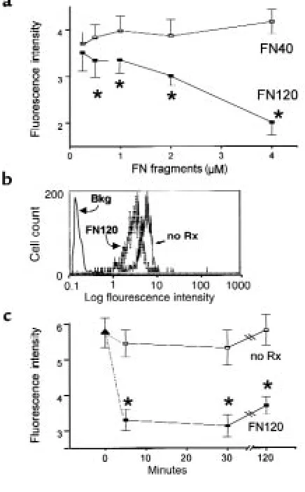
Cell-binding FN fragments modulate monocyte VLA-5 expres-sion in vitro. We incubated heparin-anticoagulated peripheral blood at 37°C for 90 minutes, either with the 120-kDa VLA-5–binding FN fragment (FN120) or with a 40-kDa gelatin-binding fragment (FN40) that does not interact with VLA-5 (39). FN120 reduced VLA-5 in a dose-dependent manner; FN40 had no such effect (Fig-ure 5a). Fig(Fig-ure 5b shows the change in monocyte VLA-5 fluorescence intensity induced by FN120 treatment.
Incubation with FN120, but not with equimolar con-centrations of native plasma FN, significantly reduced VLA-5 expression on both blood monocytes and U937 cells (Table 1), indicating that structures revealed by frag-mentation of FN were necessary for this effect. Exposure to endotoxin also reduced monocyte VLA-5 (Table 1). But because Limulusassays of the FN120 used in these experiments were negative, it was unlikely that the effects of FN120 resulted from endotoxin contamination. To confirm this, we compared the effects of FN120 and endotoxin on U937 cells, which lack CD14, the LPS-binding protein receptor. Only FN120 decreased U937 VLA-5 expression (Table 1). Incubation with a peptide (GRGDSP) that contains the RGD sequence found in FN120 (38, 40) also decreased VLA-5 on both blood monocytes and U937 cells, whereas a control peptide (GRGESP) had no effect (data not shown).
Kinetic analyses showed that monocyte VLA-5 decreased significantly within 5–15 minutes after addi-tion of 2 µM FN120 to whole blood at 37°C (Figure 5c). The decrease in VLA-5 caused by FN120 could be demonstrated with any of 4 anti–VLA-5 mAb’s: clones P1D6, CLB-705, 16, and SAM-1. The fluorescence inten-sity for VLA-5, as measured with SAM-1, CLB-705, and P1D6, declined 35–40%, whereas fluorescence intensity measured by the rat mAb, clone 16, declined by 80% after addition of FN120.
Quantitative flow cytometric analyses showed that with-in 2 hours after the addition of 2 µM FN120 to whole blood, the number of VLA-5 binding sites available to the SAM-1 mAb fell from 12,001 ± 321 to 9,376 ± 199 per monocyte (mean ± SEM) (P = 0.002, ANOVA). Under the same conditions, SAM-1 binding sites on U937 cells decreased from 68,247 ± 300 to 58,886 ± 1,573 (P = 0.004, ANOVA).
FN fragments produced by infarcted myocardium also decrease monocyte VLA-5 expression. To examine the effects of FN fragments generated in the course of ischemia-reperfusion injury on monocyte VLA-5, we incubated 105U937 cells at 37°C for 2 hours with 50 µL
[image:7.612.290.540.50.204.2]undilut-ed postreperfusion cardiac lymph from dogs with myocardial infarctions. This treatment caused mono-cyte VLA-5 mean fluorescence intensity to fall from 6.3 ± 0.3 before treatment to 3.2 ± 0.4 after treatment (P = 0.003, Student’s ttest). When FN and its fragments were removed, the mean fluorescence intensity for VLA-5 in
Figure 4
FN and FN fragments in cardiac lymph. (a) Cardiac lymph from a dog with ischemia-reperfusion injury. Lanes 1 and 2 (positive controls) show immunoblots of human plasma FN enriched for 40- and 120-kDa FN fragments, respectively. Lanes 3–8 show lymph collected 30–300 minutes after reper-fusion. An arrow indicates 120-kDa FN fragments. Brackets indicate smaller FN fragments found in lymph 4–5 hours after reperfusion. (b) Cardiac lymph from an operated dog that, because of good collateral circulation, did not experience car-diac ischemia (lanes 1–6). Almost all FN remains in the native 220-kDa form. Lanes 7 and 8 show human FN enriched for 120- and 40-kDa FN fragments, respectively, to show that FN fragments could have been detected under the conditions of this experiment.
Table 1
Effect of various agents on VLA-5 expression
Rx Monocytes Pvalue U937 Pvalue
None 3.7 ± 0.10 15.4 ± 0.20
Native FN (2 µM) 4.3 ± 0.20 NS 16.7 ± 0.40 NS
FN120 (2 µM) 2.7 ± 0.10 0.01 13.6 ± 0.04 0.02
LPS (10 ng/mL) 2.8 ± 0.30 0.04 16.1 ± 0.30 NS
[image:7.612.57.285.611.671.2]lymph-treated samples was 7.1 ± 0.7, a value not signif-icantly different from controls. On the other hand, sham-immunoadsorbed lymph that retained a full com-plement of FN fragments reduced the VLA-5 expression to 3.1 ± 0.9 (P = 0.01). Similarly, treatment with FN-depleted lymph decreased monocyte VLA-5 expression in whole blood by 13%, whereas sham-immunoad-sorbed lymph reduced monocyte VLA-5 by 34%.
Effect of FN120 on other monocyte cell-surface molecules. Among the β1integrins, only VLA-5 was significantly
affected by treatment with FN120 (Table 2). Cell-surface molecules other than the β1 integrins became more
abundant. For example, mean fluorescence intensities for CD11b and CD14 rose, respectively, from 11.3 ± 0.26 and 6.59 ± 0.39 before treatment to 22.0 ± 1.78 and 11.8 ± 0.9 after treatment (P < 0.01, ANOVA, 3 donors).
Evidence that FN120 induces proteolysis of cell-surface VLA-5. We postulated that FN120 blocks VLA-5 antibody binding sites. But when we found no loss of VLA-5 from cells treated at 4°C with FN120, this explanation became unlikely (data not shown). To investigate whether metab-olism was necessary for the FN120 effect, monocytes were incubated with 2-deoxyglucose and sodium azide, along with FN120, at 37°C. These agents abolished the effect of FN120 on monocyte VLA-5 expression: mean VLA-5 fluorescence intensity before FN120 treatment was 5.7 ± 0.1, and afterwards it was 4.9 ± 0.2 (P < 0.01). FN120, together with 0.1% azide and 5 mM
2-deoxyglu-cose, produced a mean fluorescence intensity of 5.6 ± 0.4, a value not different from untreated controls.
To investigate the possibility that FN120 initiates hydrolysis of VLA-5, we added PMSF (which blocks ser-ine proteases), CdCl2and bestatin (which block leucine
aminopeptidases), 1,10-phenanthroline (which blocks arylamidases and aminopeptidases), and EGTA (which blocks metalloproteases) to whole blood before adding FN120 (Table 3). Among these, only PMSF blocked the FN120 effect. Subsequently, we found that aprotinin, another serine protease inhibitor, also prevents FN120-induced loss of VLA-5 (data not shown).
FN120 treatment causes fragmentation of monocyte VLA-5 molecules. We treated 107 U937 cells with 5 nmol of
[image:8.612.304.538.73.128.2]FN120 for 5 hours, and then we surface labeled these
Figure 5
Effect of 40-kDa (FN40) and 120-kDa (FN120) fragments of FN on VLA-5 expressed by CD14+monocytes in human blood. Graphs in a
and c show the mean ± SEM for data obtained from 3 different donors. (a) Dose response. Data show mean fluorescence intensity for staining with the anti-α5mAb clone SAM-1. Open squares represent
FN40 treatment; filled squares represent FN120 treatment. Asterisks indicate data that differ significantly from untreated sample values (P < 0.05, ANOVA). The mean fluorescence intensity for an isotype-matched control mouse immunoglobulin ranged from 0.21 to 0.28 for all donors and treatments. (b) Histogram compares log immunofluo-rescence intensity for monocytes treated (FN120) or not treated (no Rx) with 2 µM FN120 for 2 hours before staining with mAb clone SAM-1. The solid line represents isotype-matched control mouse immunoglobulin. (c) Kinetic analysis of response to 2 µM FN120. Filled triangle represents the mean fluorescence intensity of clone SAM-1 anti-α5on CD14+human monocytes in whole blood. Aliquots
[image:8.612.310.528.390.742.2]of this blood sample were left untreated (open squares, no Rx) or treated with FN120 (filled squares, FN120). Asterisks indicate times at which results differ significantly (P < 0.05, ANOVA) from the time 0 value. Note that VLA-5 expression was significantly reduced within 5 minutes after addition of FN120. The mean fluorescence intensity for an isotype-matched control mouse immunoglobulin ranged from 0.23 to 0.29 for all donors and treatments.
Table 2
Monocyte surface β1integrin chains modulated by FN120
Chain No treatment FN120 Pvalue
α4 1.77 ± 0.28 1.54 ± 0.19 NS
α5 5.23 ± 0.18 3.18 ± 0.13 0.02
α6 5.20 ± 0.19 4.11 ± 0.35 NS
β1 10.97 ± 0.49 9.33 ± 1.26 NS
Monocytes were stained in whole blood with or without a 2-hour treatment with 2 µM FN120 at 37°C. Data shown represent mean ± SEM of the log fluorescent intensity of CD14+cells stained with HP2/1 for α
and an equal number of untreated U937 cells with 125I.
Unbound 125I was removed and the cells were lysed.
Pro-teins reactive with antibodies to the cytoplasmic domain of the α5chain were immunoprecipitated and analyzed
by SDS-PAGE (Figure 6a). Fragments of α5were found
only in lysates of cells incubated with FN120.
We also found that 125I-labeled U937 cells, treated with
FN120 as described above, shed released VLA-5 when placed in serum-free media for 48 hours. Supernatants of these cells contained not only full-length, 150-kDa α5, but
also α5fragments that migrated under reducing
condi-tions at ∼97 kDa, ∼70 kDa, and 45 kDa (Figure 6a, lane 3). To evaluate whether this fragmentation of α5was
relat-ed to the lactoperoxidase treatment usrelat-ed in the 125I
sur-face labeling of the U937 cells, we also analyzed deter-gent lysates of unlabeled U937 cells, treated or not treated with FN120. Figure 6b shows immunoblots of U937 lysates that were immunoprecipitated with rabbit anti-β1(CD29), fractionated by SDS-PAGE, and then
[image:9.612.100.480.173.594.2]probed with anti-α5antibodies. Whereas the 150-kDa α5
Figure 6
Fragmentation of cell-surface VLA-5 after exposure to FN120. (a) Lysates of U937 cells, surface labeled with 125I, were immunoprecipitated by
affin-ity-purified antibodies to α5and then fractionated by SDS-PAGE before radioautography. Lane 1, untreated cells; lane 2, FN120-treated cells; and
lane 3, anti-α5immunoprecipitates of culture supernatants of radiolabeled cells, previously exposed to FN120, demonstrating the release of α5
frag-ments. (b) Lysates of U937 cells were immunoprecipitated with rabbit antibody to β1, fractionated, blotted to nitrocellulose, and then probed with
affinity-purified rabbit antibodies to the cytoplasmic domain of α5(lanes 2 and 3). Lane 1, lysates incubated only with the secondary antibody; lane
2, proteins from untreated cells; lane 3, proteins from FN120-treated cells in lanes 1 and 3. An arrow indicates that the 60-kDa fragment was more prominent after FN120 treatment. (c) U937 cells cultured with or without FN120. Lanes 1 and 3, supernatants from untreated cells; lanes 2, 4, and 5, supernatants from FN120-treated cells. Immunoblots probed with second antibody only (lanes 1 and 2), with anti-β1antibodies (lanes 3 and 4),
or with antibodies to the cytoplasmic domain of α5(lane 5). Note the 60-kDa fragment identified with anti-α5antibodies. (d) Serial densitometric
measurements of a 60-kDa α5fragment in immunoblots of cardiac lymph collected at intervals before occlusion and for 48 hours after reperfusion
in a dog with significant myocardial ischemia. (e) Immunoblot of cardiac lymph collected 5 hours after reperfusion of ischemic myocardium. Lane 1 was developed with only second antibody; lane 2 with anti-β1; and lane 3 with antibodies to the cytoplasmic domain of α5. The arrow beside lane
molecule was dominant, ∼60-kDa and ∼40-kDa α5
frag-ments were also found in these immunoprecipitates. Fragments of this size were only faintly evident in immunoprecipitates of cells cultured in the absence of FN120 (Figure 6b, compare lanes 2 and 3).
Differences in the gels or conditions used to fraction-ate the lysfraction-ates of the 125I-tagged and the unlabeled U937
cells may be responsible for some of the differences in the estimated molecular weights of the VLA-5 fragments we identified. Indeed, with shorter exposure times, autoradiographs of the ∼70-kDa, 125I-labeled α
5
frag-ment (Figure 6a, lane 2) appeared as a doublet similar to the ∼60-kDa fragment seen in Figure 6b, lane 3.
In supernatants of unlabeled U937 cells (Figure 6c, lane 4), proteins reactive with anti-β1antibodies were
evi-dent only after treatment with FN120. At more than 200 kDa, these molecules were not fragments. Rather, their size and resistance to reducing agents suggested that they may be the spontaneously forming β1homodimers
found by others in studies of integrins extracted from smooth muscle cells (41). Supernatants of FN120-treat-ed U937 cells, on the other hand, containFN120-treat-ed many α5
fragments (Figure 6c, lane 5). Prominent among these was an ∼60-kDa species (Figure 6c, arrow) similar in molecular weight to the fragment, reactive with anti-α5
antibodies, found in lysates of FN120-treated U937 cells (compare Figure 6b, lane 3, and 6c, lane 5).
We also found fragments reactive with anti-α5in
car-diac extracellular fluids after myocardial infarctions. Fig-ure 6e shows immunoblots of a cardiac lymph sample collected 5 hours after reperfusion. Proteins reactive with anti-β1(Figure 6e, lane 2) and anti-α5(Figure 6e, lane 3)
were identified. As in the immunoblots of FN120-treat-ed U937 cell culture supernatants, anti-β1antibodies
rec-ognized a protein larger than 200 kDa in these cardiac lymph samples (Figure 6e, lane 2). In addition, there were numerous fragments. The antibodies to α5also reacted
with many proteins in the lymph (Figure 6e, lane 3), most notably an ∼60-kDa species (arrow in Figure 6e) similar in molecular weight to the ∼60-kDa protein(s) found in lysates (Figure 6, a and b) and culture super-natants (Figure 6c) of FN120-treated U937 cells.
Although both anti-α5and anti-β1antibodies reacted
with a cluster of proteins ∼60 kDa in size in these immunoblots, it cannot be inferred that these are neces-sarily the same protein(s). It is more likely that they are
distinct α5and β1fragments with similar
electrophoret-ic mobilities. Alternatively, these may be adducts of α5
and β1fragments. Macrophages reportedly display
cell-surface transamidases that, upon activation by FN frag-ments, cause fibrin and cell membrane components to couple covalently (42). Conceivably, such enzymes could also couple α5and β1fragments.
Densitometric measurement of the ∼60-kDa α5
frag-ment(s) in cardiac lymph samples (Figure 6d) indicates that proteolysis of VLA-5 molecules on leukocytes migrating into ischemic heart tissue began during the first hour after reperfusion. This process continued throughout the first 24 hours after reperfusion, only decreasing 48 hours after reperfusion.
[image:10.612.303.536.75.149.2]How does the loss of VLA-5 affect monocyte migration on tis-sue matrix?Using the matrix interaction assay described in Methods, we counted the monocytes that sponta-neously migrated into, and remained within, polymer-ized collagen pads 4 hours after application of MNLs to the surface of the pad. Significantly more monocytes accumulated in collagen that incorporated 20 µg/mL native FN compared with collagen alone (Table 4, columns 1 and 2). Pretreatment with 2 µM FN120 reduced the numbers of monocytes that lodged in the FN-collagen pads (Table 4, compare columns 2 and 4; P < 0.04, 4 experiments). On the other hand, in 3 of 4 experiments, pretreatment with this same concentra-tion of FN120 had no significant effect on the numbers of monocytes that accumulated in collagen by itself (Table 4, compare columns 1 and 3), nor did FN120
Table 3
Effect of protease inhibitors on modulation of VLA-5 by FN120
Agent No treatment FN120 Pvalue
None 4.7 ± 0.5 2.3 ± 0.3 <0.03
PMSF 5.4 ± 0.4 5.8 ± 0.7 NS
CdCl2 4.7 ± 0.5 2.2 ± 0.3 <0.03
Bestatin 4.9 ± 0.3 2.4 ± 0.5 <0.03
Phenanthroline 4.9 ± 0.6 3.1 ± 0.6 <0.03
EGTA 5.1 ± 0.3 2.8 ± 0.6 <0.03
Monocytes in blood were exposed to 2 µM FN120 or left untreated for 2 hours at 37°C. Inhibitors were added at the initiation of treatment: 0.2 mM PMSF, 2 mM CdCl2, 40 µM bestatin, 10 µM 1,10-phenanthroline, and 0.1 mM EGTA. Val-ues represent mean ± SEM of the log fluorescent intensity of CD14+cells stained with SAM-1 for α5. Data were obtained from 3 different donors. Pvalues meas-ured by Student’s ttest. NS, not significant.
Table 4
Monocyte accumulation in collagen pads after modulation of VLA-5 with FN120
Treatment
None FN120 Pvalues (columns)
Columns 1 2 3 4
Donor Collagen Collagen + FN Collagen Collagen + FN 1 vs. 2 1 vs. 3 2 vs. 4
1 2,043 ± 157 4,185 ± 218 2,223 ± 311 707±54 0.02 NS 0.002
2 2,064 ± 448 3,402 ± 307 850±223 1,725 ± 222 0.03 NS 0.04
3 778 ± 68 2,346 ± 191 424± 87 591± 31 0.006 0.02 0.01
4 1,036 ± 155 3,741 ± 552 852± 40 1,012 ± 196 0.01 NS 0.03
[image:10.612.57.537.608.695.2]treatment influence the numbers of monocytes that migrated into collagen containing 20 µg/mL laminin. After a 4-hour incubation, we found 5,826 ± 538 untreated vs. 5,525 ± 639 FN120-treated monocytes from 1 donor, and 1,298 ± 161 untreated vs. 1,266 ± 139 FN120-treated monocytes from a second donor in the collagen-laminin matrix.
The effect of FN120 on monocyte migration in response to the chemotactic activity of MCP-1 was sub-tly different. In 1 case, FN120 treatment decreased the numbers of monocytes migrating through collagen alone from 2,997 ± 418 to 1,205 ± 198 (P = 0.017); the numbers of monocytes from this donor migrating through a collagen -FN matrix likewise decreased from 3,818 ± 767 to 1,725 ± 222 after FN120 treatment (P = 0.04). With MNLs from a second donor, we found that FN120 treatment reduced the numbers of monocytes accumulating in collagen alone in response to MCP-1 from 3,186 ± 486 to 656 ± 144 (P = 0.007). With cells from this same donor, FN120 treatment decreased monocyte migration through a collagen-FN matrix from 2,520 ± 797 to 1,312 ± 156 (P = 0.004).
Discussion
FN is a prominent component of cardiac tissue matrix (43). Previous studies had shown that ischemic injury causes focal release of proteases that break down FN, pro-ducing fragments with potentially important signaling properties (21, 22, 44, 45). For example, Clark et al. (23) showed that nanomolar concentrations of certain cell-binding fragments of FN are chemotactic for monocytes. Thus, in ischemic myocardium, we reasoned that mono-cytes migrate toward sites where proteases are actively degrading extracellular matrix proteins, and thereby pro-ducing FN fragments. On the other hand, micromolar concentrations of these same cell-binding FN fragments, perhaps sufficient to saturate all cell-surface receptors, are reportedly not chemotactic (23). Therefore, our hypothesis was that high concentrations of FN frag-ments, as would be found in the epicenter of a myocardial infarction, interfere with monocyte migration.
Data presented here support the idea that cell-binding FN fragments, acting upon the monocyte FN receptor VLA-5, help to regulate monocyte interactions with tissue matrix. However, they act not by saturating or desensitiz-ing VLA-5, but by inducdesensitiz-ing proteolysis of the α5chain of
this heterodimeric molecule. Two lines of evidence sup-port this conclusion. First, incubation with purified FN120 or postreperfusion cardiac lymph that is rich in FN fragments reduced the quantity of VLA-5 expressed by monocytoid cells and caused them to release α5fragments
extracellularly. Second, treating the cells with serine pro-tease inhibitors reduced the ability of FN120 to stimulate monocyte VLA-5 shedding. Because incubation with FN120 induced proteolysis of α5even in the absence of
plasma, and because the effect of FN120 was blocked by incubating the cells with metabolic inhibitors, it is likely that the protease responsible is an inducible product of the monocyte. Thus, VLA-5 may be among the cell-surface molecules whose expression and physiological function is modulated by endogenous proteases (46–48).
Experiments that measured the numbers of monocytes
migrating into collagen containing native FN suggested that adhesive interactions between VLA-5 and FN signif-icantly enhance both spontaneous and MCP-1–driven monocyte migration into a model tissue matrix. Treat-ment with FN120 reduced — but did not abolish — migration into collagen spiked with FN; it also reduced MCP-1–stimulated monocyte migration into polymer-ized collagen that lacked any additives. Because MCP-1 also stimulates β2and β1integrin–dependent monocyte
adherence to collagen and laminin (49), it is possible that costimulation with MCP-1 and FN120 influences the expression and/or function of other ligands that are used by monocytes when interacting with FN-deficient extra-cellular matrices. We found that FN120 did not affect spontaneous monocyte migration into polymerized col-lagen or colcol-lagen that contained 20 µg/mL of laminin. Using a very different matrix interaction assay, Pacifici et al. reported that FN120 stimulates α2β1-dependent
monocyte adherence to denatured collagen (50). It is possible that interactions with matrix proteins involving neither VLA5 nor FN, including, perhaps, adhesive interactions that depend upon CD49f (VLA-6) (51, 52) or CD36 (53), enable monocytes to migrate into infarcted myocardium that has been depleted of native FN. Indeed, proteolysis of matrix FN may promote the survival of monocytes that have entered infarcted tissue. Sugahara et al. have reported that VLA-5–mediated adhesion of monocytes to native FN induces apoptosis (54). A microenvironment that is relatively deficient in FN may therefore allow monocytes to differentiate into the tissue macrophages that are known to orchestrate repair of the damaged myocardium (13, 55, 56).
Previously, we observed that reperfusion of ischemic injury significantly increased the flow of monocytes through the heart into the cardiac lymph, where, by 5 hours after reperfusion, they are as abundant as neu-trophils (11). In this report, we show that postreperfu-sion cardiac lymph contains progressively increasing amounts of FN fragments; most notably, they are frag-ments that induce proteolysis of monocyte cell-surface VLA-5. By the 3 hours after reperfusion, relatively few of the monocytes in the cardiac lymph express VLA-5. A cause-and-effect relationship between the appearance of FN fragments in lymph and the loss of monocyte VLA-5 in vivo was suggested by experiments showing that removal of FN significantly reduced the capacity of the postreperfusion lymph to induce α5shedding in vitro.
p-glycoprotein (MDR-1), a protein previously recognized for its ability to mediate transport of molecules from the intracellular compartments of malignant cells (57), is required for cutaneous T lymphocytes and dendritic cells to migrate into dermal lymphatics. The expression of similar molecules may regulate monocyte transendothe-lial trafficking in myocardium damaged by ischemia-reperfusion injury.
Acknowledgments
This research was supported by the Houston Veterans Affairs Medical Center; a grant-in-aid from the American Heart Association, Texas Affiliate; and National Insti-tutes of Health-Department of Health and Human Ser-vices grants HL-42550, NS-32583, and CA62596. We thank Merry Lindsey for help with dog lymph collection and storage.
1. Cowan, M.J., Reichenbach, D., Turner, P., and Thostenson, C. 1991. Cel-lular response of the evolving myocardial infarction after therapeutic coronary artery reperfusion. Hum. Pathol.22:154–163.
2. Rossen, R.D., and Mann, D.L. 1996. Myocardial inflammation. In
Immunology: principles and practice.R.R. Rich, editor. C.V. Mosby and Co. St. Louis, MO. 1463–1481.
3. Boyle, M.P., and Weisman, H.F. 1993. Limitation of infarct expansion and ventricular remodeling by late reperfusion. Circulation. 88:2872–2883.
4. Morita, M., Kawashima, S., Ueno, M., Kubota, A., and Iwasaki, T. 1993. Effects of late reperfusion on infarct expansion and infarct healing in conscious rats. Am. J. Pathol.143:419–430.
5. Buckley, B.H., and Roberts, W.C. 1974. Steroid therapy during acute myocardial infarction: a cause of delayed healing and ventricular aneurysm. Am. J. Med.56:244–250.
6. Roberts, R., DeMello, V., and Sobel, B.E. 1976. Deleterious effects of methyl-prednisolone in patients with myocardial infarction. Circulation. 53:(Suppl. 1):1204–1206.
7. Hammerman, H., Kloner, R.A., Hale, S., Schoen, F.J., and Braunwald, E. 1983. Dose-dependent effects of short-term methylprednisolone on myocardial infarct extent, scar formation and ventricular function. Cir-culation.68:446–452.
8. Hammerman, H., et al. 1983. Indomethacin-induced scar thinning after experimental myocardial infarction. Circulation.67:1290–1295. 9. Sommers, H.M., and Jennings, R.B. 1964. Experimental acute
myocar-dial infarction. Histologic and histochemical studies of early myocarmyocar-dial infarcts induced by temporary or permanent occlusion of a coronary artery. Lab. Invest.13:1491–1503.
10. Dreyer, W.J., et al. 1991. Neutrophil accumulation in ischemic canine myocardium. Circulation.84:400–411.
11. Birdsall, H.H., et al. 1997. Complement C5a, TGFβ1 and MCP-1, in sequence, induce migration of monocytes into ischemic myocardium within the first 1 to 5 hours following reperfusion. Circulation. 95:684–692.
12. Weihrauch, D., Zimmermann, R., Arras, M., and Schaper, J. 1994. Expres-sion of extracellular matrix proteins and the role of fibroblasts and macrophages in repair processes in ischemic porcine myocardium. Cell. Mol. Biol. Res.40:105–116.
13. Weihrauch, D., Arras, M., Zimmermann, R., and Schaper, J. 1995. Impor-tance of monocytes/macrophages and fibroblasts for healing of micronecroses in porcine myocardium. Mol. Cell. Biochem.147:13–19. 14. Arras, M., et al. 1998. Tumor necrosis factor-alpha is expressed by mono-cytes/macrophages following cardiac microembolization and is antag-onized by cyclosporin. Basic Res. Cardiol.93:97–107.
15. Reuterdahl, C., Sundberg, C., Rubin, K., Funa, K., and Gerdin, B. 1993. Tissue localization of beta receptors for platelet derived growth factor and platelet derived growth factor B chain during wound repair in humans. J. Clin. Invest.91:2065–2075.
16. Pierce, G.F., Mustoe, T.A., Altrock, B.W., Deuel, T.F., and Thomason, A. 1991. Role of platelet-derived growth factor in wound healing. J. Cell. Biochem.45:319–326.
17. Pierce, G.F., et al. 1989. Platelet-derived growth factor and transforming growth factor-beta enhance tissue repair activities by unique mecha-nisms. J. Cell Biol.109:429–440.
18. Pierce, G.F., Brown, D., and Mustoe, T.A. 1991. Quantitative analysis of inflammatory cell influx, procollagen type I synthesis, and collagen cross-linking in incisional wounds: influence of PDGF-BB and TGF-beta 1 therapy. J. Lab. Clin. Med.117:373–382.
19. Cromack, D.T., Porras, R.B., and Mustoe, T.A. 1990. Current concepts in wound healing: growth factor and macrophage interaction. J. Trauma. 30(Suppl.):129–133.
20. Brown, E.J., and Goodwin, J.L. 1988. Fibronectin receptors of phago-cytes. J. Exp. Med.167:777–793.
21. Doherty, D.E., Henson, P.M., and Clark, R.A. 1990. Fibronectin frag-ments containing the RGDS cell-binding domain mediate monocyte migration in the rabbit lung. J. Clin. Invest.86:1065–1075.
22. Clemmensen, I., and Anderson, R.B. 1982. Different molecular forms of fibronectin in rheumatoid synovial fluid. Arthritis Rheum.25:25–31. 23. Clark, R.A., Wikner, N.E., Doherty, D.E., and Norris, D.A. 1988. Cryptic
chemotactic activity of fibronectin for human monocytes resides in the 120-kDa fibroblastic cell-binding fragment. J. Biol. Chem. 263:12115–12123.
24. Yonemasu, K., Nakanishi, A., Sasaki, T., and Kashiba, S. 1983. Stimula-tion of locomoStimula-tion of peripheral blood monocytes by human plasma fibronectin. Microbiol. Immunol.27:283–290.
25. Yamada, K.M., and Kennedy, D.W. 1984. Dualistic nature of adhesive protein function: fibronectin and its biologically active peptide frag-ments can autoinhibit fibronectin function. J. Biol. Chem.99:29–36. 26. Michael, L.H., et al. 1985. Creatine kinase and phosphorylase in cardiac
lymph: coronary occlusion and reperfusion. Am. J. Physiol.248:350–359. 27. Rossen, R.D., et al. 1988. Mechanism of complement activation after coronary artery occlusion: evidence that myocardial ischemia in dogs causes release of constituents of myocardial subcellular origin that com-plex with human C1q in vivo. Circ. Res.62:572–584.
28. Dreyer, W.J., et al. 1989. Canine neutrophil activation by cardiac lymph obtained during reperfusion of ischemic myocardium. Circ. Res. 65:1751–1762.
29. Zeng, L., Takeya, M., Ling, X., Nagasaki, A., and Takahashi, K. 1996. Interspecies reactivities of human macrophage monoclonal anti-bodies to various animal species. J. Histochem. Cytochem.44:845–853. 30. Zeng, L., Takeya, M., and Takahashi, K. 1996. AM-3K, a novel
mono-clonal antibody specific for tissue macrophages and its application to pathological investigation. J. Pathol.178:207–214.
31. Youker, K.A., et al. 1994. Molecular evidence for induction of intercellu-lar adhesion molecule-1 in the viable border zone associated with ischemia-reperfusion injury of the dog heart. Circulation.89:2736–2746. 32. Stouffer, G.A., et al. 1998. Beta3 integrins are upregulated after vascular injury and modulate thrombospondin- and thrombin-induced prolif-eration of cultured smooth muscle cells. Circulation.97:907–915. 33. Forsyth, K.D., and Levinsky, R.J. 1990. Preparative procedures of cooling
and re-warming increase leukocyte integrin expression and function on neutrophils. J. Immunol. Methods.128:159–163.
34. Birdsall, H.H., et al. 1997. Transendothelial migration of lymphocytes from HIV-1 infected donors. A mechanism for extravascular dissemina-tion of HIV-1. J. Immunol.158:5968–5977.
35. Baughn, R.E. 1986. Antibody-independent interactions of fibronectin, C1q, and human neutrophils with Treponema pallidum. Infect. Immun. 54:766–773.
36. Baughn, R.E., Demecs, M., Taber, L.H., and Musher, D.M. 1996. Epitope mapping of B-cell determinants on the 15-kilodalton lipoprotein of Tre-ponema pallidum (Tpp15) with synthetic peptides. Infect. Immun. 64:2457–2466.
37. Keski-Oja, J., Mosher, D.F., and Vaheri, A. 1977. Dimeric character of fibronectin, a major cell surface-associated glycoprotein. Biochem. Bio-phys. Res. Commun.74:699–706.
38. Wright, S.D., and Meyer, C.B. 1985. Fibronectin receptor of human macrophages recognizes the sequence arg-gly-asp-ser. J. Exp. Med. 162:762–767.
39. Ruoslahti, E., and Hayman, E.G. 1979. Two active sites with different characteristics in fibronectin. FEBS Lett.97:221–224.
40. Pierschbacher, M.D., Hayman, E.G., and Ruoslahti, E. 1982. Synthetic peptide with cell attachment activity of fibronectin. Proc. Natl. Acad. Sci. USA.80:1224–1227.
41. Belkin, V.M., Kozlova, N.I., Bychkova, V.V., and Shekhonin, B.V. 1996. Beta 1 integrin subunit dimerization via disulfide bonds. Biochem. Mol. Biol. Int.40:53–60.
42. Hormann, H., Richter, H., and Jelinic, V. 1987. The role of fibronectin fragments and cell-attached transamidase on the binding of soluble fib-rin to macrophages. Thromb. Res.46:39–50.
43. Ahumada, G.G., and Saffitz, J.E. 1984. Fibronectin in the rat heart: a link between cardiac myocytes and collagen. J. Histochem. Cytochem. 32:383–388.
44. Casscells, W., Kimura, H., Sanches, J.A., Yu, Z.-X., and Ferrans, V.J. 1990. Immunohistochemical study of fibronectin in experimental myocardial infarction. Am. J. Pathol.137:801–810.
down-regu-lated adhesion molecule. Proc. Natl. Acad. Sci. USA.87:2244–2248. 47. Moulder, K., Barton, A., and Weston, B. 1993. CD23-mediated
homo-typic cell adhesion: the role of proteolysis. Eur. J. Immunol.23:2066–2071. 48. Itzhaky, D., Raz, N., and Hollander, N. 1998. The glycosylphos-phatidylinositol-anchored form and the transmembrane form of CD58 are released from the cell surface upon antibody binding. Cell. Immunol. 187:151–157.
49. Jiang, Y., Zhu, J.F., Luscinskas, F.W., and Graves, D.T. 1994. MCP-1-stim-ulated monocyte attachment to laminin is mediated by beta 2-integrins.
Am. J. Physiol.267:1112–1118.
50. Pacifici, R., et al. 1994. Ligand binding to monocyte α5β1 integrin acti-vates the α2β1 receptor via the α5 subunit cytoplasmic domain and pro-tein kinase C. J. Immunol.153:2222–2233.
51. Shaw, L.M., and Mercurio, A.M. 1994. Regulation of cellular interactions with laminin by integrin cytoplasmic domains: the A and B structural variants of the alpha 6 beta 1 integrin differentially modulate the adhe-sive strength, morphology, and migration of macrophages. Mol. Biol. Cell. 5:679–690.
52. Penberthy, T.W., Jiang, Y., Luscinskas, F.W., and Graves, D.T. 1995. MCP-1-stimulated monocytes preferentially utilize beta 2-integrins to migrate on laminin and fibronectin. Am. J. Physiol.269:60–68.
53. Yesner, L.M., Huh, H.Y., Pearce, S.F., and Silverstein, R.L. 1996. Regula-tion of monocyte CD36 and thrombospondin-1 expression by soluble mediators. Arterioscler. Thromb. Vasc. Biol.16:1019–1025.
54. Sugahara, H., et al. 1994. Induction of programmed cell death in human hematopoietic cell lines by fibronectin via its interaction with very late antigen 5. J. Exp. Med.179:1757–1766.
55. Li, J., Brown, L.F., Laham, R.J., Volk, R., and Simons, M. 1997. Macrophage-dependent regulation of syndecan gene expression. Circ. Res.81:785–796.
56. Kluge, A., et al. 1997. Coordinate expression of the insulin-like growth factor system after microembolisation in porcine heart. Cardiovasc. Res. 33:324–331.