0095-1137/96/$04.0010
Copyrightq1996, American Society for Microbiology
Rapid Susceptibility Testing for Nontuberculosis Mycobacteria
Using Flow Cytometry
SHANNON E. BOWNDS,1,2TERRENCE A. KURZYNSKI,1MARK A. NORDEN,1,2JOANNE L. DUFEK,1,3
ANDRONALD F. SCHELL1–4*
Wisconsin State Laboratory of Hygiene,1and Departments of Medical Microbiology and Immunology,2Bacteriology,3 and Preventive Medicine,4University of Wisconsin, Madison, Wisconsin 53706
Received 15 November 1995/Returned for modification 15 February 1996/Accepted 4 March 1996
We demonstrated previously that susceptibility testing ofMycobacterium tuberculosiscould be accomplished
within 24 h after the organisms were incubated with antituberculosis agents by using fluorescein diacetate
(FDA) staining and flow cytometry. Continued studies have now shown that assay suspensions containingM.
avium, M. fortuitum, M. gordonae, or M. marinum incubated with various concentrations of ciprofloxacin, clarithromycin, erythromycin, kanamycin, rifampin, and tobramycin hydrolyzed less FDA than drug-free controls. Suspensions of treated and nontreated mycobacteria could be easily differentiated at 6 and 24 h after the initiation of the susceptibility assays by using FDA staining and flow cytometry. In addition, multiplication of the mycobacteria was not required to discern differences between drug-free suspensions of mycobacteria and those treated with antimycobacterial agents. The flow cytometric assay is simple, reproducible, and rapid.
Recently, we showed (12) that susceptibility testing of My-cobacterium tuberculosis H37Ra could be performed rapidly by using flow cytometry. Results of tests were available within 24 h after M. tuberculosis organisms were incubated with antimy-cobacterial agents. The method is based on the ability of flu-orescein diacetate (FDA) to pass through the hydrophobic cell membrane of viable mycobacteria and be rapidly hydrolyzed to free fluorescein by intrinsic esterase. As the fluorescein accu-mulates in the bacterial cells, the fluorescent mycobacteria can be easily detected by flow cytometric analysis. In contrast, dead mycobacteria or mycobacteria inhibited by antimycobacterial agents hydrolyze significantly less FDA.
The use of flow cytometry and FDA staining of mycobacteria shows considerable promise as a rapid method for obtaining test results indicating resistance or susceptibility. Conventional methods of susceptibility testing generally require 1 to 3 weeks of incubation before results are available (3, 10, 11). Although the BACTEC TB-460 system has greatly reduced the period of incubation before results are attainable, 4 to 12 days of incu-bation are still required (6, 8, 18, 20, 21). Even the luciferase reporter phage assay (7) and the gel microdrop encapsulation method, which employs flow cytometry (19), requires several days of incubation before results are available. In contrast, our assay can be completed within 6 or 24 h of the initiation of testing and does not require multiplication of mycobacteria.
In the present study, further evidence that flow cytometry is valuable for performing susceptibility testing of mycobacteria is provided. Nontuberculosis mycobacteria including M. avium, M. fortuitum, M. gordonae, and M. marinum were incubated with various concentrations of antimycobacterial agents. The results of susceptibility tests were available 6 to 24 h after the initiation of testing.
MATERIALS AND METHODS
Antimycobacterial agents. Ciprofloxacin (CIP) and clarithromycin (CLR) were obtained from Miles Pharmaceuticals, West Haven, Conn., and Abbott Laboratories, Abbott Park, Ill., respectively. Erythromycin (ERY), kanamycin
(KAN), rifampin (RIF), and tobramycin (TOB) were obtained from Sigma Chemical Co. St. Louis, Mo. Stock solutions of CIP, CLR, ERY, KAN, RIF, and TOB were prepared at 1,280mg/ml in distilled water, sterilized by filtration with a 0.22-mm-pore-size filter apparatus (Nalgene Lab Ware Division, Rochester, N.Y.), dispensed in 1-ml aliquots, and frozen at2708C until use. RIF was prepared at 1,280mg/ml in 10% methanol.
Bacteria and inoculum preparation.M. fortuitum ATCC 14467, M. avium
TMC 706, M. gordonae TMC 1324, and M. marinum TMC 1218 were obtained from the American Type Culture Collection, Rockville, Md. The bacteria were grown separately in 50 ml of Middlebrook and Cohn 7H9 broth (Difco, Detroit, Mich.) at 378C until the turbidity of the suspension was equivalent to that of a McFarland 1 standard (33108
CFU/ml). This took approximately 5 days of incubation. One-milliliter samples were dispensed into 1.5-ml screw-cap tubes (Starstedt, Newton, N.C.) containing 0.5 ml of sterile glycerol (Sigma), sealed, and stored at2708C. When needed, a frozen suspension of mycobacteria was thawed and an aliquot of 20ml was used to inoculate fresh 7H9 broth. The culture was incubated at 378C for 2 days before an aliquot of 20ml was trans-ferred to 3 ml of 7H9 broth that was incubated for an additional 3 days. Subse-quently, the culture was centrifuged (IEC, Needham Heights, Mass.) at 500 rpm for 2 min to removed large clumps of bacteria. The culture was then adjusted to a turbidity equivalent to that of a McFarland 0.5 standard by the addition of 7H9 broth. Only log-phase cultures were used for susceptibility testing.
Susceptibility tests.The agar dilution method recommended by the National Committee for Clinical Laboratory Standards (M24-P) (11) was used for the determination of the MICs for M. fortuitum. The MIC endpoint was the lowest concentration of the antimycobacterial agent that completely inhibited visible growth (106organisms) on Middlebrook and Cohn 7H10 agar medium. The MICs were determined after 3 to 4 days of incubation at 378C. The MICs were also read 7 days later for confirmation. Prior to incubation, the plates were sealed in plastic bags to reduce drying.
A broth macrodilution test was also performed with 106mycobacteria for determination of MICs and the MBCs for M. avium, M. fortuitum, M. gordonae, and M. marinum. The macrodilution method was similar to that recommended by the National Committee for Clinical Laboratory Standards (M7-A3). The MIC endpoint was the lowest concentration of the antimycobacterial agent that completely inhibited visible growth. The MICs were determined after 5 days of incubation at 378C. The MBC was determined 5 days after incubation at 378C by subculturing 0.1 ml of the test suspensions onto drug-free Middlebrook and Cohn 7H10 agar medium. The plates were then streaked for isolation and were incubated at 378C for 10 days.
Preparation of assay suspensions for flow cytometric analysis and determi-nation of viability.Serial twofold dilutions (0.5 ml) of the antimycobacterial agents were prepared with 7H9 broth in 12-by-75-mm round-bottom Falcon tubes (no. 2054; Becton Dickinson Immunocytometry Systems, Mountain View, Calif.). Each dilution was then inoculated with 0.5 ml of 7.53105
mycobacterial organisms. Drug-free suspensions of mycobacteria were also included as con-trols. The tubes were incubated for 6 or 24 h at 378C before the test suspensions were analyzed by flow cytometry.
The numbers of viable mycobacteria in the drug-free controls and in the various concentrations of CIP, CLR, ERY, KAN, RIF, and TOB were deter-mined 24 h after inoculation. Briefly, serial 10-fold dilutions of each
concentra-* Corresponding author. Mailing address: Wisconsin State Labora-tory of Hygiene, 465 Henry Mall, Madison, WI 53706. Phone: (608) 262-3634. Fax: (608) 265-3451.
1386
on May 15, 2020 by guest
http://jcm.asm.org/
tion of CIP, CLR, ERY, KAN, RIF, and TOB were prepared with 7H9 broth, and 50-ml samples were plated to determine the number of CFU per milliliter on 7H10 medium. The plates were incubated at 378C for 10 days before the number of CFU of mycobacteria per milliliter was determined. This method was also used for determination of the numbers of viable mycobacteria in the drug-free controls.
Flow cytometric data acquisition.After incubation of assay suspensions for 6 or 24 h, 10ml of FDA (10mg/ml; Sigma) was added to the suspensions to yield a final concentration of 100 ng/ml. The samples were incubated at 378C for 30 min before being analyzed with a single argon laser tuned at 488 nm (FACScan flow cytometer; Becton Dickinson Immunocytometry Systems) by using FAC Scan Lysys II software for data acquisition and analysis. Initially, viable and heat-killed M. fortuitum organisms were detected and differentiated from 7H9 particles by using forward scatter, side scatter, and FDA fluorescence. Live gating was performed on profiles of mycobacteria during data acquisition to exclude all 7H9 particles. Data were acquired over a period of between 7 and 60 s to count 10,000 bacteria or events.
Flow cytometric statistical analysis.Samples were analyzed by examining histogram profiles of FDA fluorescence with FACScan Lysys II software. Two parameters were evaluated: the number of events per second (number of labeled mycobacteria) and the mean channel fluorescence (intensity of fluorescence-labeled mycobacteria). These values were obtained as part of the flow cytometric statistical analysis provided with the FACScan Lysys II software.
Statistics.The values obtained were tested by analysis of variance. The Fisher least-significant-difference test was used to examine pairs of means when a significant F value indicated reliable mean differences (22). The alpha level was set at 0.05 before the experiments were started.
RESULTS
Detection ofM. fortuitumby flow cytometry.Initially, data
were acquired to identify viable M. fortuitum organisms in Middlebrook and Cohn 7H9 broth (Fig. 1A and B). Although background particles (less than two events per second) were detected in 7H9 broth (Fig. 1A), the number of particles did not significantly affect our ability to detect M. fortuitum organ-isms (Fig. 1B). When viable (Fig. 1C) and heat-killed (Fig. 1D) M. fortuitum organisms were incubated with FDA, only viable M. fortuitum organisms hydrolyzed FDA (Fig. 1C). The fluo-rescent viable M. fortuitum organisms had a mean channel fluorescence of 3,5006500 (Fig. 1C), while the heat-killed M. fortuitum organisms failed to hydrolyze FDA and had a mean channel fluorescence of approximately 5 (Fig. 1D). In addition, the viabilities of the M. fortuitum organisms were not affected by the duration of exposure to or to various concentrations of FDA. When these experiments were repeated, similar results were obtained.
Effects of antimycobacterial agents onM. fortuitum
organ-isms exposed to FDA and detection by flow cytometry. The
ability of M. fortuitum organisms to hydrolyze FDA after incu-bation for 6 or 24 h in various concentrations of CIP, KAN, and RIF was determined (Fig. 2). The mean channel fluores-cence decreased rapidly with increasing concentrations of CIP, KAN, and RIF. In general, the mean channel fluorescence decreased more rapidly in suspensions of M. fortuitum organ-isms exposed to CIP, KAN, and RIF for 24 h than for 6 h. The mean channel fluorescence decreased from approximately 4,000 to 500 or less after the M. fortuitum organisms were incubated with these antimycobacterial agents for 24 h. Similar results were obtained when the M. fortuitum organisms were incubated with increasing concentrations of CLR, ERY, and TOB (Fig. 3). By contrast, the mean channel fluorescence increased steadily from approximately 4,000 to 6,000 in drug-free controls.
In other experiments, the number of viable M. fortuitum organisms decreased rapidly with increasing concentrations of CIP, KAN, and RIF (Fig. 4). The decrease in the viability of the M. fortuitum organisms correlated with a decrease in the mean channel fluorescence for CIP, KAN, and RIF. The MICs were 0.15mg/ml for CIP, 2.5mg/ml for KAN, and 5mg/ml for RIF. Each MIC was associated with a 50% or more decrease in
the mean channel fluorescence compared with that for the drug-free control. No viable mycobacteria were recovered from the suspensions containing antimycobacterial agents when the mean channel fluorescence had decreased approxi-mately 70% or more compared with that for the drug-free controls. The MBCs were 2.5 mg/ml for CIP, 2.5 mg/ml for KAN, and 40mg/ml for RIF.
Effects of antimycobacterial agents on other mycobacterial
exposed to FDA and detection by flow cytometry. Figure 5
shows the effects of various concentrations of CIP, KAN, and RIF on the ability of M. avium, M. gordonae, and M. marinum organisms to hydrolyze FDA and the level of detection of fluorescent mycobacteria by flow cytometry. The mean channel fluorescence decreased rapidly with increasing concentrations of the antimycobacterial agents. Similar results were obtained when M. avium, M. gordonae, and M. marinum organisms were incubated with various concentrations of CLR, ERY, and TOB.
DISCUSSION
The pathogenicity of M. tuberculosis has been clear to clini-cians since its discovery by Koch in the 1890s. However, rec-ognition of the pathogenic potential of nontuberculosis myco-bacteria has lagged behind recognition of the pathogenic FIG. 1. Fluorescence versus side scatter of 7H9 broth (A), 7H9 broth with viable M. fortuitum (Mf) (B), 7H9 broth with viable FDA-labeled M. fortuitum (Mb) (C), and 7H9 broth with heat-killed M. fortuitum in the presence of FDA (D). The numbers on the y axis of each panel and on the x axis are 100
, 101 , 102
, 103, and 104, respectively.
on May 15, 2020 by guest
http://jcm.asm.org/
potential of M. tuberculosis (4, 13, 23). The delay in the accep-tance of the pathogenic roles of these organisms arises from their relatively low level of intrinsic virulence, their propensity to commensally inhabit the airways, and their tendency to infect patients with preexisting lung disease, in which case the features of the infections caused by these organisms may be difficult to distinguish from those of the underlying disease process (13). Previously, the prevalence and distribution of nontuberculosis mycobacterial infections have been obscure (13). Recent evidence, however, suggests that the incidence of nontuberculosis mycobacterial infections, especially those caused by the M. avium complex, is on the rise because of the AIDS epidemic. Some hospitals and public health laboratories have reported that the overall number of cases of nontuber-culosis mycobacterial infection now exceeds the number of cases of tuberculosis (4).
One must add susceptibility testing to these difficulties in
recognizing the pathogenic potential of nontuberculosis myco-bacteria. Conventional susceptibility testing for nontuberculo-sis mycobacteria is problematic (4). The clinical relevance of in vitro susceptibility testing is debated (5), even though clinicians still request them (4). The use of flow cytometry and FDA staining shows considerable promise as a rapid means of ob-taining susceptibility test results to make a correlation with the clinical outcome. Flow cytometric analysis may enhance pa-tient care by improving the designs of studies performed to assess the relevance of treatment regimens.
Our results demonstrate that susceptibility testing of nontu-berculosis mycobacteria by flow cytometry can be accom-plished rapidly, in 24 h or less after the initiation of testing. The technique depends on the entrance of FDA into viable myco-bacteria and the conversion of FDA to fluorescein by the cellular esterases (2). In dead mycobacteria or mycobacteria incubated with antimycobacterial agents, hydrolysis of FDA fails to occur or is inhibited. The use of flow cytometry permits rapid measurement (1 min) of differences in the amounts of accumulated fluorescein between susceptible and nontreated mycobacteria. Consequently, assessment of the susceptibility and resistance of mycobacteria to various antimycobacterial agents can be performed rapidly.
[image:3.612.319.554.68.371.2]The results of the current investigation extend and confirm our previous findings (12). We demonstrated previously that susceptibility testing of M. tuberculosis can be accomplished rapidly by using flow cytometry. Results of tests were available within 24 h after M. tuberculosis organisms were incubated with ethambutol, isoniazid, RIF, or streptomycin. In addition, the FIG. 2. Effects of antimycobacterial agents on the ability of M. fortuitum to
hydrolyze FDA and detection of fluorescent mycobacteria (mean channel fluo-rescence) by flow cytometry. Assay suspensions containing M. fortuitum organ-isms with various concentrations of CIP, KAN, and RIF were incubated at 378C for 6 h (■) or 24 h (E) before exposure to FDA and analysis by flow cytometry.
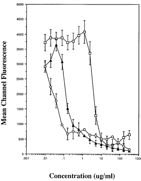
FIG. 3. Effects of CLR (E), ERY (å), and TOB (h) on the ability of M.
fortuitum to hydrolyze FDA and detection of fluorescent mycobacteria (mean
channel fluorescence) by flow cytometry. Assay suspensions were incubated with the antimycobacterial agents for 24 h at 378C before FDA was added prior to analysis by flow cytometry.
on May 15, 2020 by guest
http://jcm.asm.org/
[image:3.612.81.279.75.485.2]assay system did not require multiplication of mycobacteria in the drug-free control suspensions to differentiate them from drug-treated mycobacteria. In the present study, we showed that assay suspensions containing M. avium, M. fortuitum, M. gordonae, and M. marinum organisms incubated with various concentrations of CIP, CLR, ERY, KAN, RIF, and TOB hy-drolyzed significantly less FDA than nontreated control organ-isms. The susceptibilities of treated and nontreated mycobac-teria could be easily differentiated at 6 and 24 h after the initiation of the assays by using flow cytometry. Although mul-tiplication occurred in some of the assay suspensions, multi-plication was not necessary to discern differences between drug-free suspensions of mycobacteria and those treated with antimycobacterial agents.
We combined the sensitivity and detection systems of the flow cytometer with the ability of FDA to distinguish between viable and dead bacteria. The result was a more rapid method of performing susceptibility testing of mycobacteria. By mea-suring mean channel fluorescence (intensity of
fluorescence-labeled mycobacteria), the effects of antimycobacterial agents on nontuberculosis mycobacteria and M. tuberculosis (12) could be determined in 24 h or less after the initiation of testing. We also suggested previously (12) that an operational definition that correlated changes in the mean channel fluo-rescence with the MICs of various antimycobacterial agents for mycobacteria could be developed. The MIC, as determined by flow cytometry in the present study, is that concentration of antimycobacterial agent which yielded a reduction in the mean channel fluorescence of at least 50% compared with the drug-free control value. The MICs of CIP, CLR, ERY, KAN, RIF, and TOB for M. fortuitum were 0.15, 0.15, 0.62, 2.5, 5, and 5 mg/ml, respectively. A decrease in the mean channel fluores-cence of 50% did identify the macrodilution MIC or the MIC within one twofold dilution. Likewise, a 70% or greater de-crease in the mean channel fluorescence correlated with the bactericidal concentration of each antimycobacterial agent or the MIC within one twofold dilution. Additional experiments, however, with a larger number of each Mycobacterium species are needed to determine accurately the percent change in FIG. 4. Effects of various concentrations of CIP, KAN, and RIF on the
viability (CFU per milliliter) of M. fortuitum organisms (E) and mean channel fluorescence (■). Assay suspensions containing M. fortuitum with various con-centrations of CIP, KAN, and RIF were incubated at 378C for 24 h before exposure to FDA and analysis by flow cytometry. Assay suspensions were also inoculated on drug-free 7H10 agar to determine the viability of the mycobacte-ria.
FIG. 5. Effects of various concentrations of CIP, KAN, and RIF on the ability of M. avium (E), M. gordonae (h), and M. marinum (ç) to hydrolyze FDA, and detection of fluorescent mycobacteria (mean channel fluorescence) by flow cy-tometry.
on May 15, 2020 by guest
http://jcm.asm.org/
mean channel fluorescence that would predict the MIC or the MBC of each antimycobacterial agent.
Flow cytometry has increasingly been used to perform sus-ceptibility tests for bacteria (9, 17) and yeasts (14–16). The main advantages are accuracy, reproducibility, sensitivity, ob-jectivity, and speed. In addition, the system can be automated. A disadvantage is the identification of a suitable fluorescence indicator that accurately reflects events leading to cell inhibi-tion or death. It is known that FDA passes through the cell membranes of viable mycobacteria (1) and other bacteria (2). Once it is inside the cytoplasm, FDA is cleaved by nonspecific intracellular esterases to form fluorescein, which causes the bacteria to fluoresce intensively. By contrast, mycobacteria treated with heat or inoculated with inhibiting concentrations of effective antimycobacterial agents cleaved less FDA and fluoresced significantly less or not at all. We found that the hydrolysis of FDA was a reliable means of assessing viability. When the mean channel fluorescence had decreased 70% or more, the mycobacteria were not viable. Frequently, the cost of a flow cytometer is also stated to be a disadvantage. This may be more perception than reality. The BACTEC TB-460 instru-ment is the one most frequently used for susceptibility testing. When the high cost of supplies for performing susceptibility testing with this instrument are considered, a flow cytometer will be less expensive. The reagents used with our method were relatively inexpensive. Costs were restricted to the purchase of 7H9 broth, FDA, and the antimycobacterial agents. Finally, the speed and quality of information obtained by flow cytom-etry increase turnaround time, which improves health care.
In conclusion, we showed that flow cytometry and FDA staining can be used to perform susceptibility testing for non-tuberculosis mycobacteria. The assay is simple and reproduc-ible and can be completed in 24 h or less from the time of the initiation of testing. The technique also does not require mul-tiplication of mycobacteria for accurate and reproducible re-sults.
ACKNOWLEDGMENTS
We thank Adolf L. Gundersen for continuous support and encour-agement. We also thank the board of directors of the Gundersen Medical Foundation, Inc., La Crosse, Wis., for providing the flow cytometer.
REFERENCES
1. Bercovier, H., M. Resnick, D. Kornitzer, and L. Levy. 1987. Rapid method for testing drug-susceptibility of Mycobacteria spp. and gram-positive bacte-ria using rhodamine 123 and fluorescein diacetate. J. Microbiol. Methods
7:139–142.
2. Brunius, G. 1980. Technical aspects of the use of 3969-diacetyl fluorescein for vital fluorescent staining of bacteria. Curr. Microbiol. 4:321–323. 3. Canetti, G., S. Frosman, J. H. Grosset, P. Hauduroy, M. Langerova, H. T.
Mahler, G. Meissner, D. A. Mitchison, and L. Sula.1963. Mycobacteria: laboratory methods for testing drug sensitivity and resistance. Bull. W. H. O.
29:565–578.
4. Good, R. C. 1985. Opportunistic pathogens in the genus Mycobacterium. Annu. Rev. Microbiol. 39:347–369.
5. Heifets, L. B. 1991. Dilemmas and realities in drug susceptibility testing of M.
avium-intracellulare and other slowly growing nontuberculosis mycobacteria,
p. 123–146. In L. B. Heifets (ed.), Drug susceptibility in the chemotherapy of mycobacterial infections. CRC Press, Inc. Boca Raton, Fla.
6. Heifets, L. B., M. D. Iseman, J. L. Cook, P. J. Lindholm-Levy, and I. Drupa. 1985. Determination of in vitro susceptibility of Mycobacterium tuberculosis to cephalosporins by radiometric and conventional methods. Antimicrob. Agents Chemother. 27:11–15.
7. Jacobs, W. R., Jr., R. G. Barletta, R. Undani, J. Chan, G. Kalkut, G. Sosne,
T. Kieser, G. J. Sarkis, G. F. Hatfull, and B. R. Bloom.1993. Rapid assess-ment of drug susceptibilities of Mycobacterium tuberculosis by means of luciferase reporter phages. Science 260:819–822.
8. Lee, C., and L. B. Heifets. 1987. Determination of minimal concentrations of antituberculosis drugs by radiometric and conventional methods. Am. Rev. Respir. Dis. 136:349–352.
9. Mason, D. J., and V. A. Gant. 1995. The application of flow cytometry to the estimation of bacterial antibiotic susceptibility. J. Antimicrob. Chemother.
36:441–448.
10. McClatchy, J. K. 1978. Susceptibility testing of mycobacteria. Lab. Med.
9:47–52.
11. National Committee for Clinical Laboratory Standards. 1990. Antimycobac-terial susceptibility testing. Proposed standard M24-P. National Committee for Clinical Laboratory Standards, Villanova, Pa.
12. Norden, M. A., T. A. Kurzynski, S. E. Bownds, S. M. Callister, and R. F.
Schell. 1995. Rapid susceptibility testing of Mycobacterium tuberculosis (H37Ra) by flow cytometry. J. Clin. Microbiol. 33:1231–1237.
13. O’Brien, R. J., L. J. Geiter and D. E. Snider, Jr. 1987. The epidemiology of nontuberculosis mycobacterial diseases in the United States: results from a national survey. Am. Rev. Respir. Dis. 135:1007–1014.
14. O’Gorman, M. R. G. and R. L. Hopfer. 1991. Amphotericin B susceptibility testing of Candida species by flow cytometry. Cytometry 12:743–747. 15. Pore, R. S. 1990. Antibiotic susceptibility testing of Candida albicans by flow
cytometry. Curr. Microbiol. 20:323–328.
16. Pore, R. S. 1991. Ketoconazole susceptibility of yeast by the FCST method. Curr. Microbiol. 23:45–50.
17. Pore, R. S. 1994. Antibiotic susceptibility testing by flow cytometry. J. Anti-microb. Chemother. 34:613–627.
18. Roberts, G. D., N. L. Goodman, L. Heifets, H. W. Larsh, T. H. Lindner, J. K.
McClatchy, M. R. McGinnis, S. H. Siddiqi, and P. Wright.1983. Evaluation of the BACTEC radiometric method for recovery of mycobacteria and drug susceptibility testing of Mycobacterium tuberculosis from acid-fast smear pos-itive specimens. J. Clin. Microbiol. 18:689–696.
19. Ryan, C., B. Nguyen, and S. J. Sullivan. 1995. Rapid assay for mycobacterial growth and antibiotic susceptibility using gel microdrop encapsulation. J. Clin. Microbiol. 33:1720–1726.
20. Siddiqi, S. H., J. E. Hawkins, and A. Laszio. 1985. Interlaboratory drug susceptibility testing of Mycobacterium tuberculosis by a radiometric proce-dure and two conventional methods. J. Clin. Microbiol. 22:919–923. 21. Snider, D. E., R. C. Good, J. O. Kilburn, L. F. Laskowski, Jr., R. H. Lusk,
J. J. Marr, Z. Reggiardo, and G. Middlebrook.1981. Rapid drug suscepti-bility testing of Mycobacteria tuberculosis. Am. Rev. Respir. Dis. 123:402– 408.
22. Steel, R. G. D., and J. H. Torrie. 1960. Principles and procedures of statistics with special reference to the biological sciences, p. 481, McGraw-Hill Book Co., New York.
23. Wolinsky, E. 1979. Nontuberculosis mycobacteria and associated diseases. Am. Rev. Respir. Dis. 119:107–159.