muscle injury
William M. Mahoney Jr., Stephen M. Schwartz
J Clin Invest.
2005;
115(2)
:221-224.
https://doi.org/10.1172/JCI24272
.
For 3 decades, terms such as synthetic phenotype and contractile phenotype have been
used to imply the existence of a specific mechanism for smooth muscle cell (SMC)
responses to injury. In this issue of the
JCI
, Hendrix et al. offer a far more precise approach
to examining the mechanisms of SMC responses to injury, focused not on general changes
in phenotype but on effects of injury on a single promoter element, the CArG [CC(A/T)
6GG]
box, in a single gene encoding smooth muscle (SM)
a
-actin. Since CArG box structures are
present in some, but not all, SMC genes, these data suggest that we may be progressing
toward establishing a systematic, molecular classification of both SMC subsets and the
response of SMCs to different injuries.
Commentary
Find the latest version:
Acknowledgments
Thanks to Robert Piper, Greg Cooney, and members of the James laboratory for invaluable discussions.
Address correspondence to: David E. James, Garvan Institute of Medical Research, Darlinghurst, Sydney, New South Wales 2010, Australia. Phone: 61-2-92958100; Fax: 61-2-92958201; E-mail: D.james@garvan.org.au.
1. Bryant, N.J., Govers, R., and James, D.E. 2002. Regulated transport of the glucose transporter GLUT4. Nat. Rev. Mol. Cell Biol.3:267–277. 2. Whiteman, E.L., Cho, H., and Birnbaum, M.J. 2002.
Role of Akt/protein kinase B in metabolism. Trends Endocrinol. Metab.13:444–451.
3. Saltiel, A.R., and Pessin, J.E. 2002. Insulin signal-ing pathways in time and space. Trends Cell Biol.
12:65–71.
4. Chen, Y.A., and Scheller, R.H. 2001. SNARE-medi-ated membrane fusion. Nat. Rev. Mol. Cell Biol.
2:98–106.
5. Rizo, J., and Sudhof, T.C. 2002. Snares and
Munc18 in synaptic vesicle fusion. Nat. Rev. Neurosci.3:641–653.
6. Dulubova, I., et al. 1999. A conformational switch in syntaxin during exocytosis: role of munc18.
EMBO J.18:4372–4382.
7. Toonen, R.F., and Verhage, M. 2003. Vesicle traf-ficking: pleasure and pain from SM genes. Trends Cell Biol.13:177–186.
8. Misura, K.M., Scheller, R.H., and Weis, W.I. 2000. Three-dimensional structure of the neuronal-Sec1-syntaxin 1a complex. Nature.404:355–362. 9. Kanda, H., et al. 2005. Adipocytes from
Munc18c-null mice show increased sensitivity to insulin-stimulated GLUT4 externalization. J. Clin. Invest.
115:291–301. doi:10.1172/JCI200522681. 10. Tellam, J.T., et al. 1997. Characterization of
Munc-18c and syntaxin-4 in 3T3-L1 adipocytes. Putative role in insulin-dependent movement of GLUT-4.
J. Biol. Chem.272:6179–6186.
11. Bose, A., et al. 2004. Unconventional myosin Myo1c promotes membrane fusion in a regulated exocytic pathway. Mol. Cell. Biol.24:5447–5458.
12. van Dam, E.M., Govers, R., and James, D.E. 2005. Akt activation is required at a late stage of insulin-induced GLUT4 translocation to the plasma mem-brane. Mol. Endocrinol. In press.
13. Semiz, S., et al. 2003. Conventional kinesin KIF5B mediates insulin-stimulated GLUT4 movements
on microtubules. EMBO J.22:2387–2399. 14. Emoto, M., Langille, S.E., and Czech, M.P. 2001.
A role for kinesin in insulin-stimulated GLUT4 glucose transporter translocation in 3T3-L1 adipocytes. J. Biol. Chem.276:10677–10682. 15. Maffucci, T., Brancaccio, A., Piccolo, E., Stein,
R.C., and Falasca, M. 2003. Insulin induces phosphatidylinositol-3-phosphate formation through TC10 activation. EMBO J.22:4178–4189. 16. Slot, J.W., Geuze, H.J., Gigengack, S., James, D.E.,
and Lienhard, G.E. 1991. Translocation of the glu-cose transporter GLUT4 in cardiac myocytes of the rat. Proc. Natl. Acad. Sci. U. S. A.88:7815–7819. 17. Slot, J.W., Geuze, H.J., Gigengack, S., Lienhard, G.E.,
and James, D.E. 1991. Immuno-localization of the insulin regulatable glucose transporter in brown adipose tissue of the rat. J. Cell Biol.113:123–135. 18. Kane, S., et al. 2002. A method to identify serine
kinase substrates. Akt phosphorylates a novel adipocyte protein with a Rab GTPase-activating pro-tein (GAP) domain. J. Biol. Chem.277:22115–22118. 19. Sano, H., et al. 2003. Insulin-stimulated phos-phorylation of a Rab GTPase-activating protein regulates GLUT4 translocation. J. Biol. Chem.
278:14599–14602.
20. Zerial, M., and McBride, H. 2001. Rab proteins as membrane organizers. Nat. Rev. Mol. Cell Biol.
2:107–117.
Defining smooth muscle cells
and smooth muscle injury
William M. Mahoney Jr. and Stephen M. Schwartz
Department of Pathology, University of Washington, Seattle, Washington, USA.
For 3 decades, terms such as synthetic phenotype and contractile phenotype
have been used to imply the existence of a specific mechanism for smooth
muscle cell (SMC) responses to injury. In this issue of the
JCI
, Hendrix et
al. offer a far more precise approach to examining the mechanisms of SMC
responses to injury, focused not on general changes in phenotype but on
effects of injury on a single promoter element, the CArG [CC(A/T)
6GG]
box, in a single gene encoding smooth muscle (SM)
α
-actin (see the related
article beginning on page 418). Since CArG box structures are present in
some, but not all, SMC genes, these data suggest that we may be
progress-ing toward establishprogress-ing a systematic, molecular classification of both SMC
subsets and the response of SMCs to different injuries.
Nonstandard abbreviations used: CArG, CC(A/T)6GG; SM, smooth muscle; SMC, smooth
muscle cell; SRF, serum response factor.
Conflict of interest: The authors have declared that no conflict of interest exists.
Citation for this article:J. Clin. Invest.115:221–224 (2005). doi:10.1172/JCI200524272.
Efforts to understand the response of arte-rial smooth muscle cells (SMCs) to injury have led to confusion, in part because of the as-yet-unconfirmed implication that terms such as dedifferentiation, synthetic pheno-type, and phenotypic modulation refer to a specific, common mechanism. This issue of the JCI brings a major new perspective to
this subject with a report by Hendrix et al. (1) that builds upon their recent findings (2).
Since the 1970s, most investigators assumed that the loss of properties (i.e., the loss of contractile capacity and the appearance of proteins associated with the extracelluar matrix) observed when SMCs adapted to culture used the same mecha-nisms required for the response of arterial SMCs to vascular injury, sometimes termed phenotypic modulation (3, 4). However, this theory of a common mechanism underly-ing the response of SMCs to multiple forms of injury in vivo is largely unsubstantiated. Since the molecular mechanism
control-ling SMC differentiation and modulation in vivo was poorly understood, Hendrix et al. (1) examined the molecular control of smooth muscle (SM) α-actin expression in response to injury. Their earlier studies showed that expression of SM α-actin is regulated by promoter elements called CArG [CC(A/T)6GG] boxes, which are bound by
serum response factor (SRF) either alone or as a macromolecular complex including its specific cofactor, myocardin. Interestingly, cytoskeletal modulation regulates the SRF-myocardin interaction. Myocardin is bound by G-actin in the cytoplasm, and polymer-ization of actin releases myocardin, allow-ing it to travel to the nucleus. Once in the nucleus, it acts as a cofactor to enhance the binding of SRF to genes associated with cell replication and the dissociation from genes associated with SMC contractile proteins (5, 6). Therefore, there is a delicate balance between SMCs’ need to respond to various stimuli and the availability of proteins to mediate these processes.
of these mutations were assayed both in vitro and in vivo following injury. Hendrix et al. show that generic CArG binding sites are sufficient for regulation of SM α-actin transcription in vitro. However, SMC-spe-cific, degenerate CArG sequences (Figure 1) are required for appropriate repression of SM α-actin following injury in vivo. Fur-thermore, the relative levels of myocar-din control the interaction between SRF and the degenerate CArG boxes within the SM α-actin promoter. Thus, in vitro dedifferentiation is an imperfect “common” model for SMC response to injury in vivo.
Do these CArG sequences regulate all SM genes?
Returning to the hypothesis that there is a common mechanism controlling at least a large portion of the SM phenotype, Hendrix et al. (1) note that sequences of CArG boxes in
promoters for SM α-actin and for other SMC-specific genes, including MYH11 (encod-ing SM myosin heavy chain), SMTNA/B (encoding smoothelin A/B), SM22α, and
CHF-1 (encoding HEY2), are distinct from the canonical CArG sequences responsible for genes involved in cell replication (Figure 1). The authors’ suggestion that degenerate CArG boxes determine restructuring of the SMC contractile apparatus is supported by recent studies of the role of the cytoskeleton in regulating SRF function (5, 6). This ele-gant mechanism allows SMCs to move into a wound while decreasing synthesis of pro-teins required for cell contraction. However, the CArG element is not the sole regulator. A number of SMC-specific genes, including
NOTCH3 (encoding neurogenic locus notch homolog protein 3), APEG1 (encoding aor-tic preferentially expressed gene 1), ELN
(encoding elastin), and ACTN1 (encoding
α-actinin) (7), are not regulated through CArG elements. For example, APEG1 is spe-cifically expressed in differentiated vascular SMCs. However, its expression is dependent not upon CArG boxes, but upon the binding of members of the upstream stimulatory fac-tor family of transcription facfac-tors to E box promoter sequences (8).
In summary, though the SRF-myocar-din complex is largely responsible for the molecular regulation of some SMC-spe-cific genes, there are still many facets of this complex modulation that must be explored further. It is also reasonable to propose that different sets of SMC pro-moters are related to the need of different SMCs to respond to different kinds of inju-ry. This leads us to ask, how are the diverse SM genes controlled in response to injuries as distinct as intimal formation, hypertro-phy, polyploidization, stenotic remodeling, aneurism, and rarefaction?
Can we define SMCs? Is there a single pattern for SMC response to injury?
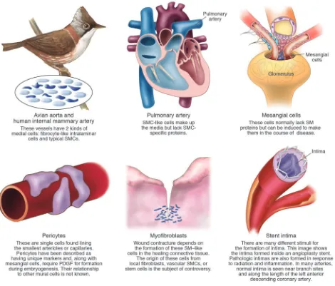
We already know that, even with no inju-ry, there is great variation in gene expres-sion in various forms of SMCs (Figure 2). For example, SM-memb, a myosin often considered characteristic of all intimal cells, is present in normal intima and in many intimas formed as a result of inju-ry but is absent from the atherosclerotic fibrous cap (9). Similarly, cells making up the fibrous cap have very little mRNA, while cells at the edges of atherosclerotic plaques have abundant mRNA, and these messages code for proteins responsible for matrix synthesis. Finally, microarrays show that the SMCs of the atherosclerotic cap have a phenotype distinct from both nonatherosclerotic intima and the pheno-type of medial SMCs (10).
Medial cells of the pulmonary artery ful-fill the morphological criteria of SMCs: they are full of contractile apparatus (Fig-ure 2). These cells, however, fail to express classical SMC genes (11). In contrast, in pulmonary hypertension, adventitial fibro-blasts have been shown to express the clas-sical SMC genes (12). Similarly, contractile myofibroblasts present in wounds express SM proteins (13). Even endothelial cells are able to delaminate from the monolayer, transdifferentiate, and express at least part of the SMC repertoire of contractile pro-teins, including SM α-actin (14).
[image:3.585.56.355.83.356.2]Perhaps the most confusing example of identification of a cell as SM arises in the
Figure 1
Regulation of CArG-specific SMC genes. (A) CArG box elements in the SM α-actin promoter. The SM α-actin promoter contains 2 consensus CArG boxes [CC(A/T)6GG]. However, the
wild-type sequences are degenerate relative to the canonical sequence first identified in c-fos
vasculopathy characteristic of organ plants. When the abdominal aorta is trans-planted across immunological barriers, the donor cells die and are entirely replaced by cells from the host. These precursors of these newly formed SMCs appear to derive from circulating stem cells or they may derive from adventitial fibroblasts (15–18). Recent studies claim that some or all of the cells in the neointima formed by mechani-cal injury or atherosclerosis also arise out-side of the vessel wall (15).
Unfortunately, the answer to the ques-tion of identificaques-tion of a cell as an SMC
remains a semantic rather than an objective decision in most cases. It is preferable, in our opinion, to describe a cell’s properties rather than to arbitrarily decide that a cell is or is not an SMC based on one or even a few molecular markers.
Where do the CArG-responsive cells originate?
Last, we need to question whether we can assume that cells reporting a change in a promoter-driven assay are the same cells that existed before injury. A num-ber of recent studies claim that some or
[image:4.585.56.526.82.483.2]all of the cells in the neointima formed as a result of mechanical injury or athero-sclerosis, like the cells seen in transplant atherosclerosis, are also of extravascular origin (16). Wamhoff et al. (2) demon-strated that a GC-rich sequence is needed for repression of SM22α, a classic CArG-regulated SMC gene, in intimal SMCs of atherosclerotic lesions. Hu et al. (19) used the same promoter to show that bone marrow–derived cells do not give rise to intimal SMCs in transplant atherosclero-sis. The obvious question is whether Hu and colleagues failed to see intimal SMCs
Figure 2
because bone marrow-derived SMCs can-not utilize this promoter in the intima. Conversely, how can we be sure that the intimal cells seen by Wamhoff et al. were not bone marrow derived?
In summary, the work by Hendrix et al. (1) may change our focus from vague notions of phenotypic modulation to studying the response by specific genes to specific stimuli. Science progresses by discoveries that change our paradigms. It remains to be seen whether detailed promoter analy-ses will lead to new paradigms to classify SMCs; to elucidate whether arterial SMCs of nonvascular origin use the CArG box mechanism to differentiate into vascular SMCs; and to ultimately explain how SMCs respond to injury.
Acknowledgments
The authors wish to acknowledge support from the NIH (grant 2 RO1 HL26405, enti-tled “Endothelial Injury in Small Vessels”). Address correspondence to: Stephen M. Schwartz, University of Washington, Department of Pathology, Box 357335, HSB Rm. 1-416, 1959 NE Pacific Street, Seattle, Washington 98195-7335, USA.
Phone: (206) 0258; Fax: (206) 543-5657; E-mail: steves@u.washington.edu.
1. Hendrix, J.A., et al. 2005. 5′ CArG degeneracy in
smooth muscle α-actin is required for injury-induced gene suppression in vivo. J. Clin. Invest.115:418–427. doi:10.1172/JCI200522648.
2. Wamhoff, B.R., et al. 2004. A G/C element medi-ates repression of the SM22alpha promoter within phenotypically modulated smooth muscle cells in experimental atherosclerosis. Circ. Res.95:981–988. 3. Thyberg, J., Blomgren, K., Roy, J., Tran, P.K., and
Hedin, U. 1997. Phenotypic modulation of smooth muscle cells after arterial injury is associated with changes in the distribution of laminin and fibronectin. J. Histochem. Cytochem.45:837–846. 4. Campbell, J.H., and Campbell, G.R. 1994. The role
of smooth muscle cells in atherosclerosis. Curr. Opin. Lipidol.5:323–330.
5. Miano, J.M. 2004. Channeling to myocardin. Circ. Res.95:340–342.
6. Wang, D.Z., and Olson, E.N. 2004. Control of smooth muscle development by the myocardin family of transcriptional coactivators. Curr. Opin. Genet. Dev.14:558–566.
7. Miano, J.M. 2003. Serum response factor: toggling between disparate programs of gene expression.
J. Mol. Cell. Cardiol.35:577–593.
8. Chen, Y.H., Layne, M.D., Watanabe, M., Yet, S.F., and Perrella, M.A. 2001. Upstream stimulatory fac-tors regulate aortic preferentially expressed gene-1 expression in vascular smooth muscle cells. J. Biol. Chem.276:47658–47663.
9. Aikawa, M., Yamaguchi, H., Yazaki, Y., and Nagai, R. 1995. Smooth muscle phenotypes in developing and atherosclerotic human arteries demonstrated by myosin expression. J. Atheroscler. Thromb.2:14–23.
10. Mulvihill, E.R., et al. 2004. Atherosclerotic plaque smooth muscle cells have a distinct phenotype.
Arterioscler. Thromb. Vasc. Biol.24:1283–1289. 11. Frid, M.G., Dempsey, E.C., Durmowicz, A.G., and
Stenmark, K.R. 1997. Smooth muscle cell hetero-geneity in pulmonary and systemic vessels. Impor-tance in vascular disease. Arterioscler. Thromb. Vasc. Biol.17:1203–1209.
12. Davie, N.J., et al. 2004. Hypoxia-induced pul-monary artery adventitial remodeling and neovascularization: contribution of progenitor cells.
Am. J. Physiol. Lung Cell Mol. Physiol.286:L668–L678. 13. Gabbiani, G. 2003. The myofibroblast in wound
healing and fibrocontractive diseases. J. Pathol.
200:500–503.
14. deRuiter, M.C., et al. 1997. Embryonic endothelial cells transdifferentiate into mesenchymal cells expressing smooth muscle actins in vivo and in vitro. Circ. Res.80:444–451.
15. Sata, M., et al. 2002. Hematopoietic stem cells differentiate into vascular cells that participate in the pathogenesis of atherosclerosis. Nat. Med.
8:403–409.
16. Hu, Y., et al. 2004. Abundant progenitor cells in the adventitia contribute to atherosclerosis of vein grafts in ApoE-deficient mice. J. Clin. Invest.
113:1258–1265. doi:10.1172/JCI200419628. 17. Hillebrands, J.L., Klatter, F.A., and Rozing, J. 2003.
Origin of vascular smooth muscle cells and the role of circulating stem cells in transplant arteriosclero-sis. Arterioscler. Thromb. Vasc. Biol.23:380–387. 18. Dor, Y., Djonov, V., and Keshet, E. 2003. Induction
of vascular networks in adult organs: implications to proangiogenic therapy. Ann. N. Y. Acad. Sci.
995:208–216.
19. Dietrich, H., et al. 2000. Rapid development of vein graft atheroma in ApoE-deficient mice. Am. J. Pathol.157:659–669.
Birth pangs: the stressful origins of lymphocytes
Shiv Pillai
Center for Cancer Research, Massachusetts General Hospital and Harvard Medical School, Boston, Massachusetts, USA.
Inositol-requiring enzyme 1 (IRE1) is a transmembrane protein that signals
from the ER and contributes to the generation of an active spliced form of the
transcriptional regulator X-box–binding protein 1 (XBP1). XBP1 is required
for the terminal differentiation of B lymphocytes into plasma cells, and IRE1
also participates in this differentiation event. A study in this issue of the
JCI
reveals, quite unexpectedly, that IRE1 is also required early in B lymphocyte
development for the induction of the machinery that mediates Ig gene
rear-rangement (see the related article beginning on page 268).
Nonstandard abbreviations used: ATF6, activating transcription factor 6; CHOP, C/EBP-homologous protein; eIF2α, eukaryotic translation initiation factor 2α; IRE1, inositol-requiring enzyme 1; PERK, double-stranded RNA-activated protein kinase–like ER kinase; TdT, terminal deoxynucleotidyl transferase; UPR, unfolded protein response; XBP1, X-box–binding protein 1.
Conflict of interest: The author has declared that no conflict of interest exists.
Citation for this article:J. Clin. Invest.115:224–227 (2005). doi:10.1172/JCI200524238.
Commitment of a common lymphoid
pro-genitor to the B lineage requires the initia- tion of Ig gene rearrangement. After a B cell encounters and responds to antigen, it eventually differentiates into an antibody-secreting plasma cell. It has become appar-ent over the past few years that evappar-ents in the ER provide important cues for the dif-ferentiation of B cells into plasma cells. A role for the ER as a source of signals that drive early events in B cell development is now beginning to emerge.
A little over a decade ago, an intriguing and novel intracellular signaling path-way was described in budding yeast (1, 2).

![Bis[2 (3 chlorobenzylidene)propanoato κ2O,O′]diethyltin(IV)](data:image/gif;base64,R0lGODlhAQABAIAAAP///wAAACH5BAEAAAAALAAAAAABAAEAAAICRAEAOw==)