http://dx.doi.org/10.4236/fns.2014.520209
The Role of Copper and Vitamin A
Deficiencies Leading to Neurological Signs
in Captive Cheetahs (
Acinonyx jubatus
)
and Lions (
Panthera leo
) in the
United Arab Emirates
Claudia Kaiser1,2, Ulrich Wernery1, Jörg Kinne1, Laurie Marker3, Annette Liesegang2,4
1Central Veterinary Research Laboratory, Dubai, United Arab Emirates 2Institute of Animal Nutrition, Vetsuisse Faculty Zurich, Zürich, Switzerland 3Cheetah Conservation Fund, Farm Elandsvreugde #367, Otjiwarongo, Namibia
4Center for Applied Biotechnology and Molecular Medicine, University of Zurich, Zürich, Switzerland
Email: claudiakaiser-vet@gmx.de
Received 24 August 2014; revised 20 September 2014; accepted 25 September 2014
Copyright © 2014 by authors and Scientific Research Publishing Inc.
This work is licensed under the Creative Commons Attribution International License (CC BY).
http://creativecommons.org/licenses/by/4.0/
Abstract
Keywords
Ataxia, Cheetah (Acinonyx jubatus), Copper, Lion (Panthera leo), Vitamin A
1. Introduction
Copper (Cu) is an essential trace element and is required for a number of body functions like hemoglobin syn-thesis, myelination of the nerve sheaths, mineralization of the skeleton, cross linking of elastin and collagen, as well as a constituent of several enzymes [1]-[3]. It also plays a synergistic and antagonistic role in other minerals and vitamins. Vitamin B1, B10, B12, D and minerals like calcium (Ca), cobalt (Co), selenium (Se), sodium (Na) and iron (Fe) enhance Cu retention whereas molybdenum (Mo), cadmium (Cd), mercury (Hg), zinc (Zn), lead (Pb), potassium (K), phosphorous (P), Vitamin A (VA), B3, B5, B6 and C as well as iron (Fe) act antagonistically to Cu. An increased corticoid production, which can arise in stress situations, decreases Cu retention as well [3]. Cu deficiency is assumed to affect the central nervous system causing demyelination of nerves in the spinal cord and therefore ataxia and other neurological signs [4] [5].
Dietary factors have been found as possible contributors to a number of health issues in cheetahs (Acinonyx jubatus) [6]-[13] and other captive bred felids like lions (Panthera leo) [14]-[18] and snow leopards (Panthera unica) [13] [19]. In cheetahs, ataxia, as well as hind limb paralysis and paresis due to degenerative lesions in the spinal cord, has been reported in zoos, private collections and wildlife parks and is known as “cheetah myelopa-thy” [13] [20]-[29]. Deficiencies of the trace element Cu [20] [21] [29] [30] and VA [14] [23] have been dis-cussed.
A similar phenomenon has been described in lions where skull malformations have been found as a cause for neurological signs, possibly caused by hypovitaminosis A [14] [15] [31].
Felids require preformed VA in their diet due to the lack of ability to convert β-carotene into retinol [32]. In captivity, a meat diet which is not supplemented and does not contain bones, viscera, fur or feathers may lead to these deficiencies. It is essential to offer a carnivorous diet to captive felids which is balanced and in-cludes all necessary nutrients, including vitamins and minerals. Since there is only limited information published about requirements of most non-domestic felid species, the domestic cat is often used as a reference [33] [34].
In the United Arab Emirates (UAE), many different felids are kept in zoos, wildlife centers and private col-lections. Some of the most commonly reported clinical signs in these felids are ataxia, swaying gait and hind limb paresis.
This study is a retrospective study, based on pathology findings, tissue and serum results of cheetahs and lions, tested at the Central Veterinary Research Laboratory (CVRL), Dubai, UAE between 2002 and 2013. Some records were incomplete and not every sample of each animal was available for each parameter tested. The study was designed to determine vitamin and trace element levels of captive cheetahs and lions in the UAE which were fed different diets and to find a correlation between neurological signs and the food these animals received, with particular focus on Cu and VA.
2. Material and Methods
2.1. Animals
Animals in this study originated from various private collections within the UAE. The age of these animals va-ried from 11 month to 12 years. Individual’s history was taken from the medical history form or from pathology records when available.
In total, 76 animals were included in this study consisting of: 61 cheetahs (49 adults and 12 sub-adults) and 15 lions (8 adults and 7 sub-adults). They were divided into three groups, according to their base diet (Table 1and
Table 2).
Thirty-nine blood samples of adult and juvenile felids including 30 cheetahs (Acinonyx jubatus sp.) and 9 lions (Panthera leo) were tested for Cu, Se, Zn, VA and Vitamin E (VE) serum concentrations.
Table 1. Number of cheetahs (Acinonyx jubatus) by age group (number of affected animals)a and diet composition grouped by base diet.
Group No. of cheetahs
adults sub-adults Main diet fed Daily food intake Extra diet Supplements
A 15 2
(0)a (0)a
Quails (fresh, including
viscera and feathers) 1000 - 1500 g b
2 - 4 times a month: pieces of a carcass (rabbit, goat, gazelle
or sheep)
Adults: Mazuri®c 3.5 g and Copper-Tritiond 1.5 g per day,
each given 6 days a week. Sub adults: Nutrobale 1 g per
day, 7 days a week
B 13 5
(1)a (1)a
Whole chicken with bones (defrosted without viscera and feathers)
1000 - 1500 gb
2 - 4 times a month: pieces of a carcass (rabbit, goat, gazelle
or sheep)
Adults and sub adults: Predatorf, 10 g per 2 kg chicken every other day
C 21 5
(11)a (4)a
Whole chicken with bones (defrosted, without viscera and
feathers) 3 days a week; pure chicken muscle meat, 3 days a week
1000 - 1500 gb None None
aAnimals were declared as affected when they showed neurological signs like ataxia and/or swaying gait and/or hind limb paresis. bDepending on age and size of the animal. cMazuri® Carnivore Supplement 58QC for Slab Meat (Dietex International Ltd., P.O. Box 705, Witham, Essex, CM8 3TH, United Kingdom). dCopper-Trition (Equine Products [U.K.] Ltd, 22 Riversdale Court, Newburn Haugh Industrial State, Newcastle upon Tyne, NE15 8SG United Kingdom). eNutrobal (VETARK Professional, P.O. Box 60, Winchester, SO23 9XN, United Kingdom). fPredator supplement (HEALTHtech laboratories (Pty) Ltd, Cnr Douglas and Old Pretoria Rd, Midrand, South Africa, P.O. Box. 12285, Vorna Valley 1686, South Africa).
Table 2. Number of lions (Panthera leo) by age group (number of affected animals)a and diet composition grouped by base diet.
Group No. of lions
adults sub-adults Main diet fed Daily food intake Extra diet Supplements
A 0 0
(0)a (0)a - - - -
B 2 4
(1)a (0)a
Whole chicken with bones (defrosted without viscera and feathers)
3000 g - 5000 gb Pieces of a carcass of a rabbit, goat, gazelle or sheep
Adults and sub adults: Predatorc, 10 g per 2 kg chicken every other day
C 6 3
(2)a (3)a
Whole chicken with bones (defrosted, without viscera and feathers) 3 days a week; chicken muscle meat, 3 days a week
3000 g - 10,000 gb None None
a
Animals were declared as affected when they showed neurological signs like ataxia and/or swaying gait and/or hind limb paresis. bDepending on age and size of the animal. cPredator supplement (HEALTHtech laboratories (Pty) Ltd, Cnr Douglas and Old Pretoria Rd, Midrand, South Africa, P.O. Box. 12,285, Vorna Valley 1686, South Africa).
2.2. Diet
Analysis of the food and supplements the three groups were fed is presented in Table 3.
Group A consisted of cheetahs only and included 17 individuals (15 adults and 2 sub-adults). Adult cheetahs of Group A were fed six days a week (Table 1). Their daily diet consisted of whole quails (1200 - 1500 g, de-pending on the size and weight of the animal), including viscera and feathers. Birds were cut open and coated with 3.5 g Mazuri® Carnivore Supplement 58QC for Slab Meat (Dietex International Ltd., P.O. Box 705, Wi-tham, Essex, CM8 3TH, United Kingdom) and 1.5 g Copper-Trition (Equine Products [U.K.] Ltd, 22 Riversdale Court, Newburn Haugh Industrial Estate, Newcastle upon Tyne, NE15 8SG, United Kingdom) per day, and 2 - 4 times per month pieces of a whole animal carcass like a rabbit or a gazelle was fed. The sub-adults (up to 1.5 years) in Group A received 1000 - 1200 g of whole quails, sprinkled with 1 g of Nutrobal (VETARK Profes-sional, P.O. Box 60, Winchester, SO23 9XN, United Kingdom) each day.
[image:3.595.89.539.366.494.2]Table 3. Food, supplement and water analysis from captive cheetahs (Acinonyx jubatus) and lions (Panthera leo) in the United Arab Emirates which were divided into three groups according to their base diet.
Caa Pa Cua Sea Zna VAa VEa
Meat analysis (in g/100g dry matter) (in IU/100g) (in IU/100g)
Recommended values for domestic catsb 0.59 0.50 0.0005 0.00003 0.0075 33,300 380
Whole quail 1.3 1.3 0.0003 <0.0001 0.0021 nac nac
Whole chicken with bones 1.53 1.05 <0.0001 <0.0001 0.0044 nac nac
Chicken muscle meat 0.0036 0.1 <0.0001 <0.0001 0.0048 nac nac
Beef muscle meat 0.002 0.11 0.0002 <0.0001 0.0078 nac nac
Beef on bone 1.14 0.98 0.0002 <0.0001 0.0044 nac nac
Supplement analysis (in g/100g dry matter)
Mazuri® carnivored 22.3 0.4 0.012 <0.0001 0.11 18,960 760
Copper-tritione 0.049 0.0096 0.21 0.0022 0.016 nac 4000
Nutrobalf 23.7 0.39 0.075 0.00045 0.24 50,000 2000
Predatorg 29.51 2.1 0.0001 <0.0001 0.0001 27,500 365h
Water analysis (in mg/L)
Reference valuesi <100 <5 <0.01 <5.0 <2 nac nac
Group A 19 <1 <0.004 <0.01 0.01 nac nac
Group B 10.8 <1 <0.004 0.09 0.066 nac nac
Group C 15.8 <1 <0.004 <0.01 0.013 nac nac
aCa = calcium, P = phosphorus, Cu = copper, Se = selenium, Zn = zinc, VA = Vitamin A, VE = Vitamin E. bF. E. D. I. A. F. Nutritional Guidelines for complete and complementary pet food for dogs and cats. cna = not analyzedd Mazuri® Carnivore Supplement # 58QC for Slab meat (Dietex Inter-national Ltd., P.O. Box 705, Witham, Essex, CM8 3TH, United Kingdom). eCopper-Trition (Equine Products [U.K.] Ltd, 22 Riversdale Court, New-burn Haugh Industrial Estate, Newcastle upon Tyne, NE15 8SG, United Kingdom). fNutrobal (VETARK Professional, P.O. Box 60, Winchester, SO23 9XN, United Kingdom). gPredator supplement (HEALTHtech laboratories (Pty) Ltd, Cnr Douglas and Old Pretoria Rd, Midrand, South Africa, P.O. Box 12285, Vorna Valley 1686, South Africa). hVitamin E from mg/100g, 1.49. i[47] Kölle W. (2007) Wasseranalysen-richtig beurteilt. Wiley- VCH GmbH & Co. KGaA Hannover, Germany.
group received Predator supplement (HEALTHtech laboratories (Pty) Ltd, Cnr Douglas and Old Pretoria Rd, Midrand, South Africa, P.O. Box 12285, Vorna Valley 1686, South Africa), 10 g per 2 kg chicken every other day. Felids of this group were fed pieces of a whole carcass (without viscera and fur) of a gazelle, sheep or a goat 2 - 4 times a month.
Group C consisted of 35 animals (26 cheetahs: 21 adults and 5 sub-adults; 9 lions: 6 adults and 3 sub-adults). Animals were fed six days a week (Table 1 and Table 2). Three days a week they received chicken with bones, and three days pure chicken muscle meat. Cheetahs got fed 1000 - 1500 g and lions 3000 - 10,000 g (de-pending on the age and size of the animal). No additional supplements were given.
All animals had access to water ad libitum.
2.3. Analytical Chemistry of Food and Water Samples
One hundred mL of water samples and 200 g of food samples from three facilities were tested for Ca, Cu, P, Se, and Zn concentrations at CVRL (Table 3).
The food and water samples were analyzed by emission spectrometry in a Thermo Scientific iCAP 6000 Se-ries ICP Spectrometer (Thermo Fisher Scientific Inc., 81 Wyman Street, Waltham, Massachusetts, 02454 USA).
2.4. Analytical Chemistry of Serum
Table 4. Blood vitamin and trace element analysis (mean ± standard deviation [SD] (range); µM/L) [n cheetahs: lions] of captive cheetahs (Acinonyx jubatus) and lions (Panthera leo) in the United Arab Emirates compared to published serum pa-rameters for cheetahs; liver and kidney analysis (mean ± standard deviation [SD] (range); ppm wet weight) [n] of captive fe-lids in the United Arab Emirates and published reference values for domestic cats. Conversion factors were used to change reference values into µM/L.
Vitamin A in serum
Vitamin E in serum
Selenium in serum
Copper in
serum Zinc in serum Copper in liver Zinc in liver
Reference values µM/L µM/L µM/L µM/L µM/L ppm wet weight ppm wet weight
Becherta
1.85 ± 0.1 (1.40 - 3.28)
[14]
14.85 ± 0.46 (6.26 - 24.36)
[14]
5.66 ± 0.28 (3.18 - 6.78)
11.62 ± 0.79 (2.51 - 21.7)
[14]
14.1 ± 2.1 (8.42 - 37.18)
[14]
- -
Bechertb (3.12 - 6.60) 4.4 ± 0.21 [18]
40.6 ± 1.6 (22.5 - 67.7)
[18]
5.98 ± 0.06 (4.95 - 7.52)
[18]
15.54 ± 0.16 (11.6 - 24.3)
[18]
10.56 ± 0.31 (7.65 - 13.77)
[18]
- -
Dierenfeldc
1.82 ± 0.11 (0.25 - 4.20)
[88]
18.14 ± 0.53 (4.87 - 39.46)
[88]
-
15.06 ± 0.5 (4.52 - 60.24)
[81]
13.13 ± 0.51 (7.58 - 19.7)
[81]
- -
Beckmannd
2.2 ± 0.22 (0.5 - 5.4)
[27]
35.6 ± 3.9 (6.5 - 66.1)
[27]
3.1 ± 0.16 (1.4 - 5.5)
[27]
12.53 ± 0.84 (5.0 - 20.2)
[27]
10.87 ± 0.52 (7.6 - 20.2)
[27]
- -
Pulsee - - - - - 37.0 - 45.0 25.0 - 80.0
Groups
A
1.85 ± 0.68 (0.2 - 2.9)
[13:0]
27.31 ± 5.69 (16.2 - 36.4)
[13:0]
3.68± 1.08 (1.84 - 5.58)
[13:0]
12.16 ± 3.42 (5.1 - 17.8)
[14:0]
10.48 ± 1.86 (8.3 - 14.7)
[13:0]
16.01 ± 17.51 (2.2 - 35.7)
[3:0]
34.1 ± 0.0 (34.1)
[1:0]
B
1.28 ± 1.45 (0.0 - 4.0)
[4:2]
12.56 ± 7.98 (6.3 - 24.6)
[4:1]
2.49 ± 1.05 (1.02 - 3.87)
[2:3]
12.74 ± 4.26 (8.0 - 19.0)
[4:4]
18.84 ± 6.26 (11.0 - 26.6)
[3:2]
9.41 ± 6.41 (3.9 - 23.9)
[13:1]
39.93 ± 19.94 (15.0 - 84.3)
[9:1]
C
0.45 ± 0.17 (0.2 - 0.6)
[3:1]
15.88 ± 4.04 (10.5 - 20.6)
[4:1]
3.17 ± 0.34 (2.74 - 3.71)
[4:2]
0.41 ± 0.71 (0.0 - 1.8)
[6:3]
10.7 ± 7.51 (2.1 - 16.0)
[3:0]
2.16 ± 0.95 (1.02 - 5.4)
[20:6]
43.75 ± 16.48 (24.8 - 78.2)
[9:2]
a,b
Bechert et al., 2002, supplemented meata and commercial diet. b(Retinol, from µg/mL, 3.49; α-tocopherol, from µg/mL, 2.32; copper, from µg/mL, 15.7; selenium, from ng/mL, 0.0127; zinc, from µg/mL, 15.3; serum). cDierenfeld, 1993 (copper, from mEq/L, 502; zinc, from mEq/L, 505;serum). d
Beckmann et al., 2013. ePuls, R. 1994. Mineral Levels in Animal Health, Diagnostic Data, 2nd Edition, Sherpa International, Clearbrook, BC, Canada.
For serum analysis, 5 - 6 mL of blood were collected either during an annual routine health check or before euthanasia by venopuncture of the V. jugularis or V. cephalica, using a 19-gauge needle and a 10-mL disposable syringe. The blood was collected into serum vacutainers (BD Vacutainer® Blood Collection tubes, BD Vacu-tainer Systems, Plymouth, Devon, PL6 7BP, United Kingdom) and transported in cooled conVacu-tainers to CVRL for analysis.
At CVRL the sera were diluted 10-times with de-ionized water. VA (retinol) and VE (α-tocopherol) concen-trations were determined in serum, using an Alliance Waters 2695 Separations Module High-performance liquid chromatography (HPLC), and a Waters 2487 Dual Absorbance Detector (Waters Corporation, 34 Maple Street, Milford, Massachusetts, 01757, USA).
Serum Cu and Zn concentrations were determined using a Thermo Elemental iCE 3000 Series Atomic Ab-sorption Spectrometer (AAS) (Thermo Fisher Scientific Inc., 101 Constitution Blvd, Franklin, Massachusetts, 02038, USA). For Cu, a wavelength of 324.8 nm was set and 213.9 nm for Zn. Three standards were performed for both elements for plotting a standard curve. For deuterium background correction a STAT (slotted tube atom trap) was used to increase the sensitivity of the analysis.
2.5. Pathology and Histopathology
Necropsies were performed on 43 carcasses immediately after arriving at CVRL (Table 5). Fresh tissue samples were taken from kidney, liver and spinal cord of the lumbar region. Tissue samples were fixed in a 10% forma-lin solution. After using routine methods for the sample preparation, the samples were then stained with Hema-toxylin and Eosin (H & E) and histological examined.
2.6. Analytical Chemistry of Liver Tissue Samples
Forty-three liver samples of 43 dead felids were processed and analyzed at CVRL after necropsy (Table 4). For the analysis of Cu, Se and Zn concentrations in liver tissue samples were digested with 5 mL 65% nitric acid and 1 mL 31% H2O2 in Ethos Milestone Microwave Labstation (200˚C, 800 W for 10 minutes) (Milestone Inc., 25 Controls Drive, Shelton, Connecticut, 06484, USA). After the digestion the samples were made up to 10 mL or 100 mL with deionised water, depending on the concentration of minerals present. After sample preparation so-lutions were measured with the same method as performed with serum samples.
2.7. Statistics
For each blood and tissue parameter the mean concentrations (µM/L for serum and ppm wet weight for tissue samples), standard deviation (SD) and range were determined. All data are expressed as mean ± SD. The three groups (A, B and C) were compared to each other for serum Cu, Se, VA, VE and Zn as well as for liver Cu and Zn concentrations by one-way ANOVA using Tukey Post hoc tests in R (R Core development team [2013]: a language and environment for statistical computing and graphics. www.r-project.org). Values of p < 0.05 were considered as statistically significant.
3. Results
3.1. Clinical Signs
Out of the 76 individuals tested, 30.3% (23 animals) were affected with clinical neurological signs including ataxia, lack of coordination, swaying gait and hind-limb weakness from moderate to severe (Table 5). These animals all showed normal appetite. In the final stages they were unable to stand, developed hind limb paresis and finally died or were euthanized due to poor prognosis.
None of the animals in Group A were affected with neurological signs whereas 4.0% (3 animals) in Group B were affected. In Group C 26.3% (20 animals) of the animals were affected including three animals (one sub- adult cheetah, one sub-adult lion and one adult lion) which also showed signs of ataxia but were still alive until the date tested (Table 5).
3.2. Diet
[image:6.595.88.538.620.688.2]Table 3 shows the results of the five parameters tested (Ca, P, Cu, Se, and Zn) in five different meat products and of the four different dietary supplements (including VA and VE levels of the supplements, received from the manufacturers) feed to Group A and B. Reference values for domestic cats are also presented for comparison [33].
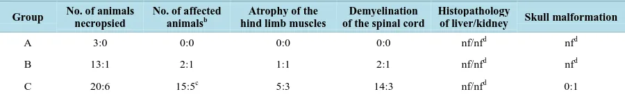
Table 5. Gross pathology and histopathology results of cheetahs (Acinonyx jubatus): lions (Panthera leo) in the United Arab
Emirates which were split into three different groups (A, B and C)a.
Group No. of animals necropsied
No. of affected animalsb
Atrophy of the hind limb muscles
Demyelination of the spinal cord
Histopathology
of liver/kidney Skull malformation
A 3:0 0:0 0:0 0:0 nf/nfd nfd
B 13:1 2:1 1:1 2:1 nf/nfd nfd
C 20:6 15:5c 5:3 14:3 nf/nfd 0:1
Meat on bone (beef and chicken) including quails, had a Ca:P level of 1:1 whereas pure muscle meat had a Ca:P ratio of 1:55 (beef) and 1:28 (chicken). All five meat products were low in Cu, Se and Zn. Quail, beef muscle meat and beef on bone possessed the highest Cu values (0.0003 g/100g dry matter; 0.0002 g/100g dry matter respectively). When compared with the supplements, most of the elements were above the reference val-ues for cats, especially Ca, Cu, and Zn [33]. However, Predator supplement (HEALTHtech laboratories (Pty) Ltd, 79 Landmarks Ave., Centurion 0157, South Africa) had a very low Cu content (0.0001 g/100g dry matter) and Copper-Trition (Equine Products [U.K.] Ltd, 22 Riversdale Court, Newcastle upon Tyne, NE I5 8SG, Unit-ed Kingdom) a very low Ca content (0.049 g/100g dry matter).
Tab water samples from three different private collection breeding places showed similar ranges (Table 3). Ranges fell within the WHO values for standard water [35].
3.3. Analytical Analysis of Serum
[image:7.595.136.462.278.688.2]Reference values for serum and hepatic VA, VE and trace elements are limited for lions and other big felids, hence the results were compared to recently published data from cheetahs [8] [36] [37]. The values are presented in Table 4and Figure 1.
Overall there were significant differences between the three groups for VA (F2,20 = 3.84, p = 0.039), VE (F2,20 = 14.06, p < 0.001), Cu (F2,28 = 45.29, p < 0.001) and Zn (F2,18= 3.97, p = 0.037). Group A had the highest VA (1.85 ± 0.68 µM/L) and VE (27.31 ± 5.69 µM/L) concentrations whereas Group C had the lowest VA (0.45 ± 0.17 µM/L) and Cu (0.41 ± 0.71 µM/L) levels of all groups. There was no significant difference for serum Se between the three groups (F2,21 = 2.909, p = 0.077). Group A was significantly higher than Group C for serum VA, VE (27.31 ± 5.69 µM/L; 15.88 ± 4.04 µM/L) and Cu (12.16 ± 3.42 µM/L; 0.41 ± 0.71 µM/L) values but not for serum Zn (10.48 ± 1.86 µM/L; 10.7 ± 7.51 µM/L). There were no significant differences between Group A and B for serum VA and Cu. Group A had significantly higher serum VE levels than Group B (27.31 ± 5.69 µM/L; 12.56 ± 7.98 µM/L) whereas Group A had significantly lower levels for serum Zn than Group B (10.48 ± 1.86 µM/L; 18.84 ± 6.26 µM/L). Group B was significantly higher than Group C for serum Cu (12.74 ± 4.26 µM/L; 0.41 ± 0.71 µM/L) but not for serum VA, VE and Zn (Figure 1).
3.4. Pathology and Histopathology
The results of the gross pathology and histopathology of necropsies are presented in Table 5.
Forty-three percent (n = 10) of the affected animals (n = 23) showed slight hind limb muscle atrophy.
In histopathology, significant pathological changes were found in the central nervous system. Demyelination of the spinal cord, characterized by bilateral dilated myelin sheath of the white matter and vacuolization of the tissue was seen in all 20 felids that showed neurologic signs before death. Diffuse vacuolated myelin sheaths were seen in the spinal cord (Figure 2and Figure 3).
[image:8.595.201.395.356.501.2]Skull malformation was seen in one lion of Group C. No amyloid was found in the liver or the kidney and no inflammation was seen in any of the cases.
Figure 2. Transverse section of the lumbar region of the spinal cord from a cheetah (Acinonyx
jubatus) showing severe vacuolation due to demyelination (H & E, bar = 100 µm).
[image:8.595.200.398.541.693.2]3.5. Analysis of Liver Tissue Samples
The results of liver tissue sample analysis are shown in Table 4.
Overall there were significant differences between the three groups for liver Cu (F2,41 = 14.40, p < 0.001) but not for liver Zn (F2,19 = 0.204, p = 0.817) concentrations. Group A was significantly higher for liver Cu concen-trations than Group C (16.01 ± 17.51 ppm wet weight; 2.16 ± 0.95 ppm wet weight) but not significantly higher than Group B (9.41 ± 6.41 ppm wet weight).
4. Discussion
Neurological disorders like ataxia, paresis and hind limb weakness have been described numerous times in cheetahs. Some authors associated their findings with Cu deficiency [21] [29] [30], while others with viral infec-tions in combination with high stress levels [27] [28], where animals started showing ataxia and respiratory signs like nasal discharge and sneezing after stressful events like hand capture, transport or relocation, but no virus was found. However, an increased corticoid production in stress situations can occur which may decrease Cu retention [3] and could trigger Cu deficiency.
Cheetahs with ataxia and hind limb paresis in the above mentioned studies, showed atrophy of the hind limb muscles and severe demyelination with vacuolation of the spinal cord, which validates our findings [12] [21] [26] [29] [38]. The neurological disease in cheetahs, characterized by degenerative lesions of the spinal cord, causing ataxia and paresis is called “cheetah encephalomyelopathy” [39]. This disease is responsible for 25% of all deaths in the European Endangered Species Programme (EEP) cheetah population [39] however the exact etiol-ogy is still uncertain [13].
Drug therapy had no real influence on the progress of this disease in previous cases [25] [26] except in one animal where progression of ataxia was inhibited by an anti-herpes drug [27]. Copper sulfate supplementation had no impact on the improvement of the clinical signs in cheetah cubs with ataxic signs at Nuremberg Zoo, Germany [27] but in another case in the USA, where cubs received oral and injectable Cu supplements, signs of ataxia improved within four weeks [20].
Liver Cu values, which are only available through liver biopsy, are a better indicator for the Cu status than serum values and are the most responsive to changes in dietary Cu intake [40]. Serum Cu levels may remain within normal ranges whereas liver levels may fluctuate according to the needs of the animals. Therefore, liver values provide the best analysis to detect Cu deficiency [41], though, with continued deficient Cu intake, serum Cu concentration will decline over time, which was shown to occur in animals of Group C. Since this study is a retrospective study, liver vitamin levels were unfortunately not available.
This study shows that felids which were only fed pure poultry diet developed ataxia and hind limb paresis more often than animals that were fed an appropriately balanced diet with regular supplements. Since muscle meat does not contain a high amount of trace elements and vitamins, [15] not only Cu but other minerals like Ca and P should be monitored closely. The Ca: P ratio, which is ideally between 1:1 - 2:1 [34], is too low if only pure muscle meat is fed (Table 3) and can result in metabolic bone disease [42] as well as Cu deficiency since high P intake depletes Cu retention [3]. As cheetahs are not able to consume large bones [43], Ca needs to be supplemented when only pure muscle meat is fed.
A primary cause of Cu deficiency, which can be assumed for animals of Group C, can occur when not enough Cu is fed [44]. A secondary cause can be due to an excessive intake of antagonistic acting mineral such as Zn [1] [2] not only when fed in large amounts but also when animals do extensive licking at cages and therefore absorb a lot of Zn [44]. This behavior has been seen in some of the animals in this study.
Chicken muscle meat does not only contain an inappropriate amount of Cu for a balanced diet for captive fe-lids, but it also has a high level of Zn (Table 3) and therefore, a diet based on chicken muscle meat without sup-plements may be a cause for the etiology of ataxia [19] [20]. Although serum Cu levels are not the best indicator for Cu deficiency, they should be analyzed when common signs like ataxia or paresis are visible. An early start on Cu supplements is recommended because felids will more likely respond to therapy [30], therefore prognosis seems better for a reversal in signs of ataxia.
Pre-toria Rd, Midrand, South Africa, P.O. Box 12,285, Vorna Valley 1686, South Africa). The serum Cu level was 8.00 µM/L before the diet was changed. After feeding beef on bone with supplements for four weeks, serum Cu concentration reached 17.80 µM/L. The same was seen with serum Se levels with 2.80 µM/L before and 4.79 µM/L after the diet change. VE levels were 5.40 µM/L and 16.20 µM/L respectively. There was not a big change in Zn (14.7 µM/L before and 13.6 µM/L after the diet change) and VA levels (0.20 µM/L before and 0.30 µM/L respectively). It is assumed that VA serum levels may react slower to diet changes than Cu serum levels. If the animal has a VA deficiency, serum VA levels may still be adequate for an uncertain amount of time [31].
VA deficiency has also been associated with cases of ataxia in cheetahs [12] [13] and lions [14]-[18] [45], particularly when fed a lean red meat diet. Hypovitaminosis A has especially been reported in lions under one year of age. The rapid growth in that age seems a causal factor for that deficiency [15]. Skull malformations have been found in affected lions and was also seen in one of the tested lions in this study which showed neuro-logical disorders before death [14] [15] [17] [31].
A balanced nutrition for these obligate carnivores is important because hyper VA intake has shown to anta-gonize ingestion of VE and Cu [3]. Some supplements contain a high level of VA, therefore the level of VE and Cu needs to be monitored and it needs to be considered that VA can be toxic when fed >10,000 IU/kg in a diet [34]. Since the liver is the major storage organ for VA, liver VA concentrations are more significant than serum values. Therefore, a liver biopsy would be recommended as a diagnostic tool. Since this study is a retrospective study, liver VA values were unfortunately not available. Only an extreme depletion or overconsumption of VA is reflected in serum VA concentrations [31].
It is very important that animals, especially cubs and infants, get a balanced nutrition with an adequate level of VA to avoid deficiencies. Blood mineral and vitamin concentrations may vary according to gender, age, en-vironmental factors and nutritional intake (interactions with other minerals and vitamins) [8].
In the wild, carnivores consume a wide variety of whole vertebrate prey (e.g. antelopes, birds) including skin, fur/feathers, viscera and bones. These prey animals eat a different diet in the wild than captive animals that are then fed to captive carnivores. For instance, a wild bird eats a more insectivorous diet in the wild than what a captive chicken is fed [46]. Hence a wild predator like a cheetah or a lion gets a more balanced diet in the wild than in captivity. Therefore, feeding whole carcass diets is more natural and enhances the animal’s physical and psychological well-being.
5. Conclusions
In summary, the results in this study show that multiple nutritional deficiencies appear to be the cause for neu-rological disorders in these species. Not all animals in this study were tested for all analysis conducted in this paper due to the opportunistic collection of the samples.
Further investigations and correlations to minerals and vitamins influencing Cu and VA absorption need to be conducted. Because this study is a retrospective one, not all Cu and VA influencing factors are analyzed.
This paper provides an overview of the correlation between mineral and vitamin serum and tissue levels and further investigations are important and to be conducted.
Acknowledgements
The authors would like to thank H.H. Sheikh Mohammed bin Rashid Al Maktoum for his support at the Central Veterinary Research Laboratory, the staff of CVRL for their help with the sample-preparation and analysis and for their help to create this study. They also thank Alan Stephenson, Christina Hebel, Corina Berner-Schultheiss, Birger Kruse, Claudia Szentiks, Eli Walker, Kathrine Forsythe and Fabiano Ezekiel from the Cheetah Conserva-tion Fund for their help during the preparaConserva-tion of this publicaConserva-tion, and are thankful to the reviewers for their helpful comments on the development for this manuscript.
References
[1] Jubb, K.V.F. and Huxtable, C.R. (1993) The Nervous System. In: Jubb, K.V.F., Kennedy, P.C. and Palmer, N., Eds.,
[2] Turnlund, J.R. (1994) Copper. In: Shils, M.E., Olson, J.A. and Shike, M., Eds., Modern Nutrition in Health and Dis-ease, Vol. 8, Lea and Febiger, Philadelphia, 231-241.
[3] Watts, D.L. (1989) The Nutritional Relationships of Copper. Journal of Orthomolecular Medicine, 4, 99-108.
[4] Goodman, B.P., Chong, B.W., Patel, A.C., Fletcher, G.P. and Smith, B.E. (2006) Copper Deficiency Myeloneuropathy Resembling B12 Deficiency: Partial Resolution of MR Imaging Findings with Copper Supplementation. American
Journal of Neuroradiology, 27, 2112-2114.
[5] Prodan, C.I., Holland, N.R., Wisdom, P.J., Burstein, S.A. and Bottomley, S.S. (2002) CNS Demyelination Associated with Copper Deficiency and Hyperzincemia. Neurology, 59, 1453-1456.
http://dx.doi.org/10.1212/01.WNL.0000032497.30439.F6
[6] Allan, G., Portas, T., Bryant, B., Howlett, R. and Blyde, D. (2008) Ulnar Metaphyseal Osteochondrosis in Seven Cap-tive Bred Cheetahs (Acinonyx jubatus). Veterinary Radiology Ultrasound, 49, 551-556.
http://dx.doi.org/10.1111/j.1740-8261.2008.00430.x
[7] Allen, M.E. and Ullrey, D.E. (2004) Relationships among Nutrition and Reproduction and Relevance for Wild Animals.
Zoo Biology, 23, 475-487. http://dx.doi.org/10.1002/zoo.20029
[8] Bechert, U., Mortenson, J., Dierenfeld, E.S., Cheeke, P., Keller, M., Holick, M., Chen, T.C. and Rogers, Q. (2002) Diet Composition and Blood Values of Captive Cheetahs (Acinonyx jubatus) Fed Either Supplemented Meat or Commercial Food Preparations. Journal of Zoo and Wildlife Medicine, 33, 16-28.
[9] Bolton, L.A. and Munson, L. (1999) Glomerulosclerosis in Captive Cheetahs (Acinonyx jubatus). Veterinary Pathology,
36, 14-22. http://dx.doi.org/10.1354/vp.36-1-14
[10] Depauw, S., Hesta, M., Whitehouse-Tedd, K., Stagegaard, J., Buyse, J. and Janssens, G.P.J. (2012) Blood Values of Adult Captive Cheetahs (Acinonyx jubatus) Fed Either Supplemented Beef or Whole Rabbit Carcasses. Zoo Biology,
31, 629-641. http://dx.doi.org/10.1002/zoo.20427
[11] Gosselin, S.J., Setchell, K.D.R., Harrington, G.W., Welsh, M.B.B., Pylypiw, H., Kozeniauskas, R., Dollard, D., Tarr, M.J. and Dresser, B.L. (1989) Nutritional Considerations in the Pathogenesis of Hepatic Veno-Occlusive Disease in Captive Cheetahs. Zoo Biology, 8, 339-347. http://dx.doi.org/10.1002/zoo.1430080404
[12] Palmer, A.C. and Cavanagh, J.B. (1995) Encephalo Myelopathy in Young Cats. Journal of Small Animal Practice, 36, 57-64. http://dx.doi.org/10.1111/j.1748-5827.1995.tb02822.x
[13] Robert, N. (2008) Neurologic Disorders in Cheetahs and Snow Leopards. In: Fowler, M.E. and Miller, R.E., Eds., Zoo and Wild Animal Medicine: Current Therapy 6. Saunders Elsevier, St. Louis, 265-271.
http://dx.doi.org/10.1016/B978-141604047-7.50035-X
[14] De Risio, L., Beltran, E., Matiasek, K., de Stefani, A. and Holloway, A. (2010) Neurological Dysfunction and Caudal Fossa Overcrowding in a Young Cheetah with Hypovitaminosis A. Veterinary Record, 167, 534-536.
http://dx.doi.org/10.1136/vr.c4802
[15] Hartley, M.P., Kirberger, R.M., Haagenson, M. and Sweers, L. (2005) Diagnosis of Suspected Hypovitaminosis A Us-ing Magnetic Resonance ImagUs-ing in African Lions (Panthera leo). Journal of the South African Veterinary Association,
76, 132-137. http://dx.doi.org/10.4102/jsava.v76i3.414
[16] Maratea, K.A., Hooser, S.B. and Ramos-Vara, J.A. (2006) Degenerative Myelopathy and Vitamin A Deficiency in a Young Black-Maned Lion (Panthera leo). Journal of Veterinary Diagnostic Investigation, 18, 608-611.
http://dx.doi.org/10.1177/104063870601800617
[17] O’Sullivan, B.M., Mayo, F.D. and Hartley, W.J. (1977) Neurologic Lesions in Young Captive Lions Associated with Vitamin A Deficiency. Australian Veterinary Journal, 53, 187-189.
http://dx.doi.org/10.1111/j.1751-0813.1977.tb00168.x
[18] Wenker, C.J. and Robert, N. (2012) Stargazing in Lions. In: Fowler, M.E. and Miller, R.E., Eds., Zoo and Wild Animal Medicine, 7th Edition, W.B. Saunders Co., Philadelphia, 470-476.
[19] Stidworthy, M.F., Lewis, J.C.M., Penderis, J. and Palmer, A.C. (2008) Progressive Encephalo Myelopathy and Cere-bellar Degeneration in a Captive-Bred Snow Leopard (Uncia uncia). Veterinary Record, 162, 522-524.
http://dx.doi.org/10.1136/vr.162.16.522
[20] Eldridge, R.(1997) LIOC Endangered Species Conservation Federation: Maintaining Mineral Balance. Newsletter, 41, 8-10.
[21] Hafner, A., Schmidt, P., Hänichen, T. and Schmahl, W. (1996) Degenerative Myelopathie bei Geparden (Acinonyx jubatus) [Degenerative Myelopathy in Cheetahs (Acinonyx jubatus)]. Berliner und Münchener tierärztliche Wochen-schrift, 109, 403.
23-26 May 2001, 333-334.
[23] Palmer, A.C. and Franklin, R.J.M. (1999) Review of Neuropathological Material from Ataxic Cheetah Cubs at Fota Wildlife Park. Workshop on Ataxia in Cheetah Cubs, Dublin, 13-15.
[24] Palmer, A.C., Callanan, J.J., Guerin, L.A., Sheahan, B.J., Stronach, N. and Franklin, R.J. (2001) Progressive Encepha-lomyelopathy and Cerebellar Degeneration in 10 Captive-Bred Cheetahs. Veterinary Record, 149, 49-54.
http://dx.doi.org/10.1136/vr.149.2.49
[25] Robert, N., Walzer, C., Petit, T. and Vitaud, C. (2006) Neurological Diseases in Cheetah. Proceedings of EAZWV 6th Scientific Meeting, Budapest, 24-28 May 2006.
[26] Walzer, C. and Kübber-Heiss, A. (1995) Progressive Hind Limb Paralysis in Adult Cheetahs (Acinonyx jubatus).
Journal of Zoo and Wildlife Medicine, 26, 430-435.
[27] Walzer, C., Kuebber-Heiss, A., Gelbmann, W., Suchy, A., Bauder, B. and Weissenboeck, H. (1998) Acute Hind Limb Paresis in Cheetah (Acinonyx jubatus) Cubs. Proceedings of the 2nd Scientific Meeting of the European Association of Zoo- and Wildlife Veterinarians (EAZWV), Chester, 21-24 May 1998, 267-274.
[28] Walzer, C., Url, A., Robert, N., Kübber-Heiss, A., Nowotny, N. and Schmidt, P. (2003) Idiopathic Acute Onset Mye-lopathy in Cheetah (Acinonyx jubatus) Cubs. Journal of Zoo and Wildlife Medicine, 34, 36-46.
[29] Zwart, P., von der Hage, M., Schotman, A.J.H., Dorrestein, G.M. and Rens, J. (1985) Copper Deficiency in Cheetah (Acinonyx jubatus). Verhandlungsbericht des 27. Internationalen Symposiums über die Erkrankungen der Zootiere, St Vincent/Torino, 9-13 June 1985, 253-257.
[30] Brand, D.J. (1980) Captive Propagation at the National Zoological Gardens of South Africa, Pretoria. International Zoo Yearbook, 20, 107-112.
[31] Shamir, M.H., Rubin, G., Aizenberg, Z., Berkovich, Z., Reifen, R., Horowitz, I., Bdolah-Abram, T. and Aroch, I. (2012) Needle Biopsy for Hepatic Vitamin A Levels in Lions (Panthera leo). Journal of Zoo and Wildlife Medicine, 43, 207- 213. http://dx.doi.org/10.1638/2010-0038.1
[32] Schweigert, F.J., Raila, J., Wichert, B. and Kienzle, E. (2002) Cats Absorb ß-Carotene, but It Is Not Converted to Vi-tamin A. Journal of Nutrition, 132, 1620-1612.
[33] FEDIAF (2011) Nutritional Guidelines for Complete and Complementary Pet Food for Cats and Dogs. European Pet Food Industry Federation, 17.
[34] National Research Council of the National Academies (2006) Nutrient Requirements of Dogs and Cats. The National Academic Press, Washington DC.
[35] Kölle, W. (2007) Wasseranalysen-Richtig Beurteilt [Evaluation of Water Analysis]. Wiley-VCH GmbH & Co. KGaA, Hannover.
[36] Beckmann, K.M., O’Donovan, D., McKeown, S., Wernery, U., Basu, P. and Bailey, T.A. (2013) Blood Vitamins and Trace Elements in Northern-East African Cheetahs (Acinonyx jubatus soemmeringii) in Captivity in the Middle East.
Journal of Zoo and Wildlife Medicine, 44, 613-626. http://dx.doi.org/10.1638/2011-0156R2.1
[37] Dierenfeld, E.S. (1993) Nutrition of Captive Cheetahs (Acinonyx jubatus): Food Composition and Blood Parameters.
Zoo Biology, 12, 143-150. http://dx.doi.org/10.1002/zoo.1430120113
[38] Shibly, S., Schmidt, P., Robert, N., Walzer, C. and Url, A. (2006) Immunohistochemical Screening for Viral Agents in Cheetah (Acinonyx jubatus) Myelopathy. Veterinary Record, 159, 557-561. http://dx.doi.org/10.1136/vr.159.17.557 [39] Robert, N. and Walzer, C. (2009) Pathological Disorders in Captive Cheetahs. In: Vargas, A., Breitenmoser-Würsten,
C. and Breitenmoser, U., Eds., Iberian Lynx ex Situ Conservation: An Interdisciplinary Approach, Fundación Biodi-versidad, Madrid, 265-272.
[40] Doong, G., Keen, C.L., Rogers, Q., Morris, J. and Rucker, R.B. (1983) Selected Features of Copper Metabolism in the Cat. Journal of Nutrition, 113, 1963-1971.
[41] Fascetti, A.J., Rogers, Q.R. and Morris, J.G. (2000) Dietary Copper Influences Reproduction in Cats. Journal of Nutri-tion, 130, 1287-1290.
[42] Fowler, M.E. (1978) Metabolic Bone Disease. In: Fowler, M.E., Ed., Zoo and Wild Animal Medicine, Saunders, Phila-delphia, 53-76.
[43] Phillips, J.A. (1993) Bone Consumptions by Cheetahs at Undisturbed Kills: Evidence of Lack of Focal-Palatine Ero-sion. Journal of Mammalogy, 74, 487-492. http://dx.doi.org/10.2307/1382408
[44] Hendriks, W.H., Allan, F.J., Tarttelin, M.F., Collett, M.G. and Jones, B.R. (2001) Suspected Zinc-Induced Copper De-ficiency in Growing Kittens Exposed to Galvanized Iron. New Zealand Veterinary Journal, 49, 68-72.
http://dx.doi.org/10.1080/00480169.2001.36205
Leukoencephalo-myelopathy in Specific Pathogen-Free Cats. Veterinary Pathology, 44, 912-916. http://dx.doi.org/10.1354/vp.44-6-912 [46] Bell, K.M. (2010) Nutrient Requirements, Utilization and Metabolism. In: Tucker, L.A. and Thomas, D.G., Eds., Spot
the Difference: Are Cheetahs Really Just Big Cats? Nottingham University Press, Nottingham, 67-91.


![Table 4. Blood vitamin and trace element analysis (mean ± standard deviation [SD] (range); µM/L) [n cheetahs: lions] of captive cheetahs (Acinonyx jubatus) and lions (Panthera leo) in the United Arab Emirates compared to published serum pa-rameters for che](https://thumb-us.123doks.com/thumbv2/123dok_us/8124768.795181/5.595.91.536.142.443/analysis-deviation-cheetahs-acinonyx-panthera-emirates-published-rameters.webp)