R E S E A R C H
Open Access
Prognostic significance of solitary lymph node
metastasis in patients with squamous cell
carcinoma of middle thoracic esophagus
Jie Wu
1,2, Qi-Xun Chen
2, Xing-Ming Zhou
2, Wei-Ming Mao
2, Mark J Krasna
3and Li-Song Teng
1*Abstract
Background:The aim of this study is to compare clinical outcomes between patients with solitary lymph node metastasis and node-negative (N0) patients in squamous cell carcinoma of the middle thoracic esophagus. Methods:A series of 135 patients with squamous cell carcinoma of the middle thoracic esophagus were
retrospectively investigated. There were 33 patients with solitary lymph node metastasis and 102 N0 patients. Skip metastasis in 33 patients with solitary lymph node metastasis was defined according to three criteria: Japanese Society for Esophageal Disease (JSED), American Joint Commission on Cancer (AJCC), and the anatomical compartment.
Results:In 33 patients with solitary lymph node metastasis, skip metastasis was shown in 13, 23, and 8 patients according JSED, AJCC and anatomical compartment respectively. The 5-year survival rates for N0 patients and patients with solitary lymph node metastasis were 58% and 32% respectively (P=0.008). Multivariate analysis revealed that skip metastasis was not an independent prognostic factor.
Conclusions:For patients with middle thoracic esophageal squamous cell carcinoma, solitary lymph node
metastasis has a negative impact on survival compared with N0 disease; skip metastasis, however, is comparable to N0 diseases in predicting prognosis.
Keywords:Esophageal cancer, Solitary lymph node, Prognosis, Lymphadenectomy
Background
Middle thoracic esophageal squamous cell carcinoma is characterized by a bidirectional lymphatic spread and lymph node metastasis [1,2]. Lymphadenectomy is an important component of surgical procedures aiming for complete resection of lesions for patients with esopha-geal carcinoma. The sentinel lymph node concept for lymphadenectomy has recently been developed in order to individualize the indication for lymphadenectomy and therefore to reduce the surgical stress of thoracic esophageal carcinoma surgery [3]. However, this concept is questioned by some authors [4-6]. For example, the site of solitary lymph node metastasis, which is thought to be the site of initial lymph node metastasis, is difficult
to predict in esophageal carcinoma because the location of solitary lymph node metastasis is extensively distribu-ted in the neck, mediastinum, and upper abdomen [4-6]. Similarly, metastasis to anatomically distant lymph nodes, known as skip metastasis, could develop even in the early phase of lymphatic invasion in patients with esophageal carcinoma [4,5,7,8].
The number of metastatic lymph nodes is one of the most important prognostic factors in esophageal cancer [9]. The new seventh tumor, node, metastasis (TNM) staging system reclassified the N stage according to the number of metastatic lymph nodes [10]. Esophageal car-cinoma has been regarded as in the earliest lymph node invasion when only one lymph node is involved [4,8]. It is important to investigate the prognosis of solitary lymph node metastasis before the presence of multiple metastatic nodes, which has already meant systematic disease and poor prognosis [11]. However, there are few * Correspondence:lsteng@zju.edu.cn
1
Department of Surgical Oncology, First Affiliated Hospital, Zhejiang University School of Medicine, 79 Qingchun Road, Hangzhou 310033, China Full list of author information is available at the end of the article
reports analyzing the prognosis of solitary lymph node metastasis in esophageal cancer.
In this study, the clinical outcomes of 33 patients with solitary lymph node metastasis in squamous cell carcin-oma of the middle thoracic esophagus were retrospect-ively investigated and compared with node-negative (N0) patients to evaluate the prognostic significance.
Patients and methods
Patients
Between January 2003 and December 2009, a series of 235 patients with middle thoracic esophageal squamous cell carcinoma underwent curative esophagectomy in the Department of Thoracic Surgery, Zhejiang Cancer Hospital, Hangzhou, China. Patients who had a history of cancer, had a synchronous cancer, or had previously received chemotherapy or radiotherapy were not included in this series. Of the 235 patients, 33 had solitary lymph node metastasis, and 102 patients had no lymph node metastasis. These 135 patients were analyzed in this retro-spective study. The institutional review board of Zhejiiang Cancer Hospital and First Affiliated Hospital, Medical College, Zhejiang University approved the study and the need for individual patient consent was waived.
Preoperative evaluation
All patients had endoscopy with biopsy, barium swallow examination, computed tomography of the chest and upper abdomen, and ultrasound of the neck. Patho-logical diagnosis of squamous cell carcinoma was con-firmed before the operation. Pulmonary and cardiac function testing were routinely performed to assess med-ical operability. Tumor location was defined according to the 6th edition of the American Joint Commission on Cancer (AJCC) Cancer Staging Manual [12]. Recurrent laryngeal nerve palsy and the presence of clinical supra-clavicular or cervical nodal involvement were considered a contraindication for surgery. The patients underwent surgery after completing the preoperative evaluation and providing written informed consent.
Surgical procedure
In our institute, two types of lymphadenectomy were carried out as a standard procedure for middle thoracic esophageal cancer. The choice of lymphadenectomy depended on the surgeon’s preference. Four surgeons performed two-field lymphadenectomy while two per-formed three-field lymphadenecotmy. The surgical indi-cation was the same for operations using these two types of lymphadenectomy.
All patients underwent right transthoracic esophagect-omy with either two- or three-field lymphadenectesophagect-omy. In principle, two-field lymphadenectomy included total mediastinal and upper abdominal lymphadenectomy.
Three-field lymphadenectomy added resection of cervical paratracheal, cervical paraesphageal and supraclavicular lymph nodes to two-field lymphadenectomy. Gastro-intestinal reconstruction was achieved by stomach bypass through the posterior mediastinal route and anastomosis was performed in the neck for all patients. After surgery, all removed nodes were labeled by their anatomical loca-tions by the operating surgeon.
Definition of skip metastasis
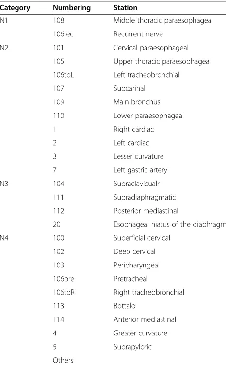
Three criteria were taken to define skip metastasis as follows: 1. Lymph node classification based on the guidelines of the Japanese Society for Esophageal Disease (JSED), which divides the locations of lymph node me-tastases into four categories (N1 through N4) [13]. Skip metastasis was diagnosed when solitary lymph node metastasis was observed in N2, N3 or N4 beyond N1. For a middle thoracic tumor, skip metastasis was defined
Table 1 Lymph node classification of middle thoracic carcinoma based on the guidelines of the Japanese Society for Esophageal Disease (JSED)
Category Numbering Station
N1 108 Middle thoracic paraesophageal
106rec Recurrent nerve
20 Esophageal hiatus of the diaphragm
when metastatic lymph node occurred in any locations other than 106rec and 108 (Table 1). 2. Lymph node classification based on the 6th edition of the AJCC Can-cer Staging Manual [12], which numbers the location of lymph node metastasis. For a middle thoracic tumor, skip metastasis was defined when solitary lymph node metastasis was observed in the upper mediastinum, upper abdomen and neck, that is, metastatic lymph node that occurred in any locations other than station 5 through 10 (Table 2). 3. Lymph node classification based on the anatomical compartment. Skip metastasis was diagnosed when a solitary lymph node metastasis was observed in the abdomen and neck rather than in the mediastinum.
Follow-up
In general, a follow-up examination was performed in our outpatient department every 3 months for the first 2 years and 6 months thereafter. The routine follow-up examination included a physical and routine blood examinations, blood chemistry, measurement of tumor markers (carcinoembryonic antigen, squamous cell car-cinoma antigen), radiograph of the chest, and ultra-sound. Computed tomography of the chest and upper abdomen were done every 6 months. Endoscopy was
done yearly. Complete follow-up information until death or March 2012 was available for all patients.
Statistical analysis
The distribution of continuous variables was described with the median and range. Categorical variables were compared between groups using the chi-square test and Fisher’s exact test if necessary. Survival curves were constructed by the Kaplan-Meier method [14] and the log-rank test [15] was used to determine significance. Significant variables identified by univariate analyses were assessed by multivariate analyses using Cox’s pro-portional hazard model [16]. A backward method was used to eliminate insignificant variables. P- values less than 0.05 were considered statistically significant.
Results
Comparison of clinicopathological characteristics between patients with solitary lymph node metastasis and patients with no lymph node metastasis (N0)
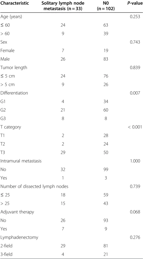
Clinicopathological characteristics of these 135 patients with N0 or solitary lymph node metastasis are listed in Table 3. In patients with solitary lymph node metastasis, advanced tumors (T3) were significantly more common than in N0 patients. There was no significant difference in any other variables between these two groups. How-ever, if the same comparison was made among the entire 235 patients, who were divided into three groups includ-ing N0 patients, patients with solitary lymph node me-tastasis and patients with more than one lymph node metastasis, all variables except age and sex were signifi-cantly different among the three groups. Tumor length > 5 cm, G3, T3 and T4 stage and intramural metastasis were more frequent in patients with more than one lymph node metastasis than in the other two groups of patients.
Distribution of solitary lymph node metastasis
The distribution of solitary lymph node metastasis based on JSED, AJCC and anatomical compartment is sum-marized in Table 4 and Table 5. Recurrent nerve node (12 cases) and middle thoracic paraesophageal node (eight cases) were the most common metastatic sites in solitary lymph node metastasis. Skip metastasis was shown in thirteen, twenty-three, and eight patients according JSED, AJCC and anatomical compartment respectively.
Survival
The 5-year survival rates for N0 patients, patients with solitary lymph node metastasis and patients with ≥ two lymph node metastases were 58%, 32%, and 15.9% respectively (P < 0.001) (Figure 1). There were signifi-cant differences in survival between N0 patients and patients with solitary lymph node metastasis (P= 0.008), Table 2 Lymph node classification of middle thoracic
carcinoma based on AJCC
Region Numbering Station
Middle and lower mediastinum 7 Subcarinal
8 M Middle paraesophageal
Upper mediastinum 2R Right upper paratracheal
2 L Left upper paratracheal
3P Posterior mediastinal
4R Right lower paratracheal
4 L Left lower paratracheal
between patients with solitary lymph node and with
≥ two lymph node metastases (P = 0.048), and between N0 patients and patients with≥two lymph node metas-tasis (P< 0.001).
Patients with solitary lymph node metastasis were fur-ther analyzed. According to the JSED, the 5-year survival rates for skip (+) and skip (−) patients were 36% and 29% respectively, both of which were significantly differ-ent from the 5-year survival for N0 patidiffer-ents (P = 0.047 and P = 0.031 respectively) (Figure 2A). According to the AJCC, the 5-year survival rates for skip (+) and skip (−) patients were 37% and 20% respectively. There was a significant difference in survival between skip (−) patients and N0 patients (P= 0.004); however, there was
no significant difference in survival between skip (+)
patients and N0 patients (P = 0.094) (Figure 2B).
According to the anatomical compartment, the 5-year survival rates for skip (+) and skip (−) patients were 58% and 23% respectively. Survival of Skip (+) patients was not significantly different from that of N0 patients (P= 0.740). There was a significant difference in survival be-tween N0 patients and skip (−) patients (P = 0.002) (Figure 2C).
Univariate and multivariate survival analysis
Univariate survival analysis showed that T category and N status were significantly associated with prognosis (Table 6). Multivariate analysis was performed including T category and N status. Owing to the different criter-ion of skip metastasis, three models were constructed. In a model according to the JSED, T category was the independent prognostic factor. In the other two models according to the AJCC and anatomical compart-ment, N status was the independent prognostic factor. In these two models, however, skip metastasis was not significantly different from N0 in predicting prognosis (Table 7).
Discussion
Patients with solitary lymph node metastasis in esopha-geal cancer has been considered a distinct prognostic subgroup with cancer outcomes closer to node-negative disease than any other node-positive subgroup [17]. It has even been believed that there is no difference in sur-vival between patients with solitary lymph node metasta-sis and N0 patients with esophageal squamous cell carcinoma, and that solitary lymph node metastasis does not affect the prognosis [5]. In this report, except for T category, all clinicopathological variables did not differ between N0 patients and patients with solitary lymph node metastasis. Solitary lymph node metastasis had a worse survival compared with N0 disease. These results may partly explained by the fact that a majority of patients with solitary lymph node metastasis were at the T3-stage. But skip metastasis was not a risk factor for poor prognosis. According to the JSED criterion both skip (+) and skip (−) patients showed worse survival than N0 patients, but it was T category not N status that was the independent prognostic factor in multivariate analysis. Although N status was the independent prog-nostic factor according to the other two criteria, there was still no significant difference between N0 and skip metastasis in predicting prognosis.
The prognostic impact of lymph node skip meta-stasis in esophageal carcinoma has been unclear. Some reports have shown a favorable prognosis of skip metas-tasis (compared with adjacent or continuous lymph node metastasis) and others have not [7,18]. Prenzel Table 3 Comparison of clinicopathological characteristics
between patients with solitary lymph node metastasis and N0 patients
Number of dissected lymph nodes 0.739
et al. [7] attributed the different results to the different lymph node classification used to define skip metastasis. In this series, three criteria were used to define skip metastasis. Skip metastasis did not show a favorable prognosis compared with adjacent metastasis. But multi-variate analysis did show that skip metastasis was not a prognostic factor, whereas adjacent metastasis was prognostic in two of the three models. It must be noted that skip metastasis could be caused by ineffi-cient histopathologic examination by missing out small metastasis in positive nodes. In addition, micrometasta-sis detected by immunohistochemistry in histologically tumor-free nodes is not a rare event in esophageal can-cer [6,18]. Natsugoe et al. [6] reported micrometastasis was found in 33 of 59 patients (55.9%) with esophageal
carcinoma, who were diagnosed as N0 by routine histo-logical examination.
As previous reports have shown [4-6,8], solitary lymph node metastasis in this report was widely distributed, but was mainly limited to the recurrent nerve region and middle thoracic paraesophagus region. It was reasonable that the JSED classified these two regions as N1 in middle thoracic squamous cell carcinoma. Some authors have found that solitary lymph node metastasis in esophageal carcinoma is predominately located in the abdomen along the lesser curvature and the left gastric artery [2]. But their series included lower thoracic tumors and upper mediastinal lymphadenectomy was not routinely performed. The wide distribution of soli-tary lymph node metastasis was closely associated with the tumor site and unique features of lymphatic spread from the esophagus, which has been discussed in many Table 4 Distribution of solitary lymph node metastasis
in 33 patients based on the guidelines of the Japanese Society for Esophageal Disease (JSED) compared with the American Joint Commission on Cancer (AJCC)
AJCC
Skip (−) (n = 10) Skip (+) (n = 23)
7 8 M 8 L 2R 2 L 3P 4 16 17 1
JSED - - -
-Skip (−) (n = 20) - - -
-106recR - - - 10 - - -
-106recL - - - - 2 - - - -
-108 - 8 - - - -Skip (+) (n = 13) -105 - - - 2 - - -
-107 1 - - - -110 - - 1 - - -
-1 - - - 2 -
-2 - - - 1 -
-3 - - - 1
-7 - - - 2
-104R - - - 1
104 L - - - 1
106pre - - - 1 - -
-Table 5 Summary of solitary lymph node metastasis in thirty-three patients defined by three criteria
JSED AJCC Anatomy
Skip (−) (n = 20) Skip (+) (n = 13) Skip (−) (n = 10) Skip (+) (n = 23) Skip (−) (n = 25) Skip (+) (n = 8)
upper mediastinum 12 3 15 15
Middle mediastinum 8 1 9 9
Lower mediastinum 1 1 1
Upper abdomen 6 6 6
Cervix 2 2 2
JSED, Japanese Society for Esophageal Disease; AJCC, American Joint Commission on Cancer.
Patients at risk N0
(n=102)
102 94 83 67 55 44 32 23 11 6
1 (n=33) 33 26 20 15 12 9 6 4 3 1
2 (n=100) 100 63 40 23 15 8 5 5 3 0
Patients at risk
N0(n=102) 102 94 83 67 55 44 32 23 11 6
Skip(+) (n=13)
13 11 8 4 3 3 2 2 1 0
Skip(-) (n=20)
20 15 12 11 8 5 4 2 2 1
Patients at risk
N0(n=102) 102 94 83 67 55 44 32 23 11 6
Skip(+) (n=23)
23 18 15 10 9 7 4 3 2 1
Skip(-) (n=10)
10 8 5 5 3 2 2 1 1 0
Patients at risk
N0(n=102) 102 94 83 67 55 44 32 23 11 6
Skip(+) (n=8)
8 7 7 4 4 4 2 2 1 0
Skip(-) (n=25)
25 19 13 11 8 5 4 2 2 1
A
B
C
previous reports [4,5,8,19,20]. In addition, middle thor-acic tumors have a more obvious tendency to develop bidirectional metastasis than upper and lower thoracic tumors of the esophagus [1,19,20], so it is still hard to predict the location of solitary lymph node metastasis, even if sentinel navigation surgery is used [4].
Three-field might be more appropriate than two-field lymphadenectomy for middle thoracic esophageal squa-mous cell carcinoma in terms of the wide distribution of solitary lymph node metastasis. But there was no survival difference between patients treated with these two types of lymphadenectomies (data not shown). Many similar results reported earlier have also been summarized in reviews of the literature [21,22]. So far, controversy exists as to the extent of lymphadenectomy required in
esophageal cancer. There is no doubt that more exten-sive lymphadenectomy provides more accurate nodal staging [21], but whether it improves survival still needs to be tested in well-designed randomized con-trolled trials [22].
Several potential shortcomings in this retrospective study deserve attention. Surgeons chose different types of lymphadenectomy depending on their preferences. A certain selection bias can therefore not be excluded. It is also possible that variation in the quality of lymphade-nectomy among different surgeons may interfere with the results. In addition, the sample size was limited by nature of the single institution of the study. Validation from other institutions is needed. Nonetheless, this series proved to be homogenous with regard to many Table 6 Univariate analysis of survival in 135 patients with squamous cell carcinoma of the middle thoracic esophagus
Variable Number of patients 5-year survival rate Median survival (months) P-value
Age (years) 0.939
≤60 87 51% 63
> 60 48 52% 61
Sex 0.500
Female 26 48% 63
Male 169 52% 60
Tumor length 0.903
≤5 cm 100 51% 63
> 5 cm 35 50% 88
Differentiation 0.553
G1 38 59% 74
G2 81 48% 59
G3 16 49% 60
T category 0.041
T1 30 67% 86
T2 26 55% 77
T3 79 44% 45
Intramural metastasis 0.135
No 131 52% 63
Yes 4 25% 29
Number of dissected lymph nodes
≤25 77 53% 66 0.929
> 25 58 48% 60
Lymphadenectomy 0.206
2-field 110 48% 60
3-field 25 66% 87
N
Negative 102 58% 74 0.008
clinical variables, such as tumor site, pathological type, and preoperative management.
Conclusions
For patients with middle thoracic esophageal squamous cell carcinoma, solitary lymph node metastasis has a negative impact on survival compared with N0 disease; skip metastasis, however, is comparable to N0 disease in predicting prognosis.
Competing interests
The authors declare that they have no competing interests.
Authors’contributions
JW conceived this study, collected data, performed analysis and drafted the manuscript. QXC participated in study design, literature search and coordination. JW, QXC, XMZ and WMM participated in the treatment of these patients. MJK performed data analysis and helped to draft the
manuscript. LST participated in study design and helped to draft the manuscript. All authors read and approve the final manuscript.
Author details
1Department of Surgical Oncology, First Affiliated Hospital, Zhejiang
University School of Medicine, 79 Qingchun Road, Hangzhou 310033, China. 2Department of Thoracic Surgery, Zhejiang Cancer Hospital, Hangzhou
310022, China.3Meridian Cancer Care, Jersey Shore University Medical Center, Neptune, NJ 07753, USA.
Received: 12 August 2012 Accepted: 18 September 2012 Published: 4 October 2012
References
1. Chen J, Liu S, Pan J, Zheng X, Zhu K, Zhu J, Xiao J, Ying M:The pattern and prevalence of lymphatic spread in thoracic oesophageal squamous cell carcinoma.Eur J Cardiothorac Surg2009,36:480–486.
2. Schröder W, Prenzel K, Baldus SE, Mönig SP, Schneider PM, Hölscher AH:
Localization of isolated lymph node metastases in esophageal cancer–does it influence the sentinel node concept?
Hepatogastroenterology2007,54:1116–1120.
3. Takeuchi H, Fujii H, Ando N, Ozawa S, Saikawa Y, Suda K, Oyama T, Mukai M, Nakahara T, Kubo A, Kitajima M, Kitagawa Y:Validation study of radio-guided sentinel lymph node navigation in esophageal cancer.Ann Surg 2009,249:757–763.
4. Matsubara T, Ueda M, Kaisaki S, Kuroda J, Uchida C, Kokudo N, Takahashi T, Nakajima T, Yanagisawa A:Localization of initial lymph node metastasis from carcinoma of the thoracic esophagus.Cancer2000,89:1869–1873. 5. Kunisaki C, Makino H, Kimura J, Oshima T, Fujii S, Takagawa R, Kosaka T,
Ono H, Akiyama H, Endo I:Therapeutic strategy for esophageal cancer based on solitary lymph node metastasis.Hepatogastroenterology2011,
58:1561–1565.
6. Natsugoe S, Matsumoto M, Okumura H, Nakashima S, Higashi H,
Uenosono Y, Ehi K, Ishigami S, Takao S, Aikou T:Initial metastatic, including micrometastatic, sites of lymph nodes in esophageal squamous cell carcinoma.J Surg Oncol2005,89:6–11.
7. Prenzel KL, Bollschweiler E, Schröder W, Mönig SP, Drebber U, Vallboehmer D, Hölscher AH:Prognostic relevance of skip metastases in esophageal cancer.
Ann Thorac Surg2010,90:1662–1667.
8. Shimada H, Okazumi S, Matsubara H, Nabeya Y, Shiratori T, Shuto K, Shimizu T, Akutsu Y, Tanizawa Y, Hayashi H, Ochiai T:Location and clinical impact of solitary lymph node metastasis in patients with thoracic esophageal carcinoma.Am J Surg2006,192:306–310.
9. Lin CS, Chang SC, Wei YH, Chou TY, Wu YC, Lin HC, Wang LS, Hsu WH:
Prognostic variables in thoracic esophageal squamous cell carcinoma.
Ann Thorac Surg2009,87:1056–1065.
10. Rice TW, Blackstone EH, Rusch VW:7th edition of the AJCC Cancer Staging Manual: esophagus and esophagogastric junction.Ann Surg Oncol2010,
17:1721–1724.
11. Peyre CG, Hagen JA, DeMeester SR, Van Lanschot JJ, Hölscher A, Law S, Ruol A, Ancona E, Griffin SM, Altorki NK, Rice TW, Wong J, Lerut T, DeMeester TR:Predicting systemic disease in patients with esophageal cancer after esophagectomy: a multinational study on the significance of the number of involved lymph nodes.Ann Surg2008,248:979985. 12. American Joint Committee on Cancer:AJCC Cancer Staging Manual. 6th
edition. Philadelphia: Lippincott-Raven; 2002.
13. Japanese Society for Esophageal Diseases:Guidelines for Clinical and Pathologic Studies on Carcinoma of the Esophagus. Tokyo: Kanehara & Co, Ltd: 9th edition; 2001.
14. Kaplan EL, Meier P:Nonparametric estimation from incomplete observations.J Am Stat Assoc1958,53:457–481.
15. Peto R:Pike MC, Armitage P, Breslow NE, Cox DR, Howard SV, Mantel N, McPherson K, Peto J.Smith PG: Design and analysis of randomized clinical trials requiring prolonged observation of each patient. II. analysis and examples. Br J Cancer1977,35:1–39.
16. Cox DR:Statistical significance tests.Br J Clin Pharmacol1982,14:325–331. 17. O'Riordan JM, Rowley S, Murphy JO, Ravi N, Byrne PJ, Reynolds JV:Impact
of solitary involved lymph node on outcome in localized cancer of the esophagus and esophagogastric junction.J Gastrointest Surg2007,
11:493–499.
Table 7 Multivariate analysis of survival in 135 patients with squamous cell carcinoma of the middle thoracic esophagus
Variable Hazard ratio 95% CI P-value
JESD
T category 0.047
T1 (reference) 1
T2 1.442 0.685, 3.033 0.335
T3 2.060 1.126, 3.769 0.019
N 0.185
N0 (reference) 1
Skip (+) 1.586 0.780, 3.223 0.203
Skip (−) 1.610 0.900, 2.882 0.109
AJCC
T category 0.205
T1 (reference) 1
T2 1.474 0.700, 3.105 0.308
T3 1.768 0.943, 3.317 0.076
N 0.013
N0 (reference) 1
Skip (+) 1.591 0.915, 2.769 0.100
Skip (−) 2.533 1.291, 4.970 0.007
Anatomy
T category 0.156
T1 (reference) 1
T2 1.517 0.720, 3.918 0.273
T3 1.843 0.988, 3.433 0.055
N 0.010
N0 (reference) 1
Skip (+) 1.145 0.458, 2.860 0.772
Skip (−) 2.178 1.320, 3.593 0.002
18. Hosch SB, Stoecklein NH, Pichlmeier U, Rehders A, Scheunemann P, Niendorf A, Knoefel WT, Izbicki JR:Esophageal cancer: the mode of lymphatic tumor cell spread and its prognostic significance.J Clin Oncol 2001,19:1970–1975.
19. Tachimori Y, Nagai Y, Kanamori N, Hokamura N, Igaki H:Pattern of lymph node metastases of esophageal squamous cell carcinoma based on the anatomical lymphatic drainage system.Dis Esophagus2011,24:33–38. 20. Motoyama S, Maruyama K, Sato Y, Usami S, Nakatsu T, Saito H, Minamiya Y,
Ogawa J:Status of involved lymph nodes and direction of metastatic lymphatic flow between submucosal and T2-4 thoracic squamous cell esophageal cancers.World J Surg2009,33:512–517.
21. Darling G:The role of lymphadenectomy in esophageal cancer.J Surg Oncol2009,99:189–193.
22. Jamieson GG, Lamb PJ, Thompson SK:The role of lymphadenectomy in esophageal cancer.Ann Surg2009,250:206–209.
doi:10.1186/1477-7819-10-210
Cite this article as:Wuet al.:Prognostic significance of solitary lymph node metastasis in patients with squamous cell carcinoma of middle thoracic esophagus.World Journal of Surgical Oncology201210:210.
Submit your next manuscript to BioMed Central and take full advantage of:
• Convenient online submission
• Thorough peer review
• No space constraints or color figure charges
• Immediate publication on acceptance
• Inclusion in PubMed, CAS, Scopus and Google Scholar
• Research which is freely available for redistribution