DOI: 10.1534/genetics.106.059238
Using Substrate-Binding Variants of the cAMP-Dependent Protein Kinase
to Identify Novel Targets and a Kinase Domain Important for
Substrate Interactions in
Saccharomyces cerevisiae
Stephen J. Deminoff, Susie C. Howard, Arelis Hester, Sarah Warner and Paul K. Herman
1Department of Molecular Genetics, Ohio State University, Columbus, Ohio, 43210 Manuscript received April 10, 2006
Accepted for publication May 23, 2006
ABSTRACT
Protein kinases mediate much of the signal transduction in eukaryotic cells and defects in kinase function are associated with a variety of human diseases. To understand and correct these defects, we will need to identify the physiologically relevant substrates of these enzymes. The work presented here de-scribes a novel approach to this identification process for the cAMP-dependent protein kinase (PKA) in Sac-charomyces cerevisiae. This approach takes advantage of two catalytically inactive PKA variants, Tpk1K336A/H338A
and Tpk1R324A, that exhibit a stable binding to their substrates. Most protein kinases, including the
wild-type PKA, associate with substrates with a relatively low affinity. The binding observed here was specific to substrates and was dependent upon PKA residues known to be important for interactions with peptide substrates. The general utility of this approach was demonstrated by the ability to identify both previously described and novel PKA substrates inS. cerevisiae. Interestingly, the positions of the residues altered in these variants implicated a particular region within the PKA kinase domain, corresponding to subdomain XI, in the binding and/or release of protein substrates. Moreover, the high conservation of the residues altered and, in particular, the invariant nature of the R324 position suggest that this approach might be generally applicable to other protein kinases.
E
UKARYOTIC cells utilize an elaborate network of signaling pathways to bring about the proper phys-iological response to changes in the extracellular envi-ronment. These pathways generally consist of a series of proteins that are responsible for amplifying the original signal and for moving it to different regions of the cell. Members of one particular family of enzymes, the protein kinases, are important mediators of much of this signal transduction (Hunter 2000; Manninget al.2002). These enzymes catalyze the transfer of the terminal phosphate from ATP to a distinct set of pro-tein targets. This phosphorylation results in specific changes in the activities associated with each of these target proteins. The net effect of activating any protein kinase therefore is due to the collective actions of the substrates of that kinase.
The cAMP-dependent protein kinase (PKA) is one of the best-characterized members of this protein family ( Johnsonet al.2001). PKA was the first protein kinase
structure to be described and much is known about the biochemical properties of this enzyme (Adams 2001;
Taylor et al. 2004). PKA activity is found in all
eu-karyotes and influences a wide variety of biological processes, from basic glucose metabolism to the
mod-ulation of long-term memory (Picket-Giesand Walsh
1986; Arnsten et al.2005). In the budding yeast
Sac-charomyces cerevisiae, PKA is a key regulator of cell growth and proliferation (Thevelein and de Winde 1999;
Herman 2002). In particular, this enzyme appears to
be a critical component of a central control mechanism that ensures that overall cell growth is properly co-ordinated with the available nutrient supply (Broach
1991; Herman2002). These activities are mediated by
three different PKA catalytic subunits, Tpk1, Tpk2, and Tpk3, that appear to have both overlapping and distinct cellular functions (Todaet al.1987; Robertson et al.
2000; Ptaceket al.2005). As with any protein kinase, a
complete understanding of the biological role of the S. cerevisiae PKA will require the identification of the targets of these enzymes.
Over the years, a number of innovative strategies have been developed to identify protein kinase sub-strates (Shahet al.1997; Zhuet al.2001; Manningand
Cantley2002; Budovskayaet al.2005). However,
de-spite these advances, this process is still often a dif-ficult and labor-intensive task, and we tend to know few, if any, of the physiologically relevant substrates of any given protein kinase (Manning and Cantley 2002;
Johnson and Hunter 2005). Here, we describe a
strategy for identifying PKA substrates that takes advan-tage of catalytically inactive variants that exhibit an increased binding to their in vivotargets. These Tpk1 1Corresponding author:Department of Molecular Genetics, Ohio State
University, 484 West Twelfth Ave., Room 984, Columbus, OH 43210. E-mail: [email protected]
variants were able to bind all substrates tested and showed no association with proteins that were not phosphory-lated by PKA. In addition, no significant binding was observed between substrates and the wild-type Tpk1. This latter result was not unexpected as protein kinases typically interact with their substrates with relatively low affinity and few kinase targets have been identified by their ability to bind to these enzymes (Manning and
Cantley 2002). The general utility of this substrate
binding was demonstrated here by the identification of both previously described and novel PKA substrates in S. cerevisiae. Interestingly, our results suggest that a par-ticular region at the C-terminus of the conserved pro-tein kinase domain might be important for substrate interactions with PKA. Moreover, the conserved nature of the residues altered in this study suggests that the results obtained here might be generally applicable to other protein kinases.
MATERIALS AND METHODS
Yeast strains and growth conditions:Standard yeast genetic methods and growth conditions were used throughout this study (Kaiseret al.1994). The yeast strains used were PHY1220 (MATahis3-D200 leu2-3,112 lys2-801 trp1-101 ura3-52 suc2-D9), PJ69A-4A (MATa gal4D gal80D his3-D200 leu2-3,11 trp1-101 ura3-52 LYS2TGAL1-HIS3 GAL2-ADE2 met2TGAL7TlacZ), and TVY614 (PHY1220prc1THIS3 pep4DTLEU2 prb1DThisG) (James et al.1996 ; Gerhardtet al.1998; Changet al.2001). The yeast rich growth medium (YPAD), synthetic complete medium (SC), and yeast minimal medium (YM) have been described (Kaiseret al.1994; Hermanand Rine1997).
Co-immunoprecipitation assays: The yeast strain PJ69-4A was transformed with a low-copy plasmid encoding Tpk1 proteins tagged at their N-terminus with three copies of the hemagglutinin (HA) epitope. The strain also contained a high-copy plasmid encoding a particular substrate (or non-substrate) that had six copies of the myc epitope at its N-terminus. All of these constructs were under the control of the copper-inducible promoter from theCUP1gene (Thiele and Hamer1986). The proteins being tested for interaction included Cdc25 (codons 51–330), Cki1 (2–200), and Cki1-N (274–471). These strains were grown at 30° in SC-glucose medium to a density of 0.5 OD600units/ml. CuSO4was added to a concentration of 100mm, and the cells were incubated for 2 additional hours. The cells were harvested, washed in TBS buffer containing 1 mm PMSF and 0.5% NP40, and then converted to spheroplasts and lysed as described (Budovskayaet al.2002). The resulting protein extracts were incubated at 4°for 2 hr with 10ml of an anti-HA affinity matrix (Roche). The beads were collected with a low-speed centrifu-gation and washed six times with 1 ml of TBS containing 1 mm PMSF and 0.5% NP40. The bound protein was eluted with 30ml of SDS sample buffer, separated by SDS-polyacrylamide gel electrophoresis, and transferred to nitrocellulose mem-branes. The relative amount of substrate present was assessed by Western immunoblotting with antibodies specific for the myc epitope (Cell Signaling). Following chemiluminescent development and exposure to X-ray film, the nitrocellulose membrane was stripped by incubating in a solution of 2% SDS, 0.0625mTris–HCl, pH 6.8, and 100 mmbME for 30 min at 50°. The blot was then washed and reprobed with anti-HA antibody (Roche) to assess the relative amount of Tpk1 protein present.
PKA in vitro kinase assays and Western immunoblot ana-lyses:PKAin vitrophosphorylation assays were performed as described (Changet al.2001). In general, these assays were performed with protein A fusion proteins that were con-structed in the vector pPHY1044 that contains two repeats of the immunoglobulin-binding region of protein A from Staph-ylococcus aureus(Budovskayaet al.2002). The protein frag-ments assayed included those listed in the above section and Dot6 (170–472). For the analysis of thein vivophosphorylation levels with the anti-PKA substrate antibody (Cell Signaling), a glutathione S-transferase (GST)–Dot6 fusion protein was precipitated with an anti-GST antibody (Cell Signaling) and run out on an SDS-polyacrylamide gel (Zhuet al.2001). The precipitated proteins were then examined by a Western immu-noblot with either the anti-GST or anti-PKA substrate antibody used at a dilution of 1:5000. All site-directed mutageneses were performed as described (Kunkel 1985; Ausubel et al.1995; Budovskayaet al.2002).
Two-hybrid analyses: For the two-hybrid assays shown, the Tpk1 variants were cloned into the Gal4 activation domain plasmid pGADT7 (Clontech) and the substrates into the Gal4 DNA-binding domain vector pGBKT7 (Clontech). The sub-strates tested included Rim15 (codons 1431–1671), Yak1 (193–362), Cki1 (2–200), Cdc25 (51–330), Kin28 (2–306), Ctk1 (2–528), Bur1 (2–657), Toa2 (2–122), and Pds1 (96– 222). In general, the yeast strain PJ69-4A was transformed with derivatives of both two-hybrid plasmids and grown to midlog phase in YM-glucose minimal medium. The cells were then plated to media lacking either adenine or histidine and in-cubated for 2–3 days at 30°to assess the two-hybrid interaction. A modified two-hybrid assay system was used specifically for the experiments shown in Figure 2B. Since overexpression of the wild-type Tpk1 was found to produce a significant growth defect (see below), alleles encoding the wild-type Tpk1 and the Tpk1KHvariant fused to the Gal4 activation domain were
placed under the control of theCUP1promoter in a single-copy plasmid. For this plasmid, nucleotides corresponding to 435 to 1 of theCUP1locus (where11 is the translation start site) were ligated into the pRS416 plasmid as anEcoRI–HindIII fragment and connected to the ATG of the Gal4 activation domain ORF via the linker TGCAAAG. Two-hybrid assays were then performed as described above except that all media were supplemented with 3mmCuSO4.
RESULTS
The catalytically inactive PKA variant Tpk1KH
exhib-ited an increased binding to known substrates in S. cerevisiae:We are interested in defining the role of PKA in the control ofS. cerevisiaegrowth, and thus have been attempting to identify the physiologically relevant sub-strates of this enzyme (Howardet al. 2002; Howard
et al.2003; Changet al.2004; Budovskayaet al.2005).
During the course of these studies, we found that a particular inactive variant of Tpk1 exhibited an in-creased binding to a number of known PKA substrates, including Cdc25 and Cki1 (Figure 1A). Cdc25 is the guanine nucleotide exchange factor for the Ras pro-teins inS. cerevisiae, and Cki1 is a choline kinase (Gross
et al.1992; Yuet al.2002). In co-immunoprecipitation
(Figure 1A). In this Tpk1KHvariant, two relatively con-served residues important for catalytic activity, K336 and H338, were each replaced with an alanine. The K336 position is conserved within the AGC subfamily of protein kinases, whereas H338 is found in a somewhat broader spectrum of serine/threonine-specific kinases (Gibbsand Zoller1991b; Hanksand Hunter1995).
Consistent with previous results with peptides (Gibbs
and Zoller1991b), we found that the Tpk1KHvariant
was20-fold less active than the wild-type enzyme in an in vitrokinase assay with a protein substrate (Figure 1C). Both alterations in Tpk1KHwere required for substrate binding as neither single mutant, K336A or H338A alone, exhibited any binding to Cdc25 or Cki1 (data not shown). Therefore, the Tpk1KHvariant, unlike the wild-type Tpk1, was able to bind stably to a number of known PKA substrates.
TPK1overexpression resulted in a severe growth de-fect: During the course of the above studies, we found that we were not able to obtain stable expression of Tpk1 from clones that were under the control of constitutive promoters, like those from the ADH1 or TDH3 genes. One interpretation of these results was that the overexpression ofTPK1was detrimental to yeast cell growth and we tested this possibility with an in-ducibleTPK1construct. It should be pointed out that a previous study had shown that aTPK1construct under the control of the galactose-inducible promoter from the GAL1gene resulted in a growth defect specifically on galactose-containing media (Nehlin et al. 1992).
However, the interpretation of these experiments is complicated by subsequent work showing that elevated levels of PKA activity inhibitGALgene expression and can result in diminished growth on galactose media (Howardet al.2002; Howardet al.2006). Therefore,
we tested whether the overexpression ofTPK1from the copper-inducible promoter from theCUP1gene would also inhibit yeast cell growth. Previous work from our lab has shown thatCUP1gene expression is not significantly affected by changes in PKA activity (Budovskayaet al.
2004). We found that the elevated expression of the wild-type Tpk1, but not catalytically inactive versions like Tpk1KH, resulted in a severe growth defect on copper-containing media (Figure 1D). Thus, these results showed that elevated levels of Tpk1 are indeed delete-rious to S. cerevisiae. Moreover, they indicated the im-portance of having inducible versions of the wild-type TPK1for some of the experiments described here.
The binding to Tpk1KH was specific for PKA
sub-strates: The interactions with the Tpk1KH variant were specific for PKA substrates as no significant binding was detected with any protein that was not phosphorylated by Tpk1. For example, in the co-immunoprecipitation assay, no interaction was detected between this variant and Cki1-N, a control Cki1 fragment that lacks the two known sites of PKA phosphorylation (Figure 2A). Similar results were obtained with a two-hybrid assay Figure1.—The Tpk1KHvariant, but not the wild-type Tpk1,
was physically associated with known PKA substrates. (A) The Tpk1KHvariant was detected in immunoprecipitates with two
PKA substrates, Cdc25 and Cki1. An HA epitope-tagged ver-sion of either the wild-type Tpk1 or the Tpk1KHvariant was
precipitated from cell extracts with an anti-HA affinity matrix. The bound substrate was eluted, separated on an SDS-poly-acrylamide gel, and transferred to a nitrocellulose membrane. The relative amount of substrate present was then assessed by Western immunoblotting with antibodies specific for the myc epitope on the particular substrate present, either Cdc25 or Cki1. Three different amounts of the final immunoprecipi-tates and original inputs were loaded onto the gels, corre-sponding to 1, 3, and 10ml of the 30 and 1000ml present, respectively. (B) A Western immunoblot showing the relative levels of the wild-type Tpk1 and Tpk1KHpresent in the
immu-noprecipitates shown in A. (C) The Tpk1KHand Tpk1R324A
var-iants were defective for PKA kinase activity in vitro. A myc epitope-tagged Cdc25 protein was incubated with [g-32P]ATP
and the wild-type Tpk1, Tpk1KH, or Tpk1R324Afor 30 min at
25°. The reaction products were separated on an SDS-poly-acrylamide gel and the extent of phosphorylation was assessed by autoradiography. (Bottom) A Western immunoblot show-ing the relative levels of the Tpk1 enzymes present in the ki-nase reactions. (D) The overexpression of the wild-type Tpk1 resulted in a growth defect in yeast cells. Wild-type cells were transformed with plasmids expressing the wild-type Tpk1, Tpk1KH, or Tpk1R324Afrom theCUP1promoter. The cells were
system. Under conditions that produced equivalent lev-els of enzyme, a positive two-hybrid signal was observed with the Tpk1KHvariant but not with the wild-type Tpk1 (Figure 2, B and C). Again, this signal was specific to substrates as two control proteins, Rim15-N and Toa2, did not interact with Tpk1KH (Figure 2B). Rim15 is a protein kinase important for stationary phase entry that has been shown to be a PKA target and Toa2 is a general transcription factor that is not a substrate for PKA (Reinderset al.1998; Budovskayaet al.2005) (Figure
2D). The Rim15-N fragment lacks the known PKA sites in this protein and is not phosphorylated by PKA (data not shown). In contrast, a Rim15 fragment that contains the known sites of PKA phosphorylation exhibited a
robust interaction with the Tpk1KHvariant (Figure 2E). Therefore, the Tpk1KHvariant, but not the wild-type Tpk1, was able to interact specifically with known substrates of PKA.
We further examined the specificity of this binding with a set of.40 proteins that were not substrates for PKA (Figure 3; data not shown). These negative controls included several proteins, such as Cdc14 and Toa2, that contain a reasonable match to the PKA consensus site of R 3-R 2-x 1-S/T-B11, wherexrefers to any amino acid,
Bto a hydrophobic residue, and theSorTto the site of phosphorylation (Zetterqvistet al.1976; Smithet al.
1999). Although many known PKA substrates are phos-phorylated at a sequence that conforms to this consen-sus, most occurrences of this site in native proteins are not recognized by PKA (Shabb2001; Budovskaya
et al.2005). We found that none of these nonsubstrate proteins exhibited any binding to the Tpk1KH variant (Figure 3, A and B). Therefore, Tpk1KHwas not simply binding to proteins that contained a PKA consensus site but was instead recognizing bona fidesubstrates of this enzyme.
Substrate binding by the Tpk1KH variant was
depen-dent upon PKA residues known to be important for the interaction with substrates: We tested whether the ob-served interactions with Tpk1KH were dependent on PKA residues previously shown to be important for substrate recognition. These studies took advantage of work that had identified amino acids within PKA that
Figure2.—The interactions with the Tpk1KHvariant were specific to PKA substrates. (A) The Tpk1KHvariant did not
as-sociate with a fragment of Cki1 that lacks the sites of PKA phosphorylation. The relative amount of three different pro-teins, Cdc25 (codons 51–330), Cki1 (2–200), and Cki1-N (274–471), precipitating with the Tpk1KHvariant was assessed
as described in Figure 1A. The asterisk denotes a nonspecific protein band recognized by thea-myc epitope antibody. (B) A two-hybrid assay showing that the Tpk1KHvariant interacted
with the known substrates, Cdc25 and Cki1, but not with the nonsubstrates, Rim15-N and Toa2. The wild-type Tpk1 failed to interact with any of these test proteins. For these assays, the Tpk1 fusions to the Gal4 activation domain were under the control of the inducible promoter from the CUP1gene (seematerials and methods). Rim15-N (codons 361–657) was a negative control that was not phosphorylated by PKA. (C) A Western immunoblot showing the relative levels of the wild-type Tpk1 and Tpk1KHpresent in the two-hybrid
strains shown in B. (D) Toa2 was not phosphorylated by PKA in vitro. Protein A fusions to Toa2 and a portion of Cdc25 (codons 51–330) were precipitated from yeast cell ex-tracts and incubated with PKA and [g-32P]ATP. The reaction
products were separated on an SDS-polyacrylamide gel and the level of phosphorylation was assessed by autoradiography. (Right) A Western immunoblot showing the relative levels of the two substrates present in the kinase assays. (E) The Tpk1KHvariant interacted with a Rim15 fragment containing
were critical for the association with a peptide substrate (Gibbs and Zoller1991a; Kemp et al. 1994). For
ex-ample, replacement of E171 and E274 with alanine in the wild-type Tpk1 resulted in a 40- to 80-fold increase in the apparent Kmfor this peptide (Gibbs and Zoller 1991a; Kempet al.1994). Here, we found that the
in-troduction of these E171A and E274A alterations into the Tpk1KHvariant completely abolished binding to two PKA substrates, Cdc25 and Rim15 (Figure 4A). Thus, the Tpk1KH variant may interact with substrates in a manner similar to that used by the wild-type enzyme.
We tested the sensitivity of this binding assay with a substrate containing only a single site of PKA phos-phorylation. Most of the targets used above are likely phosphorylated by PKA at more than one position (Grosset al.1992; Reinderset al.1998; Yuet al.2002).
For these experiments, we used a fragment of Yak1 that encompassed codons 243–361 and was phosphorylated by PKA at the serine residue at position 295 (Figure 4C). As with the other PKA substrates tested, we found that this Yak1 fragment exhibited a stable interaction specif-ically with the Tpk1KH variant (Figure 4D; data not shown). Thus, Tpk1KHwas able to associate with targets containing only a single site of PKA phosphorylation.
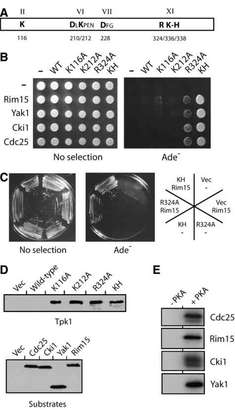
A second alteration within the C-terminal region of the Tpk1 kinase domain resulted in an increased af-finity for substrates: We also tested whether the sub-strate binding observed with the Tpk1KH variant was a general property of catalytically inactive versions of PKA. For these studies, we analyzed several different Tpk1 variants, including Tpk1K116A, Tpk1D210A, Tpk1K212A, Tpk1D228A, and Tpk1R324A, that alter residues that are highly conserved in protein kinases (Hanksand Hunter
1995; Hanks 2003) (Figure 5A). A previous study had
shown that each of these variants exhibited diminished
activity toward a peptide substrate and we confirmed these results here with protein substrates (Gibbs and
Zoller1991b) (see Figure 1C; our unpublished data).
We found that none of these inactive versions of Tpk1, except Tpk1R324A, exhibited any binding to the PKA substrates tested (Figure 5, B–E) (data not shown). In general, the two-hybrid signal with the Tpk1R324Avariant was less than that observed with Tpk1KH(Figure 5, B and C). However, it should be pointed out that more subtle alterations that changed the R324 residue to either a lysine or a histidine residue resulted in a stronger binding with specific substrates (data not shown). In all, this Tpk1R324Avariant is interesting for a couple of reasons. First, the residue altered, R324, is in the same C-terminal region of the protein kinase domain (i.e., Figure3.—The Tpk1KHvariant did not interact with
pro-teins that were not substrates of PKA, including those that possessed sequences matching the PKA consensus phosphor-ylation site. (A) A two-hybrid assay examining the interactions between the Tpk1KHvariant and a number of proteins that
were not phosphorylated by PKA in vitro. Rim15 served as the positive control in these assays. (B) A Western immuno-blot showing the relative levels of the different substrate pro-teins tested.
Figure 4.—Tpk1KH binding to substrates required amino acid residues known to be important for the interaction be-tween the wild-type PKA and its substrates. (A) A two-hybrid assay assessing the interactions between Tpk1KHvariants and
two substrates, Rim15 and Cdc25. The Tpk1KH(E/A)
var-iant has replaced both E171 and E274 with an alanine residue. These glutamic acid residues have been shown previously to be important for Tpk1 interactions with peptide substrates (Gibbsand Zoller1991a; Kempet al.1994). (B) A Western immunoblot showing the relative levels of the two Tpk1 en-zymes present in the two-hybrid strains with Cdc25 as sub-strate. (C) A Yak1 fragment containing codons 243–361 is phosphorylated at a single site by PKAin vitro. Protein A fu-sions to Yak1 with either a wild-type (Yak1) or altered PKA site [Yak1( )] were precipitated from yeast cell extracts and incu-bated with PKA and [g-32P]ATP. The reaction products were
separated on an SDS-polyacrylamide gel and the level of phos-phorylation was assessed by autoradiography. (Right) A West-ern immunoblot showing the relative levels of the two Yak1 proteins present in the kinase assays. (D) A Yak1 fragment containing only a single site of PKA phosphorylation (codons 243–361) was recognized by the Tpk1KH variant in a
within subdomain XI) as the K336 and H338 residues altered in Tpk1KH(Figure 5A). Thus, this region of PKA might play a specific role in the binding and/or release of protein substrates. Second, this arginine is conserved in all protein kinases and therefore it will be important to test if alterations of this position in other kinases result in a similar increase in substrate binding.
Dot6, a protein implicated in telomere function, is a substrate for PKA: A preliminary screen with the Tpk1KH variant identified the Dot6 protein as a candi-date substrate for Tpk1. As with other substrates, Dot6 was found to interact with the Tpk1KH and Tpk1R324 variants, but not with the wild-type Tpk1 (Figure 6A; data not shown). Although the precise biological role of Dot6 is unknown, previous work has shown that the overexpression of this protein disrupts the transcrip-tional silencing that normally occurs at yeast telomeres (Singer et al.1998). Interestingly, the predicted Dot6
protein sequence contains five consensus PKA phos-phorylation sites, the most sites present in any S. ce-revisiaeprotein (Budovskayaet al.2005) (Figure 6B).
In addition, these sites are evolutionarily conserved as all five sites are found in the likely Dot6 ortholog present in the distantly related Saccharomyces species S. kluyveri (Budovskaya et al. 2005). Consistent with
these observations, Dot6 was phosphorylated efficiently in vitroby both bovine and yeast PKA enzymes (Figure 6C). To assess whether Dot6 was also phosphorylated by PKA in vivo, we took advantage of a commercially available antibody that recognizes phosphorylated PKA sites. This antibody has been used to demonstrate that several proteins, including Srb9 and Cdc20, are phos-phorylated by PKA in vivo(Changet al. 2004; Searle
et al.2004). Here, we found that this antibody specifi-cally recognized a GST–Dot6 protein that was precipi-tated from yeast cell extracts (Figure 6D). In contrast, this antibody failed to recognize several proteins, such as Gcs1, that were not substrates for PKAin vitro (see Figure 6D) (Budovskayaet al.2005). This recognition
of Dot6 was lost upon phosphatase treatment of the precipitated fusion protein and was subsequently re-stored following incubation with PKA and ATP (Figure 6E). In all, these data suggested that Dot6 was both an in vitroand anin vivosubstrate for PKA in S. cerevisiae. Further work will be necessary to define precisely how PKA phosphorylation influences the normal functions of the Dot6 protein.
DISCUSSION
A complete understanding of the biological role of any protein kinase requires the identification of the relevant substrates of that enzyme. This study describes a novel approach to this identification process that takes advantage of protein kinase variants that bind stably to substrates. The two PKA variants identified here, Tpk1KH and Tpk1R324A, were able to bind to all substrates tested
Figure5.—Tpk1KHand Tpk1R324Awere the only inactive var-iants of Tpk1 that interacted with known substrates of PKA. (A) A schematic representing the protein kinase domain of Tpk1 (aa 90–390). The Tpk1 residues altered here are shown in boldface type; the relative positions of these residues in Tpk1 are indicated by the numbers below the diagram. The roman numerals above the diagram indicate the Hanks and Hunter subdomain designations for the indicated regions of the protein kinase domain (Hanks and Hunter 1995). (B) A two-hybrid analysis showing the interactions observed between different Tpk1 variants and four known PKA sub-strates, Rim15, Yak1, Cki1, and Cdc25. An interaction is indi-cated by growth on the YM minimal medium lacking adenine (Ade ). (C) Plates showing the relative two-hybrid interaction observed for the Tpk1KH and Tpk1R324A variants with the
Rim15 substrate. (D) Western immunoblots showing the rela-tive levels of the different Tpk1 variants and substrates pres-ent in the strains used for the two-hybrid analyses in B and C. Note that very low levels of the wild-type Tpk1 were found in these two-hybrid strains. (E) The substrates used in the above two-hybrid analysis were all efficient in vitro substrates for PKA. Each of the indicated substrate proteins was precipitated from yeast cell extracts and incubated with the yeast Tpk1 and [g-32P]ATP as described inmaterials and methods. The
but exhibited no interactions with proteins that were not phosphorylated by PKA. Moreover, these interac-tions were dependent upon PKA residues previously shown to be required for the recognition of peptide substrates (Gibbsand Zoller1991a; Kempet al.1994).
Thus, these variants appear to interact specifically with substrates in a manner that may be similar to that used by the wild-type enzyme. The general utility of these variants was demonstrated by the identification of the Dot6 protein as a new substrate for PKA inS. cerevisiae.
In addition to identifying new substrates, the binding assay described here should also assist efforts to identify the substrate sequences that are required for a pro-ductive interaction with PKA. To date, most studies examining these elements have focused on the rec-ognition and phosphorylation of peptide substrates (Songyanget al.1994; Manningand Cantley2002).
The identification of the R-R-x-S/T-B consensus phos-phorylation site discussed above is due, at least in part, to these efforts (Shabb2001). However, one limitation
of these types of studies is that they would miss any sequence elements in substrates that are distal to the actual site of PKA phosphorylation. In fact, although many PKA substrates are phosphorylated at sequences that generally conform to this consensus, most occur-rences of these sequence elements in native proteins are not recognized by PKA (see,e.g., Budovskayaet al.
2005). Therefore, there must be additional sequence information present in substrates that governs the interaction with PKA. We feel that the use of the Tpk1 variants described here should facilitate the identifica-tion of these domains, and we are presently testing for
the existence of such sequence elements in a number of PKA substrates fromS. cerevisiae.
This assay may also provide complementary informa-tion about the sequences within PKA that are important for the interaction with substrates. In fact, the results presented here already implicate a small region at the C-terminal end of the PKA kinase domain in the bind-ing and/or release of protein substrates. In general, all protein kinases possess a highly conserved, catalytic core domain of 250–300 amino acids that contains a number of signature residues critical for catalytic ac-tivity (Hanksand Hunter1995; Manninget al.2002;
Johnsonand Hunter 2005). This kinase domain has
been further divided into 11 subdomains and all of the residues altered in the Tpk1KHand Tpk1R324Avariants fall into subdomain XI (see Figure 5A). Inactivating alter-ations in other subdomains of the PKA catalytic core did not result in a concomitant increase in substrate binding. Interestingly, the arginine corresponding to position 324 in Tpk1 is thought to contribute to the stabilization of a peptide-binding loop in PKA that is in direct contact with peptide substrates ( Johnsonet al.
2001). Thus, this region of PKA may play an impor-tant role in coordinating the interactions between this enzyme and its in vivo substrates. This possibility could be tested directly if we could obtain co-crystals that contain the Tpk1KHvariant with a relevant protein substrate.
An obvious question for the protein kinase field at large is whether the results obtained here might be generally applicable. Clearly, the key to success in this endeavor will be the ability to isolate kinase variants, Figure 6.—The Dot6 protein was identified as a candidate substrate for PKA. (A) Dot6 interacted with the Tpk1KH variant in a two-hybrid assay.
analogous to Tpk1KHand Tpk1R324A, that can bind stably to their cognate substrates. In this regard, it is important to point out again that the arginine residue altered in the Tpk1R324Avariant is found at an analogous position in all other protein kinases (Hanksand Hunter1995).
Thus, it will be important to test whether this invariant residue might play a role in regulating substrate in-teractions in other protein kinases and if alteration of this site might result in a similar increase in substrate binding. Alternatively, it might be necessary to alter distinct residues in other kinases to obtain an enzyme that binds stably to substrates. The important point is that the work presented here with PKA demonstrates the feasibility of this type of an approach and can serve as a framework for future studies with other protein kinases.
It is interesting also to speculate on the possibility that the substrate binding observed here could serve as a platform for the identification of a new class of protein kinase inhibitors. Most of the protein kinase inhibitors in use today target the conserved active site of these enzymes and act as competitive inhibitors of ATP (Cohen 2002). However, because of the highly
con-served nature of the ATP-binding pocket, it can be difficult to obtain inhibitors of this type that have the specificity needed for clinical applications. In contrast, molecules that disrupt the substrate binding observed here would be interfering with the ability of the protein kinase to interact with its protein substrates. Since dif-ferent families of protein kinases interact with substrates in quite distinct ways (Hardieand Hanks1995), these
latter types of inhibitors might exhibit a higher degree of specificity. In fact, this approach could potentially identify inhibitors that are substrate-specific,i.e., inhib-itors that affect the phosphorylation of only one, or a subset, of the targets of a given protein kinase. The availability of such selective inhibitors would be invalu-able in our attempts to understand which substrates are responsible for the particular actions of a protein kinase and could ultimately allow for a more precise treatment of the consequences of protein kinase malfunction associated with human disease.
In summary, this article describes how novel substrate-binding variants of protein kinases may be used to identify physiologically relevant targets of these en-zymes. Although we presently do not know the mecha-nistic basis for the increased affinity toward PKA substrates observed here, previous studies with the Tpk1KH and Tpk1R324A variants suggested that these enzymes were specifically defective for catalytic activity (Gibbsand Zoller1991b). The alterations examined
resulted in a significant defect in the catalytic constant orkcatof the altered enzymes relative to the wild type, but had no effect on the observedKmfor either ATP or a peptide substrate. One possibility suggested by these data is that these altered enzymes might bind normally to substrates but a specific defect prevents effective
release of these targets. However, it is important to remember thatKmvalues are not simply equivalent to dissociation constants and cannot be regarded as accu-rate gauges of enzyme/substaccu-rate affinity. Indeed, there are other possible explanations for the apparent in-creased substrate binding observed here. It may be es-pecially important to emphasize that our studies were performed with protein substrates, whereas previous work utilized model peptides exclusively. Thus, it is pos-sible that not yet identified aspects of the enzyme interaction with larger protein substrates, not reflected in the smaller peptides, might influence turnover. If this were the case, the kinetic parameters described pre-viously for these Tpk1 variants might not apply. Further experimentation will therefore be necessary to deter-mine the precise mechanisms underlying the increased substrate binding observed here.
We thank Joseph Heitman for the GST–Tpk1 plasmid; Michael Snyder for the yeast GST–Dot6 and GST–Gcs1 fusion proteins; Komudi Singh for the phosphorylation analysis of Toa2; Jack Dixon, Russell Hille, and Susan Taylor for helpful discussions and advice; and Joseph Stephan for comments on the manuscript. This study was supported by a grant from the National Institutes of Health (GM-65227) to P.K.H.
LITERATURE CITED
Adams, J. A., 2001 Kinetic and catalytic mechanisms of protein
kinases. Chem. Rev.101:2271–2290.
Arnsten, A. F., B. P. Ramos, S. G. Birnbaum and J. R. Taylor,
2005 Protein kinase A as a therapeutic target for memory dis-orders: rationale and challenges. Trends Mol. Med.11:121–128. Ausubel, F. M., R. Brent, R. E. Kingston, D. D. Moore, J. G.
Seidmanet al., 1995 Current Protocols in Molecular Biology.John
Wiley & Sons, New York.
Broach, J. R., 1991 RAS genes in Saccharomyces cerevisiae: signal
transduction in search of a pathway. Trends Genet.7:28–33. Budovskaya, Y. V., H. Hama, D. B. DeWald and P. K. Herman,
2002 The C terminus of the Vps34p phosphoinositide 3-kinase is necessary and sufficient for the interaction with the Vps15p protein kinase. J. Biol. Chem.277:287–294.
Budovskaya, Y. V., J. S. Stephan, F. Reggiori, D. J. Klionskyand
P. K. Herman, 2004 The Ras/cAMP-dependent protein kinase
signaling pathway regulates an early step of the autophagy pro-cess in Saccharomyces cerevisiae. J. Biol. Chem. 279: 20663– 20671.
Budovskaya, Y. V., J. S. Stephan, S. J. Deminoffand P. K. Herman,
2005 An evolutionary proteomics approach identifies sub-strates of the cAMP-dependent protein kinase. Proc. Natl. Acad. Sci. USA102:13933–13938.
Chang, Y. W., S. C. Howard, Y. V. Budovskaya, J. Rine and P. K.
Herman, 2001 Theryemutants identify a role for Ssn/Srb
pro-teins of the RNA polymerase II holoenzyme during stationary phase entry inSaccharomyces cerevisiae.Genetics157:17–26. Chang, Y. W., S. C. Howardand P. K. Herman, 2004 The Ras/PKA
signaling pathway directly targets the Srb9 protein, a component of the general RNA polymerase II transcription apparatus. Mol. Cell.15:107–116.
Cohen, P., 2002 Protein kinases—The major drug targets of the
twenty-first century? Nat. Rev. Drug Discov.1:309–315. Gerhardt, B., T. J. Kordas, C. M. Thompson, P. Pateland T. Vida,
1998 The vesicle transport protein Vps33p is an ATP-binding protein that localizes to the cytosol in an energy-dependent man-ner. J. Biol. Chem.273:15818–15829.
Gibbs, C. S., and M. J. Zoller, 1991a Identification of electrostatic
Gibbs, C. S., and M. J. Zoller, 1991b Rational scanning mutagenesis
of a protein kinase identifies functional regions involved in catal-ysis and substrate interactions. J. Biol. Chem.266:8923–8931. Gross, E., D. Goldbergand A. Levitzki, 1992 Phosphorylation of
the S. cerevisiae Cdc25 in response to glucose results in its disso-ciation from Ras. Nature360:762–765.
Hanks, S. K., 2003 Genomic analysis of the eukaryotic protein
ki-nase superfamily: a perspective. Genome Biol.4:111.
Hanks, S. K., and T. Hunter, 1995 Protein kinases 6. The
eukary-otic protein kinase superfamily: kinase (catalytic) domain struc-ture and classification. FASEB J.9:576–596.
Hardie, G., and S. Hanks(Editors), 1995 The Protein Kinase
Facts-book: Protein-Serine Kinases.Academic Press, San Diego. Herman, P. K., 2002 Stationary phase in yeast. Curr. Opin.
Microbiol.5:602–607.
Herman, P. K., and J. Rine, 1997 Yeast spore germination: a
require-ment for Ras protein activity during re-entry into the cell cycle. EMBO J.16:6171–6181.
Howard, S. C., Y. V. Budovskaya, Y. W. Changand P. K. Herman,
2002 The C-terminal domain of the largest subunit of RNA polymerase II is required for stationary phase entry and function-ally interacts with the Ras/PKA signaling pathway. J. Biol. Chem.
277:19488–19497.
Howard, S. C., A. Hesterand P. K. Herman, 2003 The Ras/PKA
signaling pathway may control RNA polymerase II elongation via the Spt4p/Spt5p complex inSaccharomyces cerevisiae.Genetics
165:1059–1070.
Howard, S. C., S. J. Deminoffand P. K. Herman, 2006 Increased
phosphoglucomutase activity suppresses the galactose growth de-fect associated with elevated levels of Ras signaling in S. cerevi-siae. Curr. Genet.49:1–6.
Hunter, T., 2000 Signaling—2000 and beyond. Cell100:113–127.
James, P., J. Halladayand E. A. Craig, 1996 Genomic libraries and
a host strain designed for highly efficient two-hybrid selection in yeast. Genetics144:1425–1436.
Johnson, D. A., P. Akamine, E. Radzio-Andzelm, M. Madhusudan
and S. S. Taylor, 2001 Dynamics of cAMP-dependent protein
kinase. Chem. Rev.101:2243–2270.
Johnson, S. A., and T. Hunter, 2005 Kinomics: methods for
deci-phering the kinome. Nat. Methods2:17–25.
Kaiser, C., S. Michaelis and A. Mitchell, 1994 Methods in Yeast
Genetics. Cold Spring Harbor Laboratory Press, Cold Spring Harbor, NY.
Kemp, B. E., M. W. Parker, S. Hu, T. Tiganisand C. House, 1994
Sub-strate and pseudosubSub-strate interactions with protein kinases: deter-minants of specificity. Trends Biochem. Sci.19:440–444. Kunkel, T. A., 1985 Rapid and efficient site-specific mutagenesis
without phenotypic selection. Proc. Natl. Acad. Sci. USA 82:
488–492.
Manning, B. D., and L. C. Cantley, 2002 Hitting the target:
emerg-ing technologies in the search for kinase substrates. Sci. STKE
162:1–4.
Manning, G., G. D. Plowman, T. Hunter and S. Sudarsanam,
2002 Evolution of protein kinase signaling from yeast to man. Trends Biochem. Sci.27:514–520.
Nehlin, J. O., M. Carlbergand H. Ronne, 1992 Yeast SKO1 gene
encodes a bZIP protein that binds to the CRE motif and acts as a repressor of transcription. Nucleic Acids Res.20:5271–5278. Picket-Gies, C. A., and D. A. Walsh, 1986 Phosphorylase kinase,
pp. 395–459 inThe Enzymes, edited by P. D. Boyerand E. G.
Krebs. Academic Press, San Diego.
Ptacek, J., G. Devgan, G. Michaud, H. Zhu, X. Zhuet al., 2005 Global
analysis of protein phosphorylation in yeast. Nature 438: 679– 684.
Reinders, A., N. Burckert, T. Boller, A. Wiemkenand C. DeVirgilio,
1998 Saccharomyces cerevisiae cAMP-dependent protein kinase controls entry into stationary phase through the Rim15p protein ki-nase. Genes Dev.12:2943–2955.
Robertson, L. S., H. C. Causton, R. A. Youngand G. R. Fink,
2000 The yeast A kinases differentially regulate iron uptake and respiratory function. Proc. Natl. Acad. Sci. USA97:5984– 5988.
Searle, J. S., K. L. Schollaert, B. J. Wilkins and Y. Sanchez,
2004 The DNA damage checkpoint and PKA pathways con-verge on APC substrates and Cdc20 to regulate mitotic progres-sion. Nat. Cell Biol.6:138–145.
Shabb, J. B., 2001 Physiological substrates of cAMP-dependent
pro-tein kinase. Chem. Rev.101:2381–2411.
Shah, K., Y. Liu, C. Deirmengianand K. M. Shokat, 1997
Engi-neering unnatural nucleotide specificity for Rous sarcoma virus tyrosine kinase to uniquely label its direct substrates. Proc. Natl. Acad. Sci. USA94:3565–3570.
Singer, M. S., A. Kahana, A. J. Wolf, L. L. Meisinger, S. E. Peterson
et al., 1998 Identification of high-copy disruptors of telomeric silencing in Saccharomyces cerevisiae. Genetics150:613–632. Smith, C. M., E. Radzio-Andzelm, K. R. Madhusudan, P. Akamine
and S. S. Taylor, 1999 The catalytic subunit of
cAMP-dependent protein kinase: prototype for an extended network of communication. Prog. Biophys. Mol. Biol.71:313–341. Songyang, Z., S. Blechner, N. Hoagland, M. F. Hoekstra,
H. Piwnica-Wormset al., 1994 Use of an oriented peptide
li-brary to determine the optimal substrates of protein kinases. Curr. Biol.4:973–982.
Taylor, S. S., J. Yang, J. Wu, N. M. Haste, E. Radzio-Andzelmet al.,
2004 PKA: a portrait of protein kinase dynamics. Biochim. Biophys. Acta1697:259–269.
Thevelein, J. M., and J. H.deWinde, 1999 Novel sensing
mecha-nisms and targets for the cAMP-protein kinase A pathway in the yeast Saccharomyces cerevisiae. Mol. Microbiol. 33: 904– 918.
Thiele, D. J., and D. H. Hamer, 1986 Tandemly duplicated
up-stream control sequences mediate copper-induced transcription of the Saccharomyces cerevisiae copper-metallothionein gene. Mol. Cell Biol.6:1158–1163.
Toda, T., S. Cameron, P. Sass, M. Zollerand M. Wigler, 1987 Three
different genes in S. cerevisiae encode the catalytic subunits of the cAMP-dependent protein kinase. Cell50:277–287.
Yu, Y., A. Sreenivas, D. B. Ostranderand G. M. Carman, 2002
Phos-phorylation of Saccharomyces cerevisiae choline kinase on Ser30 and Ser85 by protein kinase A regulates phosphatidylcholine syn-thesis by the CDP-choline pathway. J. Biol. Chem. 277: 34978– 34986.
Zetterqvist, O., U. Ragnarsson, E. Humble, L. Berglundand
L. Engstrom, 1976 The minimum substrate of cyclic
AMP-stimulated protein kinase, as studied by synthetic peptides repre-senting the phosphorylatable site of pyruvate kinase (type L) of rat liver. Biochem. Biophys. Res. Commun.70:696–703. Zhu, H., M. Bilgin, R. Bangham, D. Hall, A. Casamayor et al.,
2001 Global analysis of protein activities using proteome chips. Science293:2101–2105.