A Unique Class of Conditional
sir2
Mutants Displays Distinct Silencing
Defects in
Saccharomyces cerevisiae
Sandra N. Garcia and Lorraine Pillus
1Division of Biology, UCSD Cancer Center and Center for Molecular Genetics, University of California, San Diego, California 92093-0347
Manuscript received April 9, 2002 Accepted for publication July 25, 2002
ABSTRACT
Silencing provides a critical means of repressing transcription through the assembly and modification of chromatin proteins. The NAD⫹-dependent deacetylation of histones by the Sir2p family of proteins lends mechanistic insight into howSIR2contributes to silencing. Here we describe three locus-specific sir2mutants that have a spectrum of silencing phenotypes in yeast. These mutants are dependent onSIR1 for silencing function at theHM silent mating-type loci, display distinct phenotypes at the rDNA, and have dominant silencing defects at the telomeres. Telomeric silencing is restored if the mutant proteins are directly tethered to subtelomeric regions, via aGal4p DNA-bindingdomain (GBD), or are recruited by tethered GBD-Sir1p. Thesesir2mutations are found within conserved residues of theSIR2family and lead to defects in catalytic activity. Since one of the mutations lies outside the previously defined minimal catalytic core, our results show that additional regions of Sir2p can be important for enzymatic activity and that differences in levels of activity may have distinct effects at the silenced loci.
S
ILENCING is a process by which transcriptional re- telomeres (Shouet al.1999;Straightet al.1999;Ghi-pression occurs in a regional, promoter-nonspecific delliet al.2001;Hoppeet al.2002). The Sir2, Sir3, and manner. Chemical modifications to DNA, histones, and Sir4 proteins also mediate silencing at theHMloci with other nuclear proteins can lead to specific alterations in the aid of the targeting protein Sir1p.
chromatin structure that may disrupt or promote trans- In contrast to the variegated silencing states observed
criptional silencing (reviewed inWolffeandGuschin for PolII transcribed genes at the telomeres and within
2000;RiceandAllis 2001). In Saccharomyces cerevisiae the rDNA array, the HMloci are distinct in that they at least three loci are subject to silencing: telomeres, the normally appear completely repressed. This difference silent mating-type loci (HMRandHML, theHMloci), and may be due in part to the interactions between Sir1p the ribosomal DNA (rDNA) repeats. Although manycis- and Orc1p and to their participation in silencing at the andtrans-acting factors have been implicated in silenc- HMloci (PillusandRine1989;TrioloandSternglanz ing in yeast (reviewed inGartenberg2000), few factors 1996;Foxet al.1997;Gardneret al.1999). One puzzle
appear required at all three loci; among these is the aboutsir1⌬mutant phenotypes has been that a
subpop-silentinformation regulator, Sir2p. ulation of cells maintains normal silencing. This
sug-The Sir2 protein is an NAD⫹-dependent protein gests that otherSIR1-independent mechanisms are
in-deacetylase that is conserved across all kingdoms of life volved in the establishment of silencing at HML and
(reviewed in Gottschling 2000; Guarente 2000; HMR. To gain insight into these mechanisms, a genetic
Shore2000;Moazed2001). The Sir2 family of proteins screen to isolate enhancers of thesir1⌬ mating defect
is the first example of enzymes that couple NADase and was performed, yielding several alleles of genes known deacetylase activities. This unique activity may be what to function in silencing, includingSIR2,SIR3, andSIR4 renders cells with aberrant levels of Sir2p defective in (
Reifsnyderet al. 1996;Stoneet al. 2000). The work
a wide range of cellular processes such as silencing,
presented here describes the characterization of the chromosome segregation, DNA repair and
recombina-sir2eso(enhancers ofsir-one) mutants. tion, cell cycle checkpoints, and senescence (reviewed
In previous studies, several sir2 alleles were
demon-inShore2000). Sir2p is found in at least two complexes,
strated to have a wide range of silencing defects that theregulator ofnucleolarsilencing andtelophase exit
could be locus specific, dominant, and in some cases (RENT) complex and the Sir2/4 complex, which
ap-correlated with decreases in enzymatic activity (
Sher-pear, respectively, to act within the rDNA array or at
manet al.1999;Tannyet al.1999;Cuperuset al.2000;
Imaiet al. 2000;Perrod et al.2001; Armstrong et al. 2002;Hoppeet al.2002). In this study, we have
character-1Corresponding author:Division of Biology, 9500 Gilman Dr.,
Univer-ized a unique class of conditionalSIR2alleles that can
sity of California, San Diego, CA 92093-0347.
E-mail: lpillus@ucsd.edu silence theHMloci only in the presence of Sir1p.
TheSIR2wild-type gene on a plasmid was digested in three
though two of the mutants contain mutations within
different ways: Gap1 (G1), pLP284 withClaI andEcoNI; Gap2
the conserved catalytic core domain, all are dominantly
(G2), pLP285 withNcoI and NruI; Gap3 (G3), pLP285 with
defective in silencing at the telomeres, but can function NruI and BlpI. The mutation within the open reading frame if targeted to the locus as Gal4p DNA-bindingdomain (ORF) of the sir2eso LPY655 was cloned using gaps G1
(pLP1101) and G3 (pLP1102). The mutations in LPY667,
(GBD) fusion proteins or via GBD-Sir1p. We
demon-LPY733, and LPY1418 were cloned with G2, pLP1112,
strate that robust enzymatic activity is not necessary for
pLP1111, and pLP1110, respectively. The mutation in LPY727
silencing telomeric and rDNA reporter genes and
pro-was cloned with G3 (pLP1137). Plasmids bearing the lesions
pose that wild-type levels of activity may be required for that resulted in the originalesophenotype were isolated and initiating stable silencing at the telomeres but not within the entire open reading frames of the gap-repaired plasmids
were sequenced. In the course of this sequencing, we
identi-the rDNA array.
fied several polymorphisms between the GenBank sequence data for the S288C background and the W303 background of these studies. Four silent changes were shown with numbering MATERIALS AND METHODS relative to the ORF start: ACA–ACG at nucleotide (nt) 339;
CCA–CCG at nt 624; UUG–UUA at nt 774; and CCT–CCC at
Yeast methods and strains:Yeast strains and plasmids are
nt 1404. Four polymorphisms resulted in amino acid substitu-listed in Table 1or described below. Strains were grown at
tions. These included P65T (CCA–ACA, nt 458), K320N 30⬚and standard manipulations were performed as described
(AAC–AAA, nt 1224), M424V (GTG–ATG, nt 1534), and (Rose et al. 1989). Yeast extract/peptone/dextrose (YPD),
T527A (ACG–GCG, nt 1848). The number of polymorphisms synthetic selective media, and minimal media were prepared
observed from comparative analyses is within the range pre-as described (Sherman1991).
dicted between these two strain backgrounds (Primig et al.
Plasmids:The pRS313 (pLP60) and pRS315 (pLP62)
vec-2000). The oligonucleotides used for sequencing were: tors were used for subcloning (SikorskiandHieter1989).
Inserting a 2.7-kbBstNI genomic fragment ofSIR2into the No. 55 5⬘-ATCGCTTCGGTAGACAC-3⬘ SmaI site of pLP60 created the plasmids pLP284 and pLP285. No. 56 5⬘-AACGTCTTGGGGATCAT-3⬘ pLP1102, pLP1110, and pLP1112 are described below inGap No. 87 5⬘-AACGTCTTGGGGATCAT-3⬘
repair and sequencing. Inserting anEagI-SalI fragment of pLP285 No. 88 5⬘-GAAGGAACCAAGCTTACGATTTC-3⬘ into pLP62 created the plasmid pLP1237. The plasmids No. 163 5⬘-TCCTTAACTCATATGGCG-3⬘ pLP1187, pLP1188, and pLP1189 result from insertingPvuII No. 164 5⬘-TGAAACTATGCAATGGAG-3⬘. fragments of the corresponding gap-repaired plasmids
pLP1102, pLP1110, and pLP1112, respectively, into pLP62. Mutations were identified by comparison to the wild-type The pJR1061/pKL5 (GAL4(1-147)-SIR1) vector is also known SIR2sequence reported in theS. cerevisiaeGenome Database as pLP114 (Chienet al.1993). The plasmid pLP118 is vector (http://genome-www.stanford.edu/Saccharomyces/). pYSR102 containing SIR1. The plasmid pLP762 is a 6.6-kb Qualitative and quantitative mating assays: For qualitative SacII-EcoRI genomic fragment ofSIR4inserted into pRS424 mating assays, cells were patched onto YPD plates for 12–18 (SikorskiandHieter1989). The pGBD-C1 vector (Jameset hr and then replica plated to YPD to assay for growth and al. 1996) is also known as pLP956. The plasmid pLP1073 onto a lawn of cells of the opposite mating type, LPY78 or contains the SIR2core domain (BclI-NruI sites) cloned into LPY142, on minimal medium to assay for successful diploid theSmaI site of pLP956. The plasmid pLP1074 contains the formation. Thesir2esomating efficiencies were determined by
ClaI fragment ofSIR2from pLP285 inserted into pGBD-C3 performing quantitative assays as described (Stone et al. (James et al. 1996), also known as pLP958. The plasmids 2000). Briefly, sir2⌬ (LPY1557 and LPY4627), sir1⌬ sir2⌬ pLP1369, pLP1370, and pLP1371 were created by inserting a (LPY3439 and LPY3440), wild-type (LPY5 and LPY79), and SacI-NcoI fragment from pLP1102, pLP1110, and pLP1112, sir1⌬(LPY6 and LPY80) strains were transformed with vector respectively, into pLP1074. The pGex-4T-1 is also known as control, pLP60; wild-typeSIR2, pLP285;sir2-R139K, pLP1102; pLP1334. The plasmid pDM111a contains wild-typeSIR2in- sir2-G270E, pLP1110; orsir2-F296L, pLP1112 (see supplemen-serted into pGex-4T-1 (Straightet al.1999) and is also known tal data for transformed yeast strain numbers at http:// as pLP1275. The plasmids pLP1335, pLP1336, and pLP1337 www.genetics.org/supplemental/). Cells were grown to mid-contain a SacI-NcoI fragment from pLP1102, pLP1110, and logarithmic phase under selection, serially diluted, and plated pLP1112, respectively, inserted into pLP1275. Mutations in onto plates lacking histidine, to assay for growth, or mixed pLP1187, pLP1369, and pLP1335 were confirmed by sequence with tester cells of the opposite mating type and plated onto analysis using primer no. 56 listed below. Mutations in pLP1188, minimal plates, to evaluate mating. Colonies were counted pLP1189, pLP1370, pLP1371, pLP1336, and pLP1337 were and mating efficiencies were determined by dividing the num-confirmed by sequence analysis using primer no. 164 listed ber of colonies on minimal plates by the number of colonies
below. on selective plates. Mating efficiencies were calculated by
aver-The eso screen:Details of the mutagenesis are described aging three independent experiments done in duplicate. Stan-elsewhere (Reifsnyderet al.1996;Stoneet al.2000). Thirty- dard deviations were determined using preset options on Mi-nine independent mutants were isolated on the basis of their crosoft Excel.
ability to mate in the presence of aSIR1plasmid but not in Telomeric and rDNA silencing assays:Telomeric and rDNA its absence. Several of the esomutants contained mutations silencing assays were performed as described previously inSIR2as assessed by standard linkage analysis and comple- (Gottschlinget al.1990;SmithandBoeke1997). Silencing mentation tests. Five alleles of sir2 were isolated from this of a URA3 reporter gene placed proximal to telomere VII screen and were cloned by gap repair as described below. The was assayed as described (Gottschlinget al.1990), with the original sir2eso allele designations prior to gap repair were: modification that the strains tested were grown in liquid
me-LPY655, 6.2k; LPY667, G16a⫺; LPY727, J9a; LPY733, M5b⫺; dium for 4 days at 30⬚ instead of on solid medium prior to and LPY1418, Gi. testing. For telomeric silencing assays, LPY1953 and LPY1954
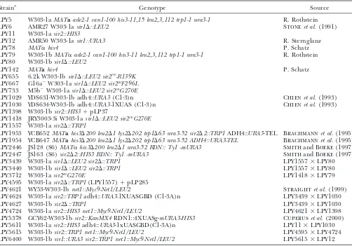
Yeast strains used in this study
Straina Genotype Source
LPY5 W303-1aMATaade2-1 can1-100 his3-11,15 leu2,3,112 trp1-1 ura3-1 R. Rothstein
LPY6 AMR27 W303-1asir1⌬::LEU2 Stoneet al.(1991)
LPY11 W303-1asir2::HIS3
LPY12 AMR50 W303-1asir1::URA3 R. Sternglanz
LPY78 MAT␣his4 P. Schatz
LPY79 W303-1bMAT␣ade2-1 can1-100 his3-11 leu2,3,112 trp1-1 ura3-1 R. Rothstein LPY80 W303-1bsir1⌬::LEU2
LPY142 MATahis4 P. Schatz
LPY655 6.2k W303-1bsir1⌬::LEU2 sir2eso-R139K
LPY667 G16a⫺W303-1asir1⌬::LEU2 sir2esoF296L
LPY733 M5b⫺W303-1asir1⌬::LEU2 sir2esoG270E
LPY1029 YDS631-W303-1b adh4::URA3(C1-3)n Chienet al.(1993)
LPY1030 YDS634-W303-1b adh4::URA3-1XUAS (C1-3)n Chienet al.(1993)
LPY1398 W303-1bsir2::HIS3⫹pLP37
LPY1418 JRY3003-3i W303-1asir1⌬::LEU2 sir2esoG270E
LPY1557 W303-1asir2⌬::TRP1
LPY1953 YCB652MATahis3⌬200 leu2⌬1 lys2⌬202 trp1⌬63 ura3-52 sir2⌬2::TRP1ADH4::URA3-TEL Brachmannet al.(1995) LPY1954 YCB647MATahis3⌬200 leu2⌬1 lys2⌬202 trp1⌬63 ura3-52 ADH4::URA3-TEL Brachmannet al.(1995) LPY2446 JS128 (S6)MAT␣his3⌬200 leu2⌬1 ura3-52 RDN:: Ty1 -mURA3 SmithandBoeke(1997) LPY2447 JS163 (S6)sir2⌬2::HIS3 RDN:: Ty1 -mURA3 SmithandBoeke(1997)
LPY3439 W303-1asir1⌬::LEU2 sir2⌬::TRP1 LPY1557⫻LPY80
LPY3440 W303-1bsir1⌬::LEU2 sir2⌬::TRP1 LPY1557⫻LPY80
LPY3712 W303-1asir2esoG270E LPY1418⫻LPY79
LPY4595 W303-1asir2⌬::TRP1(LPY1557)⫹pLP285
LPY4621 WY53-W303-1bnet1::Myc9-Net1/LEU2 Straightet al.(1999)
LPY4624 W303-1asir2::TRP1adh4::URA3-1XUASGBD (C1-3A)n LPY3439⫻LPY1030
LPY4627 W303-1bsir2⌬::TRP1 LPY3439⫻LPY1030
LPY4724 W303-1asir2::HIS3 net1::Myc9-Net1/LEU2 LPY4621⫻LPY1398
LPY5378 GCY62-W303-1bsir2::KanMX4RDN1::4XUASg-mURA3-HIS3 Cuperuset al.(2000) LPY5611 W303-1asir2::HIS3adh4::URA3-1xUASGBD(C1-3A)n LPY11⫻LPY1030
LPY5615 W303-1bsir2::TRP1 net1::Myc9-Net1/LEU2 LPY4595⫻LPY4724
LPY6400 W303-1bsir1::URA3 sir2::TRP1 net1::Myc9-Net1/LEU2 LPY5615⫻LPY12
aExcept where indicated, strains used were constructed in the course of these experiments or are part of the lab collection.
The LPY numbers for transformed strains used in this study can be found in the supplemental data at http://www.genetics.org/ supplemental/.
LPY4624 and LPY1030 were cotransformed with pLP114 and analysis as described (StoneandPillus1996). Cell extracts fromⵑ2.0⫻107cells were loaded per well. Sir2p was detected
the same set of plasmids listed above (pLP62-pLP1189). For
rDNA silencing assays, LPY2446 and LPY2447 were trans- using a 1:1000 dilution of the polyclonal antiserum (2916/8) raised to a C-terminal peptide of Sir2p (Smithet al.1998). formed with the same set of plasmids listed above
(pLP62-pLP1189). All transformed strains were grown for 4 days at Goat anti-rabbit secondary antibody was used at 1:10,000 and Western blots were developed using a standard alkaline phos-30⬚, serially diluted fivefold, and plated onto selective plates to
assay growth. Diluted cells were also plated onto the following phatase detection system (Promega, Madison, WI).
Centromeric plasmids bearing thesir2esomutations and
wild-silencing test plates: plates lacking leucine supplemented with
0.1% 5-fluoroorotic (5-FOA), plates lacking leucine and histi- type SIR2were transformed intosir2⌬(LPY1557) andsir1⌬ sir2⌬(LPY3439) strains. Immunofluorescence using affinity-dine containing 0.1% 5-FOA for telomeric silencing assays,
and plates lacking uracil for assaying rDNA silencing. purified antibody raised to the C terminus of Sir2p (2916/8) was performed as described (StoneandPillus1996;Gotta
Targeting assays: Yeast strains LPY1030, LPY5611, and
LPY5378 were transformed with pLP956, pLP1073, pLP1074, et al.1997;Ersfeld1999). The other antibody used was anti-Nop1p (D77;ArisandBlobel1988). Fluorescein-conjugated pLP1369, pLP1370, and pLP1371 and assayed for telomeric
and rDNA silencing as described above. All transformed strains anti-rabbit and Texas-red-conjugated anti-mouse secondary antibodies ( Jackson Immunoresearch, West Grove, PA) were were grown for 4 days at 30⬚and serially diluted fivefold on
plates lacking tryptophan to assay growth and onto test plates preadsorbed against spheroplasted mutant and wild-type yeast cells. Microscopy was performed on an Applied Precision opti-lacking tryptophan and containing 0.1% 5-FOA. To be certain
that the mutants expressed at high-copy (2) levels retained cal sectioning microscope to collect images spaced at 0.2-m increments. The images were deconvolved using the Delta theiresophenotype, the strains were tested for mating ability
in asir1⌬sir2⌬andSIR1 sir2⌬background. As expected, they Vision deconvolution software as previously described ( Pogli-anoet al.1999).
were able to mate only in the presence of Sir1p (data not
shown). NADⴙ hydrolysis assays: Glutathione S-transferase (GST;
pLP1334), GST-Sir2p (pLP1275), GST-sir2-R139Kp (pLP1335),
Immunoblot and immunofluorescence analyses:Levels of
Figure1.—Thesir2esomutations result in
substi-tutions of highly conserved amino acids, includ-ing a subset within the catalytic core domain of Sir2p. (A) Thesir2esoalleles were cloned onto
plas-mids using gap repair (pLP1102-R139K, pLP1110-G270E, and pLP1112-F296L). Sequence analysis of each sir2eso ORF revealed mutations altering
amino acids that lie within conserved residues of the Sir2p family. Two mutations (G270E and F296L) changed amino acids within the highly conserved core ofSIR2(dark shading), a domain essential for silencing function. (B) The crystal structure ofA. fulgidus-Sir2 withsir2esoamino acid
changes mapped using Cn3D, the National Cen-ter for Biotechnology Information Entrez struc-ture retrieval and analysis program (http:// www.ncbi.nlm.nih.gov / Structure / CN3D / cn3d. shtml). The R139K substitution is not shown since the Sir2-Afl enzyme lacks the N-terminal domain of ySir2p.
fusion proteins were expressed inEscherichia coliBL21 (DE3) until it reached an A600of 0.7–0.8. The cells were harvested and
lysed as described (Straightet al.1999). Three microliters during a 4- to 5-hr induction with 0.5 mmisopropyl-d
-thioga-lactoside at room temperature. Proteins were purified on glu- of ␣-Sir2p polyclonal antiserum (above) or 4 l of ␣-Sir4p polyclonal antiserum (7795, raised against a-gal-Sir4 fusion tathione Sepharose beads as directed by the manufacturer
(Pharmacia, Piscataway, NJ). Purified proteins were dialyzed protein) was used and incubated at 4⬚for 3–4 hr. One hundred microliters of 10% (w/v) protein-A Sepharose (Pharmacia) against 50 mmsodium phosphate (pH 7.2) and stored at 4⬚
in 50 mmsodium phosphate (pH 7.2), 0.5 mmdithiothreitol in lysis buffer was added and mixed at 4⬚for 1 hr. Immune complexes were harvested and washed as described (Straight
(DTT), and 10% glycerol (Landryet al.2000b). Protein
con-centration was deduced from extinction coefficient measure- et al. 1999) and resuspended in 50l of 2.5⫻ SDS sample buffer. Ten microliters of sample was loaded on 12 cm 9% ments as described (Gillandvon Hippel1989) and by
SDS-PAGE, comparing Coomassie brilliant blue staining of purified SDS polyacrylamide. Immunoblots were probed with a 1:20 dilution of ␣-myc (9E10) hybridoma supernatant, a 1:1000 GST-protein samples and various concentrations of the BSA
protein standard. NAD⫹-hydrolysis assays to measure histone dilution of␣-Sir2p polyclonal antiserum (2916/8), or a 1:1000 dilution of␣-Sir4p polyclonal antiserum raised against a Sir4p deacetylation were performed as described (Landry et al.
2000a). Reactions were carried out in 1 ml with 50 mmglycine C-terminal peptide (2913/8;Palladinoet al.1993). Second-ary antibodies, horseradish peroxidase-coupled anti-rabbit (pH 9.0), 0.5 mm DTT, 5 mm tetrasodium pyrophosphate
(NaP2O7), 0.1 mg/ml BSA, 1 mg calf thymus histones (Sigma, (for Sir2p and Sir4p) and anti-mouse (for 9E10; Promega)
were used at 1:10,000 and detected using the enhanced chemi-St. Louis), 2 Ci [4-3H]NAD⫹ (Amersham TRA298; 4.3 Ci/
mmol, 1 mCi/ml), and 3.7g of purified proteins. The reac- luminescence reagents (Pharmacia). tions were performed in triplicate and incubated at 30⬚. After
10 min and 3, 7, 24, and 33 hr, 185l of the total reaction was transferred to tubes containing 135l 0.5 mboric acid
RESULTS (pH 8.0) to quench the reaction. A total of 1 ml of ethyl
acetate was added and vortexed for 5 min and 700l of the Identification of thesir2esomutations and
characteriza-ethyl acetate phase was transferred to 3 ml Ecoscint fluid
tion at theHMloci:Fivesir2mutants were isolated from
(National Diagnostics, Atlanta) and analyzed by scintillation
theesomutant screen described previously (Reifsnyder
counting. Radioactivity released from Sir2p wild-type control
et al.1996;Stoneet al.2000). Thesir2esomutations were
reactions lacking histones was subtracted. The slope, or rate
of change, for activities through five time points was calculated cloned onto centromeric (CEN) plasmids using
stan-and shown as percentage of wild-type Sir2p activity with stan- dard gap repair (Rothstein1991). The repaired plas-dard deviations indicated.
mids were tested for their ability to confer mating in a Immunoprecipitation reactions:A total of 25 ml of LPY5615
SIR1 sir2⌬strain but not in asir1⌬sir2⌬strain to confirm
and LPY6400 transformed with pLP60, pLP285, pLP1102,
Figure 2.—The sir2eso mutants enhance the
sir1⌬ defect and the mutant proteins are ex-pressed at wild-type levels. (A) Thesir2esomutants
are completely mating defective only insir1⌬cells. MATa strains were patched and replica plated onto YPD plates (growth control) and onto mini-mal plates top spread with cells of the opposite mating type. The sir2-G270Emutant, in the ab-sence of Sir1p and in the preab-sence of Sir1p, is shown. The strains used are wild-type, LPY5;sir1⌬, LPY6; sir2⌬, LPY11; sir1⌬ sir2–G270E, LPY1418; andsir2–G270E, LPY3712. (B) Immunoblot analy-sis ofsir2esomutant whole-cell extracts using
anti-Sir2p antisera (2916/8). The sir2esomutant
pro-teins are expressed from their chromosomal locus in a sir1⌬ background. The three sir2eso alleles
characterized in this article are expressed at levels comparable to wild type in both theSIR1wild-type andsir1⌬backgrounds (thesir1⌬background is shown). The strains from left to right are sir2⌬ (LPY11), wild type (LPY5), sir1⌬ sir2-R139K (LPY655), sir1⌬ sir2-F296L(LPY667), sir1⌬ sir2-G270E(LPY1418), andsir1⌬sir2-G270E(LPY733).
five sir2eso
alleles were identified by sequence analysis steady-state levels. Thus, it appears that the mutant de-fects are due to more specific influences on silencing. and three are shown in Figure 1A. Two of the strains
(LPY655 and LPY667) had mutations affecting amino To evaluate the effects of these mutations relative to
proposed enzymatic activities, we considered the struc-acids that are highly conserved among Sir2p family
members at positions R139K and F296L, respectively. tures ofArchaeoglobus fulgidusSir2 (Sir2-Afl) complexed
with NAD⫹and of a human homolog, SIRT2 (Finnin
Although theesomutants were isolated from
indepen-dently mutagenized cultures, two additional strains et al. 2001; Min et al. 2001). The two superimposed
enzymes show similarities to the regions containing the (LPY1418 and LPY733) contained an identical mutation
changing a highly conserved glycine to a glutamic acid, Rossman fold domain, commonly found in NAD(H)/
NADP(H)-binding proteins, and to the smaller catalytic G270E. Characterization of one strain (LPY1418) was
extended as representative of both of these mutants. domain containing a structural zinc atom (Finnin et
al.2001) reviewed in DutnallandPillus (2001). The Another mutant isolated from the screen, LPY727,
contained a nonsense mutation at amino acid 15. The structure of the yeast Sir2 protein has not yet been deter-mined. However, on the basis of the structural similari-mutant protein was presumed to be translated using a
downstream methionine since a truncated version of ties between the Sir2-Afl and SIRT2, it appears that two
of the sir2eso mutants contain amino acid changes in the protein was detected by immunoblot analysis (data
not shown). This mutant protein also appeared to be conserved residues within regions that contribute to
the Rossman fold domain (Sir2-Afl in Figure 1B). The expressed at lower levels, perhaps due to three
addi-tional mutations found in the promoter region. Previous equivalent amino acid changes in Sir2-Afl are at amino acids G28 (yG270E) and A48 (yF296L). The fact that evidence indicates that silencing is sensitive to Sir2p
dosage (Holmeset al.1997;Smithet al.1998). There- this domain is the postulated NAD⫹-binding portion of the enzyme suggests that these two mutants might be fore, it is possible that this mutant’sesophenotype is due
to some combination of decreases in levels of expression impaired in catalytic activity. The R139K mutation lies in a region N-terminal of the conserved core domain and altered N-terminal sequence. This mutant has not
been pursued further. Immunoblot analysis showed that anda prioriwas not expected to influence activity. This region is not present in Sir2-Afl or SIRT2 structures and levels of the other sir2 mutant proteins were equivalent
to wild type in the presence and absence of Sir1p (sir1⌬ therefore the location of R139K could not be inferred
(Finnin et al. 2001; Min et al. 2001). The N-terminal
background shown in Figure 2B). Since the sir2 mutant
proteins appear to be expressed comparably to wild type, region is also variable in members of the yeastSIR2family
members and might be expected to influence SIR2
-the phenotypes observed cannot be due to decreased
TABLE 2
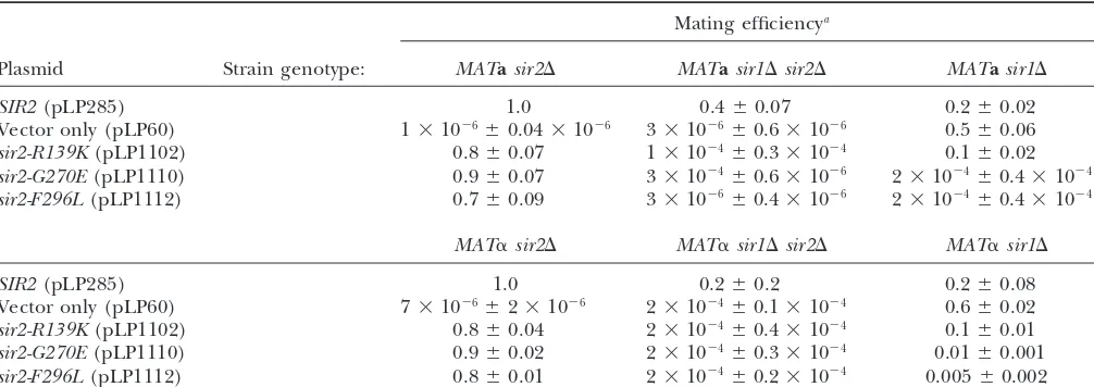
Thesir2esoquantitative mating efficiencies
Mating efficiencya
Plasmid Strain genotype: MATasir2⌬ MATasir1⌬sir2⌬ MATasir1⌬
SIR2(pLP285) 1.0 0.4⫾0.07 0.2⫾0.02
Vector only (pLP60) 1⫻10⫺6⫾0.04⫻10⫺6 3⫻10⫺6⫾0.6⫻10⫺6 0.5⫾0.06
sir2-R139K(pLP1102) 0.8⫾0.07 1⫻10⫺4⫾0.3⫻10⫺4 0.1⫾0.02
sir2-G270E(pLP1110) 0.9⫾0.07 3⫻10⫺4⫾0.6⫻10⫺6 2⫻10⫺4⫾0.4⫻10⫺4
sir2-F296L(pLP1112) 0.7⫾0.09 3⫻10⫺6⫾0.4⫻10⫺6 2⫻10⫺4⫾0.4⫻10⫺4
MAT␣sir2⌬ MAT␣sir1⌬sir2⌬ MAT␣sir1⌬
SIR2(pLP285) 1.0 0.2⫾0.2 0.2⫾0.08
Vector only (pLP60) 7⫻10⫺6⫾2⫻10⫺6 2⫻10⫺4⫾0.1⫻10⫺4 0.6⫾0.02
sir2-R139K(pLP1102) 0.8⫾0.04 2⫻10⫺4⫾0.4⫻10⫺4 0.1⫾0.01
sir2-G270E(pLP1110) 0.9⫾0.02 2⫻10⫺4⫾0.3⫻10⫺4 0.01⫾0.001
sir2-F296L(pLP1112) 0.8⫾0.01 2⫻10⫺4⫾0.2⫻10⫺4 0.005⫾0.002
aMating efficiencies from three independent experiments are shown with standard deviations and normalized to eitherMATa
sir2⌬orMAT␣sir2⌬strains transformed with a plasmid containing wild-typeSIR2(pLP285).
the sir2eso
mutants on catalysis were investigated as de- telomeric silencing, it was possible that thesir2eso alleles would be competent in telomeric silencing. To test this scribed below.
By virtue of the design of the eso screen, the sir2eso hypothesis, a
sir2⌬strain marked at telomere VII with
URA3 was transformed with CEN plasmids containing
mutants displayed a characteristic mating defect in the
absence of Sir1p. A qualitative mating assay with one the sir2eso alleles (pLP1187-1189), SIR2 (pLP1237), or vector only (pLP62). Telomeric silencing was assayed representativesir2esomutant is shown in Figure 2A. It is
notable that asir1⌬strain mates comparably to a wild- on 5-FOA-containing medium as described previously
(Gottschlinget al.1990). All threesir2esoalleles were
type strain by this assay despite its known epigenetic
silencing defects at theHMloci (PillusandRine1989). sensitive to 5-FOA, demonstrating that they were com-pletely defective in silencing the telomeric reporter Therefore, it remained a possibility that thesir2esoalleles
displayed silencing defects at HML andHMR even in gene (Figure 3A).
Recently, it was determined that there are onlyⵑ30 the presence of Sir1p at levels detectable only through
quantitative analysis. Mating efficiencies for strains trans- molecules of Sir1p per cell (GardnerandFox2001), suggesting that Sir1p may be limiting in the cell. There-formed with plasmids bearing thesir2esomutations,
wild-typeSIR2, or vector only were calculated and normalized fore, we tested whether increased SIR1 gene dosage
suppressed thesir2esotelomeric silencing defects. Expres-to wild type. The results showed that thesir2esomutants
were modestly defective in silencing in the presence of sion ofSIR1using a high-copy 2plasmid did not sup-press their defects; thus this simple possibility does not
SIR1 (Table 2). Although they mated at 70–90%
effi-ciency in comparison to wild type, the sir2eso mutants explain the sir2eso phenotype (data not shown). Next, we directed Sir1p to the telomeres using a GBD-Sir1p were slightly more efficient in mating than the sir1⌬
SIR2mutant strain. Thesir1⌬sir2esomutants are as defec- fusion protein and a modified telomeric reporter gene containing an adjacent Gal4p DNA-binding site (UASg). tive as asir1⌬sir2⌬mutant, with mating efficiencies four
to seven orders of magnitude lower than those of wild- Previous results demonstrated improvement in
silenc-ing upon tethersilenc-ing GBD-Sir1p at a telomeric reporter. type strains. In addition, two lesions within the core
domain, sir2-G270E and sir2-F296L, conferred domi- However, such improved silencing is dependent on the
other Sir proteins, including Sir2p (Chienet al.1993).
nantly derepressed phenotypes in MATa sir1⌬ and
MAT␣sir1⌬cells but not inSIR1wild-type backgrounds Asir2⌬strain marked at telomere VII with a UASg-URA3 marker was cotransformed with GBD-Sir1p (pLP114) (data not shown).
Thesir2esomutants are defective in telomeric silencing and with the same set of sir2eso plasmids used for the telomeric assay shown in Figure 3A. The telomeric
de-and can be partially suppressed by tethering Sir1p to
telomeres: In contrast to silencing at the HM silent fects for all three alleles were suppressed by tethering Sir1p, albeit to differing degrees (Figure 3B). Thesir2– mating-type loci, loss of Sir1p has no effect on silencing
reporter genes at telomeres (Aparicioet al.1991). Since R139Kdefect was fully suppressed whereas the two core mutants were only partially suppressed. Therefore, the the sir2esodefects at theHM loci were revealed only in
Figure3.—Thesir2esomutants show defects in
TPE. (A) Asir2⌬strain (LPY1953) containing a URA3marker proximal to telomere VII was trans-formed with aLEU2CEN vector (LPY4859),SIR2 (LPY4860), sir2-R139K (LPY4861), sir2-G270E (LPY4862), orsir2-F296L(LPY4863). Fivefold di-lutions of the transformants were plated on 5-FOA, growth on 5-FOA indicating silencing of theURA3 reporter, and on SC–leu plates, to control for growth differences. (B) Tethering Sir1p to a telo-meric reporter partially suppresses thesir2esoTPE
defect. A sir2⌬ strain (LPY4624) containing a URA3reporter at telomere VII, in addition to a copy of the Gal4p DNA-binding site, was cotrans-formed with a Gal4p DNA-binding domain (GBD)-SIR1 hybrid HIS3 2 vector (pLP114) and an emptyLEU2CEN vector (pLP62) or the vector con-taining SIR2 (pLP1237), sir2-R139K (pLP1187), sir2-G270E(pLP1188), orsir2-F296L(pLP1189). The transformants were assayed for silencing as in A. (C) Thesir2esomutants demonstrate
domi-nance by disrupting telomeric silencing even in the presence of Sir2p. A SIR2strain (LPY1954) was transformed and assayed as in A. (D) Teth-ering Sir1p suppresses the dominantsir2eso
pheno-type. ASIR2strain (LPY 1030) was transformed and assayed as in B.
and can function if Sir1p is directed to the telomeres. mids were transformed into a telomere-marked strain
containing wild-type Sir2p. Somewhat surprisingly, the This suggested that at some level, the sir2eso mutant
proteins had the capacity to function in telomeric silenc- sir2eso mutants disrupted silencing in the presence of
SIR2(Figure 3C). However, tethering Sir1p to the telo-ing, although this function appeared limited.
To evaluate further the nature of sir2eso function at meres overcame this dominant phenotype (Figure 3D).
A potential molecular explanation for the dominance telomeres, we performed a dominance test. In this
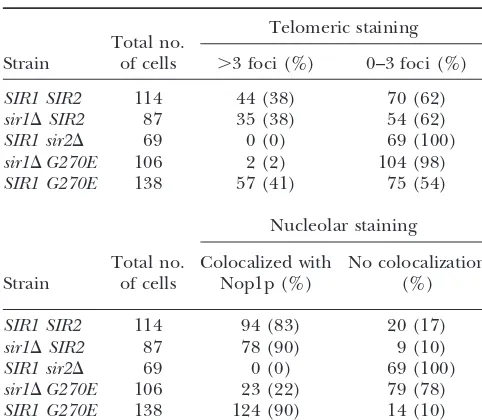
TABLE 3 calize with Nop1p, this mutant protein showed no local-ization to the nucleolus inⵑ30% of the cells analyzed Localization of sir2-G270Ep at telomeres and nucleolus
relative to wild-type Sir2p (Table 3). Furthermore, the few cells with wild-type localization showed decreased
Telomeric staining
Total no. staining intensity, represented in Figure 4. In marked
Strain of cells ⬎3 foci (%) 0–3 foci (%) contrast, inSIR1strains, the sir2-G270Ep localization was
restored to telomeres and the nucleolus in a manner
SIR1 SIR2 114 44 (38) 70 (62)
sir1⌬SIR2 87 35 (38) 54 (62) indistinguishable from wild type. Therefore, the
local-SIR1 sir2⌬ 69 0 (0) 69 (100) ization pattern of this mutant does not account for its sir1⌬G270E 106 2 (2) 104 (98) defects in telomeric silencing since comparable defects SIR1 G270E 138 57 (41) 75 (54)
are observed in bothSIR1 (Figure 3) and sir1⌬ (data not shown) backgrounds. Perhaps the sir2esomutant
pro-Nucleolar staining
teins do reach the telomeres and the nucleolus but their
Total no. Colocalized with No colocalization association to the chromatin is functionally inadequate Strain of cells Nop1p (%) (%) at these silenced regions.
Sir1p has been shown to function in the establishment
SIR1 SIR2 114 94 (83) 20 (17)
of silencing (PillusandRine1989), serving as a
recruit-sir1⌬SIR2 87 78 (90) 9 (10)
ment factor for the other Sir proteins at the silent
mat-SIR1 sir2⌬ 69 0 (0) 69 (100)
ing-type loci (Foxet al.1997;GardnerandFox2001).
sir1⌬G270E 106 23 (22) 79 (78)
SIR1 G270E 138 124 (90) 14 (10) Because Sir1p does not ordinarily serve as a recruitment protein at the telomeres, we hypothesized that Sir2p
Thesir2esomutant strains were analyzed by
immunofluores-might also function in recruiting Sir3p and Sir4p to the
cence in sir1⌬ and SIR1 strain backgrounds. The total cell
HMloci and the telomeres. Thesir2esomutants may be
number was derived from four independent experiments.
Im-ages were blinded and the number of telomeric foci counted defective in this hypothesized recruitment function,
ren-and represented as⬎3 foci and 0–3 foci. Percentages are the dering them fully dependent on other recruitment pro-number of cells with a specific pattern divided by the total
teins. This is consistent with thesir2eso dependence on
number of cells analyzed for that sample.
Sir1p at theHMloci and GBD-Sir1p at the telomeres. To test this hypothesis, we fused the sir2esomutant pro-teins to the GBD to tether the mutants directly through mislocalized in the cell and thus titrated Sir4p or
wild-type Sir2p away from the telomeres. To test this possibil- engineered binding sites on the chromosome. If the sir2eso mutants are defective in a recruitment function, ity sir2esomutant proteins were localized using
immuno-fluorescence analysis. then their defects might be suppressed if targeted to a
reporter gene at the telomeres via a GBD domain. A
Localization of the sir2eso mutant proteins: Genetic
and biochemical studies place Sir2p at the HM loci, sir2⌬strain marked at telomere VII with aURA3reporter and an adjacent Gal4p DNA-binding site was transformed the telomeres, and the rDNA repeats that serve as a
nucleolar organizer. Consistent with this, by indirect with a vector expressing GBD, GBD-Sir2pcore(210-440), GBD-⌬73NSir2p(73-562), R139Kp, GBD-⌬73Nsir2-immunofluorescence Sir2p localizes in a crescent or
cup-shaped form in the nucleolus and as clustered spots G270Ep, or GBD-⌬73Nsir2-F296Lp and the transformants
were tested for silencing. The GBD and GBD-Sir2pcore at telomeric foci found at the nuclear periphery directly
opposite the nucleolus (Gotta et al. 1997). Using a constructs served as negative controls since it has been shown that the core domain of Sir2p is necessary but polyclonal antibody raised to a peptide specific for the
C terminus of Sir2p (2916/8; Smith et al. 1998), we not sufficient for silencing even when tethered to a
reporter (Cockellet al.2000). determined the localization of the sir2esomutant
pro-teins insir1⌬andSIR1strains. To assess nucleolar local- In all cases, tethering thesir2esomutants restored telo-meric position effect (TPE) to wild-type levels (Figure ization, we evaluated colocalization with the nucleolar
marker Nop1p, the yeast homolog of fibrillarin (Aris 5). This restoration was fully dependent on tethering
since the constructs were unable to silence at telomeres
andBlobel1988). To assess telomeric localization, we
quantitated the number of telomeric foci in the various in strains lacking the Gal4p DNA-binding site (UASg)
adjacent to theURA3reporter (data not shown). There-strain backgrounds.
Two of the mutant proteins, sir2-R139Kp and sir2- fore, the sir2eso mutants are properly localized to the telomeres by immunofluorescence, yet can fully func-F296Lp, showed localization indistinguishable from
wild-type Sir2p in both SIR1 and sir1⌬ backgrounds tion only if targeted to the locus either directly via a
Gal4p DNA-binding domain or indirectly via GBD-Sir1p. (data not shown). In contrast, proper localization of
sir2-G270Ep appeared to depend on the status ofSIR1. The sir2eso mutants show distinct phenotypes at the
rDNA: PolII-transcribed reporter genes engineered The sir2-G270E mutant protein localized to telomeres
colo-Figure4.—The sir2-G270E mutant protein is mislocalized in cells lacking Sir1p. (Top: a, b, and c)SIR1 sir2⌬transformed withSIR2on aHIS3 CEN plasmid (LPY4595). (Middle: d, e, and f)sir1⌬sir2⌬strain transformed withsir2-G270Eon aHIS3 CEN plasmid (LPY4602). (Bottom: g, h, and i)SIR1 sir2⌬ transformed with sir2-G270Eon aHIS3 CEN plasmid (LPY4597). Strains (a, d, and g) were stained with anti-Sir2p affinity-purified antisera (2916/8) and detected by FITC-conjugated secondary antibodies (b, e, and h) with anti-Nop1p antibodies detected by a Texas-red-conjugated secondary antibody and (c, f, and i) the merge of Nop1p, Sir2p, and DNA staining with 4⬘,6-diamidino-2-phenylindole.
in silencing at the rDNA locus, asir2⌬strain containing porter (Figure 6A). However, the mutantsir2-R139K func-tioned fully at this locus, silencing as well as or better than aURA3cassette inserted in the rDNA was transformed
with the varioussir2esoplasmids as described above. Si- wild-typeSIR2.
Transcriptional and recombinational silencing within lencing was assayed by evaluating growth on plates
lack-ing uracil with less growth indicatlack-ing more silenclack-ing the rDNA is distinct because it is fully dependent on
SIR2, yet is independent of the otherSIRgenes (
Gott-(Smithand Boeke1997). The twosir2esomutants that
carry a mutation within the conserved core domain of liebandEsposito1989;Bryket al.1997;Fritzeet al. 1997;SmithandBoeke1997). Because thesir2esoalleles
SIR2 were defective in silencing the URA3 rDNA
re-Figure5.—Tethering thesir2esoalleles
directly to the telomeres rescued their telomeric silencing defects. A sir2⌬ strain (LPY5611) marked at telomere VII with aURA3reporter gene with an adjacent Gal4p DNA-binding site was transformed with GBD(1-147) (LPY5777),
GBD-Sir2-pcore(210-440) (LPY5778), GBD-⌬
73NSir2-p(73-562)(LPY5779), GBD-⌬73NSir2-R139Kp
Figure6.—Thesir2esomutants have distinct
silencing phenotypes within the rDNA locus. (A) A sir2⌬strain (LPY2447) marked at the rDNA with aURA3 cassette was transformed with wild-typeSIR2on a LEU2CEN plasmid (pLP1237), empty vector (pLP62),sir2-R139K (pLP1187), sir2-G270E (pLP1188), or sir2-F296L(pLP1189), and fivefold dilutions of the transformants were assayed for growth on Sc-leu or silencing on Sc-ura. Thesir2esomutants
with mutations in the conserved domain of Sir2p are defective in rDNA silencing. (B) A SIR2 strain (LPY2446) transformed and as-sayed as in A. Thesir2-G270Eand sir2-F296L mutants are dominantly defective in rDNA si-lencing. (C) A sir2⌬ strain (LPY5378) con-taining four Gal4p DNA-binding sites adjacent to the rDNAURA3reporter was transformed with GBD (LPY5637), GBD-Sir2pcore210-440
(LPY5638), GBD-⌬73NSir2p73-562 (LPY5639),
GBD-⌬73NSir2-R139Kp (LPY5640), GBD-⌬ 73N-Sir2-G270Ep (LPY5641), or GBD-⌬ 73NSir2-F296Lp (LPY5642). Fivefold dilutions were as-sayed for growth and silencing. GBD alone or fused to the conserved core domain of Sir2p failed to silence the reporter. Thesir2-G270E mutant is rescued when tethered to the rDNA; however, thesir2-F296Lmutant remains defec-tive. Note that mutantsir2-R139Kbecomes de-fective in silencing at the rDNA when tethered to the locus.
had a conditional dependence onSIR1at theHMloci, four adjacent Gal4p DNA-binding sites (4X-UASg) within
the rDNA locus (Cuperuset al.2000). Because of inher-we tested whether this dependence might also exist at
the rDNA. The absence of Sir1p had no effect on the ent variability in silencing assays, in each case multiple independent transformants were evaluated. We
ob-sir2eso phenotypes observed at the rDNA (data not
shown). Since thesir2-G270Eandsir2-F296Lmutations served, as expected, that the GBD-⌬73NSir2p
consis-tently silenced when tethered. Neither GBD-Sir2pcore
caused dominant derepression at theHMloci and the
telomeres, the dominance test was repeated at the rDNA nor GBD constructs alone silenced. The modest
differ-ences between these controls (Figure 6) demonstrate locus. The sir2eso genes on plasmids were transformed
into aSIR2strain marked within the rDNA and, as be- the occasional variability noted above. The results with thesir2esoalleles were somewhat surprising. One of the fore, transformants were assayed for growth on plates
lacking uracil. The two mutants defective in rDNA si- two rDNA silencing-defectivesir2esomutants,sir2-G270E, was rescued when tethered, whereas the other mutant, lencing, sir2-G270E andsir2-F296L, also showed
domi-nant effects at this locus (Figure 6B). sir2-F296L, was not. Further, the rDNA
silencing-compe-tentsir2esomutant, sir2-R139K, became impaired for si-We proposed in the section above that thesir2eso
mu-tants could be defective in a type of recruitment func- lencing when tethered to the rDNA reporter (Figure
6C). We observed that simply expressing these con-tion at the telomeres. To extend this hypothesis to the
rDNA, we tested the ability of thesir2esostrains to silence structs in a strain with a silencing reporter but no UAS had no effect (data not shown); thus the effects observed the rDNA if targeted directly to this locus. The
GBD-⌬73NSIR2 constructs were transformed into a sir2⌬ are completely dependent on the tethering site.
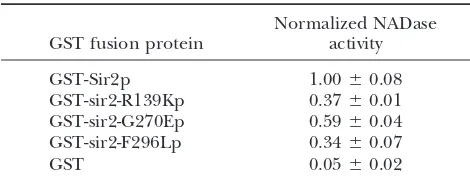
Thesir2 mutants are defective in NADase activity
paired in enzymatic activity (Table 4 and data not shown). The GST-sir2-R139Kp and GST-sir2-F296Lp
Normalized NADase
GST fusion protein activity mutant proteins consistently had ⬍50% activity when compared to wild-type GST-Sir2p. The GST-sir2-G270E
GST-Sir2p 1.00⫾0.08
mutant protein was slightly more active than the other
GST-sir2-R139Kp 0.37⫾0.01
two mutant enzymes but never achieved activities⬎70%
GST-sir2-G270Ep 0.59⫾0.04
of wild-type activity. The activities shown in Table 4
GST-sir2-F296Lp 0.34⫾0.07
GST 0.05⫾0.02 reflect consistent differences of mutant activity relative
to wild type. Further distinctions between the GST-sir2eso
Purified proteins GST-Sir2p (pLP1275), GST-sir2R139K
mutant proteins may become apparent with extensive
(pLP1335), GST-sir2G270E (pLP1336), GST-sir2F296L
kinetic analyses or with comparisons between NADase
(pLP1337), or GST (pLP1334) were tested for their ability to
convert NAD⫹to nicotinamide and ADP-ribose in a histone- and deacetylase activities. Since NADase and deacetylase
dependent manner. Reactions were performed in the pres- activities are coupled, however, the decreases in activity ence of [3H]NAD⫹and 3.7g of purified enzymes.
Radiola-for thesir2esomutant proteins suggest that they are not
beled [3H]nicotinamide was detected as described byLandry
significant enough to completely abolish silencing
be-et al. (2000a; see materials and methods). The rate of
cause these mutants were capable of silencing when
change over five time points (10 min and 3, 7, 24, and 33 hr)
was calculated and normalized to wild-type Sir2p. Multiple tethered to a reporter or when tethered by Sir1p. Since
experiments were performed. The experiment shown was per- the sir2eso mutants are not catalytically dead but have
formed in triplicate and is shown with standard deviations.
only partially impaired enzymatic activity, we asked whether the silencing defects observed in vivo might also reflect inefficient complex formation or protein
trum of phenotypes with respect to rDNA silencing, interactions.
implying that they had different silencing defects at this The sir2eso mutant proteins interact with Sir4p and
locus. Although inadequate association with chromatin Net1p: Sir2p is known to interact with a number of
might explain the silencing phenotypes of the sir2eso
other proteins. Among these are Sir4p (Moazedet al.
mutants at theHMloci and the telomeres, this model 1997) and Net1p (Shou et al. 1999; Straight et al.
seemed inadequate to explain the diversity of pheno- 1999), associations with which are correlated with telo-types of the sir2eso mutants at the rDNA. A significant
meric and rDNA silencing, respectively. We considered element of Sir2p silencing function is its NAD⫹-depen- the possibility that thesir2esophenotype was caused by dent deacetylase activity. We therefore asked if the dif- impaired interactions with these proteins and tested ferences between thesir2eso silencing abilities were
re-their associations by immunoprecipitation experiments.
flected in their catalytic activities. Immunoprecipitation was performed with either
anti-Thesir2esomutants are impaired in NADase
(deacety-Sir2p or anti-Sir4p reagents. The immunoprecipitated
lase) activity: Previous reports showed that the Sir2p samples were analyzed by protein immunoblotting using
family of proteins functions as NAD⫹-dependent pro- the relevant antisera as noted (Figure 7). The results
tein deacetylases and that decreases in deacetylase activ- indicated that the sir2esomutant proteins interacted with
ity correlate with loss of silencing (Imai et al. 2000; both Sir4p and Net1p, even in the absence of Sir1p
Landry et al. 2000b; Smith et al. 2000). The role of (Figure 7 and data not shown). This suggests that Sir
NAD⫹ in the deacetylation reaction has been investi- and RENT complex formation is not grossly disrupted
gated and has led to a deeper understanding of the in the sir2eso mutants. It is also clear that Sir1p does
mechanism of catalysis (Imaiet al.2000;Landry et al. not stabilize the components of the complexes since
2000a;Borraet al.2002). On the basis of the products interactions with Sir4p and Net1p are found in both
released, the reaction is described as the hydrolysis of SIR1 and sir1⌬ backgrounds (sir1⌬ background for
one NAD⫹to form nicotinamide and the novel product Sir4p, Figure 7A; both backgrounds for Net1p, Figure
acetyl-ADP-ribose (AADPR), for each acetyl group re- 7B). One difference observed in thesir1⌬background
moved. This results in a 1:1:1 molar ratio of acetyl-ADP- was the consistent loss of an additional␣-myc-reactive
ribose (AADPR), nicotinamide, and a deacetylated pep- band that commonly migrates with the Net1p in
immu-tide substrate with an enzyme-ADP-ribose intermediate noblot analyses (Straightet al.1999; Figure 7B). The
(Landry et al. 2000a). This finding allows for a direct band could represent a protein modification of Net1p
correlation between NADase activity and histone deacety- that, either directly or indirectly, requires Sir1p,
al-lation for the Sir2 family members. thoughSIR1has not been previously implicated inNET1
To determine whether the sir2eso proteins retained function. Further experiments will be necessary to
ex-wild-type deacetylase activity, recombinant GST-sir2eso plore these ideas, but together they suggest that the mutant fusion proteins were expressed inE. coli, puri- sir2esophenotypes do not result from gross disruptions
Figure7.—The sir2esomutant proteins
inter-act with Sir4p and Net1p. Extrinter-acts were pre-pared fromSIR1 sir2⌬net1⌬::Myc9-NET1-LEU2 andsir1⌬sir2⌬net1⌬::Myc9-NET1- LEU2strains transformed with vector (LPY6402), SIR2 (LPY6403),sir2-R139K(LPY6404),sir2-G270E (LPY6405), or sir2-F296L (LPY6406). (A) Strain LPY6400sir1⌬sir2⌬net1⌬ ::Myc9-NET1-LEU2. Sir4p was immunoprecipitated and tested for coimmunoprecipitation of Sir2p and sir2esomutant proteins by immunoblot analysis.
Immunoprecipitations were performed with anti-Sir4p (7795) and immunoblots were probed with antisera against Sir4p (2913/8; top) and Sir2p (2916/8; bottom). (B) Strains LPY5615 SIR1 sir2⌬ net1⌬::Myc9-NET1-LEU2 (left) and LPY6400 sir1⌬ sir2⌬ net1⌬ ::Myc9-NET1-LEU2 (right). Immunoblot analysis of Net1-myc9 immunoprecipitated with anti-Sir2p (2916/8) and probed with anti-myc monoclonal antibody 9E10 (top) and against Sir2p (2916/8, bottom) is shown; it is also shown in aSIR1-sir2⌬background (left).
DISCUSSION in the absence of Sir1p, wild-type activity levels are
re-quired to maintain a silenced state at the HMloci or Sir2p is an NAD⫹-dependent deacetylase whose
asso-to initiate a stable silenced chromatin structure that can ciation with Sir4p and Net1p correlates with
transcrip-then be propagated. Our data do not yet distinguish tional silencing (reviewed inGartenberg2000;Moazed
these possibilities. 2001). This study describes three newsir2mutants
iso-Telomeric silencing is disrupted in thesir2eso
mutants:
lated as enhancers of sir-one⌬, the sir2eso mutants. These
Although thesir2esomutants were identified through a mutants retain interactions with Sir4p and Net1p and
screen for silent mating-type defects, the mutants were are only partially compromised for catalytic activity.
also dominantly defective in telomeric silencing in both They do, however, display distinct phenotypes at three
sir1⌬ (data not shown) and SIR1 backgrounds.
Teth-distinct silenced loci, including dominant effects at
telo-ering directly to the telomeres, or indirectly through meres and within the rDNA. Many of these defects can
GBD-Sir1p, rescued these silencing defects and reversed be ameliorated by molecular targeting strategies. Thus
the dominant effects. This raised the possibility that the a range of catalytic activity may be compatible with
si-mutant proteins were not properly localized in the cell. lencing functions, as long as that activity is appropriately
However, immunofluorescence analysis revealed that directed to its required sites of action.
the sir2eso proteins were localized indistinguishably to
Thesir2esomutants show mating defects in the absence
telomeric foci from wild-type localization inSIR1cells.
of SIR1: The sir2eso mutants are defective in silencing
Recent studies have also evaluated silencing protein
lo-HMLandHMRonly in the absence of Sir1p and encode
calization by the independent technique of chromatin mutations in residues that are conserved in the Sir2
immunoprecipitation. In these studies, it was observed protein family. In quantitative mating analyses, thesir2eso
that Sir2p’s enzymatic activity not only is necessary for mutants were only slightly impaired in their mating
abil-the spread of abil-the Sir proteins but also may influence ity in the presence ofSIR1. In contrast, in asir1⌬
back-efficient association of the Sir proteins with chromatin ground, the sir2eso mutants were as defective as sir2⌬
at the telomeres (Armstrong et al.2002;Hoppeet al. strains. Although the mutants were not dominant in the
2002;Luo et al.2002). Subtle quantitative distinctions presence ofSIR1(data not shown),sir2-G270Eand
sir2-in association or dynamic occupancy may result from F296L displayed moderate mating defects in a sir1⌬
the decreased activity of sir2esomutant proteins. Indeed,
SIR2 background. Considering that Sir1p is required
that the mutant proteins are somehow impaired in func-for targeting the Sir2/4 complex to modified synthetic
tional association with chromatin is supported by the
silencers (Fox et al. 1997; Gardner and Fox 2001),
observation that the sir2eso mutants became silencing
sir2esosilencing defects may arise from unstable targeting
competent when tethered via GBD or GBD-Sir1p, show-to theHMloci. Such instability, when coupled with the
ing that their weakened enzymatic activity did not ren-sir1⌬ establishment defect, may synergistically lead to
der these mutants incapable of promoting silencing. the complete loss of silencing atHMLandHMR.
An activity-dependent model for function at the silent
Thesir2esomutants are impaired, yet not totally
meric silencing. The Sir2/4 com-plex is recruited to the silent mat-ing-type loci and telomeres via Rap1-Sir4p interactions (Sir1p also participates in recruitment at HML/HMR and Ku70p and Ku80p do so at telomeres). After initial recruitment and assembly, Sir2p activity is required to deacet-ylate (⫺Ac) histones H3 and H4, thereby recruiting additional Sir3 and Sir4 proteins leading to the spread of condensed chromatin. As the Sir proteins accumulate, subsequent spreading becomes less dependent on Sir2p activity. Although many aspects of sir2esop
function support previous models ofHM and telomeric chromatin, distinct from these models, sir2esop
functions suggest that there may be locus-specific threshold re-quirements for NAD⫹-dependent catalytic activity. Thus, robust NAD⫹-dependent deacetylase ac-tivity is not necessary in all circum-stances for nucleating stable silenced chromatin. For example, high levels of Sir2p enzymatic activity may not be critical when Sir1p or another targeting molecule such as GBD ensures that the Sir proteins remain associated with the locus. Thesir2eso
mutants, impaired in enzymatic activity, rely heavily on a targeting factor such as Sir1p or GBD for stable retention of the Sir proteins at the locus and for propagation of silencing.
for the establishment of silent chromatin at the telo- actions and serving as one method of Sir3p recruitment even when Sir2p activity is limiting. Since Sir1p is not meres involve the recruitment of the Sir2/4 complex to
DNA, followed by propagation of condensed chromatin present to strengthen Sir-nucleosome interactions at the telomeres, stable spreading of the Sir proteins may rely through interactions among Sir3p, Sir4p, and
deacety-lated histone tails (Ghidelliet al.2001;Carmenet al. solely on robust Sir2p enzymatic activity. An occasional successful deacetylation event, which can then be propa-2002;Hoppeet al.2002;Luo et al.2002; and reviewed
inMoazed2001). The recruitment of Sir3p to the telo- gated, may underlie the variegation of silencing that is
the hallmark of telomeric position effects. meres appears to be a regulated step since stable
Sir3p-Sir4p interactions are detected only when the N termi- Our model, outlined in Figure 8, may also explain why npt1⌬ mutants, required for the nuclear NAD⫹ salvage nus of Sir4p is removed or under conditions that
strengthen Sir-nucleosome interactions (Morettiet al. pathway, are selectively defective in silencing, only slightly affecting theHMloci (Smithet al.2000). Per-1994;Moazedet al.1997;Strahl-Bolsingeret al.1997;
Ghidelliet al.2001). We propose that if Sir3p is indeed haps even modest decreases in enzymatic activity have
amplified effects at telomeres that would be masked at also recruited through strengthened Sir-nucleosome
in-teractions, its recruitment could become progressively HMLandHMRin the presence of Sir1p.
Is dominance a threshold effect? If it is, this might less dependent on Sir2p enzymatic activity as the
num-ber of associated Sir complexes increases and spreads. explain why CEN-plasmid dosage of the sir2esomutants has dominant phenotypes.In these cases of marginally This idea is consistent with the observation that
overex-pression of Sir3p can extend telomeric silencing into increased amounts of mutant proteins, a critical thresh-old of wild-type activity may not be met. One possibility regions of chromatin that do not contain Sir2p and
Sir4p (Renauld et al. 1993; Hecht et al. 1996) and is that even slightly increased amounts of mutant
pro-teins are sufficient to limit availability of Sir2p inter-provides an explanation for our observation that
silenc-ing of a reporter gene is restored by targetsilenc-ing enzymati- acting molecules such as NAD⫹, acetylated substrates, or Sir4p, thereby interfering with proper Sir2p function. cally impaired sir2esomutant proteins to a single Gal4p
DNA-binding site. Therefore, although thesir2esomutants are impaired in
enzymatic activity, they may be sufficiently active to initi-We suggest that at theHMloci, Sir1p recruits the Sir
GBD ensures that the Sir complex remains associated inHsiehandFire2000). It is not clear whether compo-nents of the RENT complex, or other proteins as yet with the locus for the silenced state to be propagated. In
the absence of the targeting factor, the sir2esoenzymatic unidentified, directly bind histones and thereby target and promote the spread of silenced chromatin at partic-activity may become limiting and unable to overcome
a threshold required for stable initiation of chromatin ular rDNA repeats. Such additional targeting might ex-plain why sir2-R139Kp, which is enzymatically impaired decondensation.
Distinct sir2eso phenotypes in the rDNA underscore and fails to initiate silencing at the telomeres, might still function slightly more efficiently thanSIR2in
silenc-mechanistic differences at this locus: Although
other-wise similar, thesir2esomutants differ from one another ing at the rDNA. The enzymatically inactive mutant sir2-H364Yp did not immunoprecipitate telomeric chroma-in their rDNA phenotypic profiles. For chroma-instance,
al-though the sir2-G270E and sir2-F296L mutant strains tin efficiently, yet it was able to immunoprecipitate
rDNA chromatin (Tannyet al.1999;Armstronget al. were dominantly defective in rDNA silencing, they
dif-fered in their localization and their ability to rescue 2002;Hoppeet al.2002). Together, these mutant analy-ses support the idea that optimal levels of NAD⫹ -depen-silencing when tethered to the rDNA array.
Thesir2-G270Emutant was defective in rDNA silenc- dent activity appear to be required for the initiation steps of telomeric silencing but may be of less importance in ing in the presence or absence of Sir1p but was rescued
if tethered to the locus via a GBD. In addition, in the initiating stable silencing within the rDNA array.
When catalysis is not enough: Other non-null sir2 absence of Sir1p this mutant protein did not localize
normally to the nucleolus. Therefore, this mutant is the mutant alleles with locus-specific defects that raise intri-guing possibilities, but leave some key unanswered ques-only sir2eso mutant that displays similar phenotypes at
theHMloci, the telomeres, and the rDNA and appears tions, have been described (Shermanet al.1999;Tanny
et al.1999;Cuperuset al.2000;Imaiet al.2000;Perrodet to be impaired in its ability to associate with chromatin
at all three silenced loci. In contrast, the sir2-F296L al.2001). One engineered mutant protein in particular, sir2-G270Ap, was shown to have enzymatic activities al-mutant protein localized properly to the nucleolus in
the presence or absence of Sir1p and had decreased most identical to those reported in this study for
sir2-G270Ep (Imai et al. 2000; Tannyand Moazed2001).
levels of silencing and increased levels of recombination
(data not shown) in both backgrounds. However, it did Despite their enzymatic similarities, there are
pheno-typic differences betweensir2-G270Eandsir2-G270A mu-not function in silencing at the rDNA when tethered
directly to the locus. Therefore, the cause for the sir2- tants. Althoughsir2-G270Ewas defective at all silenced loci unless tethered via a GBD or Sir1p,sir2-G270Awas F296L defects in the rDNA likely differs from the
im-paired associations postulated for it at the telomeres. mating proficient, showed partial silencing defects at the telomeres, and silenced as well as wild-typeSIR2at In further contrast,sir2-R139Krepressed
recombina-tion normally at the rDNA (data not shown) in the the rDNA (Imaiet al.2000). The amino acid difference
at this residue does not appear to cause major changes presence or absence of Sir1p and, within the limits of
the silencing bioassay, was even more efficient than wild- in relative enzymatic activity and both mutants share an isogenic background. One possible explanation for the
type Sir2p at silencing the URA3 reporter. However,
synthetically tethering this mutant to the rDNA array phenotypic differences is thatsir2-G270Ais also an eso mutant. Thus, it might display mating defects only in a via a GBD abrogates its function. These paradoxical
effects in rDNA silencing by sir2-R139K may be ex- sir1⌬background, a condition not tested in the original
report (Imaiet al.2000). However, this explanation does plained by inefficient interaction with Net1p.
Coimmu-noprecipitation analyses showed that sir2-R139Kp con- not account for the differences observed in silencing
within the rDNA array and at the telomeres. Further sistently immunoprecipitated Net1p less efficiently than
did wild-type Sir2p. Perhaps the decreased sir2-R139Kp- exploration of thesir2-G270Amutant and its molecular associations may provide additional clues to the mecha-Net1p interaction allows function of the RENT complex
at the rDNA only when the sir2-R139K mutant protein nism of silencing at the rDNA.
In summary, the sir2eso phenotypes highlight the re-is targeted to the nucleolus exclusively through Net1p.
Targeting via a GBD may abolish the already weakened quirement for a targeting molecule such as Sir1p or
GBD to initiate stable silenced chromatin at the HM
interaction with Net1p and/or interactions with other
RENT complex members, thereby disrupting silencing. loci and the telomeres when Sir2p enzymatic activity is
limiting. Our studies also identified a residue outside Together, these observations underscore and extend
the growing view that there are fundamental differences the conserved core domain of Sir2p (R139), which,
when mutated, results in impaired enzymatic activity yet between Sir2p function within the rDNA compared to
the HM loci and telomeres. First, rDNA silencing re- wild-type levels of silencing within the rDNA array. The phenotypic differences observed among the sir2eso mu-quires a distinct (RENT) complex including Sir2p,
Net1p, and Cdc14p (Shouet al. 1999;Straightet al. tants suggest that wild-type levels of activity, although essential for stable initiation of silencing at the telo-1999). Also silenced rDNA repeats are in a region