The mammary myoepithelial cell
MEJDI MOUMEN, AURÉLIE CHICHE, STÉPHANIE CAGNET, VALÉRIE PETIT, KARINE RAYMOND,
MARISA M. FARALDO, MARIE-ANGE DEUGNIER and MARINA A. GLUKHOVA*
CNRS, UMR 144 Institut Curie "Compartimentation et Dynamique Cellulaires", Paris, France
ABSTRACT Over the last few years, the discovery of basal-type mammary carcinomas and the association of the regenerative potential of the mammary epithelium with the basal myoepithelial cell population have attracted considerable attention to this second major mammary lineage. Howe-ver, many questions concerning the role of basal myoepithelial cells in mammary morphogenesis, functional differentiation and disease remain unanswered. Here, we discuss the mechanisms that control the myoepithelial cell differentiation essential for their contractile function, summarize new data concerning the roles played by cell-extracellular matrix (ECM), intercellular and paracrine interactions in the regulation of various aspects of the mammary basal myoepithelial cell functional activity. Finally, we analyze the contribution of the basal myoepithelial cells to the regenerative potential of the mammary epithelium and tumorigenesis.
KEY WORDS:
differentiation, extracellular matrix, intercellular signaling, stem cells, tumorigenesis
Introduction
Functionally differentiated mammary gland consists of secretory alveoli organized into lobules and connected by branching ducts leading to the surface of the body. The most important portion of the mammary development occurs postnatally. Only a few ducts are formed late in embryonic development, and at birth, the mam-mary gland remains rudimentary. Intensive growth and branching morphogenesis take place during sexual maturation leading to the establishment of the mammary ductal system. The secretory tis-sues, the mammary alveoli, develop during pregnancy. The gland attains its fully differentiated state at lactation.
The mammary epithelium is composed of two cell layers, the luminal and basal myoepithelial. During lactation, luminal cells produce and secrete milk, whereas basal myoepithelial cells con-tract to eject the milk from the body. Mammary morphogenesis and differentiation are controlled by systemic hormones, soluble growth factors, cell-cell and cell-ECM interactions. In the mammary epithelium, hormonal receptors essential for gland development and homeostasis, such as estrogen receptor-a (ERa), progester-one receptor (PR) and prolactin receptor, are expressed mostly by luminal cells rather than basal myoepithelial cells (Brisken and O’Malley 2010). However, as discussed below, myoepithelial cells in the mammary tissue respond to hormonal stimulation in vivo. In addition, these cells express various receptors for soluble and cell surface-associated signaling molecules. They also produce most of the basement membrane components and are involved
www.intjdevbiol.com
*Address correspondence to: Marina A. Glukhova. UMR144 CNRS/Institut Curie, 26 rue d’Ulm, 75248 Paris cedex 05, France. Tel: +33-1-5624-6331. Fax: +33-1-5624-6349. e-mail: marina.glukhova@curie.fr
Final, author-corrected PDF published online: 24 August 2011.
ISSN: Online 1696-3547, Print 0214-6282 © 2011 UBC Press
Printed in Spain
Abbreviations used in this paper: a-SMA, a-smooth muscle actin; ECM, extracellular matrix; K5, K8, K14, cytokeratins 5, 8 and 14, respectively; MLC, myosin light chain; MKL1, MKL2, megakaryoblastic leukemia 1 and 2, respectively; ROCK, Rho-associated coiled-coil-containing protein kinase; SRF, serum response factor; TGF-b, transforming growth factor b.
in permanent reciprocal interactions with the connective tissue surrounding the mammary epithelium.
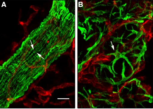
The myoepithelium is organized differently in the ducts and alveoli. Ductal myoepithelial cells are arranged in a more or less continuous monolayer, whereas alveolar myoepithelial cells are stellate-shaped and do not form a continuous layer between the secretory epithelium and the surrounding basement membrane (Fig. 1).
cytokeratins 5, 14 (K5 and K14, respectively) and cytokeratin 17, P-cadherin and high levels of DNp63.
Two discoveries reported a few years ago suggested that mam-mary basal epithelial cells might play a key role in normal mammam-mary development and tumorigenesis. First, the transcriptional profiling of mammary carcinomas revealed a subset of tumors expressing basal cell markers and characterized by a particularly poor clinical outcome (Gusterson 2009; Foulkes et al., 2011 and references therein). Second, several reports describing the isolation and partial characterization of stem/progenitor cells from adult mouse mammary gland and mammary epithelial cell lines indicated that these cells might reside in the basal compartment of the mammary epithelium (Deugnier et al., 2006; Shackleton et al., 2006; Stingl et al., 2006; Sleeman et al., 2007).
In this review, we focus essentially on the aspects of myoepi-thelial cell functional activity investigated by our team. We provide an overview of studies dealing with the control of myoepithelial cell differentiation, discuss the role of cell-cell and cell-ECM interactions involving basal myoepithelial cells in mammary gland development and, finally, summarize current knowledge concerning the contribu-tion of basal myoepithelial cells to the regenerative potential of the mammary epithelium and tumorigenesis. An important physiological property of myoepithelial cells, their tumor-suppressor potential, has been discussed elsewhere (Barsky and Karlin 2006; Panday et al., 2011). Here, we refer to the studies carried out in mouse, rat and human. Mammary gland development and pathology are clearly not identical in different mammalian species. Of note, the quiescent mammary gland of rodents consists of ramified ducts only, comprising small lateral or tertiary branches that give rise to alveoli in pregnancy, whereas adult human mammary gland, even in the absence of pregnancy, contains variable amounts of lobulo-alveoli (Anbazhagan et al., 1998; Naccarato et al., 2000). Another notable difference between rodent and human mammary tissue concerns the stroma, the connective tissue surrounding the mammary epithelium. In mouse and rat, the stroma is fatty, rich in adipocytes, whereas human breast contains much more fibrous
connective tissue. However, overall, the organization of the mam-mary parenchyma, at the cellular level, is similar in rodents and humans, and data obtained in rodent models provide essential information relevant to human breast development and disease.
Differentiation of mammary myoepithelial cells
The basal and luminal cells from the pseudostratified mammary epithelium are functionally and phenotypically distinct. Segregation of the two mammary epithelial compartments begins during em-bryonic development. In mammary buds from E15-mouse embryo, most, if not all, cells express basal cytokeratin K5, numerous cells stain positive for both, K5 and luminal cytokeratin 8 (K8), whereas expression of a basal cell marker p63 is already restricted to 2-3 basal cell layers (Fig. 2 A,B). By E18, expression of K5, is higher in basal cells and lower in the cells concentrated in the future luminal part of the ducts, K8 displays the opposite pattern of expression, and p63 is restricted to the basal cell layer (Fig. 2 C,D). Only few mammary basal cells from E18 mouse embryos express first smooth muscle-specific protein, a-smooth muscle actin (a-SMA (Fig. 2E). In newborn mouse mammary glands, we have detected mammary basal epithelial cells positive for a-SMA and calponin, whereas another contractile protein, caldesmon, was absent from the neonatal mouse mammary epithelium and was detected in myo-epithelial cells from three-week-old mice only (Fig. 2F and data not shown). In rat embryo, at E15 and E18, basal epithelial cells from mammary rudiments stained negative for smooth muscle markers. a-SMA and smooth muscle-myosin were first detected in mammary epithelial basal cells from newborn rat females (Deugnier et al., 1995). Thus, in rodents, basal cells from the embryonic mammary buds do not express smooth muscle markers, and myoepithelial cell differentiation begins during the perinatal period.
In human fetal breast, the first smooth muscle marker, a-SMA, was detected in basally located ductal cells after 22 to 23 weeks of gestation (Anbazhagan et al., 1998; Friedrichs et al., 2007). At this developmental stage, luminal and basal cell layers could be clearly distinguished due to differences in expression of K8, the transcription factor AP2-a, the transcription factor AP2-g and HER1, the first two of these markers being restricted to luminal cells and the last two restricted to basal cells (Friedrichs et al., 2007).
The differentiated phenotype, with the induction and upregulation of smooth muscle-specific contractile and cytoskeletal proteins, is acquired gradually by myoepithelial cells, essentially during postnatal mammary gland development. Changes in the adhesion system, including the integrin repertoire and the production of ECM components, accompany the maturation of mammary myoepithelial cells (Deugnier et al., 1995).
In growing pubertal mouse mammary gland, the cap cells of the terminal end buds give rise to new ductal myoepithelial cells (Williams and Daniel 1983). The terminal end buds are bulbous structures found at the growing tips of mammary ducts advanc-ing into the fat pad, with the cap cells formadvanc-ing a monolayer at the front. The cap cells proliferate and, as the duct grows into the stroma, they progressively relocate to the underlying part of the duct, where they differentiate into myoepithelial cells (Williams and Daniel 1983). Cap cells have a particular phenotype: they express a-SMA, P-cadherin and p63, but stain only weakly for basal-type cytokeratins K5 and K14. In addition, a recent study identified a specific marker of cap cells, s-SHIP, a protein of unknown biological
Fig. 1. Morphology of ductal and alveolar myoepithelial cells. Double immunofluorescence labelling of sections through 10-week-old virgin
(A) and 2-day-lactating (B) mouse mammary glands with the antibodies against cytokeratin 5 (K5, green) and CD31 (red). Arrows indicate elongated ductal (A) and stellate-shaped alveolar (B) myoepithelial cells. CD31 reveals capillaries. Bar, 10 mm.
function structurally related to SHIP1, an SH2-containing inositol 5’-phosphatase (Bai and Rohrschneider 2010). The terminal end buds are present only in rapidly growing pubertal mammary glands, but cell populations similar to cap cells may exist at the extremities of all growing buds, at various developmental stages.
The smooth muscle marker expression of myoepithelial cells is not only regulated temporally during development, it is also regulated spatially. In adult quiescent human breast, a specific caldesmon variant implicated in the regulation of smooth muscle contraction is expressed by the myoepithelial cells of large ducts and galac-tophorous sinuses, but absent from intralobular small ducts and acini (Lazard et al., 1993). Similarly, in the lactating rat mammary gland, ductal and alveolar myoepithelial cells differ in their
pat-terns of contractile and cytoskeletal smooth muscle marker expression (Deugnier et al., 1995). At least in part, this heterogeneity may reflect transient differences in the degree of maturation of the myoepithelial cells located in the various parts of the mammary tree, or differences in the functional properties of ductal and alveolar myoepithelial cells, such as contractile activity. For instance, it seems plausible that the expression of contractile proteins is upregulated in alveolar myoepi-thelial cells from lactating glands. Jolicoeur (2005) suggested that the heterogeneity of myoepithelial cells “may reflect an innate ability of the mammary basal myoepithelial lineage to adapt to a wide range of environ-ments”, enabling these cells to mediate epithelial–stromal interactions adequately.
Contractile function and the control
of mammary myoepithelial cell
dif-ferentiation
The intrinsic contractile activity of mam-mary myoepithelial cells is essential for their physiological function. In lactating mammary gland, myoepithelial cell contraction is in-duced by the pulsatile release of oxytocin from the pituitary gland. Oxytocin binds to a promiscuous G protein-coupled receptor on the surface of the myoepithelial cell (reviewed in Reversi et al., 2005). As in smooth muscle cells, the contraction of myoepithelial cells is regulated by myosin light chains (MLC). Phosphorylation of the MLC induces myo-sin ATPase activity, the binding of myomyo-sin to actin and contraction (Hartshorne et al., 1989). The subsequent dephosphorylation of MLC by a specific phosphatase leads to relaxation. We recently showed that, in addition to the phospholipase C/Ca2+/ MLC
pathway (Reversi et al., 2005), the RhoA/ ROCK signaling cascade is essential for the oxytocin-induced contraction of myoepithelial cells (Raymond et al., 2011). We found that ROCK inhibition completely prevented the
Fig. 2. Distribution of epithelial and smooth muscle markers in developing mouse mammary gland. Double immunofluorescence labelling of sections through developing mammary glands from mouse embryo (A-E) and newborn female mouse (F) with antibodies against K5, K8, p63, a-SMA and calponin. The nuclei were stained with DAPI. (A,A’), serial sections through a mammary bud from E15.5 embryo, (B) section through another bud from the same embryo. (C-E), sections through a mammary rudiment from an E18 female embryo. C’’’ shows merged C, C’ and C’’ images. F’’ shows merged F and F’ images. The arrows in E indicate basal cells positive for K5 and a-SMA, those in F, F’ and F”, indicate basal cells positive for a-SMA and calponin. Tissues were fixed in paraformalde-hyde, embedded in paraffin, sectioned and processed for immunolabelling as described elswhere (Taddei et al., 2008). Bars, 45 mm.
contractile response of myoepithelial cells to oxytocin.
The contractile activity of myoepithelial cells requires the expres-sion of smooth muscle proteins and appropriate cell-ECM interac-tions. Similar to smooth muscle cells, differentiation of myoepithelial cells is dependent on serum response factor (SRF), a transcription factor that binds a DNA sequence known as the CArG box, as-sociated with smooth muscle structural genes, such as the a-SMA and smooth muscle myosin heavy chain genes. SRF interacts with members of the myocardin family of transcriptional coactivators that enhance the expression of SRF-dependent genes (see references in Pipes et al., 2006). Myocardin itself is expressed specifically in cardiac and smooth muscle cells, whereas mammary myoepithelial cells express the myocardin-related transcription factors Mkl1 and
A
A’
B
C
C’
C’’
C’’’
D
Mkl2 (see references in Pipes et al., 2006).
The mammary glands of MKL1-deficient mice develop normally, but mutant dams fail to feed complete litters, due to impairment of the contractile function of the myoepithelial cells. In the absence of MKL1, mammary myoepithelial cells contain only very low amounts of proteins essential for contraction, such as smooth muscle variants of actin, myosin heavy chains, tropomyosin, transgelin, caldesmon and myosin light chain kinase. Mkl2 is upregulated in mutant tissue, but cannot replace MKL1 (Pipes et al., 2006).
Unlike myocardin, which localizes to the cell nucleus, myocardin-related proteins are associated with cytoplasmic monomeric G-actin. Rho GTPases positively regulate smooth muscle-specific Srf target gene expression, due to their ability to induce F-actin polymerization, leading to the release of cytoplasmic MKL1. Subsequent nuclear accumulation of MKL1 enables the protein to act as SRF co-activator, promoting smooth muscle differentiation (Pipes et al., 2006).
a-SMA is the most abundant actin isoform in mammary myoepi-thelial cells. A recent study showed that a-SMA was necessary for the contractile function of myoepithelial cells and for generation of the contractile force required for milk ejection. Mice lacking a-SMA presented lactation failure, despite the normal development of their mammary glands (Haaksma et al., 2011). Experiments in vitro revealed that the contractile response to oxytocin of myoepithelial cells lacking a-SMA was impaired. Expression of other contractile proteins was not analyzed in this study, but the authors suggested that the lack of a-SMA might alter actin dynamics, leading to G-actin accumulation, thereby decreasing the transcriptional activity of MKL1 and the expression of other smooth muscle contractile proteins.
Another known regulator of smooth muscle-specific protein ex-pression is TGF-b. A transcriptional regulator activated by TGF-b, SMAD3, interacts with MKLs, thereby controlling smooth muscle lineage differentiation (reviewed in Pipes et al., 2006). In addi-tion to their role in the regulaaddi-tion of smooth muscle-specific SRF targets, MKL1 and SMAD3 participate in control of the expression of Slug (Morita et al., 2007), a transcription factor essential for the epithelium-to-mesenchyme transition. In mammary epithelium, Slug has been implicated in control of the basal cell phenotype. This protein is found only in the basal myoepithelial cell layer (Mani et al., 2008; Proia et al., 2010).
The Notch pathway has been reported to contribute to the control of the smooth muscle phenotype (Morrow et al., 2008) and impli-cated in the amplification of mammary myoepithelial progenitors (Dontu et al., 2004). The impact of Notch signaling on control of the balance of smooth muscle (and, probably, myoepithelial) cell proliferation and differentiation is context dependent. On the one hand, Notch signaling directly activates the transcription of smooth muscle-specific genes, whereas, on the other hand, its downstream targets, acting in concert with other signaling pathways, can inhibit the expression of smooth muscle markers inducing dedifferentiation (Morrow et al., 2008). A recent report has suggested a role for the EGFR-ERK1/2 signaling pathway in the control of the propagation of the mammary basal cell population (Pasic et al., 2011) . However, the effects of EGFR ligands on the expression of smooth muscle-specific proteins has not been examined in this study.
Myoepithelial cell-ECM interactions
Cell-ECM interactions play important roles in the control of various aspects of the mammary epithelial cell functional activity.
The mammary epithelium is surrounded by a basement membrane consisting essentially of collagen IV, various laminin variants and nidogen (reviewed in Muschler and Streuli 2010). Due to their direct contact with the basement membrane, myoepithelial cells are particularly rich in integrins. Early immunohistochemical studies have revealed the expression of various integrin dimers, including collagen receptors a1b1, a2b1 and fibronectin receptor a5b1, avb3 integrin and high levels of laminin receptors a3b1, a6b1 and a6b4, in rodent and human mammary myoepithelial cells (see references in Taddei et al., 2003 and in Muschler and Streuli 2010).
Mice lacking the a1 or a2 integrin chain are viable. The germline deletion of the a1 integrin gene has no effect on mammary develop-ment, whereas ablation of the a2 integrin gene results in a slight decrease in mammary ductal branching complexity in virgin mice (reviewed in Hynes 2002). A lack of a3b1 or a6b4 integrins leads to perinatal lethality (reviewed in Hynes 2002). Therefore, tissue transplantation technique has been employed to study the roles played by these integrins in mammary development. Mammary epithelium deficient for a3 or a6 integrin chain (i.e. depleted of a3b1 or a6b1 and a6b4 integrin dimers), when transplanted into cleared mouse mammary fat pads, produced ducts and alveoli similar to those developed from control tissue, suggesting that these integrins are dispensable for mammary morphogenesis. One possible explanation for these results is the functional redundancy of the a3b1, a6b1 and a6b4 integrins.
To study the roles played by integrins expressed in the mammary myoepithelial cells in mammary gland development, we employed conditional gene deletion involving Cre-Lox approach. We have obtained mouse mutants presenting deletion of b1 or a3 integrin chains in the basal (K5-positive) cell population. The expression of Cre was driven to basal epithelial cells by the K5 promoter (Taddei et al., 2008; Raymond et al., 2011).
The deletion of b1 integrin from basal myoepithelial cells af-fected mammary branching morphogenesis in virgin mice and lobulo-alveolar development in pregnancy (Taddei et al., 2008). The mutant epithelium was characterized by a largely disorganized general branching pattern, with few side branches. Lobulo-alveolar development was significantly retarded and secretory alveoli devel-oped only late in pregnancy, after 14.5 p.c. from integrin-positive progenitor cells residing in the luminal layer. However, the deletion of b1 integrins from the basal myoepithelial cell compartment did not impede ductal growth or differentiation of the two major mam-mary lineages, the basal myoepithelial and the luminal.
Furthermore, in the mutant epithelium, the lack of b1 integrin altered the orientation of the basal cell division axis and the progeny of b1 integrin-depleted basal cells, identified by a genetic marker, was found in the luminal compartment, by contrast to what was observed for control tissue. These observations led to the conclu-sion that, in the developed ducts, basal mammary epithelial cells divided parallel to the basement membrane, whereas basal cells lacking b1 integrin escaped this rule. In this case, orientation of the basal cell division plane appeared to be random, so that part of the progeny localized to the luminal compartment and differen-tiated into luminal cells. These data suggested, that interactions between basal cells and ECM may contribute to cell fate decisions in mammary epithelium.
how cell-ECM interactions mediated by b1 integrins contribute to maintenance of the mammary stem cell population, and whether the deletion of b1 integrin affects stem cells, directly, or indirectly, by modifying their survival or self-renewal capacity or disturbing the stem cell niche. Numerous studies have shown that cell-ECM adhesion plays an essential role in various stem cell niches (Ray-mond et al., 2009).
The conditional deletion of a laminin receptor, a3b1 integrin, from myoepithelial cells did not interfere with the integrity or func-tional differentiation of the mammary epithelium, but led to low rates of milk ejection due to impaired myoepithelial cell contractil-ity (Raymond et al., 2011). This study revealed that in mammary myoepithelial cells, a3b1 integrin-mediated interactions with the ECM play an essential role in the control of FAK-Rac-PAK path-way activation thereby participating in the regulation of the myo-epithelial cell post-contraction relaxation. In the mammary glands of lactating mice presenting deletion of a3b1 integrin from basal myoepithelial cells, we observed sustained MLC phosphorylation, low levels of FAK activation/phosphorylation and altered Rho/ Rac balance. Cultured mammary myoepithelial cells depleted of a3b1 contracted in response to oxytocin, but were unable to maintain the state of post-contractile relaxation. The expression of constitutively active Rac or its effector PAK, or treatment with MLC-kinase inhibitor rescued the relaxation capacity of mutant cells, strongly suggesting that a3b1-mediated stimulation of the Rac/PAK pathway is required for the inhibition of MLC-kinase ac-tivity, permitting completion of the myoepithelial cell contraction/ relaxation cycle and successful lactation. This study provided the first in vivo evidence implicating integrin signaling in the control of myoepithelial cell contractile function.
Intercellular and paracrine interactions involving
myo-epithelial cells
Like other epithelial cells, mammary basal myoepithelial cells form junctional complexes, including desmosomes and adherens junctions, between them and with luminal cells. Of note, numer-ous cell-cell-adhesion molecules are expressed differentiatially in basal and luminal cells of the mammary epithelium. Many cell-cell-adhesion molecules display differential expression between the basal and luminal cells of the mammary epithelium. Runswick and coworkers provided evidence that desmosomes play an important role in the establishment and maintenance of the bilayer organiza-tion (Runswick et al., 2001). Desmosomal cadherins, desmocolin 3 and desmoglein 3 are restricted to the myoepithelium. Perturbation of the cell-cell interactions involving these myoepithelium-specific molecules interfere with the cell type-specific positioning of luminal and basal mammary epithelial cells.
Cadherins are adherens junction components essential for the maintenance of epithelial tissue architecture. P-cadherin is expressed in the basal cell layers of stratified and pseudostratified epithelia. In mammary gland, P-cadherin is restricted to the basal cell layer, including ductal and alveolar myoepithelial cells and cap cells from terminal end buds. P-cadherin-deficient mice present unscheduled lobulo-alveolar development (Radice et al., 1997). Alveolus-like structures, similar to those seen early in pregnancy, develop in mutant virgin females, and luminal cells present signs of lactogenic differentiation. Late in life, P-cadherin-deficient mice develop hyperplastic and dysplastic lesions (Radice et al., 1997).
This study suggested that the deletion of P-cadherin, an adhe-sion molecule expressed by basal cells, affected the luminal cell population. Further studies are required to determine how basal cells contribute to the control of luminal cell proliferation and dif-ferentiation.
The molecular mechanisms that control P-cadherin expression remain poorly understood. Using mammary epithelial cell lines and mouse mutants obtained in our laboratory, we have shown that the Wnt/b-catenin signaling pathway is involved in regulation of P-cadherin expression independently of the Lef/Tcf transcription factors (Faraldo et al., 2007). High levels of P-cadherin were found in mammary glands from transgenic mice presenting a constitu-tive activation of b-catenin signaling in basal myoepithelial cells (Faraldo et al., 2007).
Novel information about the nature of cell-cell interactions in the mammary epithelium has been provided by Hinck’s laboratory. This team was the first to implicate neural guidance molecules in basal-luminal cell adhesion and crosstalk. In the mammary epi-thelium, Netrin1, a secreted guidance cue molecule is expressed by luminal cells, whereas, its receptor, Neurogenin is present on the surface of cap and differentiated myoepithelial cells. Loss-of-function mutations in genes coding for Netrin1 and Neurogenin resulted in the disorganization of terminal end buds (Srinivasan et al., 2003). Another neural guidance molecules Slit2 is expressed in both mammary epithelial cell layers, whereas its receptor, Robo1 is restricted to the basal cell population, cap and myoepithelial cells. Deletion of Slit2 or Robo1 from the mammary epithelium results in a phenotype similar to that induced by perturbation of Netrin1/ Neurogenin couple, a lack of adhesion between luminal and cap cell layers (Strickland et al., 2006). Furthermore, simultaneous loss-of-function mutations of slit2 and ntn1 genes resulted in an enhanced phenotype with separated luminal and basal cell lay-ers in the mammary ducts suggestive of synergy between Slit2 and Netrin 1 during ductal morphogenesis. A recent report from the same laboratory implicated Slit/Robo1 signaling in the control of mammary branching morphogenesis (Macias et al., 2011). The authors suggested that basal myoepithelial cells control the formation of new ductal branches via the production of mitogens for the luminal cells. Macias and coworkers revealed that the TGF-b1-induced expression of Robo1 in basal myoepithelial cells and the interaction of Slit2 with Robo1 inhibited b-catenin signaling, limiting basal cell proliferation and preventing the formation of new branches (Macias et al., 2011).
In turn, luminal cells also affect the homeostasis of the basal cell population. Wnt signaling is essential for proliferation and functional activity of mammary stem cells localized in the basal compartment (reviewed in Incassati et al., 2010). Whilst genes coding for several Wnt ligands (Wnt4, Wnt5a, Wnt7b) were found to be expressed by luminal cells, expression of the receptors and co-receptors associ-ated with Wnt/b-catenin signaling (Frizzled 1, 2, 3, 7, and 8, Lrp5 and 6) was localized to the basal myoepithelial cell compartment suggesting paracrine interactions (Kendrick et al., 2008).
In quiescent mammary gland of virgin mice, myoepithelial cells do not proliferate. By contrast, stimulation with ovarian hormones, either under experimental conditions (hormone injection), or physi-ologically (during pregnancy or estrus), induces amplification of the basal myoepithelial cell population and expansion of the mam-mary stem cell population via paracrine mechanisms involving the Wnt and RANK pathways (Brisken and O’Malley 2010; Incassati et al., 2010; Asselin-Labat et al., 2010; Joshi et al., 2010). Wnt4 is induced upon stimulation of the mammary epithelium by pro-gesterone (see references in Brisken and O’Malley 2010). Most hormonal receptors are expressed by luminal cells and basal myoepithelial cells do not express ERa at all, but Wnt/b-catenin pathway-associated genes, including targets and receptors, were found to be upregulated in the mammary basal cell population of animals stimulated by estrogen/progesterone injection (Joshi et al., 2010). Moreover, ovarian hormones induced the expression of RANKL in luminal cells and the expression of its receptor, RANK, in luminal and, particularly, basal cells (Asselin-Labat et al., 2010; Joshi et al., 2010).
Large-scale microarray analysis of gene expression patterns in various mammary epithelial cell populations performed with freshly isolated cells from virgin mouse and human mammary glands revealed numerous genes differentially expressed in basal and luminal cells (Stingl et al., 2006; Kendrick et al., 2008; Lim et al., 2010). These studies provided important information for future work aiming to unravel the molecular mechanisms underlying crosstalk between the two major compartments of the mammary epithelium, the basal and luminal compartments. The microarray data complemented functional studies and indicated signaling pathways potentially involved in basal-luminal paracrine or direct cell-cell interactions and revealed several transcription regulators characterized by a cell type-specific expression pattern.
Mammary myoepithelium and stem cells
Studies performed with a mammary cell line CommaD1b, pos-sessing a morphogenetic potential, and with dissociated mammary epithelial cells have strongly suggested that the regenerative capacity of the mammary epithelium, as evaluated by transplanta-tion assays, is associated with the basal epithelial compartment (Deugnier et al., 2006; Shackleton et al., 2006; Stingl et al., 2006; Sleeman et al., 2007). Cell populations enriched in stem cells can be isolated from freshly dissociated mammary epithelium, by flow cytometry, due to the strong expression of a6 or b1 integrins (Shackleton et al., 2006; Stingl et al., 2006). Of note, relatively high integrin expression levels are characteristic of the entire basal mammary cell population, consisting essentially of differentiated myoepithelial cells, whereas stem cells would be expected to display phenotypic characteristics different from those of differenti-ated cells. Consistently, the morphogenetic cell population isoldifferenti-ated
from the CommaD1b cell line expresses basal cell markers, such as basal cytokeratins, P-cadherin and DNp63, but is negative for the smooth muscle-characteristic proteins (Deugnier et al., 2006). Putative mammary stem cells have not yet been physically separated from the differentiated myoepithelial cells, the major cell population in the mammary basal compartment. Stingl and coworkers have reported that repopulating activity is associated with a small cell fraction with higher levels of a6 integrin than the rest of the basal myoepithelial cell population. However, a comparative transcriptome analysis revealed that there were no significant differences in gene expression between these two basal cell subsets (Stingl et al., 2006). Smooth muscle-specific contractile and cytoskeletal proteins were present in similar amounts in both cell populations. The myoepithelial cell layer is heterogeneous, but undifferentiated cells negative for smooth muscle markers have not yet been clearly identified in vivo, in rodent or human mammary basal epithelial cell layer.
Consistent with the transplantation data, lineage tracing ex-periments with the mouse reporter strain Rosa 26 led to the con-clusion that all mammary epithelium cells originated from stem/ progenitor cells expressing the basal cytokeratins K5/K14 (Choi et al., 2009; Moumen and Faraldo, unpublished data). However, there is no experimental evidence to confirm that multipotent stem cells localized in the mammary basal cell layer contribute to the luminal compartment during ductal growth at puberty and lobulo-alveolar development in pregnancy. The results of lineage tracing experiments are consistent with the hypothesis that multipotent stem cells with basal characteristics (K5/K14-positive) give rise to basal myoepithelial and luminal cell lineages during early stages of mammary gland development, and that once the system of branching ducts is formed, the basal cell population makes no further contribution to the luminal cell layer.
Thus, it is still unclear whether an undifferentiated stem cell population exists in pubertal and adult mammary glands. It also remains to be determined whether cells possessing the regenerat-ing potential revealed in transplantation assays, are phenotypically different from the surrounding differentiated myoepithelial cells, or whether at least some of the myoepithelial cells can dedifferentiate and acquire stem cell properties after transplantation.
Although differentiated myoepithelial cells from adult animals may be devoid of stem cell activity, they seem very likely to contribute to the mammary stem cell niche, by producing the necessary growth factors or ECM components. A recent analysis of gene expression patterns in basal and luminal cell populations isolated from hu-man and mouse mammary epithelium confirmed that basal cells expressed numerous ECM proteins in large amounts, including most of the mammary basement membrane constituents, such as laminins, collagens, fibronectin, heparan sulphate proteoglycans and SPARC (Kendrick et al., 2008; Lim et al., 2010).
Contribution of myoepithelial cells to mammary
tu-morigenesis
Mammary myoepitheliomas are rare malignant tumors consid-ered to originate from myoepithelial cells. These tumors are char-acterized by high-level expression of basal epithelial cell markers and, in some cases, the expression of smooth muscle-specific proteins (Buza et al., 2010 and references therein).
carcinomas led to the discovery of a tumor subset characterized by the expression of basal epithelial cell markers, the absence of ER- and PR-positive cells and the lack of HER2 overexpression. These tumors are usually referred to as basal-type or triple negative (reviewed in Gusterson 2009; Foulkes et al., 2011). These tumors were initially thought to originate from mammary progenitor/stem cells. However, several recent studies have provided evidence that, at least, partially contradicted this hypothesis. Hereditary mammary carcinomas associated with the brca1 gene mutation belong to the basal subtype. It has been demonstrated that luminal progeni-tors are amplified in the mammary epithelium of brca1-mutation carriers suggesting that this cell population might be at the origin of basal-type BRCA1-mammary carcinomas (Lim et al., 2009). Interestingly, the basal cell population was notably decreased in the mammary epithelium of these individuals (Lim et al., 2009). Further, experiments employing a mouse model of breast carci-nogenesis demonstrated that deletion of the brca1 gene from the luminal layer of the mammary epithelium of mice heterozygous for p53 was led to the development of tumors phenotypically similar to human BRCA1-associated carcinomas (Molyneux et al., 2010). The deletion of brca1 from the basal myoepithelial cell layer led to the development of adenomyoepitheliomas and squamous metaplastic carcinomas. Although the tumors developed after the disruption of brca1 gene expression in the basal cell layer were of the basal subtype, based on their transcriptional profiles, their histological characteristics differed from human BRCA1 tumors (Molyneux et al., 2010). Finally, a study from the Kuperwasser laboratory revealed that the transcriptional repressor Slug accumulated in BRCA1-associated breast cancer may be responsible for the basal phenotypic characteristics of the tumor cells (Proia et al., 2010).
The Wnt/b-catenin signaling pathway is activated in basal-type mammary carcinomas (Khramtsov et al., 2010). We investigated the role of this pathway in mammary gland development and tumorigenesis, by generating transgenic mice expressing in the basal epithelial cell layer, an “activated” (N-terminally truncated and, thus, stabilized) b-catenin under control of the K5-promoter (transgenic lines K5-DNbcat). K5-DNbcat mice have an abnormal mammary phenotype, including precocious side branching in pregnancy, associated with an increase in proliferation and a de-crease in the apoptosis of luminal epithelial cells and accelerated post-lactational involution (Faraldo et al., 2005). By the age of 12 to 16 months, 75% of K5-DNbcat nulliparous females develop mammary hyperplasia comprising K5-positive (basal) cells nega-tive for luminal and myoepithelial cell markers. Most multiparous K5-DNbcat mice develop invasive mammary carcinomas consisting essentially of undifferentiated basal epithelial cells or presenting signs of differentiation towards epidermal lineages (Faraldo et al., 2005). We suggest that the activation of b-catenin signaling in mammary basal epithelial cell induces the amplification of basal-type progenitors, and, that basal-basal-type progenitor/stem cells may contribute to the development of a subset of basal-type mammary carcinomas, metaplastic lesions characterized by the expression of epidermal lineage markers.
Conclusions and perspectives
Myoepithelial cells are no longer considered to be a second-class mammary cell population of almost no interest. In addition to its contractile function, which is essential for lactation, the basal
cell layer harbors the regenerative potential of the mammary epi-thelium. Basal myoepithelial cells modulate the proliferation and differentiation of luminal cells, and affect the surrounding stroma. Various aspects of myoepithelial cell biology therefore merit further investigation.
Recent studies have shed light on the nature of the reciprocal paracrine and adhesion-mediated regulatory signals between the basal and luminal compartments of the mammary epithelium. How-ever, our knowledge of these complex interactions is still limited, and detailed descriptions and analysis of the basal-luminal cell crosstalk are required to define the roles played by the myoepi-thelial cells in normal development and tumorigenesis. Several of the studies discussed above suggested that triple-negative breast tumors might originate from luminal progenitors (Lim et al., 2009; Molyneux et al., 2010; Proia et al., 2010). However, some of the signals essential for the maintenance of this cell population are thought to be provided by basal cells (Bouras et al., 2008) imply-ing that the question of the contribution of the mammary basal cell compartment to tumorigenesis is still open.
A particularly important subject for further studies is to define the effects of the myoepithelium on the stroma during development and in disease. One aspect of the myoepithelium-stroma crosstalk yet to be unraveled is the contribution of the mammary myoepithelial cells to the establishment of the vascular network in the connec-tive tissue surrounding the epithelium. As discussed elsewhere (Barsky and Karlin 2006), myoepithelial cells are considered to exhibit the anti-angiogenetic properties, however, they express VEGFa, and its deletion from the basal cell layer significantly at-tenuated the angiogenesis accompanying mammary development (Rossiter et al., 2007).
Another intriguing issue is the relationships between myoepi-thelial and stem/progenitor cells. It is not known yet whether upon transplantation, rare undifferentiated cells possessing stem cell properties give rise to mammary epithelial outgrowths, or, alterna-tively, all or a subset of myoepithelial cells can dedifferentiate and acquire the repopulating potential. We therefore believe that the phenotypic heterogeneity of the basal myoepithelial cell popula-tion in quiescent and hormone-stimulated glands, and control of the smooth muscle differentiation program in the myoepithelium merit further investigation.
Acknowledgments
Due to space limitations, in several cases, we have referred to reviews, rather than the original studies. We apologize to colleagues for omission of the original references. The work in MAG laboratory is supported by La Ligue Nationale Contre le Cancer (Equipe Labelisée 2009) and a grant from Agence Nationale de la Recherche ANR-08-BLAN-0078-01. MM received funding from Association pour la Recherche sur le Cancer; AC, from Institut Curie and Servier Laboratories; SC, from Cancéropôle Ile de France; VP, from Agence Nationale de la Recherche; KR, from La Fonda-tion pour la Recherche Médicale and Institut NaFonda-tional du Cancer. MAG is Directeur de Recherche, MMF and MAD are Chargé de Recherche at the Institut National de la Santé et de la Recherche Médicale (INSERM).
References
ANBAZHAGAN R, OSIN PP, BARTKOVA J, NATHAN B, LANE EB and GUSTERSON BA (1998). The development of epithelial phenotypes in the human fetal and infant breast. J Pathol 184: 197-206.
ASSELIN-LABAT ML, VAILLANT F, SHERIDAN JM, PAL B, WU D, SIMPSON ER, YASUDA H, SMYTH GK, MARTIN TJ, LINDEMAN GJ and VISVADER JE (2010). Control of mammary stem cell function by steroid hormone signalling. Nature 465: 798-802.
BAI L and ROHRSCHNEIDER LR (2010). s-SHIP promoter expression marks activated stem cells in developing mouse mammary tissue. Genes Dev 24: 1882-1892. BARSKY SH and KARLIN NJ (2006). Mechanisms of disease: breast tumor
patho-genesis and the role of the myoepithelial cell. Nat Clin Pract Oncol 3: 138-151. BOURAS T, PAL B, VAILLANT F, HARBURG G, ASSELIN-LABAT ML, OAKES SR,
LINDEMAN GJ and VISVADER JE (2008). Notch signaling regulates mammary stem cell function and luminal cell-fate commitment. Cell Stem Cell 3: 429-441. BRISKEN C and O’MALLEY B (2010). Hormone action in the mammary gland. Cold
Spring Harb Perspect Biol 2: a003178.
BUZA N, ZEKRY N, CHARPIN C and TAVASSOLI FA (2010). Myoepithelial carci-noma of the breast: a clinicopathological and immunohistochemical study of 15 diagnostically challenging cases. Virchows Arch 457: 337-345.
CHOI YS, CHAKRABARTI R, ESCAMILLA-HERNANDEZ R and SINHA S (2009). Elf5 conditional knockout mice reveal its role as a master regulator in mammary alveolar development: failure of Stat5 activation and functional differentiation in the absence of Elf5. Dev Biol 329: 227-241.
DEUGNIER MA, FARALDO MM, TEULIERE J, THIERY JP, MEDINA D and GLUK-HOVA MA (2006). Isolation of mouse mammary epithelial progenitor cells with basal characteristics from the Comma-Dbeta cell line. Dev Biol 293: 414-425. DEUGNIER MA, MOISEYEVA EP, THIERY JP and GLUKHOVA M (1995). Myoepithelial
cell differentiation in the developing mammary gland: progressive acquisition of smooth muscle phenotype. Dev Dyn 204: 107-117.
DONTU G, JACKSON KW, MCNICHOLAS E, KAWAMURA MJ, ABDALLAH WM and WICHA MS (2004). Role of Notch signaling in cell-fate determination of human mammary stem/progenitor cells. Breast Cancer Res 6: R605-615.
FARALDO MM, TEULIERE J, DEUGNIER MA, BIRCHMEIER W, HUELSKEN J, THIERY JP, CANO A and GLUKHOVA MA (2007). beta-Catenin regulates P-cadherin expression in mammary basal epithelial cells. FEBS Lett 581: 831-836. FARALDO MM, TEULIERE J, DEUGNIER MA, TADDEI-DE LA HOSSERAYE I,
THIERY JP and GLUKHOVA MA (2005). Myoepithelial cells in the control of mammary development and tumorigenesis: data from genetically modified mice.
J Mammary Gland Biol Neoplasia 10: 211-219.
FOULKES WD, SMITH IE and REIS-FILHO JS (2011). Triple-negative breast cancer.
N Engl J Med 363: 1938-1948.
FRIEDRICHS N, STEINER S, BUETTNER R and KNOEPFLE G (2007). Immunohis-tochemical expression patterns of AP2alpha and AP2gamma in the developing fetal human breast. Histopathology 51: 814-823.
GUSTERSON B (2009). Do ‘basal-like’ breast cancers really exist? Nat Rev Cancer 9: 128-134.
HAAKSMA CJ, SCHWARTZ RJ and TOMASEK JJ (2011). Myoepithelial cell contrac-tion and milk ejeccontrac-tion are impaired in mammary glands of mice lacking smooth musclealpha-actin. Biol Reprod DOI:10.1095/biolreprod.110.090639
HARTSHORNE DJ, ITO M and IKEBE M (1989). Myosin and contractile activity in smooth muscle. Adv Exp Med Biol 255: 269-277.
HYNES RO (2002). Integrins: bidirectional, allosteric signaling machines. Cell 110: 673-687.
INCASSATI A, CHANDRAMOULI A, EELKEMA R and COWIN P (2010). Key sig-naling nodes in mammary gland development and cancer: beta-catenin. Breast
Cancer Res 12: 213.
JOLICOEUR F (2005). Intrauterine breast development and the mammary myoepi-thelial lineage. J Mammary Gland Biol Neoplasia 10: 199-210.
JOSHI PA, JACKSON HW, BERISTAIN AG, DI GRAPPA MA, MOTE PA, CLARKE CL, STINGL J, WATERHOUSE PD and KHOKHA R (2010). Progesterone induces adult mammary stem cell expansion. Nature 465: 803-807.
KENDRICK H, REGAN JL, MAGNAY FA, GRIGORIADIS A, MITSOPOULOS C, ZVELEBIL M and SMALLEY MJ (2008). Transcriptome analysis of mammary epithelial subpopulations identifies novel determinants of lineage commitment and cell fate. BMC Genomics 9: 591.
KHRAMTSOV AI, KHRAMTSOVA GF, TRETIAKOVA M, HUO D, OLOPADE OI and GOSS KH (2010). Wnt/beta-catenin pathway activation is enriched in basal-like breast cancers and predicts poor outcome. Am J Pathol 176: 2911-2920.
LAZARD D, SASTRE X, FRID MG, GLUKHOVA MA, THIERY JP and KOTELIANSKY VE (1993). Expression of smooth muscle-specific proteins in myoepithelium and stromal myofibroblasts of normal and malignant human breast tissue. Proc Natl
Acad Sci USA 90: 999-1003.
LIM E, VAILLANT F, WU D, FORREST NC, PAL B, HART AH, ASSELIN-LABAT ML, GYORKI DE, WARD T, PARTANEN A, FELEPPA F, HUSCHTSCHA LI, THORNE HJ, FOX SB, YAN M, FRENCH JD, BROWN MA, SMYTH GK, VISVADER JE and LINDEMAN GJ (2009). Aberrant luminal progenitors as the candidate target population for basal tumor development in BRCA1 mutation carriers. Nat Med 15: 907-913.
LIM E, WU D, PAL B, BOURAS T, ASSELIN-LABAT ML, VAILLANT F, YAGITA H, LINDEMAN GJ, SMYTH GK and VISVADER JE (2010). Transcriptome analyses of mouse and human mammary cell subpopulations reveal multiple conserved genes and pathways. Breast Cancer Res 12: R21.
MACIAS H, MORAN A, SAMARA Y, MORENO M, COMPTON JE, HARBURG G, STRICKLAND P and HINCK L (2011). SLIT/ROBO1 signaling suppresses mammary branching morphogenesis by limiting basal cell number. Dev Cell 20: 827-840. MANI SA, GUO W, LIAO MJ, EATON EN, AYYANAN A, ZHOU AY, BROOKS M,
REINHARD F, ZHANG CC, SHIPITSIN M, CAMPBELL LL, POLYAK K, BRISKEN C, YANG J and WEINBERG RA (2008). The epithelial-mesenchymal transition generates cells with properties of stem cells. Cell 133: 704-715.
MOLYNEUX G, GEYER FC, MAGNAY FA, MCCARTHY A, KENDRICK H, NATRAJAN R, MACKAY A, GRIGORIADIS A, TUTT A, ASHWORTH A, REIS-FILHO JS and SMALLEY MJ (2010). BRCA1 basal-like breast cancers originate from luminal epithelial progenitors and not from basal stem cells. Cell Stem Cell 7: 403-417. MORITA T, MAYANAGI T and SOBUE K (2007). Dual roles of myocardin-related
transcription factors in epithelial mesenchymal transition via slug induction and actin remodeling. J Cell Biol 179: 1027-1042.
MORROW D, GUHA S, SWEENEY C, BIRNEY Y, WALSHE T, O’BRIEN C, WALLS D, REDMOND EM and CAHILL PA (2008). Notch and vascular smooth muscle cell phenotype. Circ Res 103: 1370-1382.
MUSCHLER J and STREULI CH (2010). Cell-matrix interactions in mammary gland development and breast cancer. Cold Spring Harb Perspect Biol 2: a003202. NACCARATO AG, VIACAVA P, VIGNATI S, FANELLI G, BONADIO AG,
MONTRUC-COLI G and BEVILACQUA G (2000). Bio-morphological events in the develop-ment of the human female mammary gland from fetal age to puberty. Virchows
Arch 436: 431-438.
PANDEY PR, SAIDOU J and WATABE K (2011). Role of myoepithelial cells in breast tumor progression. Front Biosci 15: 226-236.
PASIC L, EISINGER-MATHASON TSK, VELAYUDHAN BT, MOSKALUK CA, BRENIN DR, MACARA IG, LANNIGAN DA (2011). Sustained activation of the HER1–ERK1/2–RSK signaling pathway controls myoepithelial cell fate in human mammary tissue. Genes Dev 25: 1641-1653.
PIPES GC, CREEMERS EE and OLSON EN (2006). The myocardin family of transcriptional coactivators: versatile regulators of cell growth, migration, and myogenesis. Genes Dev 20: 1545-1556.
PROIA TA, KELLER PJ, GUPTA PB, KLEBBA I, JONES AD, SEDIC M, GILMORE H, TUNG N, NABER SP, SCHNITT S, LANDER ES and KUPERWASSER C (2010). Genetic predisposition directs breast cancer phenotype by dictating progenitor cell fate. Cell Stem Cell 8: 149-163.
RADICE G, FERREIRA-CORNWELL MC, ROBINSON SD, RAYBURN H, CHODOSH LA, TAKEICHI M, HYNES RO (1997). Precocious mammary gland development in P-cadherin-deficient mice. J Cell Biol 139: 1025-1032.
RAYMOND K, CAGNET S, KREFT M, JANSSEN H, SONNENBERG A and GLUK-HOVA MA (2011). Control of mammary myoepithelial cell contractile function by alpha3beta1 integrin signalling. EMBO J 30: 1896-1906.
RAYMOND K, DEUGNIER MA, FARALDO MM and GLUKHOVA MA (2009). Adhesion within the stem cell niches. Curr Opin Cell Biol 21: 623-629.
REVERSI A, CASSONI P and CHINI B (2005). Oxytocin receptor signaling in myoepithelial and cancer cells. J Mammary Gland Biol Neoplasia 10: 221-229. ROSSITER H, BARRESI C, GHANNADAN M, GRUBER F, MILDNER M, FODINGER
D and TSCHACHLER E (2007). Inactivation of VEGF in mammary gland epithe-lium severely compromises mammary gland development and function. FASEB
J 21: 3994-4004.
Nat Cell Biol 3: 823-830.
SHACKLETON M, VAILLANT F, SIMPSON KJ, STINGL J, SMYTH GK, ASSELIN-LABAT ML, WU L, LINDEMAN GJ and VISVADER JE (2006). Generation of a functional mammary gland from a single stem cell. Nature 439: 84-88. SLEEMAN KE, KENDRICK H, ROBERTSON D, ISACKE CM, ASHWORTH A and
SMALLEY MJ (2007). Dissociation of estrogen receptor expression and in vivo stem cell activity in the mammary gland. J Cell Biol 176: 19-26.
SRINIVASAN K, STRICKLAND P, VALDES A, SHIN GC and HINCK L (2003). Netrin-1/ neogenin interaction stabilizes multipotent progenitor cap cells during mammary gland morphogenesis. Dev Cell 4: 371-382.
STINGL J, EIREW P, RICKETSON I, SHACKLETON M, VAILLANT F, CHOI D, LI HI and EAVES CJ (2006). Purification and unique properties of mammary epithelial stem cells. Nature 439: 993-997.
STRICKLAND P, SHIN GC, PLUMP A, TESSIER-LAVIGNE M and HINCK L (2006). Slit2 and netrin 1 act synergistically as adhesive cues to generate tubular bi-layers during ductal morphogenesis. Development 133: 823-832.
TADDEI I, DEUGNIER MA, FARALDO MM, PETIT V, BOUVARD D, MEDINA D, FASSLER R, THIERY JP and GLUKHOVA MA (2008). Beta1 integrin deletion from the basal compartment of the mammary epithelium affects stem cells. Nat
Cell Biol 10: 716-722.
TADDEI I, FARALDO MM, TEULIERE J, DEUGNIER MA, THIERY JP and GLUKHOVA MA (2003). Integrins in mammary gland development and differentiation of mam-mary epithelium. J Mammam-mary Gland Biol Neoplasia 8: 383-394.
WILLIAMS JM and DANIEL CW (1983). Mammary ductal elongation: differentiation of myoepithelium and basal lamina during branching morphogenesis. Dev Biol 97: 274-290.
Further Related Reading, published previously in the Int. J. Dev. Biol.
Signalling molecules involved in mouse bladder smooth muscle cellular differentiation
Benchun Liu, Dongxiao Feng, Guiting Lin, Mei Cao, Yuet Wai Kan, Gerald R. Cunha and Laurence S. Baskin Int. J. Dev. Biol. (2010) 54: 175-180
Epithelial-Mesenchymal Transitions in development and disease: old views and new perspectives
M. Angela Nieto
Int. J. Dev. Biol. (2009) 53: 1541-1547
The stroma reaction myofibroblast: a key player in the control of tumor cell behavior
Alexis Desmoulière, Christelle Guyot and Giulio Gabbiani Int. J. Dev. Biol. (2004) 48: 509-517
Transcriptional regulation of cadherins during development and carcinogenesis
Héctor Peinado, Francisco Portillo and Amparo Cano Int. J. Dev. Biol. (2004) 48: 365-375
Integrin function and regulation in development
G Tarone, E Hirsch, M Brancaccio, M De Acetis, L Barberis, F Balzac, S F Retta, C Botta, F Altruda, L Silengo and F Retta Int. J. Dev. Biol. (2000) 44: 725-731