Comparative analysis of p4ha1 and p4ha2 expression
during Xenopus laevis development
DAVIDE MARTINI
1,#, MARTINA GIANNACCINI
1,#, VIVIANA GUADAGNI
1,#, SILVIA MARRACCI
1,
GUIDO GIUDETTI
2and MASSIMILIANO ANDREAZZOLI*
,11Unità di Biologia Cellulare e dello Sviluppo, Dipartimento di Biologia, Università di Pisa, Pisa, Italy and 2Istituto di Biorobotica, Scuola Superiore Sant’Anna, Pisa, Italy
ABSTRACT Collagen prolyl 4-hydroxylases (c-P4Hs) are evolutionary conserved enzymes whose activ-ity is essential for the correct folding of stable triple helical molecules of collagen and collagen-like proteins. They play crucial roles in embryo development, connective tissue functional organization, tumor growth and metastasis. Despite the important function of these enzymes, little is known about their expression during vertebrate development. In this study, we determine and compare the previously undescribed spatio-temporal expression patterns of the p4ha1 and p4ha2 genes, which encode the main subunits containing the enzyme active site, during Xenopus development. The two genes are maternally inherited and share expression in dorsal mesoderm, branchial arches and their derivatives, as well as in the central nervous system, although with distinct spatio-temporal patterns. A major co-expression domain for p4ha1 and p4ha2 is represented by the developing notochord, where these genes are transcribed from early neurula stage to stage 42 tadpole, thus paralleling the profile of collagen II production and suggesting a coordination between collagen synthesis and its post-translational modifications.
KEY WORDS:
prolyl-hydroxylase, gene expression, development, Xenopus laevis, collagen
Collagen prolyl 4-hydroxylases (c-P4Hs), located within the lu-men of the endoplasmic reticulum (ER), are pivotal enzymes for the maturation of extracellular matrix proteins. They catalyze the 4-hydroxylation of the proline in -X-Pro-Gly- sequences of collagen and proteins with collagen-like sequences (Holster et al., 2007). This reaction is essential for the correct folding of stable triple heli-cal molecules of collagen, the most abundant protein in animals and the major component of connective tissue extracellular matrix (Shoulders and Raines, 2009). Accordingly, c-P4Hs catalyze the single most prevalent post-translational modification in animals and play a crucial role in embryo development, connective tissue functional organization, as well as tumor growth and metastasis (Holster et al., 2007, Winter and Page, 2000). These enzymes are composed of dimers or tetramers of a and b subunits. The
catalytic subunits are located in the a subunits while the b subunits
are identical to the protein-disulfide isomerase (PDI) (Helaakoski
et al., 1989, Kivirikko et al., 1990).
These enzymes are evolutionary conserved among animals from invertebrate to humans, as also shown by the ability of
*Address correspondence to: Massimiliano Andreazzoli. Unità di Biologia Cellulare e dello Sviluppo, SS12 Abetone e Brennero, 4, I-56127 Pisa, Italy. Tel. +39-050-2211485; Fax: + 39-050-2211495. E-mail: [email protected] - https://orcid.org/0000-0001-9633-204X
#Note: The indicated authors contributed equally to this work.
Submitted: 24 April, 2019; Accepted: 15 May, 2019.
ISSN: Online 1696-3547, Print 0214-6282 © 2019 UPV/EHU Press
Printed in Spain
Abbreviations used in this paper: c-P4H, collagen prolyl 4-hydroxylase.
heterodimers made by D. melanogaster or C. elegans a subunits
and human PDI to catalyze collagen hydroxylation (Annunen et
al., 1999, Winter and Page, 2000).
In C. elegans two a subunits, namely phy-1 and phy-2, as well
as two b subunits, pdi-1 and pdi-2, have been identified. In the
nematode, the active enzyme is a ab dimer rather than a a2b2
tetramer as in D. melanogaster and in vertebrates. phy-1 and phy-2 expression starts from midelongation stage and persist throughout adult stage. The highest expression of phy-1 and phy-2 is found in the hypodermal cells, which are also responsible for collagen synthesis and display a higher concentration of phy-2-positive cells. Consistently, the temporal expression pattern of phy-1 and
phy-2 shows two peaks corresponding to that of cuticle collagen
phy-1 expression leads to a dumpy phenotype with shorter and
fatter animals at larval L4 and adult stages. The dumpy phenotype is caused by abnormal cuticle deposition leading to improper exo-skeleton morphology, probably due to collagen retention inside the cells as a consequence of the lack of prolyl hydroxylation. On the contrary, disruption of phy-2 mRNA does not cause evident effects, possibly due to a partial redundancy of the enzymatic activity of the two subunits. However, the double knockdown of c-P4Ha subunits
displays more severe effects on C. elegans development leading to coiled larvae, dumpier adults and embryonic lethality in 89% of cases (Winter and Page, 2000).
In vertebrates, the a subunit is present in three isoforms: P4Ha1,
P4Ha2, and P4Ha3. P4Ha1 represents the main form in most cell
types, while the P4Ha2 is the prevalent one in chondrocytes,
os-teoblasts, endothelial and epithelial cells responsible for cartilage, cartilaginous bone and endothelium formation (Myllyharju, 2003). In humans, P4HA1 is found to be highly expressed in fetal brain and liver. In the adult, the highest expression levels are observed in skeletal muscle, although P4HA1 expression is detectable also in heart, placenta, liver and kidney as well as, at lower level, in the brain, lung and pancreas (Kukkola et al., 2003). The P4HA2 gene is highly expressed in adult chondrocytes and endothelial cells. Lower levels of P4HA2 were found in developing glomeruli of fetal kidney, according to histochemical analysis (Nissi et al., 2001). Interestingly, in the liver the bile ducts express the a2 isoform
while hepatocytes express at low level the a1 isoform, indicating a
different expression pattern and suggesting potential distinct roles for the two isoforms (Nissi et al., 2001). Indeed, it was shown that
P4HA1 is expressed preferentially by cells of mesenchymal origin
and in developing tissues, whereas P4HA2 is mainly expressed in differentiated cells (Nissi et al., 2001). Finally, the human P4HA3 is expressed in both adult and fetal tissues but at a much lower level compared to the other two (Kukkola et al., 2003).
In vertebrates, the in vivo function was deeply investigated only for P4Ha1 in mouse knockout by Holster and colleagues in 2007.
In contrast to heterozygotic mice, which appear to be normal, null embryos develop normally until stage E9, but soon after their
development begins to be retarded from stage E10.5 and they eventually die between stages E10.5-E11.5. In null embryos col-lagen IV is correctly synthetized but, without the prolyl hydroxylation by P4Ha1, is not properly assembled in functional triple helices,
leading to the rupture of the basement membrane. Moreover, these collagen IV fibers have an increased diameter probably due to a reduced contact between the triple helices, which, together with their retention inside the cells, causes the dilatation of ER (Holster et al., 2007). As for C. elegans, developmental problems in mouse appear when collagen synthesis becomes crucial. Although the synthesis of collagen IV starts at blastula stage, the collected data indicate that the incorrect assembly of triple helices becomes incompatible with life only after stage E10.5, possibly because the mechanical pressure increases during development (Holster et al., 2007).
Accordingly, human mutations in P4Ha genes are associated
to specific genetic diseases. P4HA1 mutations cause a disorder of connective tissue involving tendon, bone, muscle affecting also the eye (Zou et al., 2017) with the induction of high myopia. Similarly, mutations in P4HA2 have been described as a cause for non-syndromic high myopia (Guo et al., 2015).
Moreover, several diseases are produced by c-P4Hs malfunc-tioning. For instance, inhibition of c-P4Hs by ascorbate deficiency causes scurvy (Smith and Talbot, 2010), while excessive levels of collagen production generates unwanted fibrotic response after cardiac infarction or wound healing (Fan et al., 2012 and references therein). Recently, an interesting role of c-P4Hs in tumor growth and metastasis has been demonstrated as well. It is known that collagen deposition and alignment enhance tumor cell proliferation and migration (Gilkes et al., 2013, Xiong et al., 2014). Accordingly, it was shown that hypoxia-inducible factor 1, a crucial factor for malignant cancer growth, induces enhanced invasion and me-tastasis also by increasing the expression of P4Ha1 and P4Ha2,
(Gilkes et al., 2013). Moreover, the inhibition of P4Ha2 suppresses
the aggressive phenotype and reduces cell proliferation in breast cancer (Xiong et al., 2014).
Because of the relevance of Xenopus laevis as a model system in embryo development and in biomedical research, in this study
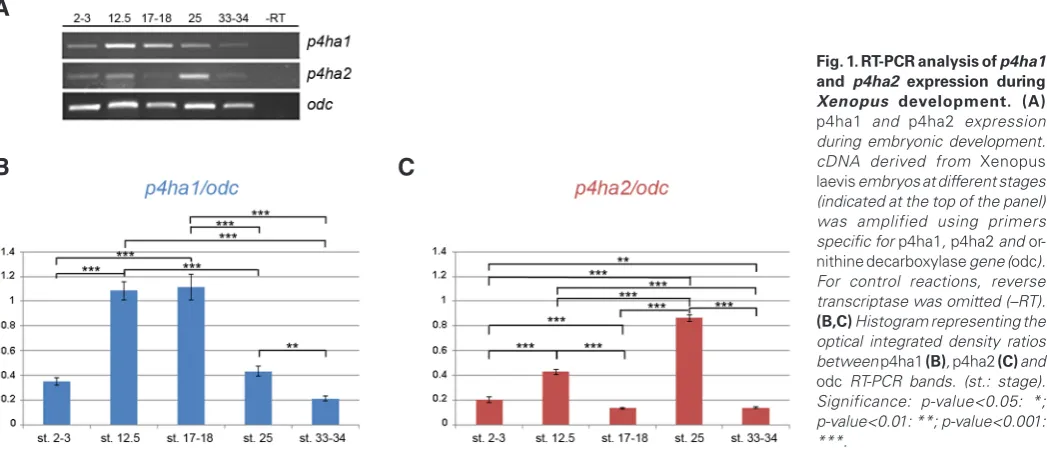
Fig. 1. RT-PCR analysis of p4ha1 and p4ha2 expression during Xenopus development. (A)
p4ha1 and p4ha2 expression during embryonic development. cDNA derived from Xenopus laevis embryos at different stages (indicated at the top of the panel) was amplified using primers specific for p4ha1, p4ha2 and or-nithine decarboxylase gene (odc). For control reactions, reverse transcriptase was omitted (–RT).
we determine and compare the spatio-temporal expression pattern of p4ha1 and p4ha2, encoding for the main subunits containing the peptide-substrate–binding domain and the enzyme active site, during Xenopus development.
Results and Discussion
Temporal expression
To analyze the temporal expression pattern of p4ha1 and
p4ha2 during embryonic development, we performed RT-PCR
assay on cDNAs obtained from Xenopus embryos at different developmental stages.
The two genes display both maternal and zygotic expression, being detected before and after the midblastula transition (stage 8, Fig. 1A). However, they show a quite different temporal expression profile. p4ha1 expression is already detected at 2-3 cell stage and reaches the highest levels between early neurula (stages 12.5) and late neurula (stage 17-18), decreasing at later stages. (Fig. 1B). Differently, p4ha2 displays a more dynamic expression during early development (Fig. 1C). It is expressed at 2-3 cell stage with an increase at stage 12.5, followed by a decrease at stages
17-18 and reaching a maximum peak at stage 25. At stage 33-34, its expression declines again to the level observed at stages 17-18.
Spatial expression
The maternal transcripts of both genes are visible in the animal pole at blastula stage (stage 7) (Fig. 2 A,C) with a very similar pattern, although p4ha2 displays lower expression levels at this stage. In particular, the intracellular localization of both transcripts is restricted to the perinuclear region (Fig. 2 A’,C’). Also the zygotic expression at early gastrula stage (stage 10) is very similar for both genes (Fig. 2 B,D), diffusely expressed in the ectoderm and in the dorsal involuting mesoderm (Fig. 2 B’,D’, brackets).
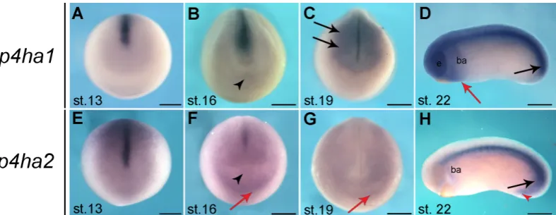
At early neurula stage (stage 13), p4ha1 and p4ha2 are mainly expressed in the developing notochord, in line with the onset of collagen II production at stage 12.5 (Su et al., 1991). Moreover, both genes show a weak expression in anterior regions of the neural plate, which extends dorsally in the case of p4ha2 (Fig. 3 A,E). This pattern is maintained at stage 16 with p4ha2 display-ing a more diffuse expression in the anterior neural plate and in the cardiac progenitors’ territory (Fig. 3F, red arrow)(Cizelsky et
al., 2013), excluding the cement gland primordium (Fig. 3 B,F ar-tion and gastrulaar-tion. Stages
of embryos are indicated at the bottom left corner of each panel (st.: stage), while the analyzed gene is indicated to the left of each row. (A,C) Lateral view of stage 7 embryos; animal pole to the top, vegetal pole to the bottom.
(A’, C’) Sagittal sections of the hybridized embryos shown in (A)
and (C), respectively; the insets in (A’,C’), indicated by the dashed line square in each panel, show the intracellular localization of the two
transcripts in animal blastomeres; white arrowheads indicate Hoechst-stained nuclei, white arrows point to the cytoplasmic perinuclear expression.
(B,D) Lateral view of stage 10 embryos; animal pole to the top, vegetal pole to the bottom. (B’,D’) Sagittal sections of the hybridized embryos shown in (B,D), respectively; black arrows point to the blastopore lip; brackets indicate the dorsal involuting mesoderm. Scale bars, 250 mm.
rowhead). At late neurula stage (stage 19), p4ha1 expression is clearly activated in anterior central nervous system (Fig. 3C) and the migrating neural crest cells (Fig. 3C, black arrows). At the same stages, p4ha2 expression appears to be remarkably low, detectable as a diffuse in situ hybridization signal in the anterior neural plate and in the cardiac progenitors’ territory (Fig. 3G, red arrow). At early tailbud stage (stage 22), both genes are highly expressed in the notochord (Fig. 3 D,H) and in the tail bud, which is composed by the chordoneural hinge and the posterior wall (Fig. 3D, H black arrows), with p4ha2 extending in the proctodeum (Fig. 3H, red arrowhead)(Beck and Slack, 1998). Furthermore, p4ha1 is expressed in the central nervous system, including the eyes, in neural crest cells migrated into the branchial arches, which in part express also p4ha2 (Fig. 3 D,H), and, posteriorly to the cement gland, in the presumptive cardiac progenitors’ territory (Fig. 3D, red arrow). As development proceeds, the predominant notochord expression is maintained for both transcripts, accordingly with collagen gene expression and protein synthesis (Su et al., 1991).
At stage 27, p4ha1 is expressed in the eye, more intensively in its dorsal peripheral region, branchial arches and faintly in somites and proctodeum (Fig. 4 A, A’). At this stage the notochord expres-sion of p4ha1 displays a gradient decreasing from the posterior to the anterior end (Fig. 4A), whereas p4ha2 is highly expressed in the entire notochord (Fig. 4B). Compared to p4ha1 (Fig. 4A’),
p4ha2 shows a lower expression level in the head and in the neural
crest streams (Fig. 4B’).
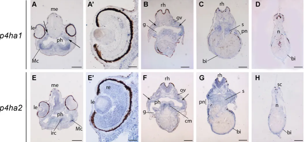
At stage 33/34 the expression profile of the two genes displays many similarities, but also some differences. Both genes are expressed in different regions of nervous system, lens, branchial arches, notochord and in the tip of the growing tail (Fig. 4 C,C’,D,D’). However, hybridization signal of p4ha1 marks also the otic vesicle and, to a lower extent, the dorsal and ventral borders of the somitic mesoderm (Fig. 4 C’,F,G). Differently, p4ha2 is expressed in the heart, the blood islands and the dorsal somitic mesoderm of the caudal region (Fig. 4 D,D’). Histological sections of hybridized embryos show a weak p4ha1 signal in the central nervous system, while p4ha2 displays a more specifically localized expression in the prosencephalon (Fig. 4 E-L). Moreover, p4ha1 expression signal is visible in branchial arches, otic vesicles, notochord and weakly in somites (Fig. 4 E-H), while p4ha2 is mainly detected in lens, branchial arches, and notochord (Fig. 4 I-L).
Finally, we compared the expression profile of p4ha1 and p4ha2 in stage 42 embryos. In the head, p4ha1 is expressed in a region corresponding to the presumptive Meckel’s cartilage, in discrete sites located dorso-laterally with respect to the pharynx (arrows in Fig. 5A) and in otic vesicles (Fig. 5B), while p4ha2 hybridiza-tion signal is present in infrarostral cartilage and cranial muscles, as well as in the mesencephalon, rhomboencephalon and otic
vesicles (Fig. 5E-G). Both genes also display specific expression in regions that bilaterally surround the internal gills (arrows in Fig. 5B, F). Moreover, p4ha2, but not p4ha1, is expressed also in the eye (Fig. 5A’, 5E’), where it is detected in the retina and, consis-tently with immunolocalization of prolyl 4-hydroxylase in rabbit, in the lens (Saika et al., 1998).
In the trunk-tail regions, expression of p4ha2 and p4ha1 is detected in somites, pronephros and blood islands, although at different expression levels. In particular, p4ha2 is also expressed in the spinal cord. Notably, at this stage both genes are not expressed in the notochord, except for the most terminal region (Fig. 5 D,H), coherently with the decrease of collagen gene expression at similar stages in Xenopus laevis embryos (Su et al., 1991).
In conclusion, we found that the mRNA expression of both subunits of the a prolyl hydroxylase is spatially and temporally
regulated during Xenopus embryogenesis, showing many sites of co-expression in embryos at different developmental stages, especially in the notochord and in neural crest-derived regions. However, each of the two genes also displays some unique expres-sion sites suggesting that they are also differentially regulated and may potentially play specific roles during development. In particular,
p4ha2 displays a specific expression in early heart territory, as well
as in blood islands and in the lens, allowing to speculate a puta-tive function in differentiation of these structures. Remarkably, we observed that the expression of both genes coherently correlate with the presence of collagen in the notochord during Xenopus development, indicating the existence of a coordination between tropocollagen synthesis and its post-translational modifications to generate mature fibrillary collagen (Gorres and Raines, 2010)
Materials and Methods
RNA extraction and RT-PCR analysis
Total RNA was extracted from embryos using Mini RNA Isolation Kit (Nucleospin RNA XS, Macherey Nagel). First strand cDNA was synthesized using Superscript II Reverse Transcriptase (Invitrogen), from 1mg of total RNA. RT-PCR analysis was performed using the following gene-specific sets of primers:
p4ha1-forward: 5’-TCATTTCCCCGCAAAATACA-3’ p4ha1-reverse: 5’-TTCCCTTGCACCTTTCAATG-3’ p4ha2-forward-: 5’-ACGAGCTGGAAGCAATCTGA-3’ p4ha2-reverse: 5’-TTGACTCCCTCTCCTCGACA-3’ odc-forward: 5’-GGGCTGGATCGTATCGTAGA-3’ odc-reverse: 5’-CTTCAGGGAGAATGCCATGT-3’
Ornithine decarboxylase (odc) was used as housekeeping gene for nor-malization. The optical density was calculated using the ImageJ software. Data are expressed as mean ± standard deviation; n=9 samples for each embryonic stage in three independent experiments. Statistical analysis was carried out using ANOVA followed by post-hoc Tukey’s Multiple range test. p-values < 0.05 were considered to be statistically significant.
In situ hybridization
Xenopus embryos were generated and staged as previously described
(Marracci et al., 2015). Whole-mount in situ hybridization was performed as previously described (Watanabe et al., 2002). Probes for p4ha1 was transcribed from cDNA clone XL014a11 (GenBank Acc. n. BJ046645) and
p4ha2 from cDNA clone XL094g09 (GenBank Acc. n. BJ071438).
Antisense transcripts were obtained by plasmid linearization with EcoRI and in vitro transcription using T7 RNA polymerase (Roche). Sense tran-scripts were used as negative control (data not shown). in situ hybridiza-tion on cryostat sechybridiza-tions (12 mm thick), as well as sechybridiza-tions (15 mm thick) of paraffin-embedded hybridized embryos were performed as previously
described (D’Autilia et al., 2010, Marracci et al., 2015).
Acknowledgments
The authors are grateful to Naoto Ueno, for generously providing the plasmids (XL014a11 and XL094g09) and thank Marzia Fabbri, Donatella De Matienzo and Elena Landi for technical assistance, and Salvatore Di Maria for frog care.
References
ANNUNEN, P., KOIVUNEN, P. and KIVIRIKKO, K.I. (1999). Cloning of the alpha subunit of prolyl 4-hydroxylase from Drosophila and expression and characteriza-tion of the corresponding enzyme tetramer with some unique properties. J Biol Chem 274: 6790-6796.
BECK, C.W. and SLACK, J.M. (1998). Analysis of the developing Xenopus tail bud reveals separate phases of gene expression during determination and outgrowth. Mech Dev 72: 41-52.
CIZELSKY, W., HEMPEL, A., METZIG, M., TAO, S., HOLLEMANN, T., KUHL, M. and KUHL, S.J. (2013). sox4 and sox11 function during Xenopus laevis eye develop-ment. PLoS One 8: e69372.
D’AUTILIA, S., BROCCOLI, V., BARSACCHI, G. and ANDREAZZOLI, M. (2010). Xenopus Bsx links daily cell cycle rhythms and pineal photoreceptor fate. Proc Natl Acad Sci USA 107: 6352-6357.
FAN, D., TAKAWALE, A., LEE, J. and KASSIRI, Z. (2012). Cardiac fibroblasts, fibrosis and extracellular matrix remodeling in heart disease. Fibrogen. Tissue Rep. 5: 15. GILKES, D.M., BAJPAI, S., CHATURVEDI, P., WIRTZ, D. and SEMENZA, G.L. (2013).
Hypoxia-inducible factor 1 (HIF-1) promotes extracellular matrix remodeling under hypoxic conditions by inducing P4HA1, P4HA2, and PLOD2 expression in fibroblasts. J Biol Chem 288: 10819-10829.
GORRES, K.L. and RAINES, R.T. (2010). Prolyl 4-hydroxylase. Crit Rev Biochem-MolBiol 45: 106-124.
GUO, H., TONG, P., LIU, Y., XIA, L., WANG, T., TIAN, Q., LI, Y., HU, Y., ZHENG, Y., JIN, X. et al., (2015). Mutations of P4HA2 encoding prolyl 4-hydroxylase 2 are associated with nonsyndromic high myopia. Genet Med 17: 300-306.
HELAAKOSKI, T., VUORI, K., MYLLYLA, R., KIVIRIKKO, K.I. and PIHLAJANIEMI, T. (1989). Molecular cloning of the alpha-subunit of human prolyl 4-hydroxylase: the complete cDNA-derived amino acid sequence and evidence for alternative splicing of RNA transcripts. Proc Natl Acad Sci USA 86: 4392-4396.
HOLSTER, T., PAKKANEN, O., SOININEN, R., SORMUNEN, R., NOKELAINEN, M., KIVIRIKKO, K.I. and MYLLYHARJU, J. (2007). Loss of assembly of the main base-ment membrane collagen, type IV, but not fibril-forming collagens and embryonic
death in collagen prolyl 4-hydroxylase I null mice. J Biol Chem 282: 2512-2519. KIVIRIKKO, K.I., HELAAKOSKI, T., TASANEN, K., VUORI, K., MYLLYLA, R., PARK-KONEN, T. and PIHLAJANIEMI, T. (1990). Molecular biology of prolyl 4-hydroxylase. Ann N Y Acad Sci 580: 132-142.
KUKKOLA, L., HIETA, R., KIVIRIKKO, K.I. and MYLLYHARJU, J. (2003). Identification and characterization of a third human, rat, and mouse collagen prolyl 4-hydroxylase isoenzyme. J Biol Chem 278: 47685-47693.
MARRACCI, S., MARTINI, D., GIANNACCINI, M., GIUDETTI, G., DENTE, L. and ANDREAZZOLI, M. (2015). Comparative expression analysis of pfdn6a and tcp1alpha during Xenopus development. Int J Dev Biol 59: 235-240.
MYLLYHARJU, J. (2003). Prolyl 4-hydroxylases, the key enzymes of collagen bio-synthesis. Matrix Biol 22: 15-24.
NISSI, R., AUTIO-HARMAINEN, H., MARTTILA, P., SORMUNEN, R. and KIVIRIKKO, K.I. (2001). Prolyl 4-hydroxylase isoenzymes I and II have different expression patterns in several human tissues. J HistochemCytochem 49: 1143-1153. SAIKA, S., KAWASHIMA, Y., MIYAMOTO, T., TANAKA, S., OKADA, Y., YAMANAKA,
O., KATOH, T., OHNISHI, Y., OHMI, S., OOSHIMA, A. et al., (1998). Immunolo-calization of prolyl 4-hydroxylase in rabbit lens epithelial cells. J Cataract Refract Surg 24: 1261-5.
SHOULDERS, M.D. and RAINES, R.T. (2009). Collagen structure and stability. Annu Rev Biochem 78: 929-958.
SMITH, T.G. and TALBOT, N.P. (2010). Prolyl hydroxylases and therapeutics. Antioxid Redox Signal 12: 431-433.
SU, M.W., SUZUKI, H.R., BIEKER, J.J., SOLURSH, M. and RAMIREZ, F. (1991). Expression of two nonallelic type II procollagen genes during Xenopus laevis embryogenesis is characterized by stage-specific production of alternatively spliced transcripts. J Cell Biol 115: 565-575.
WATANABE, M., REBBERT, M.L., ANDREAZZOLI, M., TAKAHASHI, N., TOYAMA, R., ZIMMERMAN, S., WHITMAN, M., DAWID, I.B. (2002). Regulation of the Lim-1 gene is mediated through conserved FAST-1/FoxH1 sites in the first intron. Dev Dyn 225: 448-456.
WINTER, A.D. and PAGE, A.P. (2000). Prolyl 4-hydroxylase is an essential procollagen-modifying enzyme required for exoskeleton formation and the maintenance of body shape in the nematode Caenorhabditis elegans. Mol Cell Biol 20: 4084-4093. XIONG, G., DENG, L., ZHU, J., RYCHAHOU, P.G. and XU, R. (2014). Prolyl-4-hy-droxylase alpha subunit 2 promotes breast cancer progression and metastasis by regulating collagen deposition. BMC Cancer 14: 1.
https://doi.org/10.1387/ijdb.092948ow
Skin development in bony fish with particular emphasis on collagen deposition in the dermis of the zebrafish (Danio rerio)
Dominique Le Guellec, Ghislaine Morvan-Dubois and Jean-Yves Sire Int. J. Dev. Biol. (2004) 48: 217-231
http://www.intjdevbiol.com/web/paper/15272388
Role of the extracellular matrix and growth factors in skull morphogenesis and in the pathogenesis of craniosynostosis
P Carinci, E Becchetti and M Bodo Int. J. Dev. Biol. (2000) 44: 715-723
http://www.intjdevbiol.com/web/paper/11061436
Endochondral bone formation in toothless (osteopetrotic) rats: failures of chondrocyte patterning and type X collagen expression
S C Marks, C Lundmark, C Christersson, T Wurtz, P R Odgren, M F Seifert, C A Mackay, A Mason-Savas and S N Popoff Int. J. Dev. Biol. (2000) 44: 309-316
http://www.intjdevbiol.com/web/paper/10853827
Establishing laminin, collagen IV and chondroitin sulfate patterns in otocystogenesis
S Callejo, M Barbosa, J A Moro, A Gato, M I Alonso and E Barbosa Int. J. Dev. Biol. (1996) 40: S249-S250
http://www.intjdevbiol.com/web/paper/9087783
Differences in collagen and cell density during normal and dexamethasone-treated secondary palate development in two strains of mice
M A Montenegro, M Rojas, S Dominguez and J Posada Int. J. Dev. Biol. (1996) 40: S245-S246
http://www.intjdevbiol.com/web/paper/9087781
Effects of “in situ” vitamin E on fibroblast differentiation and on collagen fibril develop-ment in the regenerating tendon
R González Santander, M A Plasencia Arriba, G Martínez Cuadrado, M González-Santander Martinez and M Monteagudo de la Rosa