ISSN 2348 – 7968
A Thiocyanate Sensor Based On Ecofriendly Silver
Nanoparticles Thin Film Composite
V. Durga Praveena1 , K. Vijaya Kumar2
1Department of Chemistry, SRI ASNM Government Degree College,
Palakol-534260, A.P, India.
2Department of Physics, KL University,
Guntur-522502 State, A.P, India.
ABSTRACT
In this work, we describe the use of silver nano particles prepared by green synthesis using Achyranthes aspera L., extract
and protected by chitosan in 1% (w/v) CH3COOH for fast, efficient and selective sensor for the removal of thio cyanate ions
present in contaminated waters, at room temperature (28 ± 1◦C). The mechanism of interaction of silver nanoparticles with
SCN - ions was studied using one of the analytical technique such as ultraviolet–visible spectroscopy (UV–VIS). The
performances of optical sensor were investigated which provide the excellent result. The response time of 2–3 s and the detection limit of thiocyanate solution, 1 ppm were found at room temperature. Thus, in future this room temperature optical thiocyanate sensor can be used for clinical and medical diagnosis for detecting low thiocyanate level in water samples, pesticides and organism metabolites such as plasma, sweat, saliva, in general for various biomedical applications in human.
Keywords: Thiocyanate sensor, Achyranthes aspera. L, Silver nanoparticles , Biopolymer.
1. INTRODUCTION
Polluted water is the world's biggest health risk, and continues to threaten both quality of life and public health.
When water from factory farms, industrial plants, and activities like fracking, it picks up toxic chemicals, dirt, trash and disease-carrying organisms along the way. Many of our water resources also lack basic protections, making them vulnerable to pollution. Water pollution is undoubtedly one of the major problems faced by the world today. The World Health Organisation (WHO) has indicated that millions of deaths in worldwide are attributable to unsafe water, sanitation and poor hygiene [1].So the detection of toxic materials in a Reliable, fast, and accurate method is of practical importance in various technological fields including medical processes, environmental analysis, and industrial processes. Recent research has been devoted to explore new materials involving metal nanoparticles (NPs) to detect toxic ions present in the impure water.
ISSN 2348 – 7968 spectrometry of peptides[10], colorimetric sensor for histidine [11] , determination of fibrinogens in human plasma[12], real-time probing of membrane transport in living microbial cells[13], enhanced IR absorption spectroscopy[14], colorimetric sensors for measuring ammonia concentration[15], bio-labeling, optical imaging of cancer[16] , biosensors for detection of herbicides[17] , and glucose sensor for medical diagnostics[18] .
Classically the nanoparticles are produced by physical[19] and chemical methods [20], as these methods are costly, toxic and non eco friendly, scientists are looking forward to synthesize low cost, non toxic, eco friendly nanoparticles. Most recently, biosynthesis of nanoparticles using bacteria[21-22], fungus and plants[23] have emerged as a simple and viable alternative to more complex physical and chemical synthetic procedures to obtain nanomaterials.
SCN−is one of the pollutant which is a metabolite of cyanide detoxication with similar toxicity that has received attention from researchers in different fields such as medicine, food chemistry, and environmental sciences [24]. Thiocyanate usually exists in industrial waste waters, pesticides and organism metabolites. It is also present in low concentrations in human serum, saliva , and urine[25]. Thiocyanate toxicity causes anorexia, fatigue, and mental status changes, including psychosis, weakness, seizures, tinnitus, and hyperreflexia.
Therefore, the determination of the SCN- concentration at low levels is important. It is essential to look for new materials with high sensitivity and efficiency for the removal of SCN- ions. In this paper, the green and biological synthesis of silver nanoparticles in water media would be discussed A green method for nanoparticles preparation should be evaluated from three aspects: the solvent, the reducing agent and the stabilizing agent. In a recent research work of this lab, a very simple method is presented for the synthesis of silver nanoparticles to be used in colorimetric optical sensor to probe SCN- is one of the pollutant which is a metabolite of cyanide detoxication media. Silver nitrate salts are reduced using aqueous extract of Achyranthes aspera.L, which acts as a reducing agent. Commonly used reducing agents such as Trisodium citrate or sodium borohydride are replaced by a more environmental friendly green method using natural polysaccharide. UV–VIS spectrophotometer was used to monitor the kinetics of nanoparticles synthesis while their size and interaction of silver nano particles with chitosan was confirmed by fourier transform-infrared (FT-IR) spectroscopy .Finally, the sensing properties of the nanoparticles solution were tested against increasing thiocyanate concentration between 10 and 100 ppm by monitoring the changes in LSPR position and amplitude with a UV–VIS spectrophotometer.
ISSN 2348 – 7968
2. Experimental
2.1 Materials
Chitosan (degree of deacetylation: 79%, molecular mass: 500,000 g/mol) was purchased from seafoods (Cochin), India. AgNO3 was purchased from Aldrich and used without farther purification. The other chemicals were analytical grade from Fischer Scientific without further treatment. All aqueous solutions were made using ultrahigh purity water purified using a Mill-Q Plus system ( resistivity = 18 M Ω cm) (Millipore Co.).
2.2 Preparation of plant extract
The fresh leaves of achyranthes aspera were washed several times with millipole water to remove dust then it was cut into small pieces. 5g of thoroughly washed leaves were heated in 250 ml of milli pole water for 15min in an Erlenmeyer flask using a water bath then the solution is filtered using whatman no.4 filter paper. The filtered leaf extract was stored in a cooled atmosphere for further use.
(a) (b)
Fig.1 (a) Achyranthes aspera (b) Photograph of aqueous extract of achyranthes aspera solution.
2.3 Synthesis of AgNP s and evaluation of reducing potential of extract
The Silver nitrate solution was reduced using plant extract at room temperature, resulting in a dark brown solution indicating the formation silver nano particles.
2.4 Deposition of Silver Nano particles in Chitosan matrix
ISSN 2348 – 7968 temperature. A range of CS-AgNP concentrations were used to treat the bacteria and for other experiments in this study.
2.5 Characterization
These developed Chitosan based silver nano composites are characterized by using FTIR and UV-VIS Spectrophotometer. FTIR spectra were recorded in a Perkin Elmer Version 10.03.06. UV-VIS spectra were measured on a JASCO (V-670) double beam spectrophotometer with a range of 200-700 nm.
Sensing study of thiocyanate detection
For the sensing study 103 ppm of thiocyanate solution is used. Thiocyanate concentration is varied by diluting thiocyanate solutions of different concentrations (1 to 100 ppm in deionised distilled water) were prepared right before experiment.
3. RESULTS AND DISCUSSION
FTIR spectra of Chitosan and its derivative
ISSN 2348 – 7968 deformation in CONH plane), 1,320 cm-1(CN band stretching ,axial deformation of amino group ) and 1,140-1,026 cm-1(ether linkage , C-O-C band stretching).
In CS-AgNPs ,(Fig. 2,) shows bands are shifted to higher frequencies i.e, 3353.51 cm−1(overlap of O− H and N− H stretching vibrations), 2922.34 cm−1(C − H stretching), 1744.89 cm−1, 1728.74 cm−1(–NH2 bending ,amide linkage), 1569.07 cm−1, 1411.41 cm−1, 1070.14 cm−1, 649.17 cm−1[C-C, C-O(esters and ethers) and C-O (polyols)] more pronounced shift in the FTIR spectrum could be observed in the complexes ( Fig. 3). The major differences are: the peak at 3,526 cm-1 corresponding to the stretching vibration of amino group (-NH2) and hydroxyl group (-OH), shifted to lower frequency (3339 cm−1), and the peak of 3339 cm−1 becomes wider, which indicates hydrogen bonding is enhanced and may be explained as that the additive effect of water absorbed on the surface of Ag nano particles and the –OH group of CS. This suggests that NPs were capped by the polymer.
The polar groups O- H of polysaccharide have the good ability of coordination reaction with metal ions (e.g., with silver ions). When O-H groups and silver ions form coordination bonds, the interac-tions among the resultant Ag particles and oxygen atoms of O-H groups become stronger with increasing amount of Ag. This can lead to corresponding changes both in the positions and in the strengths of IR spectra of CS.
Fig 2. FT-IR Spectra of AgNP/CS thin film sensor
3.2 UV-VIS spectroscopy
ISSN 2348 – 7968 Silver nanoparticles absorb radiation in the visible region of the electromagnetic spectrum (380–450 nm) due to the excitation of Surface plasmon vibrations, and this is responsible for the striking yellow–brown color of silver nanoparticles in various medium [35–37] .Silver nanoparticles stability was checked up to six months and it was found that stability is nearly constant even up to six months. Due to Vander Waals forces or Coulomb’s forces of attraction the individual particles have a tendency to form large sized agglomerates during the preparation of silver nanoparticle suspension. Chitosan is used as a stabilizer inorder to prevent the agglomeration of small particles which can form a protective layer on the particle’s surface. A strong physical adsorption of the CS onto the surface of the silver nanoparticles is also an indication of better stabilization. The evolution of UV– vis absorption spectrum of silver nanoparticles embedded in chitosan film we prepared is shown in Fig. 3b. A plasmon absorbance of the film was observed between 410–450 nm.
(a) (b)
Fig 3. (a) Schematic diagram of the AgNP/CS thin film sensor; path of the light beam in the spectrometer
is
shown. (b) Surface plasmon resonance ( SPR ) spectrum, of the AgNP/CS.3.2.1. Effect of reaction duration
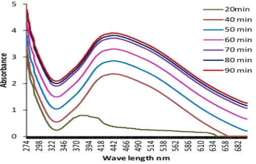
The formation of silver nanoparticles was evidenced with the change in color of the solution after addition of AgNO3 from the colorless to light yellowish color, then to the dark brown color. The formation of silver nanoparticles was monitored through UV–vis spectrophotometer at time intervals of 20, 40, 50, 60, 70, 80 and 90 min (Fig.4). The spectrum obtained at 90 min shows the absorption maximum as 439 nm. The intensity of the peak with respect to the height increases gradually with increase of time. Fig. 4 shows the UV–vis absorption spectra of silver nanoparticles colloidal solutions prepared at different durations. The data reveal several important findings which can be presented as follows: (i) at the early stage reaction duration (after 20 min) the plasmon band is broadened and simple test for silver ion using NaCl solution shows low conversion of silver ions to metallic silver nanoparticles at this duration, (ii) prolonging the reaction duration up to 70 min
1 1.5 2 2.5 3 3.5 4 4.5
Absorb
an
ce
ISSN 2348 – 7968 leads to outstanding enhancement in the Plasmon intensity showing that large amounts of silver ions are reduced and used for cluster formation, and (iii) further increase in the reaction duration up to 90 min is accompanied with marginal decrement in the absorption intensity which could be attributed to some aggregation of the formed silver nanoparticles. The silver nanoparticles formation and the size can be easily being monitored by observing peak due to SPR and blue shift position. Within a few minutes of reaction, the solution became colorless to light yellow color, indicating the synthesis of silvernanoparticles. As the reaction proceeded, the light yellowish color changes to the dark brown color. The effect of different reaction times on silver nanoparticles synthesis results in a blue shift of the absorption peak i.e. there is decrease in wavelength (increase in frequency) and shifts the color from the red end of the spectrum to the blue end, indicating that the particle size of silver nanoparticles decreases, with the reaction time changing from 0 to 90 min, when all the other reaction conditions are identical. Also there is much more amounts of silver nanoparticles were synthesized at the 90 min. It is suggested that small particle size and large quantity of particles are acquired also by controlling reaction time.
Fig. 4, Changes in the SPR band of the AgNPs at various time intervals using UV–vis spectrophotometer.
3.3 Dissolution and swelling test of chitosan based silver nano particles
The swelling studies of chitosan based silver nano particles were carried out in distilled water at room temperature for a period of 24 hrs the percentage of swelling of these films were calculated by using the eq.1:
Percentage of swelling= 100% ---(1)
Where, Ws is the weight of swollen chitosan film(g) and W is the weight of dry chitosan film.
ISSN 2348 – 7968
3.4 Thiocyanate sensing of green AgNPs/CS film
As prepared AgNPs were characterized before and after addition of different concentrations of SCN- using UV–vis spectroscopic technique which was shown in Fig 5. Decrease in the absorbance can be attributed to the oxidation of Ag atoms in the nanoparticles by scn. As in the case of SCN-, a clear blue shift of the peak is observed at higher concentrations of SCN-. We prepared aqueous solutions containing Thiocyanate up to ppm levels. The resulting solution is transparent and stable. SPR spectra of the AgNP-CS film immersed in these solutions showed trends similar to that in the previous cases.
Fig 5. UV–vis absorbtion response of green nano Ag-CS film upon addition of SCN- ions (0–16 ppm).
It is important to consider the binding forces that lead to the interaction of thiocyanate ions with the AgNP-CS sensor film. Swelling of the AgNP-CS film in the aqueous medium facilitates the diffusion of [SCN-] ions into the polymer matrix. The thin film nature of the sensor facilitates convenient monitoring of the sensor through the sensing process .
ISSN 2348 – 7968
Fig 6. Schematic representation of the swelling of the AgNP-CS film in aqueous medium and the sensitive changes in the SPR spectrum of the film induced by SCN- in the solution.
ISSN 2348 – 7968
(a) (b)
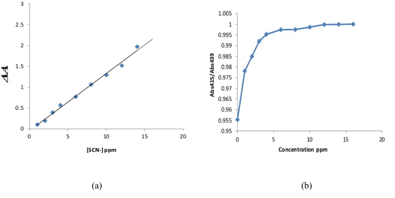
Fig. 7, (a) Plot of change in absorbance intensity versus SCN- concentration. (b) Ratio of the absorbance peak ( Abs415 / Abs439 ) for a AgNP-CS film exposed to increasing thiocyanate content showing a saturation beyond 10 ppm.
The SPR spectrum of the sensor film immersed in pure water remains constant for >1 h (Figure 5), demonstrating that even though the polymer swells in the aqueous medium, there is absolutely no leaching of Ag. This was confirmed further by the observation of an identical spectrum for the same film removed from the water and dried.
We describe first, the sensing experiments with aqueous solutions of SCN- SPR spectra of the sensor film were recorded on introducing SCN- solution. The spectra for a selected set of concentrations are shown in Figure 5. The spectra show small but definite and reproducible decrease in intensity within a few minutes, even at the lowest concentration of 1 ppm. With concentrations close to 1 ppm, the spectrum shows significant change even within 3 min. In addition to the decrease in intensity, the peak undergoes a blue shift which becomes prominent at higher concentrations. the absorbance peak of Ag NPs decrease by increasing the concentration of SCN- ion and there is a linear relationship between the absorbance intensity changes and the concentration of SCN- ion over the range from 1ppm to 15ppm (Fig. 7(a)).
We have plotted absorbance ratio ( Abs415 / Abs439 ) as a function of thiocyanate concentration in Fig. 7(b). The absorbance ratio is almost linear with ammonia concentration in the range of 1 to 15 ppm. Beyond 10 ppm, the ratio remains constant Fig. 7(b), which indicates that the silver nanoparticles based optical sensor is active for low detection of thiocyanate content in the solution.
Sensing Experiments.
ISSN 2348 – 7968 ty = 1 8 M Ω cm ) then placed in a spectrometer ; the SPR spectrum of the film was monitored at first time. The water was removed and replaced with the analyte solution. The SPR spectrum of the film was recorded. A fresh film was used for each new experiment. The reproducibility of the sensing process was examined by running repeated batches of selected experiments.
4. CONCLUSION:
In this study we have shown the feasibility of forming silver nanoparticles – chitosan composite film in which silver nano particles are produced from silver nitrate using aqueous extract of achyranthes aspera.L,. This is one of the simplest, cheapest, most rapid, environmental friendly and green processes for obtaining silver nano particles. uv-vis spectroscopy reveals the surface Plasmon property.
Further the application of nanoparticles was studied on the aqueous thiocyanate sensing properties of CS/AgNPs thin film. The silver nano particles dispersion in polymer matrix played a more important role in colorimetric sensing applications. Colorimetric assays based on the unique LSPR properties of metallic nanoparticles have showed to be very useful due to their simplicity, high sensitivity, low detection limit of 1 ppm, low cost, fast response time and great reproducibility. Keeping these significance properties in mind, in the near future we can use our room temperature optical thiocyanate sensor for clinical and medical diagnosis for detecting low thiocyanate level in human.
The present study demonstrates the unique potential of metal nanoparticle- embedded polymer thin films in chemical sensing applications. The scope for further development of this concept is extensive, in view of the flexibility and versatility of these nanocomposite materials.
5. REFERENCES:
[1].WHO and UNICEF. Progress on Sanitation and Drinking-Water ; World Health Organization and United Nations Children’s Fund: Geneva, Switzerland, 2010.
[2].D.J. Anderson, M. Moskovits (2006) J. Phys. Chem. B, 110, 13722-13727.
[3].C.N.R. Rao, G.U. Kulkarni, P.J. Thomas, P.P. Edwards (2000) Chem. Soc. Rev., 29, 27-35.
[4].Vaseashta,D.Dimova-Malinovska(2005)Sci. Technol. Adv. Mater., 6, 312-318.
[5].J. Zhong-Jie, L. Chun-Yan, S. Lu-Wei (2005) J Phys Chem B, 109, 1730-1735.
[6].M.Rai, A.Yadav, A.Gade (2009) Biotechnology Advances, 27, 76-83.
ISSN 2348 – 7968 [8].M.I.Sriram, S.B. Mani Kanth, K. Kalishwaralal, S.Gurunathan (2010) International Journal of Nanomedicine, 5, 753-762.
[9]. M. Otamiri, K.G.I.Nilsson (1999) International Journal of Biological Macromolecules 26,263–268.
[10].L. Hua, J. Chen, L. Ge, S.N. Ta (2007) Journal of Nanoparticle Research 9,1133–1138.
[11].D. Xiong, M. Chen, H. Li (2008) Chemical Communications 7 ,880–882.
[12].J. Zhejiang, C.Y. Yuan, L. Anhui, T. Hailing, T. Mingle, Z. Fuxin (2007) Science in China, Series B 50,345–350.
[13].X. Xu, W.J. Brownlow, S.V. Kyriacou, Q. Wan, J.J. Viola (2004) Biochemistry 43 ,10400–10413.
[14].S. Huo, X. Xue, Q. Li, S. Xu, W. Cai (2006) Journal of Physical Chemistry B 110 ,25721–25728.
[15].S.T. Dubas, V. Pimpan, Talanta (2008) 76, 29–33.
[16]. B.J. Wiley, Y. Chen, J.M. Mc Lellan, Y. Xiong, Z. Li, D. Ginger (2007) Nano Letters 7 1032–1036.
[17]. S.T. Dubas, V. Pimpan, (2008) Materials Letters 62 2661–2663.
[18].Y.K. Mishra, S. Mohapatra, D. Kabiraj, B. Mohanta, N.P. Lalla, J.C. Pivin, (2007) Scripta Materialia 56 , 629–632.
[19].JP Zhang, LQ Sheng, P Chen (2003) Chinese Chemical Letters, , 14, 645-648.
[20].M.G. Guzmán, J. Dille, S. Godet (2009) International Journal of Chemical and Biological Engineering, 2, 104-111.
[21].S. He, Z. Guo, Y. Zhang, S. Zhang, J. Wang, N. Gu, (2007) Materials Letters, 61, 3984-3987.
[22].M.I. Husseiny, M.A. El-Aziz, Y. Badr, M.A. Mahmoud, (2007) Spectrochimica Acta Part A, 67,1003-1006.
[23].R.W. Raut, J.R.. Lakkakula, N.S. Kolekar, V.D. Mendhulkar, S.B. Kashid (2009) Current Nanoscience, , 5, 117-122.
[24].A. F. Lavorante, M. A. Feres, and B. F. Reis (2006) “Multi-commutation in flow analysis: a versatile tool for the development of the automatic analytical procedure focused on the reduction of reagent consumption” SpectroscopyLetters ,vol.39, no.6, 631–650.
[25].G.F.Wang, M.G.Li, Y.C.Gao, and B.Fang, (2004) “Amperometric sensor used for determination of thiocyanate with a silver nanoparticles modified electrode,”Sensors , vol. 4, no. 9, 147–155.