pISSN 2320-1770 | eISSN 2320-1789
Original Research Article
A study of thyroid dysfunction in dysfunctional uterine bleeding
Santosh Kumar Verma
1*, Anita Pal
1, Saroj Jaswal
23
INTRODUCTION
DUB is a symptom complex that includes any condition of abnormal uterine bleeding in the absence of pregnancy, neoplasm, infection or other intrauterine lesion. Such bleeding is most often the result of endocrinologic dysfunction that inhibits normal ovulation.1
Dysfunctional uterine bleeding is best defined as abnormal bleeding from the uterus in the absence of organic disease of the genital tract. The term DUB applies to any abnormal uterine bleeding, including disturbances of the menstrual cycle, regular or irregular
uterine bleeding and alterations in amount and the duration of menstrual loss, but most commonly implies to excessive regular menstrual bleeding or essential menorrhagia. DUB is not one condition with one etiology, but is a group of disorders characterized by the dysfunction of the uterus, ovary, pituitary, hypothalamus or other part of reproductive system which results in abnormal or excessive uterine bleeding. In clinical practice the precise nature of the dysfunction is often not determined and the diagnosis of DUB is usually made by exclusion of organic disease of the genital tract.2
The International Federation of Gynaecology and Obstetrics in November 2010, accepted a new
ABSTRACT
Background: The objective of the study was to evaluate the prevalence of thyroid dysfunction in dysfunctional uterine bleeding and to assess the menstrual and endometrial pattern in women with thyroid disorders.
Methods: The present study was conducted on 200 patients who presented with dysfunctional uterine bleeding in gynecology OPD.
Results: Among the 200 women 39 (19.5%) had hypothyroidism, 2 (1%) had hyperthyroidism and 159 (79.55%) were euthyroid. Menorrhagia was the most common menstrual disorder in hypothyroidism and oligomenorrhoea in hyperthyroidism. In the present study 74.3% patients had proliferative endometrium,26.3% secretory endometrium, in hypothyroid patients and secretory endometrium in 2 (1%) hyperthyroid patients. A woman with hypothyroidism, commonly presents with anovulation and unopposed oestrogen activity causes endometrial hyperplasia which may outgrow the blood supply and may cause local areas of necrosis and breakdown and produces bleeding.
Conclusions: The menstrual irregularities are significantly more frequent in patients with thyroid dysfunction and menorrhagia was the commonest menstrual abnormality. The study concludes that biochemical evaluation of thyroid function should be made mandatory in all cases of abnormal uterine bleeding and this would avoid unnecessary surgeries and exposure to hormones.
Keywords: Abnormal uterine bleeding, Dysfunctional uterine bleeding, Menorrhagia, Oligomenorrhoea, Thyroid dysfunction
1Department of Obstetrics and Gynecology, 2Department of Biochemistry, KNH, IGMC, Shimla, Himachal Pradesh,
India
Received: 13 March 2017 Accepted: 07 April 2017
*Correspondence: Dr.Santosh Kumar Verma,
E-mail: [email protected]
Copyright: © the author(s), publisher and licensee Medip Academy. This is an open-access article distributed under the terms of the Creative Commons Attribution Non-Commercial License, which permits unrestricted non-commercial use, distribution, and reproduction in any medium, provided the original work is properly cited.
classification system for causes of AUB in the reproductive years. The system based on the acronym PALM COEIN (polyps, adenomyosis, leiomyoma, malignancy and hyperplasia-coagulopathy, ovulatory disorders, endometrial causes, iatrogenic, not classified) was developed in response to concerns about the design and interpretation of basic science and clinical investigation that relates to the problem of AUB.3
Dysfunctional uterine bleeding is one of the most common and significant gynaecological complaints and is seen in about 10-15% of women attending the gynaecological clinic.
According to aetiology DUB is classified as
• Primary: Pathology in endometrium or hypothalamo- pituitary-ovarian-endometrial-axis (ovulatory and anovulatory).
• Secondary: cause detected outside the hypothalmo- pituitary-ovarian-endometrial axis (endocrinopathies, hematological, vascular disease and liver disorders). Iatrogenic: caused by drugs, irregular hormone intake and/or IUCD.4
Thyroid dysfunction is a cause of nonstructural AUB. Thyroid is closely linked with the process of ovarian maturation.5 Thyroid disorders are more common in
women than in men and in older adults compared with younger age groups. Hypothyroidism is associated with a wide spectrum of reproductive disorders ranging from abnormal sexual development, menstrual irregularities and infertility. The impact of hypothyroidism on the menstrual cycle has been identified since the 1950s and leads to changes in cycle length and blood flow. Thyroid autoimmunity has been shown to have association with various kinds of thyroid dysfunction.6
Thyroid disorder is one of the endocrinopathies associated with disturbance in menstrual functions. The reported incidence of subjective menorrhagia in myxedema varies from 32-80% and menorrhagia may not infrequently be the presenting complaint. Hyperthyroidism, in contrast is associated with oligomenorrhoea and amenorrhea which are in proportion to the severity of thyrotoxicosis.7
Both conditions respond to proper therapy when the thyroid gland is the cause of the abnormality. TSH is a very sensitive indicator of thyroid action at the tissue level because it is dependent on the pituitary exposure to T4.8
METHODS
Study was conducted at Kamla Nehru state Hospital For Mother and Child, IGMC, Shimla, Two hundred patients with dysfunctional uterine bleeding during reproductive age group attending the out patient department of
Obstetrics and Gynecology were selected for the study after permission from ethical committee.
After informed consent detailed history of all patients was taken including the menstrual history.
The menstrual cycles were considered normal if the patients fulfill the following criteria
• The duration of menstrual bleeding was between 2-7 days.
• The menstrual flow was not heavy and contained by two perineal pads.
• There is no expulsion of sizeable clots during menstruation.
• The frequency of the menstrual cycle is constant ranging from 21 to 35 days.
Detailed obstetric history was taken.
Past history was taken in detail including contraception history, history of drug or hormone intake, IUCD, any disease and any bleeding disorder.
Detailed surgical and medical history with special reference to symptoms of 'thyroid dysfunctions e.g. excessive weight gain/loss, cold/heat intolerance, easy fatiguability /irritability, palpitations, hoarseness of voice, diarrhea etc, were enquired.
General physical, systemic and local examination was done.
The following investigations were done in all the patients:
Complete haemogram, urine examination and TFT and ultrasonography were done. Every patient underwent D and C in premenstrual phase or first day of cycle. The endometrial curettings were sent in 10% formalin to the Department of Pathology IGMC, Shimla for histopathological examination.
Thyroid stimulating hormone was measured by Dia Metra Kit in the department of Biochemistry 1GMC Shimla. 5 ml of venous blood was collected under all aseptic precautions. Serum was separated and stored in refrigerator at 2-8°C. The data was collected and statistically analysed.
adding stopping solution. The absorbance will be measured on a plate reader at 450 nm. This sensitive assay can detect TSH concentrations as low as 0.01µIU/L. The normal range is usually 0.45-4.5µIU/L.
RESULTS
[image:3.595.312.546.163.249.2]The mean age of the patients was 38±7 years. Majority of the patients belonged to the age group of 41 to 50 years (42.5%). The least common age group was of those between 20 to 30 years (19%).
Table 1: Distribution of patients according to age (N=200).
Age in years No. of cases Percent
20-30 38 19.00
31-40 77 38.50
41-50 85 42.50
The mean parity was 2.85±0.53. Majority of the cases were between para 2 to para 5.
[image:3.595.47.547.360.446.2]The most common complaint was menorrhagia (47.5%) followed by metrprrhagia (25%). Polymenorrhagia, Oligomenorrhoea and hypomenorrhoea were present in 8%,6% and 3.5% of patients respectively.
Table 2: Distribution of patients according to menstrual pattern (N=200).
Presenting complaint No. of patients Percent
Menorrhagia. 95 47.50
Metrorrhagia 50 25.00
Menometrorrhagia 20 10.00
Polymenorrhagia 16 8.00
Oligomenorrhoea 12 6.00
Hypomenorrhoea 07 3.50
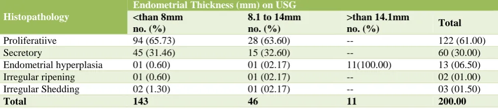
There were 11% patients with ultrasonographic diagnosis of endometrial hyperplasia with endometrial thickness of more than 14.1mm, were correlated with histopathology and all of the 11 patients had confirmed endometrial hyperplasia on histopathological examination.
Table 3: Distribution of patients according to thyroid status in relation to age groups.
Thyroid status
Age in years
Total Age group I 20-30
years no. (%)
Age group II 31-40 years no. (%)
Age group III 41-50 years no. (%)
Euthyroid 33 (20.70 65 (40.00) 61 (38.30) 159 (79.50)
Hypothyroid 08 (12.50) 15 (38.40) 16 (41.00) 39 (19.50)
Hyperthyroid 02 (100.00) 00 00 02 (01.00)
Total 43 (21.50) 80 (40.00) 77(38.70) 200
Table 4: Distribution of patients with thyroid dysfunction and relation of endometrial pathology (N=200).
Thyroid status
Endometrial Pathology
Total No. (%) Proliferative
no. (%)
Secretory no. (%)
Endometrial hyperplasia no. (%)
Irregular ripening no. (%)
Irregular shedding no. (%)
Hypothyroid 29 (74.30) 07 (17.90) 03 (07.62) 00 00 39 (19.50)
Hyperthyroid 00 02 00 00 00 2 (01.00)
Euthyroid 93 (58.40) 51 (32.00) 10 (06.20) 02 (01.20) 03 (01.80) 159 (79.50)
Total 122 60 13 02 03 200
Table 5: The patients with dysfunctional uterine bleeding according to ultrasonography in relation to histopathology (N=200).
Histopathology
Endometrial Thickness (mm) on USG <than 8mm
no. (%)
8.1 to 14mm no. (%)
>than 14.1mm
no. (%) Total
Proliferatiive 94 (65.73) 28 (63.60) -- 122 (61.00)
Secretory 45 (31.46) 15 (32.60) -- 60 (30.00)
Endometrial hyperplasia 01 (0.60) 01 (02.17) 11(100.00) 13 (06.50)
Irregular ripening 01 (0.60) 01 (02.17) -- 02 (01.00)
Irregular Shedding 02 (1.30) 01 (02.17) -- 03 (01.50)
Total 143 46 11 200.00
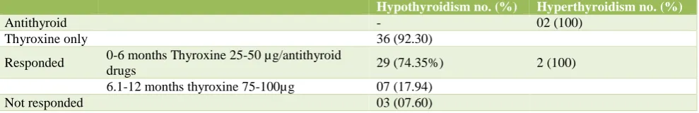
[image:3.595.54.544.477.575.2] [image:3.595.53.544.619.727.2]Table 6: Treatment and follow up of patients of dysfunctional utrine bleeding with thyrid dysfunction (N=41).
Hypothyroidism no. (%) Hyperthyroidism no. (%)
Antithyroid - 02 (100)
Thyroxine only 36 (92.30)
Responded 0-6 months Thyroxine 25-50 µg/antithyroid
drugs 29 (74.35%) 2 (100)
6.1-12 months thyroxine 75-100µg 07 (17.94)
Not responded 03 (07.60)
In the present study, the sensitivity of ultrasonography was 100% (71-100) and specificity was 97.35% (93-99). Positive predictive value was 68.75% (41-88) and negative predictive value was 100% (98-100).
79.5% of patients were euthyroid. Hypothyroidism was more common in age group of 31-40 years (38.4%) and 41-50 years (41%).There were only 2 (1%) hyperthyroid cases and they were in the age group of 21-30 years.
In hypothyroid patients 29 (74.3%) had proliferative endometrium and in 2 (1%) cases of hyperthyroidism had secretory endometrium. Out of euthyroid patients 93 (58.4%) had proliferative and 51 (32%) had secretory endometrium. 39 (19.5%) patients were found to have subclinical hypothyroidism and 2 (1%) had hyperthyroid after thyroid function test.
All of the 39 (19.5%) hypothyroid patients were put on thyroxine 25-50µg/day for six months. In 29 (74.3%) patients the TSH level fell down to normal level within six months after the initiation of treatment. 7 (17.94%) patients required a dose of 50-100µg/day for a period of upto one year for the restoration of their menstrual function. The remaining 3 (7.6%) patients were given antifibrinolytics and hormones in addition to thyroxine but they did not respond to the medical treatment and their hysterectomy was done. Both the hyperthyroid patients resumed their menstrual pattern within three months after taking antithyroid drugs. After the correction of menstrual dysfunction these patients were attached with medicine department. One patient was also an habitual aborter in addition to her menstrual dysfunction, who conceived four months after receiving the thyroxine and delivered a full term baby.
DISCUSSION
Dysfunctional uterine bleeding (DUB) is one of the most commonly encountered conditions in the OPD. Diagnosis of DUB is by exclusion. It is more common in fourth to fifth decades of life or in perimenopausal age group.3 In
present study, most of the AUB patients were in the age group of 41 to 50 years (42.50%) followed by 31 to 40 years (38.50%). N Bhavani et al observed that most of the AUB patients were in the age group of 41 to 50 years (40%) followed by 31 to 40 years (37%).9 84% of patients
were multipara. Pilli et al also reported that DUB was
seen in 87% multipara, 7% primipara and 6% in nulliparous women.10 Menorrhagia followed by
metrorrhagia was the most common menstrual complaint. According to Talukdar et al, the dominant menstrual problem in patients with abnormal uterine bleeding was menorrhagia in 44.44%.3 In the present study, 57.5%
patients had laprosterilization. The theory of increased bleeding is that delivery of hormones from the ovaries to the uterus is impaired. Ovarian functions are deficient since mid luteal progesterone levels decline significantly following this surgery.
In present study 79.55%patients were euthyroid. 19.5% patients were hypothyroid in the age group of 31-50yrs and 1% of the patients were hyperthyroid in the age group of 20-30 years. According to Sowers et-al 90.4% were euthyroid, 6.2% hypothyroid and 3.2% hyperthyroid in perimenopausal age group.11 In mid aged women, there
was a 9.6% prevalence of TSH values outside the euthyroid range and TSH levels were associated with bleeding length. Sangeeta Pahwa et al observed in their study that 22% of cases were hypothyroid, 2% hyperthyroid and 76% euthyroid.12 Hyperthyroidism
reduces menstruation and hypothyroidism causes menorrhagia. Scott and Mussay observed abnormal menstrual pattern in 56% of myxoedematous patients similar to the present study where menorrhagia and metrorrhagia combined constituted 58.95% of the abnormal pattern in patients with hypothyroidism.13 Kaur
et al observed in their study that 85% of the patients with abnormal uterine bleeding were euthyroid, 14% hypothyroid and 1% hyperthyroid.14 Main menstrual
complaint in hypothyroid patients was menorrhagia in 64.3% and hypomenorrhoea in 100% hyperthyroid patients.
Suspicion of hypothyroidism may be delayed in elderly patients because symptoms such as fatigue and constipation and other elderly manifestations of thyroid failure may be attributed to aging itself. Accoring to Goldsmith 70% of patients with hypothyroidism had proliferative and 20% had secretory endometrium.15,16 In
hyperplasia and rest 14.3% had secretory endometrium.14
Ajmani A K et al observed that hypothyroid patients mainly had proliferative endometrium (42.85%) whereas hyperthyroid had atrophic endometrium (60%).6
In present study, also 74.3% had proliferative endometrium, 26.3% secretory endometrium in hypothyroid patients. All the hypothyroid patients were put on Thyroxine 74.35% responded to dose of 25-50µg/day within 6 months and menstrual cycles became normal. 17.94% needed treatment for 6-12 months with the dose of 75-100µg/day. Ross et al reported that, in patients with myxoedema, after treatment with dessicated thyroid menstrual cycles became regular.7
According to Wilansky et al in patients of menorrhagia and hypothyroidism menorrhagia disappeared within 3-6 months and did not reappear in one to three yrs of the follow up after l- thyroxine treatment.17 According to the
study done by Talasila et al out of 100 patients with DUB 10 (10%) of the patients were hypothyroid and 1 (1%) patient was hyperthyroid.18 All the 11 (100%) of the
patients with thyroid disorders responded promptly to the medical treatment.
CONCLUSION
The menstrual irregularities most often seen in patients with dysfunctional uterine bleeding are menorrhagia metorrhagia and menometrorrhagia in patients with hypothyroidism. In hyperthyroidism irregularity seen is hypomenorrhoea. Thyroid dysfunction should be considered in the evaluation of dysfunctional uterine bleeding especially in women of age greater than or equal to 30 yrs. Thyroid disorder is the most common and treatable cause of DUB. As there is high incidence of thyroid dysfunction in our area, evaluation of thyroid in abnormal uterine bleeding would also avoid unnecessary surgeries and exposure to hormones.
Funding: No funding sources Conflict of interest: None declared
Ethical approval: The study was approved by the Institutional Ethics Committee
REFERENCES
1. Butler WJ. Normal and abnormal uterine bleeding.in Rock JA and Jones HW. Telinde’s operative gynaecology. 9th edition. PhiladelpShia:Lippinocot
Williams and Wilkins;2003:457-78.
2. Davey DA. Dysfunctional uterine bleeding. In Charles R Whitfield Textbook of obstetrics and gynaecology for postgraduates .5th edition. printed by Bombay Oxford University Press, Delhi;1995:590-607.
3. Talukdar B, Mahela S. Abnormal uterine bleeding in perimenopausal women: correlation with sonographic findings and histopathological
examination of hysterectomy specimen. J Mid-Life Health. 2016;7:73-7.
4. Lopez J. Pathophysiology of dysfunctional uterine bleeding. In: Purandare CN, Khadilkar S. Dysfunctional uterine bleeding - An update. First edition. New Delhi. Jaypee Brothers;2004:16-23. 5. Sharma N, Sharma A. Thyroid profile in menstrual
disorders. JK Sci. 2012;14(1):14-7.
6. Ajmani NS, Sarbhai V, Yadav N, Paul M, Ahmad A, Ajmani AK. Role of thyroid dysfunction in patients with menstrual disorders in tertiary care center of walled city of Delhi. J Obstet Gynaecol India. 2016;66(2):115-9.
7. Ross GT, Scholz DA, Lambert EH, Geraci. Severe uterine bleeding and degenerative skeletal muscle changes in unrecognized myxoedema. J Clin Endocrinol. 1958;18:492-500.
8. Speroff L, Fritz MA. Reproduction and thyroid . In: clinical gynaecologic endocrinology and Infertility. 7th Ed. Philadelphia:Lippincott Williams & Wilkins; 2005:805-821
9. Bhavani N, Sathineedi A, Chippa S, ReddyVSP. A study of correction between abnormal uterine bleeding and thyroid dysfunction. International J Recent Trends Sci Tech. 2015;14(1):131-5.
10. Pilli GS, Sethi B, Dhaded AV, Mathur PR, Dysfunctional uterine bleeding. J Obstet Gynaecol India. 2001;52(3):87-9.
11. Sowers M, Luborsky J, Perdue C et al.thyroid stimulating Hormone(TSH)concentrations and menopausal status in women at the mid-life. J Clin Endocrinol. 2003;58:340-7.
12. Pahwa S, Shailja G,Jasmine K. Thyroid dysfunction in dysfunctional uterine bleeding. J Adv Research Biologic Sci. 2013;5(1):78-83.
13. Scott JC, Mussey E. Menstrual patterns in myxoedema.Am J Obstet Gynaecol. 1964;90:161-5. 14. Kaur T, Aseeja V, Sharma S. Thyroid Dysfunction in
dysfunctional uterine bleeding. Webmed Central Obstet Gynaecol. 2011;2(9):002235.
15. Goldsmith RE, Sturgis SH, Lerman J, Stanbury JB Menstrual pattern in thyroid disease. J Clin Endocrinol. 1952;12:846-55.
16. Padmaleela K, Thomas V, Lavanya KM, Kiranmai D. Thyroid disorders in dysfunctional uterine bleeding (DUB) among reproductive age group women-a cross-sectional study in a territory care hospital in Andhra Pradesh India. Int J Med Pharma Sci. 2013;(1):41-6.
17. Wilansky DL, Greisman B. Early hypothyroidism in patients with menorrhagia. Am J obstet. Gynaecol 1989;160:673-77.
18. Sruthi T, Shivanna S, Gopal N. Prevalence of hypothyroidism in patients with provisional Diagnosis of DUB. J Eval Medic Dent Sci. 2014;3(11):2967-72.