RAVALIYA, KRUTI DILIP. Evaluation of a Novel Microbial Source Tracking Method to
Identify Fecal Contamination in the Fresh Produce Production Environment. (Under
the direction of Lee-Ann Jaykus, Jay Levine and Clyde Sorenson.)
Current microbiological indicators of fecal contamination do not correlate well with the
presence or absence of pathogens. Alternatives, such as library-dependent and
independent methods are becoming more widely available. Library independent
methods include tracking of molecular markers from organisms such as
Bacteroidales
.
Characterization studies have been done within different hosts, allowing researchers to
develop assays for targeting
Bacteroidales
markers in specific hosts. These studies
evaluated fecal contamination in water systems; however, none have been applied to
fresh produce systems. In developing assays for use in environmental samples,
precautions must be taken, including identifying the appropriate primers and probe,
sample preparation, assessing the efficiency of the PCR due to potential
matrix-associated inhibitors, proper use of controls, and interpretation of results. The purpose
of this study was to adapt existing methods for typing
Bacteroidales
species based on
16SrDNA for use in fresh produce and environmental samples, and to evaluate if they
might be relevant microbiological indicators of fecal contamination. Initially, the assay
was optimized for detection of total Bacteroidales with respect to pre-analytical sample
processing methods, DNA extraction and PCR primer choice. A combination of filtration
and centrifugation were chosen for sample processing, followed by DNA extraction
identify those samples demonstrating inhibition due to a variety of PCR inhibitors. The
sum total of these methods were applied in a pilot study in which samples (originating
from Northern Mexico) of fresh produce, harvester hand rinsates, and source and
irrigation water were processed for detection of evidence of fecal contamination using
the
Bacteroidales
assay and generic
E. coli
. A total of 174 samples were processed using
these methods, and the general fecal contamination marker was identified in about
36%. The marker was no identified in 55% percent of the samples. By environmental
sample type, the marker was detected in 30% of hand rinse samples, 45% of produce
rinses, and 28% of irrigation water samples. In evaluation of marker detection based
on produce type, 59% of cantaloupe samples, 31% tomato samples, and 17% pepper
samples contained the AllBac general fecal marker. Inhibitors to PCR may have been
present, and were identified through the use of the AllBac IAC; these indicated which
samples should be diluted up to 1,000-fold, and anything further was deemed
“uninterpretable.” The uninterpretable samples represented 8% of the sample pool.
Average log genome equivalence copies (GEC) for all samples was 5.5. Average log GEC
for hand rinses, irrigation waters, and produce rinses was 5.3, 6.0 and 6.1, respectively.
Based on produce type, average log GEC for peppers, melons, and tomatoes was 4.6, 6.4,
and 5.4 respectively. Average cycle threshold (Ct) value for all positive samples was
30.4. The average Ct values for hand rinse, irrigation water, and produce rinse was
between the presence of
and
in hand rinse samples,and produce
rinse samples, but strong correlation was identified in irrigation water samples (R
2=
0.99). These results suggest that of the produce types, melon is most likely to contain
fecal matter, and that produce will be more likely to contain fecal matter over other
types of environmental samples (water and hand rinses). Environmental samples can
introduce inhibitors and should be addressed, so as not to cause false reporting of
results. This data will also promote implement better contamination prevention
by
Kruti Dilip Ravaliya
A thesis Submitted to the Graduate Faculty of
North Carolina State University
in partial fulfillment of the
requirements for the Degree of
Master of Science
Food Science
Raleigh, North Carolina
2013
APPROVED BY:
_______________________________
______________________________
Dr. Lee-Ann Jaykus
Dr. Jay Levine
Committee Chair
DEDICATION
This work is dedicated to my mom, Ranjan, and my sister, Preeti. Returning to school
BIOGRAPHY
Kruti Ravaliya has attended North Carolina State University since August, 2010, and
finished her MS in January, 2013. She earned a BS from the University of Massachusetts
in 2007, in both Food Science and Spanish. Upon completion of her degree, she
accepted a food product development scientist position with the International Food
Network. While at the International Food Network, she worked on a variety of
platforms, most notably an ultra-high temperature pasteurized dairy beverage. After
developing the formulation from bench to production, this product was launched in
specific markets. She decided then that she would return to school, to pursue an
advanced degree in Food Science, with a focus in food safety. The title of her thesis,
“Evaluation of Novel Microbial Source Tracking Method for the Identification of Fecal
Contamination in the Fresh Produce Production Environment” was part of a larger
collaboration on a USDA-funded grant, between the University of Nuevo Leon, Emory
University and North Carolina State University. Upon completion of her degree at North
Carolina State University, Kruti has accepted a fellowship position through Oak Ridge
Institute for Science and Education (ORISE) at the Food and Drug Administration (FDA),
in the Office of Food Safety/Produce Safety. Professional affiliations include the
International Associate of Food Protection, the Institute of Food Technologists, the
American Society for Microbiology, and the Society for Applied Microbiology. In
ACKNOWLEDGMENTS
I would like to thank Dr. Jaykus, and all of her students and staff at NCSU, as well as the
team at UANL, and Emory University. I would also like to thank my committee
members, Dr. Clyde Sorenson and Dr. Jay Levine.
Thank you to all of my friends and family who have been there for me through this
whole experience. I could not have done this without all of you. There are a few people
who I’m going to name, specifically, so a special “thank you!” to Joey, Maggie, Stephen,
Johari, Bek, Jen, and Grace. If I have left others out, I apologize! Many people were
TABLE OF CONTENTS
LIST OF TABLES ... vii
LIST OF FIGURES ... viii
INTRODUCTION ... 1
EPIDEMIOLOGY OF FRESH PRODUCE ASSOCIATED FOODBORNE DISEASE .... 5
METHODS TO IDENTIFY FECAL CONTAMINATION
... 9
FECAL COLIFORMS AS MICROBIOLOGICAL INDICATORS
... 14
GENERIC
E. COLI
AS A MICROBIOLOGICAL INDICATOR
... 16
CLOSTRIDIUM PERFRINGENS
... 19
MALE-SPECIFIC COLIPHAGE (F+-SPECIFIC RNA COLIPHAGE
... 20
MICROBIAL SOURCE TRACKING
... 23
LIBRARY-DEPENDENT MST METHODS
... 25
LIBRARY-INDEPENDENT MST METHODS
... 29
APPLICATIONS OF BACTEROIDALES MST METHODS TO ENVIRONMENTAL STUDIES
... 33
PRIMER CHOICE
... 35
SAMPLE PREPARATION
... 58
THE IMPORTANCE OF MATRIX-ASSOCIATED INHIBITION
... 60
INCLUSION OF APPROPRIATE CONTROLS
... 62
INTERPRETATION OF RESULTS
... 66
THE VIABILITY DILEMMA
... 69
USEFULNESS OF
BACTEROIDALES
ASSAYS IN MST
... 70
CONCLUSIONS
... 71
RESEARCH PAPER ... 73
INTRODUCTION
... 73
MATERIALS AND METHODS
... 77
Study area
. ... 77
Sample Collection
. ... 78
Irrigation water
. ... 83
Produce rinsates
. ... 83
Escherichia coli screening
... 84
Sample processing for Bacteroidales detection
. ... 85
Centrifugation
. ... 85
Filtration
... 86
Combined centrifugation/filtration
. ... 86
DNA Extraction
... 91
Quantitative real-time PCR (qPCR).
... 91
Construction of an Internal Amplification Control (IAC).
... 92
AllBac qPCR Assay
. ... 93
BoBAC qPCR ASSAY ... 94
SAMPLE PROCESSING
.
... 99
LIST OF TABLES
LIST OF FIGURES
Figure 1: Geographical Representation of Study Area between the Border of Mexico and
the United States
... 78
Figure 2: Outline of Samples in a Given Chain ... 79
Figure 3: Chain Outline for Produce Samples (Combination of Produce Rinse, Hand Rinse,
and Irrigation Waters) ... 80
Figure 4: Chain Outline for Hand Rinse Samples (Combination of Produce Rinse, Hand
Rinse, and Irrigation Waters) ... 81
Figure 5: Chain Outline for Irrigation Water Samples (Combination of Produce Rinse, Hand
Rinse, and Irrigation Waters) ... 82
Figure 6: Schematic outlining Centrifugation Protocol for Sample Concentration ... 88
Figure 7: Schematic Outlining Filtration Method for Sample Concentration ... 89
Figure 8: Schematic Outlining Combination Method of Centrifugation and Filtration for
Sample Concentration ... 90
Figure 9: Construction of IAC – Diagnostic overhanging primers complementary to plasmid
pUC19, and amplified by PCR. Amplicons obtained are able to be reamplified using the
diagnostic primers ... 93
Figure 10: Jalapeno Results (Percent Positive, negative and uninterpretable) ... 103
Figure 11: Cantaloupe Results (Percent Positive, negative and uninterpretable) ... 103
Figure 12: Tomato Results (Percent Positive, negative and uninterpretable) ... 104
Figure 13: Hand Rinse Results (Percent Positive, negative and uninterpretable) ... 104
Figure 14: Produce Rinse Results (Percent Positive, negative and uninterpretable) ... 105
Figure 15: Irrigation Water Results (Percent Positive, negative and uninterpretable) ... 105
Figure 16: Concentration of AllBac Markers in All Types of Produce Rinses ... 108
Figure 17: Concentration of AllBac Markers in Hand Rinse Samples ... 108
INTRODUCTION
Each year in the United States, 9.4 million cases of foodborne illness are reportedly
caused by known disease agents (Scallan et al, 2011a). These agents include the
following:
Campylobacter
spp.,
Cryptosporidium
spp.,
Cyclospora catenanensis
,
Shiga-toxin-producing
Escherichia coli
(STEC) O157:H7, STEC non-O157:H7,
Listeria
monocytogenes
, nontyphoidal
Salmonella
spp.,
Salmonella enterica
serotype Typhi,
Shigella
spp.
Yersinia enterocolitica, Brucella
spp.,
Clostridium botulinum
,
Trichinella
spp., hepatitis A virus,
Giardia intestinalis
,
Vibrio cholerae, V. vulnificus, V.
parahemolyticus,
and other
Vibrio
spp.,
Mycobacterium bovis
,
Bacillus cereus, Clostridium
perfringens
, entertoxigenic
E. coli
(ETEC),
Staphylococcus auerus
, and
Streptococcus
spp.
Of the 9.4 million cases of foodborne illness associated with known pathogens, the
majority of them are attributed to norovirus, non-typhoidal
Salmonella
spp.,
C.
perfringens
, and
Campylobacter infections
. These illnesses result in roughly 56,000
hospitalizations annually. Although most food-related illnesses are not life threatening,
non-typhoidal
Salmonella
spp.,
T. gondii,
and
Listeria monocytogenes
infections
contribute to more than 1,400 deaths each year.
Unspecified agents also contribute to this burden of gastroenteritis. These agents
include organisms or compounds (chemicals) that are known to cause illness, for which
there may be insufficient data to estimate their contribution to the disease burden;
pathogenicity/toxicity has not yet been proven. Scallan et al (2011b) estimated that
approximately 38 million cases of food-related gastroenteritis are reported annually
due to unspecified agents.
The economic ramifications of foodborne illness are significant, and are estimated to be
about $78 billion in 2010. This value takes into account economic loss due to sick leave,
medical costs (e.g., medical treatment, hospital costs), and loss of quality of life
(including pain and suffering). The three pathogens of highest economic impact, in
terms of annual dollars, are non-typhoidal
Salmonella
spp.,
Campylobacter
spp., and
norovirus (Scharff, 2012).
Fresh produce is one of many commodity groups that can cause foodborne disease.
However, concern about the safety of these products has increased dramatically in the
past two decades. This is a function of both increases in fresh produce consumption,
and relative increases in the frequency of fresh produce-associated foodborne illness.
Clearly, consumption of fresh produce in the United States has increased; consumption
in 1970 was at 574 pounds per capita, while the same figures for 1997 were 711
pounds per capita. This 24% increase can be attributed to factors such as the
increasing recognition of fresh produce as a “healthy food” alternative; changes in
nutritional guidelines and recommendations; and increased availability of a variety of
consumption has been accompanied by an increase in fresh produce-associated
foodborne disease. For example, from the years 1973 to 1987, fresh produce was
responsible for 0.5-4.2% of all reported foodborne disease outbreaks; the number of
outbreaks doubled between the years of 1973 and 1987, and then again between 1988
and 1991. This has been accompanied by an increase in the number of reported cases,
which also doubled from 1973 to 1987, and then again from 1988 to 1991 (Johnston, et
al., 2005). According to a study done by the Center for Science in the Public Interest
(CSPI), fresh produce was responsible for 14.8% of foodborne illness outbreaks and
21% of all cases between the years 1990 and 2005. The annual cost of foodborne
illness outbreaks attributed to fresh produce in the United States is estimated to be
$38.6 billion (Scharff, 2010).
Fresh produce can be divided into two categories: fresh fruits and vegetables, defined
within the Code of Federal Regulations (CFR) and fresh-cut produce, which is defined
according to the International Fresh-cut Produce Association. According to CFR 46.2,
fresh fruits and fresh vegetables “
include all produce in fresh-form generally considered
as perishable fruits and vegetables, whether or not packed in ice or held in common or
cold storage (Agriculture CFR 46.2 1963).”
The International Fresh-cut Produce
(www.fresh-cuts.org). Within these two categories, there are many different types of
fresh produce; major groups having significant association with foodborne illness
include leafy greens, berries, melons, and fresh herbs. Relatively speaking, Anderson et
al. (2011) ranked the “riskiest” fresh produce commodity-pathogen pairs, finding leafy
greens and
E. coli
O157:H7 to rank highest, followed by tomatoes/leafy greens/melons
and
Salmonella enterica
, and crucifers/melons and
E. coli
O157:H7.
The majority of foodborne pathogens of concern in fresh produce are associated with
fecal matter (human or animal) contamination, with the exception of perhaps
Listeria
monocyotgenes
, an environmentally ubiquitous foodborne pathogen. Fresh produce can
come into contact with fecal material along the entire farm-to-fork chain.
Contamination is dependent on the type of produce item and its growing, harvesting,
packing/processing, and handling circumstances. Animal fecal material is a common
source of contamination, potentially occurring through the use of improperly
composted manures, or because of wild or domestic animal encroachment.
Fecally-contaminated irrigation or wash/chill water can also be a vehicle for introducing
pathogens (Bhagwat et al., 2004). As produce is harvested and processed, it comes into
contact with human handlers, who can contribute to the contaminant load with
human-specific pathogens, such as norovirus and
Shigella
, as well as other non-human-specific
pathogens. Poor equipment sanitation is another potential source of contamination.
can result in pathogen proliferation (Sivapalasingam et al., 2004). Clearly, there are
many ways that fresh produce can become contaminated with foodborne pathogens
that eventually reach the consuming public.
EPIDEMIOLOGY OF FRESH PRODUCE ASSOCIATED FOODBORNE DISEASE
A wide variety of produce items have been implicated in recent outbreaks of foodborne
disease, including cantaloupe, herbs, and leafy greens. Associated pathogens include
norovirus,
E. coli
O157:H7, and
Salmonella
spp (Berger et al., 2010). Several of the more
significant recent outbreaks are discussed in detail in the paragraphs below; others are
In the summer of 2011, over 3800 cases of pathogenic
E. coli
infection were reported in
Europe (mostly Germany), of which 845 patients went on to develop hemolytic uremic
syndrome (HUS), with 18 fatalities. A few unique features of this outbreak included the high
rate of HUS (20% of patients), and the fact that nearly 70% of the patients who suffered from
HUS were older (median age 42 years) and female (Frank, et al., 2011). Initially, the
produce items in question were thought to be cucumbers, leaf lettuce and tomatoes, however
further evaluation revealed sprouts as the vehicle of transmission. The causative agent was
enteroaggregative Shiga-toxin—producing
E. coli
O104:H4, a unique strain not previously
associated with this sort of outbreak. The strain was particularly interesting in that it did not
possess the
eae
gene considered necessary for adhesion to the gastrointestinal lining. It
seems that the evolution of this unique strain was associated with mutations and isolated
events of insertion and deletion (Mellman et al., 2011). Particular lots of fenugreek seeds
imported from Egypt were implicated as the likely transmission vehicle. How those seeds
became contaminated is unknown, although it can be assumed that the seeds must have been
contaminated with human or animal fecal matter. Generally, this sort of contamination
occurs at the pre-harvest phase, however contamination at other points throughout the
production chain could not be excluded (Gault, et al., 2011; EFSA Document).
In 2008, an outbreak of
Salmonella enterica
serotype Saintpaul occurred in association
with fresh, raw produce. This outbreak, which spanned 43 states, was one of the largest
Salmonella
outbreaks ever identified, with 1500 individuals affected (Behravesh et al.,
were initially identified as the vehicle of transmission. However, upon further
investigation, Serrano and Jalapeño peppers were implicated. The same
Salmonella
strain was traced to a farm in Nuevo Leon, Mexico, from which the peppers originated.
Ultimately,
Salmonella
serovar Saintpaul was isolated from the produce as well as the
associated irrigation water (Mody et al., 2011,
http://www.cdc.gov/salmonella/saintpaul/jalapeno/index.html).
Another well-documented produce-associated outbreak occurred in the U.S. in 2006,
due to
E. coli
O157:H7 contamination of bagged, fresh spinach. This outbreak spanned
26 states, but was traced back to one processing facility in California, associated with
four Californian ranches. Over 200 cases and three deaths were reported. Ultimately,
the pathogen was traced back to domesticated livestock, such as pigs and cattle, as well
as wild pigs. Feral pigs were noted to be the most abundant wildlife on the ranches,
with clear evidence of their intrusion into farm fields and fecal deposition in the crop
fields (Jay, et al., 2007).
In 2011, an outbreak of
Listeria monocytogenes
associated with cantaloupe melons
resulted in 146 cases of invasive listeriosis, spanning 28 states. Thirty deaths occurred
due to this outbreak, and one miscarriage. Initially identified by PulseNet,
environmental samples collected from a cold room associated with the cantaloupe
monocytogenes
were involved in this outbreak. Ultimately, improper sanitation in the
cold storage area, lack of a chill step, and poor packing facility design were all identified
as contributory factors for contamination (Laksanalamai et al., 2012).
Most recently, an outbreak of norovirus associated with consumption of frozen
strawberries occurred in Germany. As of mid-October, 2012, roughly 11,000 people,
mainly schoolchildren, were reported to have become ill, resulting in at least 30
hospitalizations. The allegedly contaminated, frozen strawberries were imported from
China. Although the ultimate source of contamination has yet to be identified, there is
speculation that a contaminated food handler played a role (Barfblog.com, accessed 03
November 2012;
http://www.foodproductiondaily.com/Quality-Safety/Outbreak-of-norovirus-in-Germany-over, accessed 03 November 2012).
METHODS TO IDENTIFY FECAL CONTAMINATION
The vast majority of produce-associated pathogen contamination is associated with
fecal contamination. Identification of pathogen contamination in fresh produce, and
along the fresh produce farm-to-fork chain, is not easy. There are two options: identify
the presence of a particular pathogen, or identify generic fecal contamination. The
former approach is complicated: (i) pathogen prevalence is low (usually <1%) and
pathogen concentrations, when present, are also low; (ii) most pathogens of concern
not uniform; (iii) sample sizes required to effectively capture low levels of pathogens
are large; therefore, it is necessary to enrich pathogens prior to detection, which is both
time-consuming and not amenable to in-field testing; and (iv) several important
pathogens cannot be cultivated with ease. Furthermore, detection assays are
pathogen-specific, meaning that a large number of assays would need to be applied to test for the
presence of multiple pathogens (Field and Samadpour, 2007).
As an alternative, assays have been developed to identify fecal indicator organisms. A
microbiological indicator is defined as
“a single or group of microorganisms or
alternatively, a metabolic product whose presence in a food or the environment at a given
level is indicative of a potential quality, hygiene, and/or safety problem”
(Jaykus and
McClure, 2000). Since the ecological niche for these organisms is fecal matter, their
presence on a food product or in water suggests exposure to some source of fecal
pollution. While not diagnostic per se, because enteric pathogens are derived from fecal
matter, the presence of the fecal indicators is suggestive of the potential for pathogen
contamination. Hence, the presence of fecally-associated bacteria can be used as a
proxy for the presence of enteric pathogens.
An indicator organism should fulfill a variety of criteria. Specifically, (i) the indicator
and pathogen should originate from the same source (i.e., gastrointestinal tract); (ii) the
should be more resistant to disinfection, and more persistent than the pathogen; (iv)
indicator concentrations should be higher than pathogen concentrations; (v) indicator
prevalence should be correlated with pathogen prevalence; (v) indicator concentrations
should correlate with the degree of fecal contamination; and (vi) indicators should be
easy to detect and quantify (Griffin, et al, 2001).
The most commonly used fecal indicators are fecal coliforms, generic
E. coli,
and total
Enterococcus
. For example, the U.S. Environmental Protection Agency (EPA)
recommends using
E. coli
as an indicator of fecal contamination in recreational waters,
and members of the
Enterococcus
genus as indicators of fecal contamination in both
freshwater and saltwater systems (Anderson, et al 2005). In general, water quality
management methods rely more heavily on indicator testing rather than direct
pathogen testing.
A number of different indicators are used in food systems, including the aerobic plate
count (APC), total coliforms, total
Enterococcus
, fecal coliforms, and
E. coli.
From a
regulatory perspective, these have been applied to foods such as pasteurized milk,
molluscan shellfish, and meat and poultry. For example, the Grade A Pasteurized Milk
Ordinance (PMO) provides standards for raw milk products destined for pasteurization,
as well as Grade A pasteurized milk that is bulk shipped (Pasteurized Milk Ordinance,
Administration, 2009). This regulation was implemented in the early 20
thcentury, and
has evolved over time; currently, raw milk that will be pasteurized cannot have an APC
greater than 100,000 colony forming units (cfu)/ml if derived from a single farm, or in
the case of comingling milk from multiple producers, the APC cannot exceed 300,000
cfu/ml. Pasteurized milk and milk products may not have an APC greater than 20,000
cfu/ml and coliforms may not exceed 10 cfu/ml. These standards should provide
sufficient information about hygiene and quality of the product, but do not provide any
information about safety.
The majority of indicators used by the food industry are applied to determine the
presence or absence of fecal contamination. In this regard, the major indicators used
are fecal coliforms, generic
E. coli
, and total
Enterococcus
. Members of the
Enterococcus
genus are Gram-positive cocci that are salt tolerant, and grow well at 45°C. They are
also quite resistant to environmental stresses and typical food processing practices,
such as freezing, drying, thermal processing. Two species in particular (
E. faecalis and
E. faecium
) are highly exclusive to and prevalent in fecal matter.
The most commonly used fecal indicators, however, are the fecal coliforms and
E. coli
.
Historically speaking, the use of fecal coliforms dates back to 1915 (Kornacki and
Johnson, 2001), and the establishment of the National Shellfish Sanitation Program
(National Shellfish Sanitation Program, Guide for the Control of Molluscan Shellfish,
provide guidelines on the safe production of molluscan shellfish, with a focus on
preventing enteric bacterial contamination. The program has established guidelines for
the quality/safety of shellfish harvesting waters based on the levels of microbial
indicators of fecal contamination. Standards dictate that to harvest shellfish, median
fecal coliform levels of harvest waters must not exceed 70 total per sample, or 14 cfu
per 100 ml, within appropriate confidence levels, and depending on the method of
analysis. While implementation of these standards has resulted in the desired
reduction in enteric bacterial disease associated with molluscan shellfish, there is no
relationship between the levels of fecal coliforms and those of
Vibrio
spp., or the
presence of human enteric viruses. Hence, fecal coliforms are not good indicators for
these pathogens (BioMerieux, Jaykus and McClure, 2000).
More recently, the U.S. Department of Agriculture – Food Safety Inspection Service
(FSIS) promulgated the 1996 Pathogen Reduction and HACCP Rule, in which guidelines
and criteria were established to confirm the effectiveness of processing controls in
animal slaughter practices, in preventing fecal contamination. Although it was
generally concluded that no single set of tests could be used as a sole indicator of
adequate process control,
E. coli
standards were put into place (Table 1.2). For
slaughterhouses, different sampling frequencies were recommended based on factors
such as annual production volume (small vs. large processors) and type of producer
Table 2 : E coli Standards for Pathogen Reduction in Meat and Poultry Processing Facilities
Species
Lower Limit of
Marginal
Range
Upper Limit of
Marginal
Range
# of Samples
Tested
Maximum
#
permitted
in marginal
range
Cattle
Negative
100 cfu/cm
213
3
Swine
10 cfu/cm
210,000c cfu/
cm
213
3
Chickens
100 cfu/ml
1000 cfu/ml
13
3
Turkeys
N/A
N/A
N/A
N/A
FECAL COLIFORMS AS MICROBIOLOGICAL INDICATORS
At the basic level, most fecal indicator organisms belong to the
Enterobacteriaceae
family. Members of this family are Gram-negative, asporogeneous rods that are
fermenters of glucose and lactose. They are widely found throughout the environment,
and are sensitive to methods that are commonly used to inactivate microorganisms in
foods, including heating, freezing, drying, and reduced water activity. A variety of
Enterobacteriecae
are associated with different environments, including soil, plants, and
the gastrointestinal tract of warm-blooded animals. In food systems, the presence of
Enterobacteriaceae
in general indicates poor sanitation, gross under-processing, and/or
cross-contamination, depending upon the application. As such,
Enterobacteriaceae
are
general indicators of filth.
Within the
Enterobacteriaceae
family, the coliform subgroup consists of genera and
species that ferment lactose (but not glucose) and produce gas and acid on Violet Red
foods is expected as they are environmentally ubiquitous. However, when isolated
from highly processed foods or sanitized and cleaned surfaces, they, too, provide
evidence of post-processing contamination. In the U.S., coliforms are generally used as
a hygiene indicators, however, in Europe, members of the entire
Enterobacteriaceae
family are used for this purpose (Mossell and Struijk, 1995) European standards are
based on
Enterobactericeae
rather than coliforms for the following reasons: (i)
inclusion of lactose and glucose fermenters (total
Enterobacteriaceae
) increases assay
sensitivity; (ii) coliforms are poorly defined and testing methods are variable; and (iii)
there are some lactose-negative pathogenic members of the
Enterobacteriaceae
family,
meaning that the absence of lactose-fermenting coliforms does not indicate the absence
of these pathogens.
Since many foodborne human pathogens are of animal or human fecal origin, indicators
specific to fecal material are commonly used. Fecal coliforms are members of the
Enterobacteriaceae
family that ferment lactose and produce gas and acid at 45˚C for 48
hours in EC broth. Since these organisms can tolerate higher temperatures, they are
also referred to as “thermotolerant coliforms.” Initially, the higher temperature was
thought to discriminate those coliforms that are fecally-associated, however this has
since been disproven (Kornacki and Johnson, 2001; BioMerieux). The fecal coliform
Members of this group are found in mammalian feces at levels as high as 10
9cells per
gram (Leclerc et al., 2001).
GENERIC
E. COLI
AS A MICROBIOLOGICAL INDICATOR
Despite the usefulness of the fecal coliform group, most believe that it is more useful to
use
E. coli
as an indicator of fecal contamination.
E. coli
can be discriminated from the
fecal coliforms based on the IMViC test panel. This stands for (i) Indole (the ability to
produce indole by metabolism of tryptophan); (ii) Methyl Red (the ability to ferment
glucose and produce a substantial amount of acid indicated by a pH dye, Methyl Red);
(iii) Vogues-Proskaeur reaction which involves the production of 2,3 butanediol and
possibly acetoin from the metabolism of glucose; and (iv) the use of Citrate as the sole
carbon source. Generic
E. coli
has been used historically as an indicator of fecal
contamination, and is readily quantifiable. An EPA study done in the U.S., that
attempted to correlate presence and levels of indicator organisms with enteric disease
in East Coast swimmers, found the best correlations when using
Enterococcus
and
E. coli
(Ishii and Sandowsky, 2008)
.
A variety of methods are available to detect and quantify
E. coli.
According to the Bacterial Analytical Method, a Most Probable Number (MPN) method
is the “gold standard.” In MPN, lauryl tryptose broth with
4-methylumbelliferyl-b-D-glucuronide (LST-MUG) is inoculated with serial dilutions of the sample in replicate
is scored based on the production of gas and fluorescence. During the reaction,
E. coli
produce glucuronidase, which hydrolyzes MUG, to yield a fluorogenic compound. This
can be detectable by exposure to UV-light at 366 nm. Tubes showing evidence of gas
production and fluorescence are subcultured to Brilliant Green Lactose Broth (BGLB)
and EC broths, which are used to discriminate between coliforms and fecal coliforms.
Finally, to discriminate between fecal coliforms and
E. coli
, a loopful of samples showing
evidence of growth on EC broth is plated to Levine’s eosin-methylene blue agar
(L-EMB). Positives on L-EMB agar will appear green, with a metallic sheen. Those that are
positive from L-EMB agar can be transferred to Plate Count Agar slants (PCA) and
confirmed for
E. coli.
A plating method is also available, which uses Violet Red Bile
Agar (VRBA). This agar has a pH indicator, so as lactose is fermented to lactic acid,
coliform-positive colonies will appear pink. However for specific, rapid detection of
E.
coli
, 3M offers a concise Petrifilm® product, containing a gel of VRBA. Positive
E. coli
colonies on this product will appear blue because of a precipitated blue product due to
the beta-glucuronidase reaction. Another rapid method includes the TEMPO® system
by BioMerieux. This method miniaturizes the previously described MPN technique
using LST-MUG, and can be done on one “card,” where the media is distributed over
three dilutions. The growth and subsequent fluorescence and gas production is
measured and MPN is calculated using values from each of the dilutions. In this
There are a number of advantages to using
E. coli
as a fecal indicator, not the least of
which is that it is very easy to detect and quantify, with both standardized and rapid
assays widely available. Disadvantages include conflicting information regarding its
natural prevalence and persistence in the environment. For example, it has been
reported that
E. coli
can survive in environmental settings for long periods of time
(Lopez-Torres, et al., 1987) and that it can be found in rain forest environments not
having a clear source of fecal pollution (Lopez-Torres, et al., 1987). Others have
demonstrated that
E. coli
was capable of reproducing under simulated environmental
conditions, with growth rates affected by variables such as concentration of organic
matter (Desmarais et al, 2002). However, competing studies done by Ishii et al. (2006),
found that
E. coli
from warm-blooded mammals was unable to survive well outside of
the host, particularly in soils. There may also be particular strains that are more adept
at survival outside of the gastrointestinal tract. Other limitations to using
E. coli
include
(i) that it is not well correlated to the presence of enteric pathogens (Horman et al.,
2004; Winfield and Groisman, 2003); (ii) that it does not correlate any better than other
indicators (fecal coliforms, total coliforms,
Clostridium perfringens
, F-specific coliphage
– these will be discussed below) to the presence of pathogens (Wu et al., 2011); and (iii)
that it is easily inactivated (Hurst et al., 2002). Another important limitation is that
E.
coli
cannot be used to discriminate between hosts. In other words, the presence of
E.
coli
suggests recent fecal contamination, but does not tell us anything about the source
eliminate routes of contamination in water and food supplies. In short, without
understanding how fecal contamination occurs, it is difficult to implement effective
control strategies (Field et al., 2003; Scott et al., 2002; Simpson et al., 2002).
Because
E. coli
is not an ideal indicator, scientists have searched for alternative
cultivable microorganisms that might have better correlation to pathogen presence,
and/or the ability to allow the user to discriminate between types of fecal
contamination. Two in particular show some degree of promise:
Clostridium
perfringens
and F
+-specific RNA coliphage (male-specific coliphage). These are
described briefly below.
CLOSTRIDIUM PERFRINGENS
Clostridium perfringens,
a Gram-positive, rod-shaped sporeforming bacteria, is
environmentally ubiquitous and also found in the gastrointestinal tract of many
animals.
Clostridium perfringens
has been touted as a potentially useful indicator
largely because it appears to be quite resistant to environmental stresses. Nonetheless,
its utility as an indicator is questionable. For example, Desmarais et al (2001) found
C.
perfringens
in deep and remote soils in a Florida embankment that was not impacted by
fecal contamination, suggesting that it was a natural environmental inhabitant, and not
associated with recent fecal deposition. On the other hand, other studies suggest that it
coliphage) to predict fecal contamination in recreational waters with relatively high
predictive value (Viau, et al., 2011; Curiyel-Ayala et al., 2012).
MALE-SPECIFIC COLIPHAGE (F+-SPECIFIC RNA COLIPHAGE
Coliphages are viruses that infect Gram-negative bacteria, specifically
E. coli.
Coliphages are generally categorized into two groups based on their attachment sites
for initiation of infection: (i) those that attach to the host bacterial cell wall (called
somatic coliphages); and (ii) those that attach to the host cell by means of a pilus of
“male” host strains (called F-specific coliphages). Within the latter category, there are
two classes. The first constitutes those F-specific coliphages belonging to the
Leviviridae
family. These are small, icosahedral, single stranded RNA coliphages, also called F+
RNA coliphages. The second class, called F+ DNA coliphages, belong to the
Inoviridae
family and are small, filamentous, single-stranded DNA viruses (Long et al., 2005).
Coliphages tend to be heartier than many bacterial indicators, with greater
environmental persistence, making them better indicators for some applications,
particularly for the presence of enteric viruses (Brion et al., 2002). Four genogroups of
F+RNA coliphages have been identified through nucleic acid sequence analysis. Groups
I and IV are more closely associated with
E. coli
populations in non-human animals;
group II isolates are associated with human and porcine fecal
E. coli
populations; and
group III isolates are most closely associated with human fecal matter (Griffin et al.,
Because of this apparent host specificity, many investigators have sought to use F+ RNA
coliphages as an alternative indicator, potentially to aid in the identification of fecal
contamination by source; most of this work has been done in water and sewage
systems. Long et al. (2005) showed that F-specific RNA coliphages were absent in many
individual fecal specimens obtained from a variety of animals (e.g., cow, sheep, horse,
pig), but they were found abundantly in wastewater influents, suggesting their utility
for evaluating fecal contamination at the population level but not the individual level.
Similar results were observed by Griffin et al. (2000), who were able to isolate F
+-specific RNA coliphages from lagoon waters in a wildlife park, but not in individual
animal fecal samples. Because of this, it may prove more useful to composite samples
when looking for F+ coliphages.
One issue associated with the use of coliphages as indicators of fecal contamination is
their levels and prevalence in contaminated samples. In cases of point source pollution
(i.e., wastewater influents) and relatively “dirty” matrices (such as lagoons), the
densities of F+ coliphages tend to be in the range of 10
4-10
5pfu/100 ml (Mara and
Oragui, 1981; Havelaar and Hogeboom, 1984; Donnison and Ross, 1994; Long et al.,
2005).
When used in environmental studies in developing countries (such as Bangladesh),
and the presence of pathogenic
E. coli
(Ferguson et al., 2012). In regards to F+
coliphage in the produce production environment, a study done by Endley et al. (2003)
found that coliphage were more likely to predict the presence of
Salmonella enterica
serovar. Typhimurium, than generic
E. coli
. F+ coliphages have also been isolated from
food processing facilities, such as in broiler processing, where both the F+ RNA and F+
DNA coliphage was detected (Espinosa and Pillai, 2002). They have also been detected
in a variety of produce items, including cilantro, parsley and carrots, and were found in
higher concentrations than
E. coli
(Pillai, 2006)
.
Clearly, further research remains to be
done in order to understand the utility of male-specific coliphages as alternative
indicators of fecal contamination. At the current time, they may be best used in
conjunction with other validated methods, to potentially deliver more specific
MICROBIAL SOURCE TRACKING
Microbial source tracking (MST) is an emerging tool used in environmental
microbiology to identify and, in some cases, quantify the dominant
source(s)
of fecal
contamination, usually in waters. A variety of MST techniques are being used to identify
the origins of fecal contamination. The underlying principle is that one can identify
unique targets and/or discernible traits, which can be attributed to different host
sources. As such, MST is not used to predict the presence of pathogens as is the case
with indicators, but rather to identify where fecal contamination has originated.
There are two general approaches to MST, those being “library dependent” and “library
independent” methods. The former is culture-based and entails the application of
phenotypic and/or genotypic methods to discriminate the sources of strains;
consequently, it requires a large databank of well-characterized strains of fecal origin
upon which the characteristics of unknown or test strains can be compared. Library
independent methods are typically used to classify a sample based on whether it does
or does not contain detectable fecal contamination from a particular source. This
approach is appealing because it does not require an extensive library, can oftentimes
be done in the absence of an isolate, and can usually be performed relatively quickly.
For further information on various MST approaches, the interested reader is referred to
Savichtcheva and Okabe (2006), Yan and Sadowsky (2007), Stoeckel and Harwood
LIBRARY-DEPENDENT MST METHODS
There are many library-dependent MST methods, and they overlap with methods that
are commonly used in molecular epidemiology. These are described by other sources
in much greater detail (Farber et al., 1996; Meays et al., 2004), and are summarized in
Table 3.
Ribotyping and pulsed field gel electrophoresis (PFGE) are probably the best
characterized (Meays et al., 2004). Ribotyping employs oligonucleotide probes with
complementarity to ribosomal RNA genes (rDNA), which are both abundant and highly
conserved across bacterial genera, and even families. Briefly, the procedure involves
isolation of chromosomal DNA from a bacterial isolate, followed by restriction
endonuclease digestion, and separation by gel electrophoresis. The DNA is then
transferred to a membrane and probed with labeled rDNA sequences corresponding to
E. coli
. The resulting “fingerprint” can be compared to other fingerprints in an existing
library to determine strain relatedness.
Pulsed field gel electrophoresis, the “gold-standard” for identification of
microorganisms by the U.S. Centers for Disease Control and Prevention (CDC) in
association with its PulseNet program, is another important library-dependent method.
PFGE uses “infrequent cutter” restriction enzymes that, when applied to isolated
bacterial DNA, result in large fragments that can only be resolved using a special form
orientation. Again, the resulting “fingerprint” is compared to others in an extensive
library of PFGE patterns. Note that both ribotyping and PFGE are almost exclusively
applied to foodborne pathogens, so to be useful, one would need to have a pathogen
isolate (e.g.,
Salmonella
,
E. coli
O157:H7). Even then, and with access to a
comprehensive library, there is still little information about contamination
source
, at
least with respect to particular sources of fecal contamination.
The source of contamination could potentially be addressed using
Escherichia coli
genotyping.
Escherichia coli
, a normal inhabitant of the gut flora of vertebrates displays
host-specific differential rates of colonization. Decades of work establishing the
population structure of the species reveals that most strains fall into one of four main
phylogenetic groups designated A, B1, B2, and D (Hommais et al., 2005). In 2000,
Clermont and colleagues developed a so-called universal method for
E. coli
identification, which can rapidly assign strains into one of these four phylogenetic
groups. This triplex PCR assay targets a 279 bp fragment of the
chuA
gene; a 211 bp
fragment of the
yjaA
gene; and a 152 bp fragment of TSPE4.C2, a noncoding region of
the genome. The presence and absence of combinations of these three amplicons is
used to designate phylogenetic groups (Clermon et al., 2000). A link between
phylogeny and pathogenicity has been previously reported (Juareguy et al., 2008).
Further, the phylogenetic groups A, B1, and D tend to predominate in gut microflora
with notable geographically-associated differences in population structure (Duriez et
clone (Clermont et al., 2008). Finally, recent work by Higgins et al. (2007) confirmed
differential
E. coli
genotype distribution on the basis of sample type; of particular note
were differences for waters showing differential levels of fecal contamination. The
Library-dependent methods have notable advantages, including ease of standardization
and hence, reproducibility. As they largely rely on the availability of a pure culture
isolate, the same isolate can be available for additional follow-up work if so desired.
However, these methods can be quite labor intensive and expensive; and, of course,
they require access to well-characterized libraries before being able to be effectively
used. In addition, because of a high degree of regional diversity in microbial
populations, libraries corresponding to one geographic region may not be applicable to
other regions. Further, the majority of library-dependent MST assays available are for
humans and domesticated animals, but not wildlife or birds. (Field and Samadpour,
2007).
LIBRARY-INDEPENDENT MST METHODS
Library-independent (and hence, culture-independent) methods have many advantages
over culture-dependent methods, not the least of which is the ability to sample the
entire population, without culture bias. These assays are useful because they usually
require significantly less analysis time, sometimes with time-to-results of only two to
three hours. Since these assays do not require a library, the researcher can evaluate the
entirety of the genetic material present for a variety of markers, making library
independent methods more flexible.
One type of library-independent molecular-based method that is widely used is 16S
markers) are highly conserved (allowing for detection of general fecal contamination),
and other regions are highly variable (having a high degree of host-specificity) (Layton
et al., 2006). In 16S rDNA sequence typing, PCR methods are developed for
amplification of these genes. Universal assays are able to amplify the rDNA of a wide
range of organisms, while other sets of PCR primers are used to target the variable
regions of these genes; these regions tend to display the host specificity (Dick, et al.,
2004).
Based on the observation that many of the fecal-associated anaerobic bacteria have host
specificity, rDNA assays have been used for library independent MST. Since anaerobes
constitute roughly 30% of all fecal bacteria, they are oftentimes present in higher
concentrations than are the fecal coliforms or enterococci. In years past, fecal
anaerobes have not been widely used as microbiological indicators due to difficulties
associated with their cultivation. This situation has changed over the last two decades
with the introduction of molecular techniques. Of particular interest are
Bacteroidales
,
a fecal anaerobic group for which there is substantial evidence of host specificity
(Bernhard and Field, 2000).
Bacteroidales
, an order within the phylum
Bacteroidetes
, contains at least four different
families:
Bacteroidaceae, Prevotellaceae, Porphyromonaceae,
and
Rikenellaceae
(Coyne
warm-blooded animals (mammals), as well as some birds. The most populous
Bacteroides
species in the human colon are
B. vulgatus, B. distasonis,
and
B. thetaiotaomicron
, and
the concentration of each is about 10
10CFU per gram, dry weight feces. Many other
strains are less populous, but still high in concentration (10
9per gram, dry weight
feces). Many species within the
Bacteroidales
order have yet to be cultured. However,
some have, and there are clearly clusters having high degrees of host specificity, for
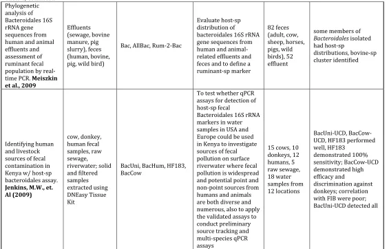
example, for cattle, swine, and humans (Mieszkin et al., 2010, Jenkins et al., 2009;
Lamendella et al., 2009; Ju-Yong et al., 2009; Mieszkin et al., 2009).
There are some major features of
Bacteriodales
that make targeting this order for MST a
promising library-independent method. For example,
Bacteriodes
appear to have a
relatively high degree of environmental persistence, with reported survival for as long
as 14 days at 4˚C, even in the presence of predators
. Even at warmer temperatures
more representative of environmental waters (14
oC), they remained detectable for 4-5
days (Kreader, 1998). In addition, as strict anaerobes, they are unable to proliferate
outside of the host environment. It is generally recognized that they are a more suitable
indicator for fecal contamination than are the fecal coliforms (Bernhard and Field,
2000; Bernhard and Field, 2000b; Layton et al., 2006; Kreader, 1995; Dick and Field,
2004). Perhaps most importantly, members of
Bacteroides
are highly-host specific, and
appear to be present in the environment at only low levels outside of the host species
Culture-independent, library-independent methods do have some disadvantages,
though. For instance, not all markers may be present in all individuals. Compositing
samples from individuals, or sampling from sewage systems eases this limitation,
however prevents distinction of host source. As is the case for culture-dependent
methods, different localities will have different microbial populations, and even though
libraries are not required for the culture-independent methods, the assays must be
validated for the locale in which they are being used (Field and Samadpour, 2007).
Aside from the detection limits of the methods themselves, other issues such as
sampling volume arise. Library-independent, culture-independent methods rely on
processing whole samples and collecting information on the profile of the genetic
material within the entire sample, so sample volume is a critical consideration for
representation purposes. Since library-independent methods almost always rely on the
application of molecular approaches to “dirty” samples, preparing the sample for
analysis such that matrix-associated inhibition is minimized can also be a challenge.
Likewise, interpretation of results obtained from environmental samples may be
difficult. Molecuar-based tools require controls, and if the marker is present in low
concentrations, the control can prevent detection of the marker (Hoorfar, et al., 2004).
Finally, the correlation of these alternative markers to currently established fecal
APPLICATIONS OF BACTEROIDALES MST METHODS TO ENVIRONMENTAL STUDIES
While members of
Bacteroidales
have been purported as potential indicators of fecal
contamination for many years, the need to maintain anaerobic conditions to facilitate
their proliferation was a deterrent to their widespread use. With the advent of
molecular-based detection approaches,
Bacteroidales-
based assays have become more
practical. Of particular interest are applications in which
Bacteroidales
host specificity
has been capitalized upon for MST purposes. Such assays work by employing PCR to
amplify variable DNA regions that show host specificity. Identifying these regions is the
crux of how
Bacteroidales
microbial source tracking assays work. The regions that are
conserved can be referred to as universal markers, or those found in all
Bacteroidales
.
Regions that are variable can be host-specific, creating the possibility for assay
development that can determine what kind of fecal matter is present by amplifying
markers that are specific to certain animals. Previous work done by Bernard and Field
(2000b) characterized host-specific regions that were specific to humans and
ruminants on the 16S rRNA genome. The polymerase chain reaction (PCR) was used to
selectively amplify these regions, and allow the researcher to directly detect the
markers of choice.
A variety of studies have been undertaken to characterize the
Bacteroidales
populations
in specific host species, including humans, cows, dogs, horses, Canada geese, chicken,
elk, gulls, and cats. Much of the early work dealt with identifying differences between
2006; Bernhard and Field, 2000; Bernhard and Field, 2000b; Seurinck et al., 2004).
Over the last 10 years, qPCR amplification assays have been developed for
Bacteroides
targets specific to humans (Layton et al., 2006; Kildaire et al., 2007; Seurinck et al.,
2005; Bernard and Field, 2000; Green et al., 2011; Seifring et al., 2008; Ju-Yong et al.,
2009), pigs (Okabe et al., 2007; Mieszkin et al., 2009), cattle (Bernard and Field, 2000b;
Kildaire et al., 2007; Layton et al., 2006; Shanks et al., 2006; Bernard and Field, 2000;
Reischer et al., 2006; Ju-Yong et al., 2009), dogs (Kildaire et al., 2007), horses (Simpson
et al., 2004; Dick et al., 2004b; Layton et al., 2006), and wild bird fecal materials (Green
et al., 2012; Dick et al., 2005). Most studies support this high degree of host specificity,
but there are exceptions (Dick et al., 2004; Ju-yong et al., 2009). Assays have been
applied to trace the origins of fecal contamination in different water systems, such as
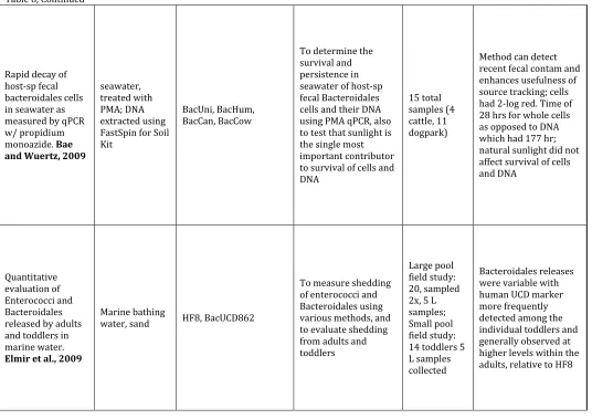
recreational water (Elmir et al., 2009; Stapleton et al, 2009; Green et al., 2010; Layton et
al., 2006) and drinking water (Sokolova et al., 2012; Layton et al., 2006), as well as
within estuarine, marine, and river waters (Walters and Field, 2008; Elmir et al., 2009;
Dick et al., 2010; Green et al., 2010; Seurinck et al., 2005). Other studies have sought to
use the
Bacteroidales
marker detection method as applied to raw sewage and
wastewater treatment plant effluents (Shanks et al., 2010).
In developing new
Bacteroidales
assays and applying them to real-world samples, many
issues must be considered, including (i) choosing the correct primers; (ii) identifying
the appropriate methods to prepare the sample prior to assay; (iii) assessing the
appropriate controls (particularly for quantification purposes); and (v) overall
interpretation of results. Each of these will be discussed in greater detail below.
PRIMER CHOICE
A variety of primers are available for general
Bacteroidales
as well as host-specific
Bacteroidales
typing. These are summarized in Table 1.5. The most commonly used
primer sets are AllBac, BacUni, and Bac32F/Bac708R (general
Bacteroidales
), HF183
(Human-specific), BoBac (Bovine-specific), and PigBac1F/PigBac1R (Pig-specific).
These assays are more widely used because they appear to have a higher success rate
based on citation frequency in the literature relative to other assays. Major
considerations when designing a qPCR assay to detect host-specific fecal material are
marker specificity (broad reactivity for general fecal markers – markers within the
highly conserved regions, and high specificity for species-specific markers – markers
within the variable regions) and the need for the target to be relatively abundant and
uniformly distributed throughout the system being evaluated. As perhaps expected, the
host-specific genes are usually not as easily detected, nor as highly prevalent, as are the
general
Bacteroidales
markers. Clearly, development of assays should take into account
relative abundance of particular markers and fecal indicator organisms (Shanks et al.,
Paper Title &
References Sample Matrix & Matrix Prep Assay Purpose # of Samples Summary of Findings
Development of Bacteroides 16S rRNA gene TaqMan-Based RT PCR assays for estimation of total, human, and bovine fecal pollution in water.
Layton, A. et al. (April, 2006) Various animal feces suspended in DNAse-free water; extracted using FastSpin for Soil.
AllBac, BoBac, HuBac
RT PCR assays was designed to detect Bacteroides 16S rRNA genes present in all mammalian fecal samples and determine whether the quantity of Bacteroides 16S rRNA genes present in a water sample was related to the fecal concentration
3 Human, 4 Swine, 4 Canine, 4 Equine, 6 Bovine
Estimation of pig fecal
contamination in a river catchment by qPCR using two pig-specific Bacteroidales 16S rRNA genetic markers.
Mieszkin, S. et. Al. (2009) River water, pig slurry, lagoon, fecal samples. Water samples filtered and extracted using DNEasy Kit; solid samples extracted using FastSpin for Soil kit
Bac, AllBac, HF183, Pig-1-Bac, Pig-2-Bac, Pig-Bac2
To design new primers for the detection and quantification of pig-specific
Bacteroidales; Validate
sensitivity/specificity of new primers and TaqMan assay using target (pig-related) and non-target (other animal related) DNA; Evaluate TaqMan for detection and quantitative estimation of pig-associated fecal pollution 24 Adults and children, 10 cows, 10 sheep, 10 horses, 25 pig, 23 slurry samples, 14 lagoon surface, 7 compost, 24 recreation water
Uncultured strains of Prevotella bacteria, Pig-2-Bac is promising marke; correlates to quantity of E. coli