ABSTRACT
CAREY, LEIAH MARIE. Probing the Structure-Function Relationship of a Multifunctional Enzyme using Crystallographic Diffraction Methods. (Under the direction of Dr. Reza Ghiladi.)
The marine annelid Amphitrite ornata possesses the ability to chemically detoxify deleterious aromatic compounds prevalent in its environment, which is afforded by its coelomic hemoglobin, Dehaloperoxidase (DHP). In addition to its oxygen transport function, DHP also possesses peroxidase, peroxygenase, oxidase and oxygenase enzymatic functions, resulting in 5 activities that coexist at a single heme reactive center. Structurally, DHP possesses the canonical -helical globin fold associated with oxygen transport, yet lacks the
structural homology with archetypical monofunctional examples of which it shares enzymatic reactivity, such as horseradish peroxidase, AaeAPO, cytochrome c oxidase and cytochrome P450 monooxygenase. The structure-function paradigm has been extensively studied and well-defined for monofunctional enzymes, however this understanding is lacking when applied to multifunctional systems. In an effort to discern how DHP accommodates this multifunctional reactivity, a structural approach utilizing crystallographic diffraction is undertaken in the investigations presented here.
effect on isoenzyme differences in reactivity, yet this isoelectric substitution has no direct contact with the heme active site. In Chapter 4, the knowledge of substrate binding sites of different mechanistic substrates is utilized in rationally designing mutagenesis studies in an effort to direct enzymatic functionality. The plasticity of the heme active center is further elaborated through DHP’s ability to preserve peroxidase reactivity, albeit at a lower capability,
even when a peroxidase substrate binding site is sterically blocked. Chapter 5 is a collection of crystallographic case studies in an effort to advance the knowledge of DHP’s representation
© Copyright 2017 Leiah Marie Carey
Probing the Structure-Function Relationship of a Multifunctional Enzyme using Crystallographic Diffraction Methods
by
Leiah Marie Carey
A dissertation submitted to the Graduate Faculty of North Carolina State University
in partial fulfillment of the requirements for the Degree of
Doctor of Philosophy
Chemistry
Raleigh, North Carolina
2017
APPROVED BY:
_______________________________ _______________________________
Dr. Reza Ghiladi Dr. Stefan Franzen
Chair of Advisory Committee
DEDICATION
BIOGRAPHY
Leiah Marie Carey was born the first child to Tim and Kathy Carey in the evening of February 16th, 1979 in Chapel Hill, NC. She called Saxapahaw, NC home until graduating from Southern Alamance High School in 1997 and went out into the world on her own. In late December 2007, her daughter Pearl was born. She obtained her Bachelor of Science in Chemistry in 2012 from the University of North Carolina at Greensboro and entered the Chemistry Ph. D. program at North Carolina State University that same year. Upon joining the Ghiladi research group, she directed her training toward a physical approach to investigate the chemistry of biological systems, from which she quickly discovered her love for crystallography. She soon realized this was exactly what she had been searching for: a career path that embraces curiosity, is intellectually stimulating and emotionally satisfying.
ACKNOWLEDGMENTS
The support I’ve received while working toward this personal goal has been instrumental to
my success and words cannot fully express my gratitude. I would like to thank my graduate mentor, Dr. Reza Ghiladi, for the opportunity to pursue my graduate studies in your lab. The insight, support and experiences you have provided me are the cornerstones of my success. In particular, thank you for allowing me to grow independently in my research and respecting my responsibility as a parent, both of which have helped me learn to balance the many facets of life while pursuing a goal. For your continuous support and interest in my research, willingness to always provide resources and assistance, and sharing your enthusiasm for scientific discovery and education, I would like to thank Dr. Stefan Franzen. Without your insistence on the need to continue crystallographic work after Junjie left and offering me the use of the different capabilities of your lab, I might not have pursued a research project outside the expertise of my lab nor realized that I truly enjoy crystallographic work. For Dr. Paul Swartz, thank you for opening you lab to me and dealing my insistent questions, which helped me develop a deeper understanding and appreciation for crystallography. For my labmates, both past and present, thank you for many insightful discussions, willingness to help, being able to share the excitement and passion for research and basically just being great people. You guys were a pleasure to work. Thank you to my parents (and Pearl’s grandparents) for your support in my decision to pursue this daunting and lengthy endeavor and for taking such great care of Pearl, which allowed me the time needed to get the work done. Thank you Bosque, because I couldn’t have returned to school without your help caring for my infant daughter. Finally, for
TABLE OF CONTENTS
LIST OF TABLES………... ix
LIST OF FIGURES……….. xii
LIST OF SCHEMES……… xix
LIST OF ABBREVIATIONS……….. xx
Chapter 1: X-ray Crystallographic and Spectroscopic Analysis of Dehaloperoxidase (DHP) on Deposited Structures……….. 1
1.1 Introduction……… 1
1.2 Global Structural Analysis……….. 2
1.3 The Multiple Functions of Dehaloperoxidase………. 4
1.4 Brief Overview of Deposited Dehaloperoxidase X-ray Crystallographic Structures……….. 6
1.5 Unusual Flexibility of the Distal Histidine: Structural and Spectroscopic Correlations………... 11
1.6 Inhibitor Binding Site Characterization……….. 13
1.7 Peroxidase Substrate Binding Site(s) Characterization………... 15
1.8 Peroxygenase Substrate Binding Site(s) Analysis……….. 19
1.8.1 Haloindoles: 5-bromoindole and 7-bromoindole…………. 19
1.8.2 4-nitrophenol………... 21
1.9 Preface of Presented Work in Proceeding Chapters……… 22
1.10 References……… 24
Chapter 2: Bridging the Functional Gap Between Reactivity and Inhibition in Dehaloperoxidase B from Amphitrite ornata: Mechanistic and Structural Studies of 2,4-dihalophenol………. 28
2.1 Abstract……….. 28
2.2 Introduction……… 28
2.3 Experimental……….. 30
2.3.1 Materials and Methods……… 30
2.3.2 Enzyme Assay Protocol………... 30
2.3.3 LC-MS Studies……… 31
2.3.4 Substrate Binding Studies……… 31
2.3.5 Stopped-flow UV-visible Studies……… 32
2.3.6 Protein Crystallization and X-ray Diffraction Studies……. 32
2.4 Results……… 33
2.4.1 Enzyme Assays……… 33
2.4.2 MS Product Characterization and 18O Labeling Studies….. 35
2.4.3 Substrate Binding Study……….. 42
2.4.4 Stopped-flow Studies of 2,4-dichlorophenol Reactivity….. 44
2.5 Discussion……….. 54
2.6 Conclusion……….. 60
2.7 Supporting Information……….. 61
2.8 References……….. 68
Chapter 3: How Nature Tunes isoenzyme Activity in the Multifunctional Catalytic Globin Dehaloperoxidase from Amphitrite ornata………... 73
3.1 Author Contributions……….. 73
3.2 Abstract……….. 73
3.3 Introduction……… 74
3.4 Experimental……….. 78
3.4.1 Materials……….. 78
3.4.2 Construction of Mutant DHP Plasmids……… 78
3.4.3 Molecular Weight Determination……… 79
3.4.4 Peroxidase Studies………... 79
3.4.5 Peroxygenase Studies……….. 80
3.4.6 5-Br-indole Binding Studies……… 80
3.4.7 Stopped-flow UV-visible Studies……… 81
3.4.8 EPR Spectroscopy………... 81
3.4.9 Protein Crystallization and X-ray Diffraction Studies……. 81
3.5 Results……… 82
3.5.1 UV-visible Spectroscopic Studies………... 82
3.5.2 EPR Spectroscopic Studies……….. 85
3.5.3 Peroxidase Studies………... 87
3.5.4 Peroxygenase Studies……….. 88
3.5.5 5-Br-indole Binding Studies……… 90
3.5.6 Non-native I9 Mutants………. 91
3.5.7 Stopped-flow UV-visible Spectroscopic Studies…………. 91
3.5.8 X-ray Diffraction Studies……… 92
3.6 Discussion……….. 98
3.7 Conclusion……….. 102
3.8 Acknowledgements……… 103
3.9 Supporting Information……….. 103
3.10 References……… 110
Chapter 4: Selective Tuning of Activity in a Multifunctional Enzyme as Revealed in the F21W Mutant of Dehaloperoxidase B from Amphitrite ornata……… 117
4.1 Author Contributions……….. 117
4.2 Abstract……….. 117
4.3 Introduction……… 118
4.4.1 Materials……….. 121
4.4.2 Construction of Mutant DHP Plasmids……… 121
4.4.3 Protein Crystallization and X-ray Diffraction Studies……. 122
4.4.4 Peroxidase Studies………... 122
4.4.5 Peroxygenase Studies……….. 123
4.4.6 5-Br-indole Binding Studies……… 123
4.4.7 Stopped-flow UV-visible Studies……… 124
4.5 Results……… 124
4.5.1 Overexpression, Purification and Characterization of DHP B (F21W)………. 124
4.5.2 Activity Assays……… 126
4.5.3 Isotopically-labeled Oxygen Studies………... 127
4.5.4 Stopped-flow UV-visible Spectroscopy……….. 128
4.5.5 X-ray Crystallographic Studies………... 131
4.6 Discussion……….. 134
4.7 Conclusion……….. 138
4.8 Acknowledgements……… 138
4.9 Supplementary Material………. 139
4.10 References……… 141
Chapter 5: Further Exploring the Structure-Function Relationship in Dehaloperoxidase Through X-ray and Neutron Crystallography: Four Case Studies………... 146
5.1 Case Study 1 – Neutron Diffraction Structure of Ferric DHP B…….. 146
5.1.1 Introduction………. 146
5.1.2 Experimental………... 149
5.1.3 Results and Discussion……… 153
5.1.4 Summary of Case Study 1……… 161
5.2 Case Study 2 – Elucidation of the 4-Nitrophenol (4NP) and 4- Nitrocatechol (4NC) Crystallographic Binding Sites in DHP B... 162
5.2.1 Abstract………... 162
5.2.2 Experimental………... 163
5.2.3 Results and Discussion……… 163
5.2.4 Summary of Case Study 2……… 169
5.3 Structural Analysis of DHP B Reconstituted with Mn Protoporphyrin IX ………... 170
5.3.1 Introduction………. 170
5.3.2 Experimental………... 170
5.3.3 Results and Discussion……… 171
5.3.4 Summary of Case Study 3……… 176
5.4.1 Introduction………. 177
5.4.2 Experimental………... 178
5.4.3 Results and Discussion……… 179
5.4.4 Summary of Case Study 4……… 183
LIST OF TABLES
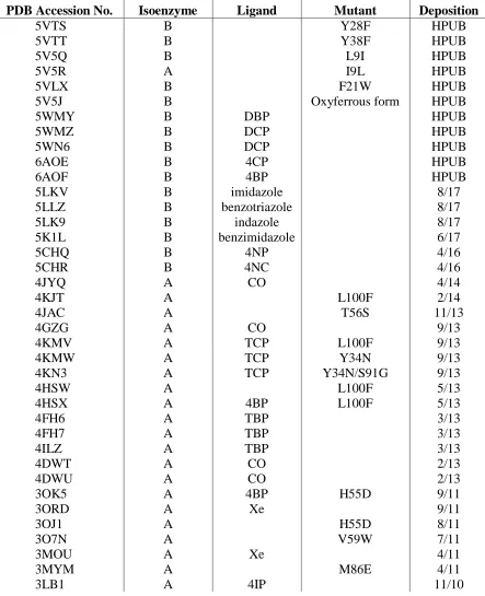
Table 1.1 Crystal structures of DHP deposited into the PDB, arranged from
most recent to initial structure……….. 8
Table 1.2 Distances (Å) given for the heme Fe and the halogen and alcohol moieties of the inhibitors (4XP) and the substrates (TXP)………... 18
Table 2.1 Substrate conversion (%), providing 2,4-DCP and 2,4-DBP reactivity with ferric DHP B and oxyferrous DHP B. Substrate conversion in the presence of 500 M 4-BP inhibitor is given in parenthesis. Reactivity of previously established substrates is provided for comparison…………... 34
Table 2.2 Kd Values for Ligand Binding to Ferric DHP B at pH 7 ………. 43 Table 2.3 X-ray Data Collection and Refinement Statistics for WT DHP B
complexed with 2,4-dichlorophenol (2,4-DCP) (5WN6 and 5WMZ), 2,4-dibromophenol (2,4-DBP) (5WMY), 4-chlorophenol (4-CP) (6AOE) and 4-bromophenol (4-BP) (6AOF)………... 51
Table 2.4 Distal pocket atomic distances of substrate (2,4-DCP or 2,4-DBP) and heme environments, given in Å. Values provided are averages of both protomers. Atomic distances for 2,4,6-trihalophenols and 4-halophenols are provided for comparison... 52
Table 3.1 UV-visible spectroscopic features of ferric DHP variants at pH 7……… 85 Table 3.2 Kinetics data for the oxidation of TCP and conversion percentage of
5-Br-indole as catalyzed by ferric DHP in the presence of H2O2 at pH 7…. 89
Table 3.3 5-Br-indole dissociation constants (Kd) for WT DHP and selected
mutants……….... 90
Table 3.4 UV-visible spectroscopic features and rate of formation of Compound ES in WT DHP and selected variants at pH 7………... 92
Table 3.6 Selected Distances and Angles for Protomers A & B (Protomer B in
parentheses)………. 96
Table S3.7 Mutagenic Primers Used to Construct the DHP A Mutants……….. 104 Table S3.8 Calculated and experimentally determined monomeric molecular
weights of the dehaloperoxidase mutants………. 104
Table S3.9 Rmsd Values for DHP Protomer C Least Squares Quadratic (LSQ)
Alignment……… 106
Table 4.1 Kinetic parameters for the oxidation of TCP, and percent substrate conversion for the oxygenation of 5-Br-indole, 7-Br-indole and 4-NP… 127
Table 4.2 Comparison of UV-visible spectroscopic data for the ferric, Compound ES, and Compound RH species in DHP B (F21W) and WT DHP isoenzymes A and B at pH 7………. 129
Table 4.3 X-ray Data Collection and Refinement Statistics for DHP B (F21W) (5VLX)……… 132
Table 4.4 Selected distances (Å) for DHP B (F21W) and WT DHP B (PDB accession 3IXF)………... 133
Table 5.1 Comparison of neutron and X-ray scattering characteristics for selected
atoms……… 148
Table 5.2 Enfors Minimal Media composition……… 151 Table 5.3 Neutron and data collection and refinement statistics for ferric WT DHP
B……….. 156
Table 5.4 Selected Distances in the Neutron Structure of Ferric DHP B for Protomer A……….. 160
Table 5.5 Data collection and refinement statistics for DHP B co-crystallized in the presence of 4-nitrophenol (4-NP) and 4-nitrocatechol (4-NC)……... 164
Table 5.7 X-ray data collection and refinement statistics for Mn-DHP B ………... 173 Table 5.8 Selected porphyrin-related atomic distances of Mn-DHP B and WT
DHP B (pdb accession 3IXF). All distances given in Å………... 176
Table 5.9 Data collection and refinement statistics for DHP B (Y28F) and DHP B (Y38F)……….. 181
LIST OF FIGURES
Figure 1.1 Image of Amphitrite ornata, from which DHP was discovered... 1 Figure 1.2 X-ray crystal structures of A) sperm whale myoglobin (SWMb, PDB
accession code 1A6G), B) DHP A (PDB accession code 1EW6) and C) cytochrome c peroxidase (CcP, PDB accession code 1ZBY). It is clearly shown that DHP possesses the conserved globin fold as
displayed in Mb……… 3
Figure 1.3 Protoporphyrin IX prosthetic group, as found in DHP, Mb and HRP. Pyrrole rings (A-D) and heme edges (-) are labeled in accordance
with convention……… 4
Figure 1.4 Xe crystallographic binding sites observed in DHP………... 10 Figure 1.5 Sites of single amino acid mutations of DHP A, utilized previously
for mechanistic and reactivity investigations……… 11
Figure 1.6 A) Room temperature resonance Raman spectrum of WT DHP A. Core markers for 6cHS and 5cHS hemes are annotated for . B) Room temperature crystal structure (1EW6) emphasizing i) the water that occupies the Fe 6th coordination site, and ii) distal histidine His55 in an equilibrium between the open (solvent exposed) and closed (internal) conformations………... 13
Figure 1.7 A) Resonance Raman spectra of WT DHP A in the presence of the indicated inhibitor. [DHP A] = 100 M; [4IP] = 1 mM; [4BP], [4CP] and [4FP] = 8 mM. B) Superposition of the para-halogenated phenolic inhibitor structures depicting orientation within the distal pocket; 4IP (red, 3LB1), 4BP (green, 6AOF), 4CP (blue, 6AOE) and 4FP (purple,
3LB4)………... 14
Figure 1.9 Resonance Raman spectra of DHP B in solution with A) bromine-substituted indoles at different positions and B) 5-haloindoles of different halogens. [DHP] = 50M, [5-haloindole] = 500M…….... 20
Figure 1.10 Geometry optimized structure for A) 5-bromoindole and B)
7-bromoindole………. 21
Figure 1.11 The binding of 4NP with DHP B is characterized through A) UV-visible spectral perturbation via titration of 4NP, B) binding isotherm yielding the dissociation constant of 260 M, and C) X-ray crystallographic binding site of 4NP in the distal pocket of DHP B... 22
Figure 2.1 HPLC chromatograms, as observed at 288 nm, of the 2,4-DCP (A) and 2,4-DBP (B) reactions with 10 M ferric DHP B, 500 M = [substrate] = [H2O2]……….. 35
Figure 2.2 Kinetic data for the reaction of pre-formed Compound I [DHP B (Y28F/Y38F)] with 2,4-dichlorophenol (2,4-DCP)………... 45
Figure 2.3 Kinetic data for the reaction of pre-formed Compound ES with 2,4-dichlorophenol (2,4-DCP)……….... 47
Figure 2.4 Kinetic data for the reaction of oxyferrous DHP B with 2,4-dichlorophenol (2,4-DCP) and hydrogen peroxide………... 48
Figure 2.6 Superposition of selected halophenol binding sites. A) 2,4-DBP (green) and 2,4-DCP (silver); B – D) Comparison of monohalophenol (blue) and trihalophenol (pink) binding sites with the elucidated dihalophenol binding sites. B) Brominated variants; C) Chlorinated variants ( heme edge proximity); D) Chlorinated variants ( heme edge proximity)………. 59
Figure S2.7 Optical difference spectra (left) and titration curves (right) of 2,4-dichlorophenol (top) and 2,4-dibromophenol (bottom) binding to 10 M DHP B (100 mM KPi, pH 7, 5% MeOH v/v). The calculated
dissociation constant is provided in each titration curve………... 62
Figure S2.8 TOP: LC chromatogram of the reaction of 2,4-dichlorophenol (500 M) with DHP B (10 M) in the presence of H2O2 (500 M) at 25 °C
(5% MeOH in 100 mM KPi, pH 7). Unlabeled peaks correspond to
dimer and trimer products of various levels of oxidation and chlorination. BOTTOM: ESI-MS total ion chromatograms obtained for single ring species………... 63
Figure S2.9 ESI-MS total ion chromatograms obtained from the 1a (left panel) and 1c (right panel) reaction products of the 18O isotopic labeled 2,4-DCP oxidation catalyzed by ferric DHP B conducted under the following conditions: A) unlabeled water and hydrogen peroxide B) labeled H218O2 and unlabeled water; C) labeled H218O and unlabeled
hydrogen peroxide; D) 18O labels on both H
218O2 and H218O………… 64
Figure S2.10 ESI-MS total ion chromatograms obtained from the 1b (left panel) and 1d (right panel) reaction products of the 18O isotopic labeled 2,4-DCP oxidation catalyzed by ferric DHP B conducted under the following conditions: A) unlabeled water and hydrogen peroxide B) labeled H218O2 and unlabeled water; C) labeled H218O and unlabeled
hydrogen peroxide; D) 18O labels on both H
218O2 and H218O………… 65
Figure S2.11 TOP: LC chromatogram of the reaction of 2,4-dibromophenol (500 M) with DHP B (10 M) in the presence of H2O2 (500 M) at 25 °C
(5% MeOH in 100 mM KPi, pH 7). Unlabeled peaks correspond to
Figure S2.12 ESI-MS total ion chromatograms obtained from the 2a (left panel), 2b (middle panel) and 2c (right panel) reaction products of the 18O isotopic labeled 2,4-DBP oxidation catalyzed by ferric DHP B conducted under the following conditions: A) unlabeled water and hydrogen peroxide B) labeled H218O2 and unlabeled water; C) labeled
H218O and unlabeled hydrogen peroxide; D) 18O labels on both H218O2
and H218O……….. 67
Figure 3.1 C superposition of the A protomers of DHP A (PDB accession code 1EW6, teal) and DHP B (PDB accession code 5V5J, purple), highlighting substitution sites between isoenzymes (isoenzyme A residue listed first): proximal side N/S81 and S/G91, distal side R/K32 and Y/N34, with I/L9 located 15 Å from the heme……… 77
Figure 3.2 UV-visible spectra of A) WT DHP A, WT DHP B, DHP A (I9L), DHP B (L9I) and DHP A (I9V), B) single mutants of DHP A, C) I9L containing double mutants of DHP A, D) double mutants of DHP A lacking the I9L substitution, and E) I9L containing triple mutants of DHP A. All spectra were recorded at 10 µM enzyme concentration in 100 mM KPi buffer (pH 7, 25 °C)………. 83
Figure 3.3 EPR spectra of ferric WT DHP A (black), DHP A (I9L) (blue), WT DHP B (green), and DHP B (L9I) (red), and the result of their deconvolution into three individual ferric heme EPR signals, rhombic high spin, axial high spin, and mixed spin.………... 87
Figure 3.4 Panels A-C: LSQ C superposition of DHP A (I9L) (green), DHP B (L9I) (teal), as viewed from the heme edge. A) Superposition of the A protomers. B) Magnified view from panel A of the Leu9 and Ile9 residues. C) Heme active site from panel A. D) LSQ C superposition of DHP B (L9I) and WT DHP B (yellow). E) LSQ C superposition of DHP A (I9L) and WT DHP A (yellow; PDB accession code
2QFN)……….. 97
Figure S3.5 Kinetic data obtained by optical spectroscopy for the reaction of DHP
A (I9L) with H2O2………. 105
Figure S3.7 LSQ C superposition of DHP A (I9L) and DHP B (L9I), focusing on the distal side of the heme cavity as well as the region containing the residue at position 9……….. 107
Figure 4.1 Superposition of substrate binding sites within the distal pocket of DHP as viewed from the heme edge: TBP (yellow, PDB 4FH7), 4-NP (purple, PDB 5CHQ), 4-BP (green, PDB 3LB2), and both internal (TCPinterior) and external (TCPexterior) conformations of TCP (cyan,
PDB 4KN3)……….. 120
Figure 4.2 UV-visible spectra of ferric (red) and oxyferrous (black) DHP B (F21W) in 100 mM KPi (pH 7)………. 125
Figure 4.3 ESI-MS total ion chromatograms obtained for the reaction product 5-Br-2-oxindole (A: H218O, H216O2; B: H216O, H218O2). Reaction
conditions: [5-Br-indole] = [H2O2] = 500 M, [enzyme] = 10 M,
100 mM KPi (pH 7), 25 C……… 128
Figure 4.4 Kinetic data obtained by optical spectroscopy for the reaction of DHP
B (F21W) with H2O2………. 130
Figure 4.5 A) DHP B (F21W) (silver) Csuperposition with WT DHP B (cyan; PDB 3IXF), highlighting the F21/W21 residues. Only one conformation for F21 is shown for clarity. B) Distal pocket of the DHP B (F21W) mutant, as viewed from the heme edge. There are two water molecules in the distal pocket, forming a H-bonding network with W21, T56, Y38, and the heme propionate arms. All distances are given in Å……… 131
Figure 4.6 DHP B (F21W) mutant distal pocket superposition with TCP [TCPinterior (PDB 4KMV) and TCPexterior (PDB 4KMW)] and 4-NP
(PDB 5CHQ) substrates……… 137
Figure S4.7 Optical difference spectra of 5-Br-indole (10-130 eq.) binding to 10 M DHP B (F21W) in 10% MeOH/100 mM KPi (v/v) at pH 7; inset
– corresponding titration curve………. 140
Figure S4.8 Distal pocket of A) WT DHP B (PDB 3IXF18), B) DHP B (F21W)
(PDB 5VLX2) and C) superposition of WT DHP B and DHP B
Figure 5.1 Representative ferric DHP B crystals of sufficient size for obtaining neutron diffraction data. A) Perdeuterated DHP B, volume ~0.2 mm3. B) Protiated DHP B crystal, volume ~2.0 mm3. C) Representative
quasi-Laue diffraction pattern of ferric DHP B. The image was obtained from 20 hour neutron exposure………... 154 Figure 5.2 Room temperature X-ray structure of the perdeuterated DHP B
protomer A heme cavity, viewed from the heme side. FO – FC
electron density maps are displayed in green and scaled to 3 Blue depicts the 2FO – FC maps. A) 2FO – FC maps scaled to 2 . B) 2FO –
FC maps scaled to 1 ……… 155
Figure 5.3 Residues Ile 6 and Phe 123 from DHP B, shown modeled into blue nuclear and red electron density maps. Panel A) Nuclear density maps of perdeuterated residues, highlighting the visibility of deuterium atoms (shown in red) at a resolution of 2.05 Å. Panel B) Electron density maps illustrating the absence of hydrogen atoms, even at a higher resolution, 1.81 Å……….. 157
Figure 5.4 Neutron structure of ferric DHP B, presenting nomenclature and atomic distances. A) Protomer A ribbon structure, with emphasis placed on the distal pocket environment. B) Proximal environment of protomer A. C) Bis-his heme ligation present in protomer B………… 159
Figure 5.5 Superposition (with atomic distances) of 2,4-dichlorophenol (DCP) binding site and distal waters found in the neutron ferric DHP B structure……… 160
Figure 5.6 As viewed from the edge, panels A and B provide atomic distances for 4-NP and 4-NC, respectively. Panels C and D present the distal superposition of 4-NP (5CHQ, green), 4-NC (5CHR, silver) and 4BP (3LB2, purple) structures……….. 166
Figure 5.7 Fitting of substrates A) 4NP and B) 4NC into their respective
2mFo-DFc electron density maps, contoured to 1………. 167
Figure 5.9 Distal cavity of Mn-DHP B protomer B. As shown, there exists two water molecules at the entrance of the cavity, participating in a hydrogen-bonding network with T56, Y38, and propionate arm A. The presence of distal waters in this orientation has only been observed previously in the DHP B (F21W) mutant………... 174
Figure 5.10 LSQ Csuperposition of WT DHP B (3IXF,17 silver) and Mn-DHP
B (teal) protomer A. Emphasis is placed on the heme cavity, where it is clearly shown that the porphyrins occupy identical orientations…... 175
Figure 5.11 Diagram portraying the dihedral angle that dictates the hyperfine splitting of the tyrosyl radical signal by the two methylene hydrogens,
H and H………... 177
LIST OF SCHEMES
Scheme 1.1 Reversible oxygen transport as carried out by globins……….. 5 Scheme 1.2 Oxidative dehalogenation of trihalophenols yielding their
corresponding quinone products as catalyzed by DHP via a peroxidase mechanism……….. 5
Scheme 1.3 Oxidation of monohaloindoles to form either 2-oxindole or 3-oxindole products. The oxygen atom inserted in the product is derived from H2O2 as necessitated by a peroxygenase pathway……… 6
Scheme 1.4 Oxidation of 2,3-dimethylindole to form either 2,3-dimethyl-3H-indol-3-ol or N-(2-acetylphenyl)acetamide products. The oxygen atom inserted in the product is derived from O2 as necessitated by an
oxygenase pathway………... 6
Scheme 1.5 Oxidation of 5-Br-3-oxindole to form 5,5’-Br2-indigo. The product
oxidation, characterized by a lack of atom incorporation and dependence on O2, is representative of an oxidase pathway………….. 6
Scheme 2.1 Proposed oxidation pathways of 2,4-dichlorophenol in the presence of DHP……….. 37
Scheme 2.2 Proposed oxidation pathway of 2,4-dibromophenol in the presence of DHP B……….. 40
LIST OF ABBREVIATIONS
2,4-DBP 2,4-dibromophenol 2,4-DCP 2,4-dichlorophenol 2,4-DXP 2,4-dihalophenol
4BP 4-bromophenol
4CP 4-chlorophenol
4FP 4-fluorophenol
4IP 4-iodophenol
4XP 4-halophenol
4NC 4-nitrocatechol
4NP 4-nitrophenol
5BI 5-bromoindole
5XI 5-haloindole
7BI 7-bromoindole
CcP Cytochrome c peroxidase
COOT Crystallographic object-oriented toolkit
DHP Dehaloperoxidase
DHP A Dehaloperoxidase A
DHP B Dehaloperoxidase B
DMSO Dimethyl sulfoxide
EPR Electron paramagnetic resonance FPLC Fast protein liquid chromatography
G Glycine
I Isoleucine
IPTG isopropyl--D-1-thioglactopyranoside ITC Isothermal calorimetry
K Lysine
Kd Dissociation constant
L Leucine
LB Lysogeny broth
LSQ Least squares quadratic
Mb Myoglobin
ML Mother liquor
MPEG Monomethyl polyethylene glycol MWCO Molecular weight cut off
N Asparagine
NMR Nuclear magnetic resonance
PDB Protein data bank
PEG Polyethylene glycol
R Arginine
r.m.s.d. Root mean square deviation
RCSB Research Collaboratory for Structural Bioinformatics
rR Resonance Raman
Rz Reinheitzahl
S Serine
SDS-PAGE Sodium dodecyl sulfate polyacrylamide gel electrophoresis SER-CAT Southeast regional collaborative access team
SP-FF Sepharose fast flow STI soybean trypsin inhibitor
SWMb Sperm whale myoglobin
TBP 2,4,6-tribromophenol
TCP 2,4,6-trichlorophenol
TFP 2,4,6-trifluorophenol
TIP 2,4,6-triiodophenol
TPCK N-p-tosyl-L-phenylalanine chloromethyl ketone TRSSA Tyrosyl radical spectra simulation algorithm
TXP 2,4,6-halophenol
Vm Matthew’s coefficient
WT Wild-type
Chapter 1
X-ray Crystallographic and Spectroscopic Analysis of Dehaloperoxidase (DHP) on Deposited Structures
1.1 Introduction
DHP was first discovered for its oxygen-transport capability in 1977 by Bonaventura et al.1 Observance of its peroxidase activity led to the rediscovery of DHP in 1996 by Lovell et al.2 DHP exists as two isoenzymes, DHP A and DHP B, with only 5 amino acids differentiating
the two. The structural and reactivity differences between the two isoenzymes will be further elaborated in Chapter 3. It was discovered in 2001 that two unique genes are responsible for DHP A and DHP B, dhpA (GenBank accession number AF284381) and dhpB (AF285090), respectively.3 Crystallographic studies revealed that the enzyme possessed a traditional globin fold despite exhibiting a very low sequence homology with other globins.4 In our lab,
peroxygenase, oxygenase and oxidase activities for DHP have been characterized, adding to the scope of this enzyme’s functionality.5 Given the multiple possible activities within this
single protein, the question thus arises as to what parameters dictate which enzymatic function is observed. Could this coexistance of 5 functions residing at a single active site be understood through structural analysis?
1.2 Global Structural Analysis
DHP is classified as a globin due to its canonical globin helical fold. Structurally, DHP has the appearance of a traditional oxygen transport protein. When compared with the prototypical globin, myoglobin (Mb), the overall folds are very similar (Figure 1.2, panels A & B). The largest difference lies in the proximal region. The proximal histidine of DHP occupies position 89, while in Mb the proximal histidine is two residues further toward the C-terminus at position 91. The proximal histidine in Mb is located on helix F, however this perturbation in sequence places DHP’s His89 in a loop region between helices F & G. Due to the shifted orientation, the imidazole ring of the distal histidine is rotated 60 in DHP when compared to Mb.6
any known globin.2,11 This lower peroxidase activity may be related to the absence of the traditionally conserved distal arginine and proximal aspartate, which are found in many peroxidases to be necessary for optimal catalytic activity.12,13 The 60° rotation of the proximal
Histidine 89, in comparison with Mb, places the Ninto a hydrogen-bonding interaction with
the carbonyl oxygen on L83. This interaction restores some of the negative charge density on the proximal His in peroxidases, yet absent in globins.14 For DHP to exhibit a biologically
relevant peroxidase activity, yet possess the conserved globin tertiary fold, the structure-function relationship must be examined on a more local view, rather than the global approach.
Figure 1.2. X-ray crystal structures of A) sperm whale myoglobin (SWMb, PDB accession code 1A6G)7, B) DHP A (PDB accession code 1EW6)4 and C) cytochrome c peroxidase (CcP, PDB accession code 1ZBY).8 It is clearly shown that DHP possesses the conserved globin fold
as displayed in Mb.
A
B
C
DHP A
Figure 1.3. Protoporphyrin IX prosthetic group, as found in DHP, Mb and HRP. Pyrrole rings (A-D) and heme edges (-) are labeled in accordance with convention.
The observance of multi-functional enzymes is not a novel concept. However, DHP exhibits functions that traditionally require different iron oxidation states to proceed. Oxygen transport requires a ferrous, FeII, heme while the peroxidase function requires a ferric, FeIII, heme to initiate the traditional catalytic cycle. DHP has developed a switching mechanism that allows the heme activation for multiple functions to coexist at a single heme center. Protoporphyrin IX, shown in Figure 1.3, is the heme cofactor that is found at DHP’s catalytic center, and is a conserved feature in many globins and peroxidases.12,15
1.3 The Multiple Functions of Dehaloperoxidase
DHP possesses a heme cavity large enough to accommodate halophenols and haloindoles for peroxidase and peroxygenase activity, respectively5,16,17. This expanded heme cavity is unnecessary for performing the globin oxygen transport function. Further insight on the multi-functional nature of DHP may be gained through structural analysis of its heme cavity, especially in regards to the distal histidine at the entrance, internal residue orientation and electronic nature, and in the orientation of the substrate itself. However, to fully understand the structure-function relationships observed and hypothesized, clarification of each function is beneficial.
A B
C D
Structurally, the primary function of DHP resides in oxygen transport. Scheme 1.1 illustrates the reversible binding of molecular oxygen with a ferrous heme. Scheme 1.2 provides the overall reaction for the oxidative dehalogenation of 2,4,6-trihalophenols, producing the corresponding 2,4-dihaloquinone product. This reaction proceeds through a peroxidase pathway, resulting with the oxygen atom inserted into the product being derived from water and not from H2O2. Scheme 1.3 depicts the oxidation of halogenated indoles to
produce either 2-oxindole or 3-oxindole. Peroxygenase activity requires that the oxygen atom inserted must be derived from the hydrogen peroxide co-substrate. Scheme 1.4 illustrates the oxidation of 2,3-dimethylindole. There is no dependence on H2O2, as the oxygen atom inserted
is derived from molecular oxygen, O2, as required for oxygenase activity. Oxidase activity is
also dependent on O2, however lacking the atom incorporation into the product, as depicted in
the oxidation of 5-Br-3-oxindole to 5,5’-Br2-indigo in Scheme 1.5. These multiple functions
were validated through anaerobic and 18O isotopic labeling studies.5
Scheme 1.1. Reversible oxygen transport as carried out by globins.
Scheme 1.3. Oxidation of monohaloindoles to form either 2-oxindole or 3-oxindole products. The oxygen atom inserted in the product is derived from H2O2 as necessitated by a peroxygenase pathway.
Scheme 1.4. Oxidation of 2,3-dimethylindole to form either 2,3-dimethyl-3H-indol-3-ol or N-(2-acetylphenyl)acetamide products. The oxygen atom inserted in the product is derived from O2 as necessitated by an oxygenase pathway.
Scheme 1.5. Oxidation of 5-Br-3-oxindole to form 5,5’-Br2-indigo. The product
oxidation, characterized by a lack of atom incorporation and dependence on O2,
is representative of an oxidase pathway.
was difficult. DHP was thought to be structurally related to the globin family due to its molecular weight and the presence of the heme. However, myoglobin at first provided a poor model for the experimental electron density.4,11 Initial geometric and phase information was
obtained by heavy metal derivatization utilizing Hg, complexation with 4-iodophenol, and the anomalous diffraction of the heme Fe. This allowed for the resolution of the heme center and several helical loops adjacent to this area. From this information, the presence of a general globin fold became apparent and a model of myoglobin was utilized to form the molecular envelope of the protein. Although this structure was not determined fully de novo, the initial steps were performed without preconceived notions and relied on the experimental data. This initial DHP structure refined to a resolution of 1.78 Å with Rwork and Rfree values of 20.7% and
22.5%, respectively. This crystal, and every DHP structure since, belonged to space group P212121, resulting in all cell angles of 90°. Unit cell lengths were found to be a=68.50 Å,
b=68.40 Å, and c=61.00 Å, values which have been similarly determined for all subsequently refined structures of DHP.4,11
Currently, there are 40 crystal structures released in the Protein Data Bank (PDB) for DHP and 12 deposited structures awaiting release upon publication (HPUB), as provided below in Table 1.1.18 Of the ~50 total structures, 34 are of isoenzyme A and the others are of the B isoenzyme. The initial DHP A structure was deposited in 2000, followed by only 4 additional structure depositions by the year 2010. However, in the past 7 years, 35 more structures were released, with 12 in 2013 alone. Every single structure since 2010 is either a mutant variant of DHP or complexed with a ligand, either as a substrate, inhibitor, or with direct ligation to the heme Fe.18 Thus, it is clear that recent crystallographic studies have been driven by the desire to explain mechanistic and functional details surrounding DHP’s heme center.
There exists a variety of ligands that have been crystallized in complex with DHP, each
Table 1.1. Crystal structures of DHP deposited into the PDB, arranged from most recent to initial structure.18
PDB Accession No. Isoenzyme Ligand Mutant
Date of Deposition
5VTS B Y28F HPUB
5VTT B Y38F HPUB
5V5Q B L9I HPUB
5V5R A I9L HPUB
5VLX B F21W HPUB
5V5J B Oxyferrous form HPUB
5WMY B DBP HPUB
5WMZ B DCP HPUB
5WN6 B DCP HPUB
6AOE B 4CP HPUB
6AOF B 4BP HPUB
5LKV B imidazole 8/17
5LLZ B benzotriazole 8/17
5LK9 B indazole 8/17
5K1L B benzimidazole 6/17
5CHQ B 4NP 4/16
5CHR B 4NC 4/16
4JYQ A CO 4/14
4KJT A L100F 2/14
4JAC A T56S 11/13
4GZG A CO 9/13
4KMV A TCP L100F 9/13
4KMW A TCP Y34N 9/13
4KN3 A TCP Y34N/S91G 9/13
4HSW A L100F 5/13
4HSX A 4BP L100F 5/13
4FH6 A TBP 3/13
4FH7 A TBP 3/13
4ILZ A TBP 3/13
4DWT A CO 2/13
4DWU A CO 2/13
3OK5 A 4BP H55D 9/11
3ORD A Xe 9/11
3OJ1 A H55D 8/11
3O7N A V59W 7/11
3MOU A Xe 4/11
3MYM A M86E 4/11
Table 1.1 continued
3LB2 A 4BP 11/10
3LB3 A 4CP 11/10
3LB4 A 4FP 11/10
3K3U A V59W 11/10
3KUN A CN 6/10
3KUO A CN 6/10
3IXF B B isoenzyme 5/10
3DR9 A Deoxy form 1/09
2QFK A Ferric form 7/08
2QFN A Oxyferrous form 7/08
1EWA A 4IP 5/00
1EW6 A 5/00
Ligand abbreviations: DBP, 2,4-dibromophenol; DCP, 2,4-dichlorophenol; TCP, 2,4,6-trichlorophenol; TBP, 2,4,6-tribromophenol; 4IP, iodophenol; 4BP, bromophenol; 4CP, 4-chlorophenol; 4FP, 4-fluorophenol; 4NP, 4-nitrophenol; 4NC, 4-nitrocatechol.
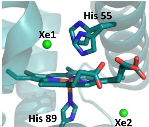
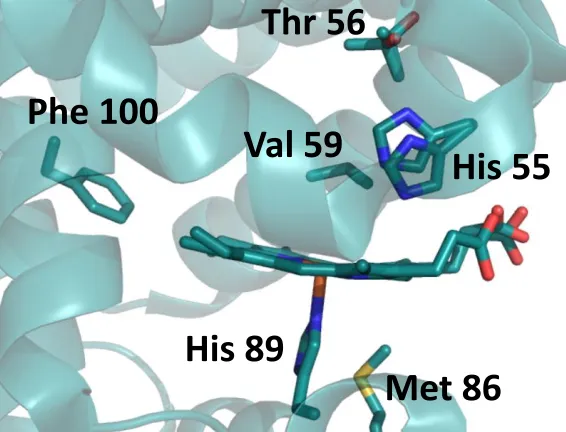
---Figure 1.4. Xe crystallographic binding sites observed in DHP.
Single amino acid variants of DHP A have been crystallized to determine the structural differences that correlate to kinetic variations in the peroxidase activity, as shown in Figure 1.5. The H55D mutant examined the role of the distal histidine, His55, in the activation and regulation of peroxidase activity given this residue’s role as a general acid-base that facilitates
O-O bond cleavage of bound H2O2 (vide infra). The T56S mutation was utilized to examine
neighboring amino acid dependency on the flexibility and function of the distal histidine 55.22 Mutations L100F and V59W investigated the role of residues inside the distal pocket and their effect on substrate and inhibitor binding.23 Mutations M86E and M86D introduced a proximal charge relay that is conserved in peroxidases but absent in globins. This latter study strove to address the structural difference but functional similarities of DHP to other peroxidases.14
Xe1
His 55
Figure 1.5. Sites of single amino acid mutations of DHP A, utilized previously for mechanistic and reactivity investigations.
1.5 Unusual Flexibility of the Distal Histidine: Structural and Spectroscopic Correlations X-ray crystallography has provided direct evidence supporting the spectroscopic assignment of enzymatic states, ligand occupation, and geometric orientation. Distal histidines are conserved features throughout both globin and peroxidase families, and are shown to play a crucial role in oxygen stabilization and enzymatic activity, respectively24–28. In globins, the distal histidine has been shown to reside in a closed, or internal, orientation when molecular oxygen is bound to the heme Fe, helping stabilize the bound ligand. In the absence of O2, the
distal histidine has been shown to reside in an open, or exposed, orientation. The environmental pH also is a factor in histidine orientation, with the open orientation dominating at lower pH. The protonation of histidine’s imidazole ring has been suggested as the key factor regarding
this pH-dependent distal histidine orientation.
Regarding peroxidase activity, the distal histidine promotes peroxide activation by serving as an acid and base for the cleavage of the peroxide O-O bond, according to the Poulos-Kraut push-pull peroxidase mechanism.29 The closed position is of adequate distance to promote
His 55
His 89
Phe 100
Thr 56
peroxide activation, while the open position is incapable of involvement due to the much greater distance. The heme Fe-distal histidine N distance for DHP A is 5.4 Å,30 which lies
intermediate between that for globins and peroxidases at 4.1 – 4.6 Å7,15,31 and 5.5 – 6.0 Å,12,13
respectively. This intermediate distance between two different classes of proteins may help explain the globin and peroxidase functional coexistance in DHP, but is not the sole deciding factor.
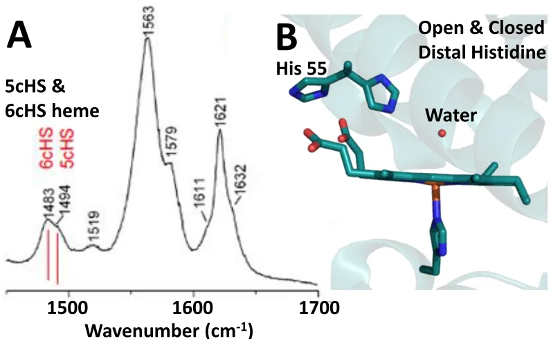
The flexibility of the distal histidine has been well documented in globins, however DHP exhibits this flexibility in a manner that is dictated by a number of different factors. The orientation of the distal histidine has been correlated to ligand occupation in the distal pocket. Smaller ligands, such as molecular oxygen and water, are stabilized by the closed orientation of the heme, whereas larger ligands, such as mono-, di- and trihalophenols, sterically push the distal histidine to the open orientation17,21,27,32,33. Through this correlation, the coordination state of the heme Fe can be directly related to the histidine conformation via spectroscopic means. In particular, resonance Raman (rR) spectroscopy possesses the ability to assess the coordination and spin states of the heme Fe. High frequency core marker bands for and denote the presence of the ferric heme iron in 5-coordinated high spin (5cHS)
and 6-coordinated high spin (6cHS) populations9,14,34 The presence of ferric 5cHS heme is observed at 1494 cm-1 (), 1568 cm-1 () and 1632 cm-1 (), while ferric 6cHS heme is
observed at 1481 cm-1 (), 1562 cm-1 (), and 1611 cm-1 () (Figure 1.6A). Through
analysis of these vibrational transitions, the coordination number of the Fe is determined. X-ray crystallographic structures have been obtained which further support the rR assignment of these bands14,20,22,34.
An equilibrium between 5cHS and 6cHS heme species exists in the metaquo state of DHP. Figure 1.6 shows the correlation between ferric DHP X-ray crystal structures and rR spectroscopy, both of which were obtained at room temperature. For each core marker band, and, there are populations represented by both 5cHS and 6cHS assigned frequencies.
Figure 1.6. A) Room temperature resonance Raman spectrum of WT DHP A. Core markers for 6cHS and 5cHS hemes are annotated for .37 B) Room temperature crystal structure (1EW6) emphasizing i) the water that occupies the Fe 6th coordination site, and ii) distal
histidine His55 in an equilibrium between the open (solvent exposed) and closed (internal) conformations.4
1.6 Inhibitor Binding Site Characterization
The inhibitor binding site has been well characterized within the distal cavity11,33,38–40 . Resonance Raman spectroscopy has shown the equilibrium shift toward a more dominant 5-coordinate high spin Fe species in the presence of para-halophenol inhibitors. Analysis of the core markers and clearly shows a population shift towards a 5cHS heme upon the
introduction of the para-halogenated phenol (Figure 1.7A). Inhibitors 4IP, 4BP, 4CP, and 4FP have all been shown crystallographically to be located above the heme Fe at a distance too great for ligation, in an orientation that is nearly perpendicular to the heme plane.4,37 The
halogen is directed toward the Xe1 binding site, with the phenolic O hydrogen toward the cavity entrance, within hydrogen bonding distance to Tyr38. Histidine 55 resides in the open, or solvent exposed, orientation in each structure. The ligand’s orientation inside the heme
5cHS &
6cHS heme
Open & Closed
Distal Histidine
His 55
Water
Wavenumber (cm
-1)
1500
1600
1700
cavity sterically prohibits the occupation of a 6th ligand for Fe, which supports the rR spectral analysis.
As the size of the halogen increases, inhibitor binding affinity increases as does the depth of the inhibitor within the distal cavity.37 Para-iodophenol possesses the strongest binding affinity and largest 5-coordinated population shift, and also coincides with the deepest penetration in the Xe1 binding site among the halogenated inhibitors analyzed. Panel B of Figure 1.7 provides the superposition of the 4 inhibitor crystallographic binding sites (3LB1), 4BP (6AOF), 4CP (6AO3) and 4FP (3LB4), and clearly shows the orientation and depth trend of the series.21,37 The 4IP and 4FP structures are complexed with DHP A with the 4BP and 4CP are in complex with DHP B. No isoenzyme effect is observed for the binding sites of p-halophenols.
Figure 1.7. A) Resonance Raman spectra of WT DHP A in the presence of the indicated inhibitor. [DHP A] = 100 M; [4IP] = 1 mM; [4BP], [4CP] and [4FP] = 8 mM.37 B) Superposition of the para-halogenated phenolic inhibitor structures depicting orientation within the distal pocket; 4IP (red, 3LB1), 4BP (green, 6AOF), 4CP (blue, 6AOE) and 4FP (purple, 3LB4).21
1.7 Peroxidase Substrate Binding Site(s) Characterization
Substrate binding is a key question that, if solved, could shed tremendous light onto numerous mechanistic questions. Figure 1.8A provides the rR spectra of WT DHP B with the traditional peroxidase substrates, 2,4,6-trihalogenated phenols, TXP (X = I, Br, Cl or F). Analysis of heme core marker bands does show a Fe coordination population shift that is halogen-dependent. However, the population shift is not nearly as pronounced as the monohalophenols, which suggests different binding sites between substrate and inhibitor. The smaller population shift in the substrate spectra suggests the binding site likely exists further from the heme center. When comparing the rR spectra of inhibitors with their corresponding halogenated substrates, in combination with NMR data, an external substrate binding site is realized as a possibility.40
The DHP B – TBP rR spectrum closely resembles the WT DHP B trace with a very slight perturbation. The addition of TCP to DHP B increases the 5cHS heme population slightly, whereas the addition of TFP greatly shifts the equilibrium toward the 6cHS heme species, most notably for and for both substrates. The TFP spectral shifts may be attributed to the smaller size of fluorine that prevents it from binding in the same manner as TBP and TCP. The possibility of an external substrate binding site is again suggested in the TFP spectra. External binding is postulated to enforce an allosteric effect on the enzyme, forcing the distal histidine into the closed position, thus increasing the 6cHS Fe population by stabilizing the distal ligand.24, 26 Another consideration is that TFP might ligate to the heme Fe itself, effectively increasing the 6cHS population by becoming the 6th ligand. Crystallographic structural
Figure 1.8. A) Resonance Raman core size marker bands of WT DHP B (100 M), and WT DHP B in the presence of TBP (200 M), TCP (3 mM) and TFP (4 mM).8 B) Crystallographic
binding site of TBP (4FH6).16 C) Internal and external binding sites of TCP in the DHP A (Y34N/S91G) mutant (4KN3).17 Sterically, it is shown that both TCP binding sites cannot be simultaneously occupied.
The DHP B – TBP rR spectrum closely resembles the WT DHP B trace with a very slight perturbation. The addition of TCP to DHP B increases the 5cHS heme population slightly, whereas the addition of TFP greatly shifts the equilibrium toward the 6cHS heme species, most
notably for and for both substrates. The TFP spectral shifts may be attributed to the smaller size of fluorine that prevents it from binding in the same manner as TBP and TCP. The possibility of an external substrate binding site is again suggested in the TFP spectra. External binding is postulated to enforce an allosteric effect on the enzyme, forcing the distal histidine into the closed position, thus increasing the 6cHS Fe population by stabilizing the distal ligand.24, 26 Another consideration is that TFP might ligate to the heme Fe itself, effectively increasing the 6cHS population by becoming the 6th ligand. Crystallographic structural
C
A
TBP
B
TCP
intanalysis, if and when it is obtained, will help resolve the TFP binding site enigma. The substrate binding affinity shows the same trend as the inhibitors, where the larger halogen results in the stronger binding.25 However, TCP does show a larger 5cHS heme population than TBP, which
could suggest different binding modes or provide further corroboration for the external binding site.
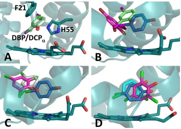
In 2013, binding locations of the traditional DHP peroxidase substrate, 2,4,6-trihalogenated phenols, were crystallographically identified. Notably, thirteen years passed between the first crystal structure of DHP (1EW6, 2000)4 and the first peroxidase substrate bound structure
(4FH6, 2013).17 The low aqueous solubility of TXP has been one of the reasons attributed to this difficulty, which also helps explains why the TBP occupancy is only 10% in this structure. The DHP A – TBP structure was obtained from the wild type protein, while the DHP A – TCP structures are all of mutants. Substrate solubility increases with the introduction of organic solvents, and both structures were obtained by soaking crystals in substrate enhanced precipitation solutions. Figure 1.8B provides the binding site of TBP, while Figure 1.8C shows the dual binding sites of TCP (TCPint & TCPext), all of which were complexed with DHP A
variants.
Both TCP and TBP have been resolved in the distal pocket, with slightly different results. Two different binding sites for TCP and one binding site for TBP have been observed. Both TCPint and TBP have been observed in an internal binding site near the -edge of the heme,
although the latter in low occupancy of 10%.27, 28 The problem of low occupancy is mainly attributed to the low solubility of TBP. In this internal binding site the substrate’s phenolic oxygen is located 3.3 Å and 3.7 Å to the heme Fe, for TCPint and TBP, respectively. There are
steric considerations when envisioning substrate binding congruent with successful hydrogen peroxide binding and resultant catalytic activation. For both TCPint and TBP structures, the
para-halogen is directed towards the Xe1 binding site, while the phenolic oxygen is positioned toward the Fe.
TCPext was also observed in a unique binding site close to the -edge of the heme.28 In this
toward the cavity entrance, within hydrogen bonding distances with Tyr38 and the heme propionate arm D. This second binding site is very similar in orientation and geometry to the inhibitor site, and therefore might perturb the Fe coordination number accordingly. The increased 5cHS heme population in the DHP B – TCP rR spectrum may also be attributed to this TCPext binding site. Both sites have been speculated as the productive substrate binding
site, while also speculated as inhibition sites. The question has yet to be fully resolved, and is still under debate.
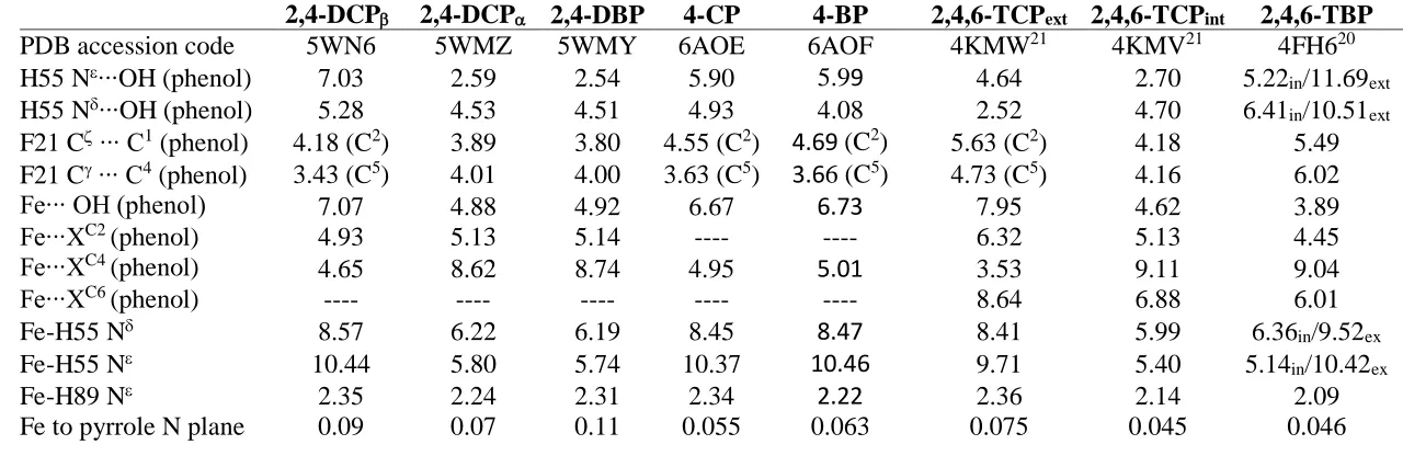
Table 1.2. Distances (Å) given for the heme Fe and the halogen and alcohol moieties of the inhibitors (4XP)33 and the substrates (TXP).16,17
Table 1.2 provides the crystallographic atomic distances between the heme Fe and halogen or phenolic O for the 4XP and TXP structures. It can be seen that the inhibitor distances are too large to be included in Fe’s coordination sphere, however their orientation above the Fe
could sterically prevent the simultaneous binding of a 6th ligand. The peroxidase substrate distances show a slightly different trend. They are still too large to be coordinated with the Fe yet are closer than the inhibitor distances, and they appear to be close enough to be affected by, or cause, electronic disturbances in the heme environment. In addition to TXP substrates, 2,4-dichlorophenol (DCP) and 2,4-dibromophenol (DBP) have been validated as peroxidase
Ligand
PDB accession
code
Fe-X dist (Å) (4XP)
Fe-OH dist (Å) (4XP)
Fe-OH dist internal (Å)
(TXP)
Fe-4X dist external (Å)
(TXP)
4IP 3LB1 5.1 6.4
4BP 6AOF 5.0 6.7
4CP 6AOE 4.9 6.7
4FP 3LB4 4.1 6.6
TBP 4FH6 3.7
substrates and their crystallographic binding sites have been determined. This is discussed in detail in Chapter 2.
1.8 Peroxygenase Substrate Binding Site(s) Analysis
1.8.1 - Haloindoles: 5-bromoindole and 7-bromoindole. DHP’s peroxidase mechanism has been shown to proceed through a radical pathway, which involves radical migration across several tyrosines.9,10,41 In regard to this, the peroxidase substrate need only be accessible to the migratory pathway, not necessarily the heme Fe. However, based on the mechanistic specifics of peroxygenase activity, the inserted oxygen must be derived from hydrogen peroxide. This implies that the substrate must bind in close proximity to the heme Fe in order to obtain the bound oxygen that originated from the heterolytic O-O bond cleavage of bound H2O2. The
acquisition of peroxygenase substrate distances will provide a more thorough examination of local structure-function relationships, and possibly ligand binding contributions.
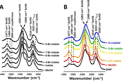
Spectral and computational data have been collected on the binding of haloindoles.5 The rR spectra of DHP – bromoindole complexes with the bromine atom located at different positions on the molecule is provided in Figure 1.9A. Through analysis of heme core marker bands and, it is observed that the 5cHS/6cHS Fe equilibrium is only perturbed when
the bromine is located at the 5-position of the indole ring, while the other brominated positional isomers showed no noticeable difference in the rR data. Interestingly, substrate conversion for 5- and 7-bromoindole are statistically equivalent at 48.1% (±2.3) and 46.1% (±1.7), respectively.5 The similar percent conversions with respect to the different rR spectra could be
Figure 1.9. Resonance Raman spectra of DHP B in solution with A) bromine-substituted indoles at different positions and B) 5-haloindoles of different halogens. [DHP] = 50M, [5-haloindole] = 500M.5
The geometry optimized calculated models of 5- and 7-bromoindole binding in the distal pocket are provided in Figure 1.10, panels A and B, respectively. Based on the similarities in the rR spectral data, the indoles were placed into crystallographic ligand binding sites to obtain the starting models. Since the presence of neither TBP nor 7BI yields substantial effects on the DHP rR spectral data, TBP from the crystal structure 4FH616 was replaced with 7BI to create the starting model for the MD calculations. Likewise, with respect to their similar perturbation of the DHP rR spectra, the 5BI starting model was obtained by replacing 4BP from the crystal structure 3LB2.33
1450 1500 1550 1600 1650
1 4 7 7 c m -1 6 c HS 1 4 9 4 c m -1 5 c HS 1 5 0 4 c m -1 o x y ferrous 1 6 1 0 c m -1 6 c HS 1 6 2 2 c m -1
νC=C
1 6 3 0 c m -1 5 c HS + 7-Br-indole + 6-Br-indole + 4-Br-indole + indole + MeOH + 5-Br-indole
Wavenumber (cm-1)
1 5 6 3 c m -1 6 c HS 1 5 6 9 c m -1 5 c HS
1450 1500 1550 1600 1650
14 77 cm -1 6c H S 1 4 9 4 c m -1 5 c HS 15 04 cm -1 o xy fer rou s 1 6 1 0 c m -1 6 c HS 1 6 2 2 c m -1
νC=C
1 6 3 0 c m -1 5 c HS + 5-Br-indole + 5-Cl-indole + 5-F-indole + MeOH DHP + 5-I-indole 1 5 6 3 c m -1 6 c HS 1 5 6 9 c m -1 5 c HS
Wavenumber (cm-1)
Figure 1.10. Geometry optimized structure for A) 5-bromoindole and B) 7-bromoindole.5
1.8.2 – 4-nitrophenol. The oxidation of 4-nitrophenol (4NP) as catalyzed by DHP has recently been characterized to proceed via a peroxygenase pathway.42 Monitoring the perturbation of the heme charge-transfer band at 615 nm showed 4NP to bind in the heme cavity with a dissociation constant (Kd) of 260 M. The crystallographic binding site of 4NP
correlates well with the spectroscopic and mechanistic data, showing the peroxygenase substrate to bind distal to the heme in a position that i) perturbs water ligation to the heme Fe, affecting the heme UV-vis absorption and ii) facilitates oxygen atom transfer due to its close proximity to the heme cofactor. Figure 1.11 provides the UV-vis spectral changes upon titration of 4NP, binding isotherm which provides the Kd value of 260 M, and crystallographic binding site. The crystallographic analysis of 4NP, in combination with its product 4-nitrocatechol, is presented in detail in Chapter 5.2.