Sequences With the Potential to Form Stem-and-Loop Structures Are Associated
With Coding-Region Duplications
in
Animal Mitochondrial DNA
David
J. Stanton,’
L. Lynne
Daehler, Craig C.
Moritz2
and Wesley M.
Brown
Department of Biology and Museum of Zoology, University of Michigan, Ann Arbor Michigan 48109-1048
Manuscript received March 19, 1993 Accepted for publication January 12, 1994
ABSTRACT
Tandem duplications of geneencoding regions occur in the mitochondrial DNA (mt DNA) of some individuals belonging to several species of whiptail lizards (genus Cnemidophorus). All or part of the duplicated regions of the mtDNAs from five different species were sequenced. In all, the duplication endpoints were within or immediately adjacent to sequences in tRNA, rRNA or protein genes that are
capable of forming energetically stable stem-and-loop structures. In two of these mtDNAs, the duplication
endpoints were also associated with a direct sequence repeat of 13 bp. The consistent association of stem-and-loop structures with duplication endpoints suggests that these structures may play a role in the duplication process. These data, combined with the absence of direct or palindromic repeats at three of the pairs of duplication endpoints, also suggest the existence of a mechanism for generating de novo duplications that is qualitatively different from those previously modeled.
S
IZE variations in animal mitochondrial (mt) DNA, although most often resulting from copy number variation of tandemly repeated sequences in noncoding regions (BROWN 1985; MORITZ et al. 1987; RAND andHARRISON 1989), sometimes result from direct tandem
duplications of coding regions (MORITZ and BROWN 1986, 1987; WALLIS 1987; MORITZ 1991; ZEVERING et al. 1991). Ten such duplications in mtDNAs from lizards in the genus Cnemidophorus were characterized by cleav-
age mapping (MORITZ and BROWN 1986, 1987). From
those data, 15 of 20 duplication endpoints appeared to be located within or adjacent to tRNA genes, supporting the hypothesis that tRNA genes can mediate mtDNA rearrangements (BROWN 1985). Further, 8 of the 10 du- plications had one end in or near a region consisting of the adjacent tRNAp“ and tRNATh‘ genes, identifylng it as a hot spot for duplication. However, these inferences were speculative, because they depended upon the as- sumption that mtDNA gene order in Cnemidophorus is the same as in most vertebrates and because the exact duplication endpoints had not been determined by se- quence analysis. In particular, it remained a possibility that the sequences mediating the duplications were near to, but not within, the tRNA genes. Also, MORITZ and BROWN (1987) had noted that five of the
20 endpoints
appeared to be in genes encoding proteins or rRNAs, rather than in or near tRNA genes. We have resolved all of these ambiguities by sequence analysis.Five of the Cnemidophorus duplications that would provide maximum information on junction sequences,
Present address: Department of Biology, Eastern Michigan University, Present address: Department of Zoology, University of Queensland, St. Ypsilanti, Michigan 48197.
Lucia, Queensland 4067, Australia. Genetics 137: 233-241 (May, 1994)
and thus on mechanisms of duplication, were chosen for sequence analysis. Attention was initally focussed on a 1.5-kb mtDNA duplication in order to test the hypoth- esized association of one of its endpoints with a tRNA gene and to characterize the other, which appeared not to be associated with a tRNA gene. This duplication had been found in mtDNAs from 13 of 32 individual Cnemidophorus uniparens (MORITZ and BROWN 1987). To locate precisely the endpoints and to confirm the gene order in this region, sequences from two individu- als of C. uniparens, one with a long ( L) , duplication- containing mtDNA and one with a standard length (S) mtDNA, were compared. Using primers designed from the C. uniparens sequence, mtDNA duplication junc- tions from four other species that had been mapped by restriction analysis (MORITZ and BROWN 1987) were also sequenced, thus providing identification of additional duplication endpoints. The endpointcontaining re- gions were examined for the presence of common se- quence features that might play a role in the duplication process.
MATERIALS AND METHODS
Details of specimen collection, mtDNA purification, and
cleavage mapping are published (MORITZ and BROWN 1986,
1987). Purified mtDNA and plasmid DNA vectors (Bluescript; Stratagene, Inc.) were digested with appropriate restriction
enzymes, ligated using T4 DNA ligase, and used to transform
Escherichia coli XL1 host cells (Stratagene, Inc.) made trans-
formation competent by the method of HANAHAN (1983).
Transformed colonies were screened for recombinant plas- mids using the X-Gal color system. Plasmid “minipreps” were prepared by alkaline lysis (KLEIN et al. 1980) in order to con-
firm insert identities. E . coli cells containing the desired
recombinant plasmids were stored as glycerinated stocks
234 D. J. Stanton et al.
STANDARD
/ I / I
-
X
E m rn
-
X
rn E
m
LONG t / I / Y I I
I
P*
T Cyt b E ND6 ND5
: S H ND4
according to MANIATIS et al. (1982) and purified by sedimen- tation equilibrium in CsCl gradients (BROWN 1981; WRIGHT et al. 1983) prior to sequencing.
Both strands of each cloned mtDNA were completely se- quenced using HATTORI and SAKAKI'S (1986) modification of the procedure of SANGER et al. (1977), in which double stranded template DNA is alkali denatured and annealed to single primers. The U.S. Biochemical Corp. Sequenase kit was used as the source of sequencing reagents. When necessary, 7deazadGTP was substituted for dGTP in order to resolve band compressions.
Primers complementary to conserved regions flanking the recombinant junctions were made with an AB1 model 380-B DNA synthesizer; the conserved regions were identified by comparisons among published mtDNA sequences. The primer sequences appear in the legend to Figure 5. Sequencing re- actions were performed according to the supplier's instruc- tions, using the Sequenase kit (U.S. Biochemical Corp.), and products were resolved by electrophoresis in 6 and 4% poly- acrylamide gels. Gels were dried, then exposed to X-ray film. For the C. uniparens sequence, each nucleotide was indepen- dently determined a minimum of six times from the three different copies indicated in Figure 1.
DNA sequences were amplified using the polymerase chain reaction (PCR) (Saiki et al. 1988). Each reaction contained approximately 10 ng of purified mtDNA in 100 pl of the fol- lowing mixture: 10 mM Tris-HC1 (pH 8.3); 50 mM KCl; 2.0 mM MgCl,; 0.01% gelatin; 2.0 mM each of the four dNTPs; 1.0 p~ each of two primers; and 1.0 unit of AmpliTaq DNA polym- erase (Perkin-Elmer Cetus). A thermal cycler (Perkin-Elmer Cetus) was used to carry out 30 cycles consisting of: 1 min at 94" for denaturation; 2 min at 37" for annealing; and 3 min at 72" for elongation. Products were phenol extracted and etha- nol precipitated, then treated with 5 units of the large (Kle- now) fragment of E. coli DNA polymerase I for 15 min at 30" to ensure the presence of blunt ends for ligation, after which they were cloned into plasmid vectors that had been digested with EcoRV, and sequenced. In the case of the Cnemidophorus opatae and Cnemidophorus exsanguis duplication junctions
(e.g., Figure 3, D and E), independent clones gave identical
junction sequences and no errors due to infidelity during PCR amplification were detected.
Sequence analysis was performed using resident programs in Eugene (MBIR) run on a Sun computer. Gene identities were inferred by comparisons with published sequences for house mouse (BIBB et al. 1981) and Xenopus laeuis (South
FIGURE 1 .-Cleavage maps of homologous re- gions in the standard and long C. uniparens mtDNAs, and the genetic map of the region. Dashed connecting lines indicate the portion of the standard mtDNA that is duplicated in long; the arrowheads indicate the endpoints of the duplicate copies C, and C,. Gene abbreviations: Cyt 6, cytochrome b oxidase apoenzyme; ND4-6, NADH dehydrogenase subunits 4-6; P, T, E, L,, S, and H, respectively, proline, t h r e e nine, glutamate, leucine (CUN), serine, and his- tidine tRNA; 6* and P*, partial duplications of ND6 and tRNAPro. All genes are transcribed right to left except P, Cyt b, E, and ND6. The shaded region at the left end of the genetic map is one end of the control (Dloop) region.
African clawed toad) (ROE et al. 1985) mtDNAs. Duplication endpoints were identified by alignment of the sequences flank- ing the internal duplication junction with corresponding se- quences from the C. uniparens S genome, or (for ND1 and rRNA) with appropriate regions of the Cnemidophorus maslini and C. exsanguis mtDNAs, respectively. Secondary structures for tRNA and rRNA gene sequences were inferred by com- parison to published secondary structure models (ROE et al. 1985; GLOTZ et al. 1981). Putative secondary structures within protein coding regions were inferred using a Macintosh ver- sion (supplied by D. GILBERT) of the program MulFold (version 2.0; JAEGER et al. 1989a,b; ZUCKER 1989). The free energies of DNA secondary structures were calculated using unpublished, empirically determined values generously provided by HEATHER D. MAYOR (Department of Microbiology, Baylor Col- lege of Medicine, Houston, Texas).
RESULTS
Gene arrangement in Cnemidophorus mtDNA: The partial gene arrangement in Cnemidophorus mtDNA
(Figure 1 )
,
was determined from the sequence shown inFigure 2 and from additional sequence data (D. J.
STANTON and W. M. BROWN, unpublished) that will be
reported separately. The gene arrangement in this re- gion is identical to those in other vertebrate mtDNAs
(BIBB et al. 1981; ANDERSON et al. 1982; ROE et al. 1985;
JOHANSEN et al. 1989), exclusive of birds (DESJARDINS and
Moms
1990; DESJARDINS et al. 1990).Duplication endpoints in C. uniparens: Comparison
of the C. uniparens L and S sequences shows that L
contains a tandem duplication extending from the
tRNAPro gene into ND6 (Figure 1). The duplicated se- quence is 1520 bp in length (Figure 2) and contains the
genes for cytochrome 6, tRNAnr, tRNAG'", and portions of
those for ND6 and tRNAp" (Figure 1). No direct or in-
verted repeats greater than 12 bp were found within or immediately adjacent to this sequence, nor were there any cases in which corresponding members of smaller (512 bp) repeats were located at each duplication endpoint.
Except for the redundancy resulting from the dupli-
C o n t r o l R.510” - - - > I [ ” ~ t R R A P Z O
T C T A G A G G A A A A G A G A T G A A T G B A C T A T T A T T A T A T G T A T A C A G C A G A A G A T A G T T T 6 0
t R N A P r o - - > I
A A A A A G A T A A A A T T T C G G T T T T G G G G A C C G G A G A T ~ G G G G G C C T C T T T T G A G G T G 120
I < - - t R N A T h r
TGGCAGAGACAGGAAAAGATTATTGCAAGGGTAAGGGGGTTCCCTTTTCCGGTTTACAAGG 180
< - - - t R K A T h r l I c - - - C y t b
CCGGCGTGTTATAGAATAATATACTATTGCAACAATaAGAGAGAAGTTTGTTTTCTATGG 240
T G G C T A T T A G G G G T A T A A G G A T A A G A A T A A T T G C G A A A T A G C T T A G C G A T G C G A T T T T T G 300
CTAGTGTAGCAAATGGTTCGTCEATTGGGGGAGCCTCCTAGTCAGGTAAGAAGAATGAGGT 360
T T G A A A T A A G A A T T C A A A A G A G A A T T T G G G A T A T T G G T C G G A A G G C A T G T G T T T G T T G T T 4 2 0
T T G A G G T G T G T A G T A T T G G G A T A A A T A G A A G A A T T A A G A T G G A G G C A A G A A G G G C T A A C A 4 8 0
C C C C T A C G A A C T T A T T T G G G A T A G A T C G C A G A A T T G C A T A G G C A A A T A G A A A A T A T C A T T 5 4 0
C T G G C T T G A T G T G T T T T G G G G T G G T T A T G G G G T T T G C T G G G A T G A A G T T T T C T G G G T C T C 6 0 0
C T A A T A T A T T A G G T G A A A A G A G G A C A A T A A T T A G G A A G A T T G T A A T G G C G A T G G C T A T T C 6 6 0
C A A A T A G G T C T T T G T A T A C T A A A T A G G G G T G A A A T A T A A T T T T A T C T G T G T T G G A G T T T A 720
A T C C A A G G G G G T T G T T T G A G C C T G T C T C G T G T A G T A T A A A T A G G T G T A A T A T T G A G A A A G 7 8 0
C G G C T A A G A T A A A A G G T A A T G T A A A G T G T A G T G T G A A A A A T C G G G T A A G G G T A G G G T T G T 8 4 0
C A A C A G A A A A G C C C C C T C A A A G C C A T T C A A C T A G G G T T T G T C C T A C A T A T G G G G T T G T T G 9 0 0
A T A G A A G G C T T G T A A T T A C T G T A G C A C C T C A G A A T G A T A T T T G G C C T C A T G G T A G A A T A T 9 6 0
A T C C C A T G A A G G C T G T T G C T A T T A G T A G G A A T A G T A G G A T G A C T C C A A T A T T T C A G G T T T 1 0 2 0
C T T T G T G G A A A T A A G A G C C G T A G T A A A G T C C T C G T C C A A T G T G T A G A T A A A T A C A A A T A A 1 0 8 0
A G A A T A T T G A C A G A C A A T T G G C G T G T A T T G T T C G T A G G A G T C A G C C A T A T T G T A C G T C T C 1 1 4 0
G A C A G A T A T T T G C T A C T G A T G A G A A G G C T A G T A G T A G T A T C T G C T G T G T A A T G T A T A G C T A 1 2 0 0
G A A A T A G T C C G G T T A G G A T T T G A A T A A T T A G T G T T A G G C C T A A T A A T G A G C C T A A G T T T C 1 2 6 0
A T C A G G C G G A T A T A T T T G A A G G T G T T G G T A A G A T A A T T A G T G A A T T G T T G A T A A T T T T T A 1 3 2 0
< - - - C y t b l I t R N A G l u - - - >
G T A A T G G G T G T G A T T T T C A T A T A T T G T G G G T C A T A T T T T T A T A G T T G A A T T A C A A C G G C A 1 3 8 0
t R N A G l u - - - r l [ N D 6 - - - >
G T T T T T C G T A T T G T T G G T C T T G G A T T G A A A G C C A A G T A A A A A T A A T G T A T G T T T T T T T T G 1 4 4 0
T A C T A A T T T T T T G T A T C G T G T T T G G T A C G A T T G G G G T A G C T T G T C A T C C T T C T C C C T T T T 1 5 0 0
T T G G T G C A G T T G G T T T A T T A T T T T G T G T A A T A G G G G G T T G T T T T T A T T T G T T A T C T C T T G 1 5 6 0
G A A T A A G T T T T G T T G G A G T G G T T T T A C T T T T A G T G T A T T T G G G A G G A G T T C T T G T A G T G T 1 6 2 0
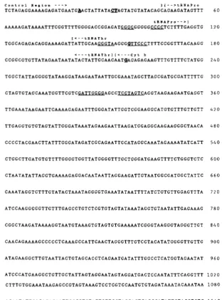
FIGURE 2.-A portion of the C. uniparens standard mtDNA sequence that includes the region duplicated in the C. uniparens long
mtDNA (see Figure 1). Positions 1-97 and
1620-1719 flank this region. Brackets above the sequence indicate gene boundaries, and arrows indicate the direction of transcription. Genes are abbreviated as in Figure 1. Nucle- otides at positions that differ between the standard and long mtDNAs, or between the two copies (C, and C,, Figure 1) in the long mtDNA are in boldface and underlined. The C, and standard sequences differ by three substitutions (T at position 23, C at 158, and
A at 217) and by the additional nucleotide (A) in C, between positions 33 and 34. The C, and standard sequences are identical, except that C , has 6 instead of 8 T’s on the interval
1432-1439; this results in a 2 bp shift of the reading frame in this (presumably inactive) partial copy of ND6. Sequences capable of forming potentially stable stem-and-loop structures (Figure 4) are underlined.
A T G C A T A T T C T A T T G C A T T G G C G T G T G A T G A A T A T T C T G A A A C T T G G G G A T C T C G T C C T G 1 6 8 0
236 D. J. Stanton et al.
A: C. uniparens (1.5 Kb duplication)
1 5 9 5 G T A T T T T G G G A G G A G T T C T T G T A G T G T A T G C A T A T A T T C T A T ~ G C A T T G G C G T 1 6 4 4
. . .
*
*
**
*
*
*
*
x ND6 G T A T T T T G G G A G G A G T T C T T G T A G T G G G C C C C T C T T T T G A G G T G T G G C A G A P r o* ***
****
*
*
*
*
* * * * * * * * * * x * * * * * * * * * * * * * * *7 7 G G T T T T G G G G A C C G G A G A T B G G G G G W T C T T T T G A G G T G T G G C A G A 1 2 7
B: C. inornatus (1.1 Kb duplication)
1 2 1 0 C G G T T A A G T T T G A A T A A T T A A T G T T A G G C ~ T A A T A A T ~ A G C C T A A G T T T C A 1 2 6 1
. . .
*
* *
*
C y t b C G G T T A A G T T T G A A T A A T T A A T G T T A G G C C C C C C T T T T G A G G T G T G G C A G A P r o7 7 G G T T T T G G G G A C C G G A G A T W G G G G G - T C T T T T G A G G T G T G G C A G A 1 2 7
* ** *
*
*
*******
* * * * * * * * * * * X * * * * * *C: C. maslini (4.9 Kb duplfcation)
1 2 9 G A C A G G A A A A G A T T A T T G C A A G G G T A A G G G G T T C C C T T T T C C G G T T T A C 1 7 7
Thr G A G A C A G G A A A T G T T A T T G C A A G G G T T G G A G T G T T C C T A T T G G G C C T A C A N D 1 A T A G T T T T A A G G G C G T C T G C G A T T G G T T G G A G T G T T C C T A T T G G G C C T A C A
x *
* *
*
*
. . .
* x*
x * * * **
* * * * * x * * * * * * ** * *
***
** * *
D: C. opatae (4.8 Kb duplications)
303 A G T G T A G C A A A T G G T T C G T C G A T T G G G G A G C C T C C T A G T C A G G T A A G A A G A 3 5 3
C y t b A G T G T G G C A A A T G G T T C G T C G A T T T G G G C T C T A T G A G G G G A T G G C G C T G T T 1 6 5 * x * * * * * * * * * * * * * x * * * * * * * * x *
*
*
* *
**
G G T C G T A A G C C C C G C T C G T C G A T ~ T G G G C T C T A A A G G G G A T G G C G C T G T T
*
******************
* * * x * * * * * * * * * * * * *E: C. exsanguis (4.8 Kb duplication)
303 A G T G T A G C A A A T G G T T C G T C G A T T G G G G A G C C T C C T A G T C A G G T A A G A A G A 353
C y t b A G T G T G G C A A A T G G T T C G T C G A T T T G G G C T C T A T G A G G G G A T C C G C G T G T T 1 6 s * * * x *
******************
***
*
* *
*
**
*
******************
x * * * * * * *****
G G T C G T A A G C C C C G C T C G T C G A T T T G G G C T C T A A G A G G G G A T G G C G C T G T TFIGURE 3.-Identifkation of duplication
endpoints. For each of the five duplications (A-E) , the sequence containing the junction between duplicate copies is shown between portions of either the C. uniparens standard
mtDNA sequence (A, B), numbered as in
Figure 2, or between a portion of that se- quence and a portion of the C . maslini ( C ) or C. exsanguis (D, E) mtDNA sequence.
Unnumbered sequences for C. maslini and
C. exsanguis were obtained by sequencing
PCR amplified regions of rntDNAs from
these species. Gene identities for the upper sequences are shown to the left of the middle sequences, and those for the lower se- quences are shown to their right. Genes are abbreviated as in Figure 1. Asterisks are placed between identical aligned nucleoti-
des. Sequences that are hypothesized to
form stable stem-and-loop structures (see
Figure 4) are underlined, Nucleotides at du- plication endpoints are in boldface; when an exact endpoint could not be determined, all nucleotides on the interval that must contain
the endpoint are in boldface. The two-
nucleotide gaps near the left ends of the u p
per and middle sequences in (C) have been
inserted for alignment purposes. The gaps are in an intergenic region that is highlyvari- able in length among vertebrates.
Within L, the duplicate copies (C, and C,, Figure 1) are also nearly identical. The most notable difference is the deletion in C, of two T’s from the run of eight T’s found in the interval 1432-1439 in both S and C, (Figure 2). This deletion disrupts the ND6 reading frame in C,.
Comparison of the junction between the tandem re- peats in the L sequence with S sequences corresponding to the ends of the duplicated region identifies the po- sitions of the endpoints to within ? l bp (Figure 3A), at positions 98 or 99 and 1618 or 1619 (Figure 2). One is in the “TC” loop of the tRNApr0 gene and the other is in the ND6 gene, 193 -+ 1 bp from its 5’ end (Figure 4A). No similarities were found between the sequences at
o r near the ends of this duplication. However, the region just beyond the endpoint in ND6 contains a 5-bp sequence separated from its inverse repeat by 8 bp (Figure
2).
These complimentary sequences can potentially interact to form a 5 bp stem with an 8 bp loop (Figure 4A), a structure whose formation is en- ergetically favorable (free energy = -3.7 kcal). Thus, although only one endpoint is within a tRNA gene,both are associated with potentially stable stem-and-
loop structures.
Duplication endpoints in other species: Sequences through four other duplication junctions (2. e . , those for duplications of 1.1 kb in
C.
inornatus, 4.9 kb in C . maslini, and 4.8 kb inC.
opatae andC.
exsanguis; M o m z and BROWN 1987) were also obtained. The se- quences of those junctions, the potential secondary struc- tures at or adjacent to their respective endpoints, and the locations of the duplicated regions on the mitochondrial genetic map are shown in Figures 3 , 4 and 5.For the 1.1-kb duplication in
C.
inornatus (Figure 3B), one endpoint is in the “TC” loop of the tRNApr0 gene, at a position on the interval 103-106 (Figure2)
that is from 1 to 5 bp from the correspondingC.
uni- parens duplication endpoint (Figures 3B and 4B). The other endpoint is near the 5‘ end of the cytochrome b gene, on the interval 1236-1239 (Figure 2); like theC.
uniparens duplication endpoint in the ND6 gene, it is associated with a potentially stable stem-and-loop struc-
4 8 c - G A - T
A 1 1 6
G : T A - T A - T
A A A A T A - T G
G - C
A T T T G C C C C G
A I l l : I I I I Q
G A A A T
G G G G G Q
A T A
T A T : G A G
C - G G - C G - C T - A
T G
T G
LEG
tRNAPro
4 8 c - G A - T G : T A - T A - T G - C
A 1 1 6
A AAA T T T G C C C C 0 0
G A T A A A A T G G G G G G
A I l l : I I I I G
TA
T: GAG
C - G G - C c - c T - A
T G
.~
T G
rn
tRNAPro
2 1 3 G : T
A 1 4 1
T - A T - A G - C C - G
A T T A A T A - T A - T
T T A T G C C C A T T C
T I I I I l l I C
c A C A C G G G A A C c
T A T A G AA
C - G A A C - G
G - C G - C
C A
T A
C A
TET
tRNAThr
TTT QQQ
A C
Q T
C T
T C
Q T
C A
T-A
G A
C - G
C - G C - G C - G
G T
C - G
A G
A - T
T:G G - C C - G G A G G T G T G T T A T
1 6 s rRNA
T C
T T
A A
T T
A - T C - G G-C T - A A - T
1614 G T A G T W TGGCGTG 1645
ND6 (-3.7 K c a l )
A A
ATA
T T
C- G
Q-C Q-C A - T T - A T - A 1227 ATTAGTG GTTTCAT 1262
Cyt b ( 5 ’ e I d ; -5.8 K c a l ) C.GA
G T
T G
A - T G-C G-C
T T
~~
C T C C G A T T W TIGGGCCTAC
T-A
ND1(5’ e n d ; -2.8 K c a l )
A C
G C
Q T
Q- C Q-C
Q T T-A T G A- T Q-C
316 G m A G G T M G 349
C y t b ( 3 ’ e n d ; -7.4 K c a l )
The endpoints of the 4.9-kb duplication in C. maslini are located within the tRNAThr and ND1 genes (Figure 3C). One endpoint is within the “TC” stem of the tRNAThr gene and the other is near the 5‘ end of ND1, immediately adjacent to a sequence capable of forming a stable stem-and-loop structure (Figure 4C).
The endpoints of the C. opatae and C. exsanguis du- plications could not be determined precisely, due to the presence of a nearly identical (12/13) 13-bp sequence found at the endpoint regions in both the cytochrome b and 16s rRNA genes. However, the endpoints for both species must be within or immediately adjacent to this 13-bp sequence (Figure 3, D and E). One endpoint is located near position 330 (Figure 2 ) , within or near a stem-and-loop structure near the 3’ end of the Cyt b gene (Figure 4D). The other is located within the 16s rRNA gene in a sequence that forms a stem-and-loop structure (Figure 4D); this structure is conserved in large subunit rRNAs from E . coli to man (GLOTZ et al.
Of the 10 duplication endpoints investigated, all are unambiguously associated with potentially stable stem- and-loop structures (Figure 4). Three of the endpoints are associated with the “TC” stem-and-loop of tRNA genes. However, their exact positions within this struc- ture are not constant; one is in the loop (Figure 4A), one is in the stem (Figure 4C), and one is in either the stem or the loop (Figure 4B). The most consistent feature shared by all five duplications is the presence of one endpoint immediately adjacent to the 3’ end of the D-loop region (Figure 5). It may also be noteworthy that there is a consistent association of the endpoints with runs of G and/or C nucleotides (Figure 3). Finally, in
two cases the endpoints were associated with stem- and-loop structures and a 13-bp direct sequence re- peat (Figure 4D).
1981).
FIGURE 4,”Potential secondary structures associated with duplication endpoints. All possible endpoint positions are in
boldface (see Figure 3). The sequences from which the pairs
of structures in A-C are formed correspond, respectively, to those found at the endpoints of the duplications whose re- combinantjunctions are shown in A-C of Figure 3; those in D correspond to both D and E of Figure 3, since those duplica- tion endpoints cannot be distinguished. Sequences are num- bered as in Figure 2. Values for the minimum energy structures shown were calculated by the MulFold program (JAEGER et al.
1989a,b; ZUCKER 1989), using empirically determined energy
values for DNA (H. ~ ~ A Y O R , unpublished data); no values were calculated for tRNA or rRNA structures. Dashes represent hy- drogen bonding between nucleotides; colons represent pair- ings with zero free energy. The structures were inferred by inspection and alignment with published vertebrate mito- chondrial tRNA and rRNA sequences (see, e.g., GLOTZ et al. 1981). All structures are based on C . uniparens standard
mtDNA sequences except those in the ND1 and 16s rRNA
238 D. J. Stanton et al.
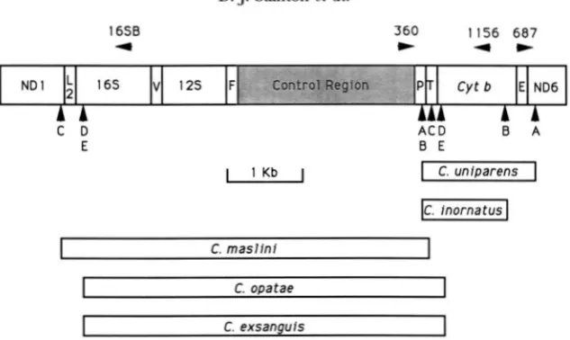
1658 4
360 1 I56 687
*
4 +C D A C D B A
E B E
I
1 K bI
IC.
fnornatusI
~~
I
C. opataeI
I
C. exsanguisI
FIGURE 5.-Duplication endpoints for Cnemidophorus mtDNA. Rectangles indicate the regions duplicated in the species in- dicated. The duplication endpoints for each of the five species (A-E) are indicated by vertical arrowheads; species designations correspond to those in Figure 3. Genes are abbreviated as in Figure 1 and as follows: NDl, NADH dehydrogenase subunit 1; 16s andlSS, respectively, large and small ribosomal subunit
RNA;
F,V,b,
respectively, phenylalanine, valine and leucine (UUR) tRNA.All genes are transcribed from left to right except P, E, and ND6. Locations of sequences correponding to primers are indicated
by horizontal arrowheads. Sequences (5' to 3') of the primer pairs that were used to PCR-amplify the recombinant junctions of duplications were: for B, 687 (AGCTAGAAATAGTCCGG) and 1 156 (GGATTAAACTCCAACAC) ; for C, D and E, 360 (GGAAAA-
GAGATGAATGTAC) and 16SB (GCCTGTlTACCAAAAACAT). The 16SB sequence was a consensus of aligned vertebrate mtDNA sequences. Note that primer orientation precludes amplification in the absence of a duplication.
DISCUSSION
This analysis of sequences both confirms and extends
the general conclusions drawn from the previous restric-
tion analysis of these duplications (MORITZ and BROWN
1987). It confirms that the assumed gene arrangement in this region of Cnemidophorus mtDNA and the gene content of the duplications are correct, and that three
of the duplication endpoints lie within the tRNApro and
tRNAnr genes. However, the sequence analyses also
show that two fewer endpoints actually lie within tRNA
genes than had been inferred in the previous study
(3/10 instead of 5/10). The most significant extension of the previous conclusions is that all 10 endpoints,
whether in tRNA, rRNA or protein genes, are either con-
tained within or less than 5 nucleotides from potential
stem-and-loop structures (Figure 4). In two cases, du-
plications that have arisen independently have identical or nearly identical endpoints, which indicates that the duplication mechanism(s) must act with a fair amount of precision.
Two assumptions underlying the conclusions about endpoints are that the five duplications analyzed r e p
resent five independent events and that the original end-
points have not been obscured by base substitutions, ad-
ditions or deletions subsequent to the duplication event.
Phylogenetic evidence addresses the issue of indepen- dent origins. The duplications most likely to be related
by common ancestry are those in C. uniparens (1.5 kb)
and C. inornatus (1.1 kb), which share a common end-
point in the tRNAPro gene (Figure 5). C. uniparens is a
hybrid parthenogenetic species whose mtDNA was in-
herited from C. inornatus (DENSMORE et al. 1989). It is
conceivable that the 1.5-kb duplication arose in C. in-
ornatus, was inherited by C. uniparens, then subse- quently reduced by deletion to 1.1 kb. This is unlikely,
however, because the C. inornatus populations to which
the maternal parent of C. uniparens most likely be-
longed (see DENSMORE et al. 1989) lack duplications, as
do most C . uniparens.
A more dramatic example of shared endpoints is pro-
vided by the 4.8-kb duplications in two other species, C.
opatae and C . exsanguis, both of which are partheno-
genetic hybrids (Figure 5 ) . Despite having identical or
nearly identical endpoints, these two duplications occur
only rarely in these distantly related (>lo% sequence
divergence) mtDNAs
(Momz
et al. 1992). The maternalparent species of C. opatae is C. inornatus (DENSMORE
et al. 1989) and that of C. exsanguis is either C. costatus
or a closely related species,
C .
burti( M o m
et al. 1989).The maternal parent species are not closely related to each other, and this duplication has not been found in other species closely related to either of them. Thus, it
seems most unlikely that the two duplications arose in
and were inherited from a common ancestor; that the two duplications arose independently is a far more par- simonious explanation. Although both duplications must end within or immediately adjacent to the same 13bp sequence (Figure 3, D and E), a more precise determination of their endpoints is impossible.
The possibility that the junction sequences have been
modified by deletions cannot be evaluated with these data. Deletions within duplicated segments of mtDNAs
have previously been reported in Cnemidophorus
1991; ZEVERING et al. 1991). Such deletions would be impossible to detect, however, if they were immediately adjacent to the internal junction of the duplication.
Sequence elements and endpoints Directly repeated
sequences are associated with both deletions (QUIGLEY
and WEIL 1985) and inversions (HOW et al. 1988) in
chloroplast DNA, and with several large deletions in hu-
man mtDNA (HOFFNER et al. 1989; SCHON et al. 1989;
DEGOUL et al. 1991). In theory, such sequences could
also promote duplication, but none of those found in or
near the
C.
uniparens duplication (Figure 2) or at thejunctions between duplicated regions in
C.
inornatusand C. maslini (Figure 3, B and C) suggest an involve-
ment in the duplication process. However, the 13-bp
direct repeat that is present at the endpoints in both
C.
opatae and
C.
exsanguis may have played a role in the formation of those two duplications.Pairs of palindromic and quasipalindromic sequences
capable of complementary base pairing often occur near duplication and deletion endpoints. Slipped-strand mi- spairing of such sequences during DNA replication has been postulated as the initiating mechanism for such
events (RIPLEY and GLICKMAN 1983; GLICKMAN and RIPLEY
1984). However, no such sequences were found at or
near the duplication endpoints in C. uniparens mtDNA
(Figure 2) or associated with the duplication junctions in the mtDNAs of the four other species examined.
Based on the analysis of the sequences presented and
on our previous cleavage mapping data (MORITZ and
BROWN 1987), the most reasonable overall hypothesis is
that there is a duplication process in Cnemidophorus mtDNA that is mediated by stem-and-loop structures. The stable formation of these stem-and-loop structures
in duplex DNA ( e . g . , by transformation of linear helices
to cruciform structures) is improbable, based on free energy considerations. Their stable formation in single-
stranded DNA is much more probable. In animal
mtDNA, a portion of the molecule is single-stranded for
a significant period during replication (reviews by
CLAWON 1982; BROWN 1985). As previously noted, all du-
plications either begin near one end of or include the control (D-loop) region, where mtDNA replication is
initiated (MORITZ and BROWN 1987). Thus, one mtDNA
strand in the region within which the duplications occur
is single-stranded for a considerable portion of the rep- lication cycle. This provides an opportunity for struc- tures with intrastrand base-pairing to form and to persist for a relatively long period.
The ability of tRNA genes to mediate rearrangement
events in DNA is well documented. In bacteria (REITER
et al. 1989), slime molds (MARSCHALEK et al. 1989), and
yeast (SANDMEYER et al. 1988; CHALKER and SANDMEYER
1990), tRNA genes appear to function as genomic land- marks for the site-specific integration of many different types of genetic elements. In chloroplasts, tRNA genes are associated with rearrangements and inversions
(QUIGLEY and WEIL 1985; HOW et al. 1988). In animal
mtDNA, gene order comparisons suggest that tRNA genes rearrange at a higher frequency than protein or
rRNA genes (BROWN 1985; WOLSTENHOLME and CLARY
1985; CANTATORE et al. 1987; MORITZ et al. 1987). Thus,
the association of tRNA genes and, by extension, stem-
and-loop structures with a rearrangement-like process in
animal mtDNA is not without precident.
Duplication mechanism: Although these results sug- gest that stem-and-loop structures might mediate se- quence duplication in mtDNA, the duplication mecha- nism remains obscure. Duplications could arise by
intermolecular recombination, transposition, or
slipped-strand mispairing during replication. Although not thoroughly studied, recombination appears to be at
most, infrequent in animal mtDNA (BROWN 1983); if
present, however, even a low frequency of recombina- tion might suffice to produce duplications. Transposi- tion is an unlikely mechanism, because it is improbable that transpositions would produce tandem duplications exclusively, and because the duplication sequences ex- amined lack the terminal direct repeats that are char-
acteristic of most transposable sequences (CALOS and
MILLER 1980). Slipped-strand mispairing (LEVINSON and
GUTMAN 1987) has been invoked to account for size
variation in mtDNA [see references in MORITZ et al.
(1987) and MORITZ (1991)l. RAND and HARRISON (1989)
found seven mtDNA size classes resulting from differ- ences in copy number of a tandemly repeated sequence in crickets. They postulated that these differences could have resulted from slipped-strand mispairing, or from an unknown process mediated by a 1 4 b p sequence with dyadic symmetry that was found at the repeat termini. The 13-bp direct repeat found at the duplication end-
points in
C.
opatae and C. exsanguis is compatible witha slipped-strand mispairing hypothesis. However, be- cause no such sequences are present at the duplication termini in the other Cnemidophorus mtDNAs, and be- cause higher order variation in copy number has not been observed in any Cnemidophorus mtDNAs (MORITZ
and BROWN 1987), the present data do not support
slipped-strand mispairing as a universal mechanism. A stem-and-loop structure appears to serve as the sig- nal for the enzyme that initiates Lstrand synthesis dur-
ing mtDNA replication (WONG and CLAWON 1985,1986;
HIXSON et al. 1986). These structures also appear to serve
as the signals that mediate processing of the polygenic
mtRNA transcript into its components (BAmYand CLAY-
TON 1980; OJALA et al. 1981). Thus, similar structures,
even those arising by chance, might be accidentally rec-
ognized by these or other enzymes and initiate a process
that occasionally leads to tandem sequence duplication.
The structures might also function in combination with
other sequences or sequence characteristics, such as the
runs of G and/or C that are associated with all ten of the
240 D. J. Stanton et al.
structure/sequence complex might be mistakenly rec- ognized by an enzyme such as endonuclease G, which appears to be capable of generating the primers re- quired to initiate replication of mtDNA. This enzyme is known to cleave DNA at double stranded (dG) (dC) and at single stranded (dC) sequences (COTE and RUIZ- CARRILLO 1993), like those associated with the duplica- tion endpoints.
These results have relevance that goes beyond dupli- cation in mtDNA alone. The presence of small palin- dromic or repeated sequence elements (either direct or inverted) has been a common feature of previous mod- els that have been invoked to explain de novo sequence duplication. The evidence presented here indicates that there must also be a second, previously undetected du- plication mechanism that does not require those ele- ments, but requires stem-and-loop structures instead.
Finally, the presence of both unduplicated and du- plicated mtDNA sequences in individuals of the same species also provides opportunities for understanding processes of genetic change within the mitochondrion and, perhaps, some of the events that may be involved in the evolution of mitochondrial gene rearrangements
(BROWN 1985).
We thank H. MAYOR for supplying unpublished energy values for DNA secondary structures, and J. BOORE, T. COLLINS, S. HOFFMAN, E. T. SNOW and W. K THOMAS for comments on the manuscript. This work was supported, in part, by grants from the U.S. Public Health Service
(GM30144) and the National Science Foundation (BSR-8517830).
LITERATURE CITED
ANDERSON, S., M. H. L. DEBRUIJN, A. R. COULSON, I. C. EPERON, F. SANGER
et al., 1982 Complete sequence of bovine mitochondrial DNA. J. Mol. Biol. 156: 683-717.
BATTEY, J.. and D. A. CLAWON, 1980 The transcription map of human mitochondrial DNA implicates transfer RNA excision as a major processing event. J. Biol. Chem. 255: 11599-11606.
BIBB, M. J., R. A. VAN ETTEN, C. T. WRIGHT, M. W. WALBERG and D. A.
CLAYTON, 1981 Sequence and gene organization of mouse mi- tochondrial DNA. Cell 26: 167-180.
BROWN, W. M., 1981 Mechanisms of evolution in animal mitochon- drial DNA. Ann. N.Y. Acad. Sci. 361: 119-134.
BROWN, W. M., 1983 Evolution of animal mitochondrial DNA, pp.
62-88 in Evolution of Genes and Proteins, edited by M. NEI and R. K. KOEHN. Sinauer, Sunderland Mass.
BROWN, W. M., 1985 Evolution of the animal mitochondrial DNA genome, pp. 95-130 in Molecular Evolutionary Genetics, edited by R. MACINTIRE. Plenum Press, New York.
CALOS, M. P., and J. M. MILLER, 1980 Transposable elements. Cell 2 0
579-595.
CANTATORE, P., M. N. GADALETA, M. ROBERTI, C . SACCONE and A. C. WIL- SON, 1987 Duplication and remoulding of tRNA genes during the evolutionary rearrangement of mitochondrial genomes. Nature 329: 853-855.
CHALKER, D. L., and S. B. SANDMEYER, 1990 Transfer RNA genes are genomic targets for de novo transposition of the yeast retrotrans- poson Ty3. Genetics 126: 837-845.
CLAYTON, D. A,, 1982 Replication of animal mitochondrial DNA. Cell
COTE, J., and A. RUIZ-CARRILLO, 1993 Primers for mitochondrial DNA replication generated by endonuclease G . Science 261:
DEGOUL, F., I. NELSON, S. AMSELEM, N. ROMERO, B. OBERMAIER-KUSSER rt al., 1991 Different mechanisms inferred from sequences of human mitochondrial DNA deletions in ocular myopathies. Nucleic Acids Res. 19: 493-496.
28: 693-705.
765-769.
DENSMORE, L. D., C. MORITZ, J. W. WRIGHT and W. M. BROWN, 1989 Mi- tochondrial DNA analyses and the origin and relative age of par- thenogenetic lizards (genus Cnemidophorus). N . Nine
Sexlineatus-group unisexuals. Evolution 43: 969-983.
DESJARDINS, P., and R. MORAIS, 1990 Sequence and gene organization of the chicken mitochondrial genome. J. Mol. B~ol. 212: 599-634.
DESJARDINS, P., V. RAMIREZ and R. MORAIS, 1990 Gene organization of
515-518.
the Peking duck mitochondrial genome. Curr. Genet. 17:
GLICKMAN, B. W., and L. S. RIPLEY, 1984 Structural intermediates of deletion mutagenesis: A role for palindromic DNA. Proc. Natl. Acad. Sci. USA 81: 512-516.
GLOTZ, C., C. ZWIEB and R. BRIMACOMBE, 1981 Secondary structure of the large subunit ribosomal RNA from Escherichia coli, Zea mays
chloroplast, and human and mouse mitochondrial ribosomes. Nucleic Acids Res. 9: 3287-3306.
HANAHAN, D., 1983 Studies on transformation of Escherichia coli with plasmids. J. Mol. Biol. 166: 557-580.
HATTORI, M., and Y. SAKAKI, 1986 Dideoxy sequencing method using denatured plasmid templates. Anal. Biochem. 152: 232-238.
HIXSON, J. E., T. W. WONG and D. A. CLAWON, 1986 Both the con- served and divergent 5"flanking sequences are required for ini- tiation at the human mitochondrial origin of light strand repli- cation. J. Biol. Chem. 261: 2384-2390.
HOFFNER, J. M., M. T. L o n , A. S. VOLJAVEC, S. A. SOUEIDAN, D. A. COSTI- CAN et al., 1989 Spontaneous Kearns-Sayre/chronic external opthaloplegia plus syndrome associated with a mitochondrial DNA deletion: a slipreplication model and metabolic therapy. Proc. Natl. Acad. Sci. USA 86: 7952-7956.
HOW, C. J., R. F. BARKER, C. M. BOWMAN and T. A. DYER, 1988 Com- mon features of three inversions in wheat chloroplast DNA. Curr. Genet. 13: 343-349.
JAEGER, J. A,, D. H. TURNER and M. ZUCKER, 1989a Improved predic- tions of secondary structures for RNA. Proc. Natl. Acad. Sci. USA
JAEGER, J. A,, D. H. TURNER and M. ZUCKER, 198913 Predicting optimal and suboptimal secondary structure for RNA. Methods Enzymol.
JOHANSEN, S., P. H. GUDDAL andT.JoHANsEN, 1989 Organization of the
mitochondrial genome of Atlantic cod, Gadus morhua. Nucleic Acids Res. 18: 411-419.
KLEIN, R. D., E. SELSING and R. D. WELLS, 1980 A rapid microscale technique for isolation of recombinant plasmid DNA suitable for restriction enzyme analysis. Plasmid 3: 88-91.
LEVINSON, G., and G. A. GUTMAN, 1987 Slipped-strand mispairing: a major mechanism for DNA sequence evolution. Mol. Biol. Evol. 4 203-221.
MmIATIs, T., E. F. FRITSCH and J. SAMBROOK, 1982 Molecular Cloning:
A Laboratory M a n u a l . Cold Spring Harbor Laboratory, Cold Spring Harbor, N.Y.
MARSCHALEK, R., T. BRECHNER, E. AMON-BOHM and T. DINGERMANN, 1989
Transfer RNA genes: landmarks for integration of mobile genetic elements in Dictyostelium discoideum. Science 244: 1493-1496.
MORITZ, C., 1991 Evolutionary dynamics of mitochondrial DNA duplications in parthenogenetic geckos, Heteronotia binoei.
Genetics 129: 221-230.
MORITZ, C., and W. M. BROWN, 1986 Tandem duplication of D-loop and ribosomal RNA sequences in lizard mitochondrial DNA. Science 233 1425-1427.
MORITZ, C., and W. M. BROWN, 1987 Tandem duplications in animal mitochondrial DNAs: variation in incidence and gene content among lizards. Proc. Natl. Acad. Sci. USA 84: 7183-7187.
MORITZ, C., T. E. DOWLING and W. M. BROWN, 1987 Evolution of ani- mal mitochondrial DNA Relevance for population biology and systematics. Annu. Rev. Ecol. Syst. 18: 269-292.
MORITZ, C., J. W. WRIGHT and W. M. BROWN, 1989 Mitochondrial DNA analyses and the origin and relative age of parthenogenetic lizards (genus Cnemidophorus). 111. C . veloxand C. exsanguis. Evolution
MORITZ, C., J. W. WRIGHT and W. M. BROWN, 1992 Mitochondrial DNA analyses and the origin and relative age of parthenogenetic
Cnemidophorus: phylogenetic constraints on hybrid origins. Evolution 46: 184-192.
OJALA, D., J. MONTOVA and G. ATTARDI, 1981 tRNApunctuation model of RNA processing in human mitochondria. Nature 290:
470-474.
8 6 7706-7710.
183: 281-306.
QUIGLEY, F., and J. H. WEIL, 1985 Organization and sequence of five tRNA genes and of an unidentified reading frame in the wheat chloroplast genome: evidence for gene rearrangements during the evolution of chloroplast genomes. Curr. Genet. 9: 495-503.
RAND, D. M., and R. G. HARRISON, 1989 Molecular population ge- netics of mtDNA size variation in crickets. Genetics 121: 551-569. REITER, W., P. PALM and S. YEATS, 1989 Transfer RNAgenes frequently
serve as integration sites for prokaryotic genetic elements. Nucleic Acids Res. 17: 1907-1914.
RIPLEY, L. S., and B. W. GLICKMAN, 1983 Unique self-complementarity of palindromic sequences provides DNA structural intermediates for mutation. Cold Spring Harbor Symp. Quant. Biol. 47:
851-861.
ROE, B. A,, D. MA, R. IC WILSON and J. F. H. WONG, 1985 The complete nucleotide sequence of the Xenopus laeuis mitochondrial ge- nome. J. Biol. Chem. 260: 9759-9774.
SAIKI, R. R, D. H . GELFAND, S. STOFFEL, S. J. SCW, R. HIGUCHI et al., 1988 Primerdirected enzymatic amplification of DNA with a thermostable DNA polymerase. Science 2 3 9 487-491.
SANDMEYER, S. B., V. W. BILANCHONE, D. J. CLARK, P. MORCOS G. F. CARLE et al., 1988 Sigma elements are position-specific for many dif- ferent yeast tRNA genes. Nucleic Acids Res. 16: 1499-1515. SANGER, F., S. NICKLEN and A. R. COUISON, 1977 DNA sequencing with
chain-terminating inhibitors. Proc. Natl. Acad. Sci. USA 7 4
5463-5467.
SCHON, E. A., R. RIZZUTO, C. T. MOWS, H . NAKASE, M. Z E ~ I et al.,
1989 A direct repeat is a hotspot for largescale deletion of hu- man mitochondrial DNA. Science 244: 346-349.
WALLIS, G. P., 1987 Mitochondrial DNAinsertion polymorphism and germ line heteroplasmy in the Triturus cristatus species complex. Heredity 58: 229-238.
WOLSTENHOLME, D. R., and D. 0. CLARY, 1985 Sequence evolution of
Drosophila mitochondrial DNA. Genetics 1 0 9 725-744. WONG, T. W., and D. A. CLAYTON, 1985 Zn vitro replication of human
mitochondrial DNA Accurate initiation at the origin of light strand synthesis. Cell 4 2 951-958.
WONG, T. W., and D. A. CLAKON, 1986 DNA p r i m a e of human mi- tochondria is associated with structural RNA that is essential for enzymatic activity. Cell 45: 817-825.
WRIGHT, J. W., C. SPOLSKY and W. M. BROWN, 1983 The origin of the parthenogenetic lizard Cnemidophorus laredoensis inferred from mitochondrial DNA analysis. Herpetologica 39: 410-416. ZEVERING, C. E., C. MORITZ, A. HEIDEMAN and R. A. STRUM, 1991 Par-
allel origins of duplications and the formation of pseudogenes in mitochondrial DNA from parthenogenetic lizards (Heteronotia
binoei; Gekkonidae). J. Mol. Evol. 33: 431-441.
ZUCKER, M., 1989 O n finding all suboptimal foldings of an RNA mol- ecule. Science 244: 48-52.