Medical Image Analysis – A Review
A.Sivaramakrishnan
1, Dr.M.Karnan
2, Dr.R.Sivakumar
31
Assistant Professor, Department of Computer Application, Karunya University, India
2
Professor and Head, Department of Computer Science and Engineering, Tamilnadu College of Engineering, India
3
Professor, Department of Information Technology, Tamilnadu College of Engineering, India
Abstract— In this survey work various automatic detection
methods of microcalcifications and brain tumor through
mammograms and MRI has been studied and compared for
the period of more than two decades. This is used to focus on
the future of developments of medical image processing in
medicine and healthcare. We have described several methods
in medical image processing and to discussed requirements
and properties of techniques in tumor detection. This work is
used to give more information about tumor detection and
segmentation. It is a milestone for analyzing all technologies
relevant to tumor from mammogram and MRI in Medical
image processing. In this work, various steps in detection of
automatic detection :i) The Preprocessing and Enhancement
Technique ii) Segmentation Algorithm iii) Feature Extraction
iv) Classification v)Performance Analysis using Receiver
Operating Characteristics and their performance have been
studied and compared.
Keywords— MRI, mammogram, Enhancement, Feature
Extraction, Receiver Operating Characteristics.
I.
I
NTRODUCTIONIn this chapter, methods of automatic detection of
tumour in digitized MRI and mammograms used in various
stages of intelligent systems for detection of masses and
brain tumour are summarized and compared. In particular,
the preprocessing and enhancement, segmentation
algorithms, feature extraction, selection and classification,
classifiers, receiver operating characteristics curve analysis
and their performance are studied and compared.
II.
E
NHANCEMENTA
NDP
REPROCESSINGSeveral authors have suggested various techniques for
preprocessing and enhancement in the last two decades.
The task of medical image enhancement is to sharpen the
edges to increase the contrast between suspicious regions
and the background. Image enhancement includes intensity
and contrast manipulation, noise reduction, background
removal, edges sharpening, filtering, etc. Table 2.1 and 2.1a
shows the overview of enhancement techniques for
mammogram and MRI
Table 2.1 An overview of enhancement techniques for
mammogram
Method Description
median filter(Lai et
al 1989)
This filter can remove the noise without
significantly distorting the signal.
Central Weighted
Median Filter (Qian
et al 1994)
A CWMF with a large central weight
preserves more image detail but
suppresses less noise than a filter with a
smaller central weight.
Method Description
Model-based, scatter
function (Highnam et
al 1994)
A weighting mask has been calculated
which represents the percentage of the
total scatter reaching the central pixel
and coming from the column of Lucite
above each pixel in a neighborhood.
First derivative and
the local statistics
(Kim et al1997)
The adaptive image enhancement
method exploits the first derivative
operations using the Sobel operators or
the compass operators and the local
statistics of a mammographic image are
used for an adaptive realization.
Fractal modeling (Li
et al 1997)
The key point of fractal modeling is to
explore the self-similarity property of
images.
Median
filter[Thangavel and
karnan 2005]
Computer aided detection of
microcalcification in digitized
mammogram using median filter and
genetic algorithm.
Fuzzy logic
(Kovalerchuk et al
1997)
Fuzzy logic has the potential of opening
a new and promising direction for
effective and early breast cancer
diagnosis.
Wavelet transform,
multiscale features
(Chang and Laine
1999)
Wavelet transform, multiscale features,
Coherence measure and dominant
orientation clearly improved
discrimination of features from complex
surrounding tissue and structure in dense
mammograms.
Filtering tech
(Kobatake et al 1999)
This filter output for the tumor is very
high and its region is well isolated from
its background.
Region based
Enhancement (Ferrari
et al 1999)
Region based contrast enhancement uses
each pixel as a seed to grow a region.
Applying an empirical transformation
based on each region’s seed pixel value,
its contrast and its background enhances
contrast.
Wavelet,
Morphological
operation (Cordella et
al 1999)
Fractal approach compared with the
partial wavelet reconstruction and the
morphological operation approaches.
Unsharp Masking,
Sobel Operators
(Bhangale et al 2000;
Enderwich and
Tzanakou 1997]
The Unsharp masking method reduces
the low frequency information while
amplified the high frequency detail.
Adaptive noise
equalization
(Veldkamp and
Karssemeijer 2000)
It gives much better results than does a
fixed noise equalization, probably
because noise characteristics are
mammogram dependent, caused by
variation of film type and film
development characteristics.
Method Description
and sub-sampling
(Mudigonda et al
2001)
intensity patterns of mass regions to
form smooth hills with respect to their
surroundings in the low resolution image
and help in estimating the approximate
extent of isolated regions present in the
image.
Quantum noise
assumption (Rogova
et al 1999)
If quantum noise is assumed the
dominant noise source present, a square
root model will provide an accurate
estimate of the noise with respect to gray
level.
Matched filtering
(Bocchi et al 2004)
In particular, Fractional Brownian
Motion (FBM) can model non-stationary
random fields with stationary
increments. In addition, a stationary
power spectrum can be attached to
FBMs leading to an approximate
implementation of the enhancement filter
via conventional matched filtering.
Table 2.1.a An overview of enhancement techniques for
MRI
Methods Description
Oliver et al (2005)
Standard Imaging Protocol
MRIs have been acquired in
the standard follow-up.after
surgical resection.
Dana et al (2007)
Statistical Parametric
Mapping,
Pipe line Approach
It provides the solution of
noise reduction, Inter-slice
intensity variation correction,
Intra-volume bias field
correction
Jayaram et al (2002)
Content Based model,Shape
based,Texture based
technique, Histogram and
Profilling Method
It showed detections of tumor
with decrease in pixel count in
binary images, increase in
image intensity, High numbers
of high intensity pixel.
Tracking algorithm [Jaya et
al 2009]
De-noising of MR brain
images using the tracking
algorithm .
Elizabeth et al (2005)
Pixel Histograms,
Morphological Process
It was more robust to noise and
it can improve the integrity
performance.
Leung et al (2003)
Boundary Detection
Algorithm, Generalized Fuzzy
operator(GFO),
To obtain the fine result in the
tumor consideration.
Zu et al (2004)
Histogram
based(HB),Sub-second imaging technique
Separate brain image, from
head image removal of residual
fragments such as sinus,
cerebrospinal/fluid, dura,
marrow.
Gray (1997)
Neural Networks, Genetic
Programming
Large volume of data
processed successfully.
Mark et al (2005)
Statistical Parametric
Mapping Method
It is used to align the image
properly and it uses
left-to-right symmetry to confer
robustness to areas of
abnormality.
Toshiharu et al (2003)
Independent Component
Analysis(ICA)
Separate the components in
MR images
Farahat et al (2006)
Head Model, Finite
Difference
Time-It is used to analyse different
Tissue types.
Methods Description
Domain(FDTD)
Kyeong et al (2004)
Discrete Wavelet
Transform(DWT)
DWT provides higher intensity
images than other.
Lim et al (1989)
Stripping algorithm
To remove the skull and scalp
portions from each axial
section.
Thangavel et al (2005)
Gradient-Based Method,
Median Filter, Normalization
Method
Shows the validity of detection
of Memmographic lesions.
Chunyan et al (2004)
Triple Quantum Filtered
Sodium MRI (TQF)
Technique
Detects neoplastic changes in
the brain before angiogenesis
and blood brain barrier (BBB)
breakdown develop.
Tsai et al (1995)
Low pass Filter
Takes care of local noisy
fluctuations from MR images.
Boada et al (2004)
Triple Quantum Filtered
(TQF) Sodium NMR
Minimizes the effects of extra
cellular fluids and Found
Non-Contrast Enhancing tissue
Aria et al (2002)
Gadolinium-Diethylenetriaminepentaacetic
acid (Gd
DTPA)Enhancement
Provides additional
independent information and
improve the accuracy.
Boada (2004)
Novel image Approach
Earlier detection of
non-contrast enhancing tissue.
Amini (2003)
Prewitt edge-finding filter
This filter enhances the tumor
tissue greatly.
Zhe chen (2003)
Morphological Filter
It is used to remove
background.
Corina et al (2005)
Gaussian Filter
Enhances image Boundaries.
Dimitris et al (2006)
Gabor Filter Bank
technique
It is used to remove the tagging
lines and enhance the
tag-patterned region.
Hideki et al (1990)
V-filter
Enhances the image by
smoothing the noise gray level
distribution while retaining the
edge.
Gordon et al (2006)
Anisotropic sample
Enhances the utility of
glyph-based tensor visualization.
Shishir et al (2006)
Non linear Filter
Non – Contrast enhancing
Brain Volumes are linearly
aligned.
Salman et al (2005)
Region Growing Filter
It is usually convenient to
preprocess the image by using
a noise reduction filter.
Sean et al (2001)
K-nearest neighbour
Algorithm
It generates enhancement data
volumes. These are highly
correlated with manually
defined standard.
Michael et al (1988)
Non linear Filter
Filter noise from source image.
Sonali et al (2012)
Median Filter
To Remove noise on the MRI.
III.
S
EGMENTATIONoperation, multi-scale analysis, fuzzy approaches and
stochastic approaches have been used for mammogram
image segmentation but with some limitations. Table 3.1
and 3.1.a shows the overview of segmentation techniques
for mammogram and MRI.
Table 3.1 An overview of segmentation techniques for
mammogram
Methods Description
Gaussian filter,
morphological filter,
conditional thickening
(Dengler et al 1993)
The weighted difference of
Gaussian makes use of the
knowledge of the approximate size
of the spots. It also requires an idea
of the inter-spot distance.
Adaptive thresholding,
MRF model-based
method, fuzzy binary
decision tree (Li et al
1995)
An MRF model-based
segmentation belongs to partitional
clustering, but it also has the ability
to model image joint distributions
in terms of local spatial interaction.
Fractal [ Li et al 1997; Li
et al 1996) model
Mammograms possess structures
with high local self-similarity that
is the basic property of fractal
object. However, the computation
time is high.
Metaheuristic algorithm
[Thangavel et al 2005,
2006]
Mammogram image analysis using
metaheuristic algorithm. Ant
Colony algorithm and genetic
algorithm is used to detect the
microcalcification in digitized
mammogram.
Region growing
approach, Surrounding
region dependency
(Kim et al 1998)
Works best when the region
homogeneity criterion is easy to
define. It depends on the selection
of seed region and the termination
conditions.
Top-hat, Morphological
filters with multi-scale
and Multi elements
[Mossi and Albiol 1999].
When using the multi-scale and
multi-structuring elements, the
results are not affected by the
complex background and the
extracted images are not distorted
much.
Histogram thresholding,
MRF (Peters and
Skowron2004)
It does not need a prior information
for the histogram thresholding of
the image and can be used widely
work very well with low
computation complexity.
Fuzzy logic (Cheng et al
1998; Cheng et al 1998 ;
Cheng et al 2004)
Due to variable shapes of masses,
it is best to use fuzzy rules to
perform approximate inference.
However, the determination of
fuzzy membership is not easy.
ACO [Subash Chandra
Bose et al 2012]
Microcalcification identification in
Mammograms using Soft
Computing Techniques
Meta Heuristic Algorithm
[Rajiv Gandhi et al 2012]
A Hybrid Meta Heuristic
Algorithm for Discovering
Classification Rule in medical
Data Mining
Enhanced Artificial Bee
Colony Optimization
[Sivakumar and Karnan
2012]
Early Breast cancer detection
through Mammogram Image using
Enhanced Artificial Bee Colony
Optimization Algorithm
FCM [Joseph Peter and
Karnan 2013]
Medical Image Analysis Using
Unsupervised and Supervised
Classification Techniques
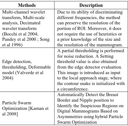
Methods Description
Multi-channel wavelet
transform, Multi-scale
analysis, Decimated
wavelet transform
(Bocchi et al 2004;
Pandey et al 2000 ; Song
et al 1996)
Due to its ability of discriminating
different frequencies, the method
can preserve the resolution of the
portion of ROI. Moreover, it does
not require the use of heuristics or
a prior knowledge of the size and
the resolution of the mammogram.
Edge detection,
thresholding, Deformable
model (Valverde et al
2004)
A partial thresholding is performed
for noise reduction. A Setting
threshold value is also obtained
from the edge detector evaluation.
This image is introduced as input
to the local approach stage, where
the contour snake is initialized with
a circumference.
Particle Swarm
Optimization [Karnan et
al 2008]
Automatically Detect the Breast
Border and Nipple position to
Identify the Suspicious Regions on
Digital Mammograms Based on
Asymmetries using hybrid Particle
Swarm Optimization
Table 3.1a. An overview of segmentation techniques for
MRI
Methods Description
Genetic Algorithm (karnan
and logeswari)
It segment tumor region from
background MRI.
Fuzzy Cmeans (FCM)
unsupervised
clustering(Philips et
al.1995)
It extracts the image edges
robustly and moves the vertices
towards the boundaries of the
desired structure.
Supervised k-nearest
neighbor(kNN)rule,
semi-supervised fuzzy
c-means(SFCM)
(vaidyanathan et.al.1997)
A sample set of pixel vectors
(ROI) is selected by an expert
observer, and the vectors are
assigned to different tissue
classes.
Level set Surface Model
(james et.al 2000)
To produce qualitative results
from several different datasets
for brain tumor segmentation.
Fuzzy C Means Clustering
Algorithm (SR
Kannan2005)
To segment tumor regions from
background MRI well.
Seed Growing
Method(1997)
(vaidyanathan et.al.1997)
Seed propagation was
independently performed.
Genetic Algorithm
(Thangavel and karnan
2007)
To segment and identify nipple
position from mammogram
image.
Pipe line approach,
Expectation Maximization
(EM) Algorithm(Jeffrey
and soloman 2004)
To estimate and processed
tumor volume successfully.
Hybrid Deformable
model,Meta Morphs model,
Novels Shape, Texture
Integration, Graphical
Model, Learning Methods
( dimitris et al.2006)
It Integrates both shape and
interior texture, its dynamics
are derived coherency from
both boundary and region
information in a common
variational framework.
Fuzzy C-means Clustering
Algorithm(FCM),Neural
Network Model (shan shen
et al.2005)
It processes seeking the optimal
labeling of the image pixels.
Atlas Matching Technique,
Finite Element
Method(FEM) ( oliver et.al
2005)
Artificial Bee Colony
[Neeraja et al 2013]
Brain Tumor Segmentation In
MRI Image Using
Unsupervised Artificial Bee
Colony And FCM Clustering
Soft Computing
[Sivaramakrishnan and
Karnan 2013]
A Novel Based Approach for
Extraction of Brain Tumor in
MRI Images Using Soft
Computing Techniques
Expectation Maximization
scheme(EM) (benedicte
et.al.2005)
Its performance is below than
Seni-Automated.
Automatic Two –
dimensional
Segmentation.( zhen chen
et.al 2003)
Each PET plane is segmented.
Ground Truth Algorithm
(marcel et.al
2003,2003,2004,
2007,2009)
It can provide the means for
objective assessment of
segmentation performance.
Texture Features,
Self-Organizing Map(SOM)
(logeswari and karnan
2010)
The tumor area is segmented
from brain MRI.
Morphological Operations,
Fuzzy model of Regions of
Interest(ROI) ( jing
et.al.2001)
It is use to represent more
appropriately the knowledge
about distance, shape and
interactions of structures.
Fuzzy C-means(Philips et
al.1995: siyal et.al.2005)
To generate segmentation
images that display clinically
important neuroanatomic tissue
and neuropathologic tissue
contrast information from raw
MR image data.
Amanpreet (2012)
To segment and deduct
suspicious region from
background using PSO
algorithm based on colony
aptitude and provide better
result than other parallel
algorithm.
Region-based method,
Region growing method,
Region-of-interest(ROI),
Multi resolution edge
detection method, modified
region segmentation
method( angel et.al 2012)
To segment brain tissue
structure from the
multi-resolution images are utilized.
Graph-Based
Method,Generative Model,
Weighted Aggregation
Algorithm(jasoncary 2006)
It Indicates the benefit of
incorporating model-aware
affinities into the segmentation
process for the difficult case of
brain tumor
Iterative Self-Organizing
Data Analysis
Techniques(ISO
DATA),Unsupervised
Computer Segmentation
Algorithm,Novel Model
( Michel Jacob 2001)
Multiparametric ISODATA
volume
was significantly
Identifies.
Spatio-Temporal
Model( Jeffery soloman et
al.2006)
The sensitivity and specificity
of tumor segmentation using
this spatio-temporal model is
improved over commonly used
spatial or temporal models
alone.
Multiscale Method,
Multiscale linking Model,
Supervised Segmentation
Method (naaaathan moon et
al.2002)
It was shown that the errors are
in the order of or smaller than
reported in literature.
Semi-Supervised Fuzzy
C-Means Clustering
Method,K nearest
neighbor(Knn),Gray level
thresholding & Seed
Growing(ISG-SG),Manual
Pixel Labelling(GT)
(vaithyanathan et.al.1995)
This method was achieved good
performance and reduction
operation time.
Hybrid level set (HLS)( xie
et.al,2005)
It provides objective,
reproducible segmentations that
are close to the manual results.
Fuzzy Model( webei
et.al.2007)
Average Probability of correct
detection was found.
Deformable
Model,Med-Volumeter( chunvan
et.al.2004)
The target area is segmented
under level set frame.
3D Variational
Segmentation
Method( dana cobzas
et.al.2007)
The tumor area was segmented
accurately.
Fuzzy k-means, GA
(maoyang et,al2005)
A thresholding is performed for
noise reduction.
Supervised
technique-Mountain
Method,Maximum
Likelihood,K-nearest
neighbour,Artificial neural
network.( Robert
et.al.1997)
Producing excellent partitions
of large data sets.
Fuzzy Connectedness &
Fuzzy sets( jayaram udupa
and punam saha 2003)
It allows the spatio-topological
concept of
hanging-togetherness of image elements
in the presence of a gradation of
intensities stemming from
natural material heterogeneities,
blurringand other
phenomenonrelated artifacts.
Expectation-Maximization
(EM)( Nathan mano
et.al.2002)
It separates WM,GM and CSF
from ti and t2 weighted image.
Classic snaks,Deformable
Contour model ( amini
et.al.2003)
To segment T1 weighted
images of the brain with low –
contrast structures and
discontinuous edges
Markov random field
model ( kabir et .al.2007)
Segmentations obtained with
single sequences to that
obtained with multiple
sequences.
Generalized fuzzy
operator(GFO),Contour
Deformable model( leung
et.al.2003)
The tumor regions are
segmented
Atlas-based segmentation
( pierreyes et.al 2005)
Propagation of the labeled
structures on to the MRI
Expectation-Maximization
Technique,Robest
Estimation,VALMET
Segmentation validation
tool ( marcel et.al.2003)
To segment tumor, edema and
ventricles.
Multilayer segmentation,
Automatic region
segmentation
( xinbai et.al.2003)
regions.
Content-based retrieval
technique ( zhechen
et.al.2003)
Image segmented successfully.
Atlas-driven segmentation
( guido 2003)
Automatically tumor region is
segmented successfully.
Fuzzy methods( zushan
et.al.2004)
Results show relatively high
accuracy.
Active Contour
Model( corina draphca et.al
2005)
The tumor regions are
segmented from MRI.
Evaluating Image
Segmentation Algorithm
( jayaram udupa 2002)
High Accuracy appeared in
segmentation.
Fuzzy mean
Algorithm(FCM),Silhovette
Method(SM)( SR
kannan2005)
Its provide Easiest way to find
appropriate structure in the data
of MRI
Contour Tracing
Algorithm, Region
Segmentation(hideki et. Al
1990)
To establish boundaries in order
to partition the image space into
meaningful regions.
Soft-Margin Support
Vector
Machine(SVM)( mark
Schmidt et.al. 2005)
It can involve millions of
training and testing instances
with a relatively small feature
set.
k-means Clustering
Algorithm(2004)
It separates background from
brain pixels accurately.
k-means clustering( ming
et.al 2007)
It is used to convert a given
gray-level MR image into a
color space image and it
separates the position of tumor
objects from MRI
Statistical Model ,Markov
Random Field, Level Set
Method, Non-Uniformity
Correction Method( ranos
et.al 2004)
Non-brain structures removed
and it estimates the tissue
intensity variation.
Multi-Scale Watershed
Segmentation( erck dam
et.al 2004)
It is used to select the subsets of
the expected regions
automatically.
Deformable Region Model,
Shrinking Method and
Snake Method (chan
et.al.1996)
It is used to locate the boundary
of an object quickly.
Hidden Markov Chain
Model(HMC) ( briq
et.al.2008)
To produce ever finer resolution
in spectral, spatial and temporal
data.
Seeded Region Growing,
Active Contour Snakes
Model ( salman et.al 2005)
It is based on extraction of a
connected set of pixels whose
pixel intensities are consistent
with pixel statistics of a seed
point.
Hybrid level Set (HLS)
Model( kai xie et.al 2005)
It is used to segment edema and
tumor.
Expectation
Maximization(EM)
Algorithm, MRF( david
gerind et.al.2002)
It is used to select the subsets of
the expected regions efficiently.
Population-Based Tissue
Maps, K Nearest Neighbor
Model. (sean haney et.al
2001)
It is used to differentiate tissue
types with high accuracy.
Level-Set Surface Model
( aaron et.al 2003)
It is used to segment target
regions from background tissue.
Support Vector
It is used to locate the boundary
machine( zhou et.al. 2005)
of an object quickly.
GPU based
Segmentation(aaron et.al
20032003)
This system found interactivity
users to produce good, reliable
segmentation on MRI and it
produced qualitative and
quantitative for brain tumor
detection
Genetic Algorithm ( karnan
and thangavel 2007)
Segment objective region from
MRI.
Self organizing Map(SOM)
( logeswari and karnan
2010)
Segment the suspicious region.
Standard Deterministic
Annealing(DA) and Fuzzy
C Means ( xi-lei yang et.al
2008)
To apply robust segmentation
on brain MRI images for
segmenting tumor pixels on
MRI.
Bacteria Foraging
Optimization Algorithm
[Ben George and Karnan
2012]
MRI Brain Image
Enhancement , Feature
Extraction and Classification of
Brain Tumor using BFOA
IV.
F
EATUREE
XTRACTIONA
NDS
ELECTIONThe textural features can be extracted from the
co-occurrence matrix. They are related to specific textural
characteristics such as the homogeneity, contrast, entropy,
energy and regularity of the structure. In this paper, the
texture analysis methods such as, Surrounding Region
Dependency Matrix, Spatial Gray Level Dependency
Matrix, Gray Level Difference Matrix, Gray Level Run
Length Matrix are used to extract the features from the
segmented image.Feature extraction is a general term for
methods of constructing combinations of the variables to
get around these problems while still describing the data
with sufficient result. It is defined as the operation to
quantify the image quality through various parameters or
functions, which are applied to the original image. Feature
extraction involves simplifying the amount of resources
required to describe a large set of data accurately.
Today, one of the main problems in machine learning
and statistics is keeping track of the most relevant
information. For this purpose, feature selection techniques
are addressed. The major aims of feature selection for
classification are finding a subset of variables those results
in more accurate classifiers and constructing more compact
models. Therefore, feature selection will filter out those
variables that are irrelevant for the specific model. The
selection should only capture the relevant features while
not over fitting the data. Also there is a reduction in the
sample size needed for good generalization.
Table 4.1 an overview for Feature Extraction and Selection
Methods Description
Back Propagation
Network(BPN), Ant Colony
Optimization technique(ACO)
( thangavel and karnan 2005)
Masses were extracted
completely.
Multi-scale Gabor-type Feature
set( dana cobaz et.al.2007)
Accurate results to be
obtained with a relatively
small number of training
images.
Methods Description
model,Active contour
model,Kolmogorov-Smirnov(KS),Point Sampling
technique.(albert law 2002)
result.
Wavelet Transform,Wavelet
Packets(azadeh et.al 2004)
Related features are extracted
from the background issue.
Fuzzy C-means(FCM)
Unsupervised clustering,
Morphological
Operators.( amini et.al.2003)
To extract the image edges
robustly.
Optimal Residual Extraction
( xin bai et.al.2003)
Masses were extracted
completely
Morphologic
Operation( zuchen et.al 2004)
To extract the sulcal images
from the brain images.
Rough Set Particle Swarm
Optimization(xiang vang et.al
2006)
It can generate more general
decision rules and better
classification quality of new
samples.
Kruskal-Wallistest, Fisher
Discriminant Criterion
,Relief-F Algorithm and Least Squares
– Support Vector
Machine(LS-SVM) ( jan luts et.al.2007)
It is used to reduce the
dimension of the input space
and can extract feature set.
Quick Reduct algorithm
[Jaganathan et al 2007]
Classification rule discovery
with ant colony optimization
and improved Quick Reduct
algorithm for the medical
images.
Relative Reduct Algorithm
[Kalyani and Karnan 2010]
Medical data Attribute
Reduction using Forward
Selection and Relative Reduct
Algorithm
Artifcial Bee colony [Mary
Jeyanthi Prem and Karnan
2013]
Business Intelligence using
Optimization techniques for
Decision Making
V.
C
LASSIFIERSClassifiers play an important role in the implementation
of intelligent system to identify the tumour from
mammogram and MRI image. The features are given as
input to the classifiers to classify the medical image into
normal and abnormal
Methods Description
Woods et al. 1993
Binary decision tree
Area under ROC curve is 0.9 for
24 images.
Woods et al. 1993
Quadratic Classifier
Area under ROC curve is 0.918
for 24 images.
Caldwell et al 1990.,
Nishikawa et al 1990. Woods
et al 1995. Nishikawa et al
1992. Nishikawa et al 1993.
Linear classifier
Maximum area 0.70 under ROC
for 70 images.
Cordella et al. 2000
Multiple expert system
The area under the ROC curve is
0.786 for 40 images.
Caldwell et al. 1990
Woods et al. 1993
Dhawan et al. 1996
Kim and Park 1999
Neural Networks
Maximum area 0.86 under ROC.
Area under ROC curve 0.935 for
24 images.
Maximum area 0.76 under ROC
for 191 images.
The area under ROC curve is
0.88 for 120 images.
Woods et al. 1993
Area under ROC curve 0.929 for
Methods Description
K-Nearest Neighbor
Classifier
24 images.
Recursive feature elimination
based on Support Vector
Machine(SVM RFE), Genetic
Algorithm (GA) ( mao yang
2005)
To determine these optimal
hyper-planes, It Solves a convex
quadratic programming problem.
It is use to get optimal values. It
Achieves high classification
accuracies with genes.
Performance is very Satisfied.
Support Vector
Machines(SVM)( dana
cobzas et.al. 2004)
To predict sub cellular
localization.
Multi-Layer Feed Forward
Neural Network, Support
Vector
Machine(SVM)( corina
drapaca 2005)
Used to classify the tumor
regions from non-tumor regions.
Bayesian Model( Jason carso
2006)
Each region is assigned
a most likely model class
according to a set of learned
model classes
3D-Expectation
Maximization Method,
Hidden Markov Model
( Jeffrey soloman 2006)
To classify the tumor Region
Voxel Classification,
Geometric Model( guido
2003)
To classify the tumor regions
from non-tumor regions.
Multilayer preceptron neural
network.( azadeh 2004)
To classify the tumor features
extracted from the spectra.
Supervised voxal
Classification.
( guido2003)
The tumors classified
successfully.
Support Vector
Machines(SVMs),Decision
Tree(DT)
( gotsos 2003)
90.8%low from high grade
tumors and 85.6% less from
highly aggressive tumors are
classified clearly. The ability of
SVM to ensure good
performance even with limited
training samples was verified.
Quadratic Discriminant
Analysis(QDA),Support
Vector Machine(SVM)
(hongmin 2007)
SVM classification is combined
with QDA based classification to
obtain a better tumor profile.
Linear Discriminant Analysis,
Least Squares Support Vector
Machines(LS-SVM),Linear
Kernel Techniques (or)
Radial Basis Function(devos
2005),( jain luts 2007)
Classifiers were evaluated over
100 stratified random splitting of
the dataset into training and test
sets.
Multi-Scale Jet -Based
Classification( erick dam
et.al.2004)
It is used to automatically select
a subset of the regions generated
by the watershed segmentation
method.
Statistical Classification
method( war field 2000)
Classify the tumor is benign ,
malignant or normal
VI.
R
OCA
NALYSISis a plot of the classifier’s true positive detection rate and
its false positive rate. True positive (TP) detection rate is
the probability of correctly classifying a target object and
false positive (FP) detection rate is the probability of
incorrectly classifying a target object. The following
figures show that the sample ROC curves. The Researchers
suggested various techniques of ROC and they are
available in the survey. Each classifier is constructed using
the training set and is evaluated by ROC Analysis.
Table 6.1 An overview of ROC Analysis
Methods Description
Devos (2005)
MRI with Peak
Integration
Performance of ROC is 0.99 for 76
patients with 142 data’s. It’s
performed higher than other classifier.
Devos (2005) 29
Principal Component
Analysis(PCA) –LS
SVM
Area Under Curve (AUC) higher than
0.94 for low versus high grade
gliomas fro 70 data sets.
Higher result than PCA/LDA
Devos(2006) 29
Linear Discriminant
Analysis(LDA)
Area Under Curve (AUC) higher than
0.91 for low versus high grade tumors.
Performs better than PCA
Devos(2006)
Least Square-Support
Vector Machine
Area Under Curve (AUC) higher than
0.91for low versus high grade
gliomas.Gives accurate result.
Devos(2006)
Least Square-Support
Vector Machine and
Radial Basis Function
Kernal(RBF)
Area Under Curve (AUC) higher than
0.99 for gliomas versus meningiomas.
LS-SVM and RBF Combination gives
better improvement than other
classifier.
VII.
C
ONCLUSIONSIn this survey work various automatic detection
methods of microcalcifications and brain tumor through
mammograms and MRI has been studied and compared for
the period of more than two decades .This is used to focus
on the future of developments of medical image processing
in medicine and healthcare. We have described several
methods in medical image processing and to discussed
requirements and properties of techniques in tumor
detection .This work is used to give more information about
tumor detection and segmentation. It is a milestone for
analyzing all technologies relevant to tumor from
mammogram and MRI in Medical image processing. In this
work, various steps in detection of automatic detection :i)
The Preprocessing and Enhancement Technique ii)
Segmentation Algorithm iii) Feature Extraction iv)
Classification v)Performance Analysis using Receiver
Operating Characteristics and their performance have been
studied and compared.
R
EFERENCES[1] Lai, S., Li, X. and Bischof, W.F. “On Techniques for Detecting Circumscribed Masses in Mammograms,” IEEE Transactions on Medical Imaging, Vol. 8, No. 4, pp. 377–386, 1989.
[2] Qian, W., Clarke, L.P., Kallergi, M. and Clark, R.A. “Tree-Structured Nonlinear Filters in Digital Mammography,” IEEE Transactions on Medical Imaging, Vol. 12, No. 1, pp. 25–36, 1994. [3] Highnam, R.P., Brady, J.M. and Shepstone, B.J. “Computing the
Scatter Component of Mammographic Images,” IEEE Transactions on Medical Imaging, Vol. 13, No. 2, pp. 301–313, 1994.
[4] Kim, J.K., Park, J.M., Song, K.S. and Park. W. “Adaptive Mammographic Image Enhancement Using First Derivative and Local Statistics,” IEEE Transactions on Medical Imaging, Vol. 16, No. 5, pp. 495-502, 1997.
[5] Li, H., Liu, K.J.R. and Lo, S.C.B. “Fractal modeling and segmentation for the enhancement of microcalcifications in digital mammograms,” IEEE Transactions on Medical Imaging, Vol.16, No. 6, pp. 785–798, 1997.
[6] Kovalerchuk, B., Traintaphyllou E.J.F. Ruiz and J. Clayton, J. “Fuzzy Logic in Computer-Aided Breast Cancer Diagnosis: Analysis of Lobulation,” Artificial Intelligence in Medicine, Vol. 11, pp. 75–85, 1997.
[7] Chang, R.F., Wu, W.J., Tseng, C.C., Chen, D.R. and Moon, W.K. “3-D Snake for US in Margin Evaluation for Malignant Breast Tumor Excision Using Mammotome,” IEEE Transactions on Information Technology in Biomedicine, Vol. 7, no. 3, pp. 197–201, 2003.
[8] Kobatake, H., Murakarni, M., Takeo, H. and Nawano, S. “Computerized Detection of Malignant Tumors on Digital
Mammograms,” IEEE Transactions on Information Technology in Biomedicine, Vol. 18, No. 5, pp. 369–378, 1999.
[9] Ferrari, R.J., De Carvalho, F., Marques, P.M.A. and Frere. A.F. “Computerized classification of breast lesions: shape and texture analysis using an artificial neural network,” 7th international conference on Image Processing and its applications, pp. 517–521, 1999.
[10] Cordella, L.P., Tortorella, F. and Vento, M. “Combing experts with different features for classifying clustered microcalcifications in mammograms,” Proceedings of 15th International Conference on Patten Recognition, pp. 324–327, 2000.
[11] Bhangale, T., Desai, U.B. and Sharma, U. “An unsupervised scheme for detection of microcalcifications on mammograms,” IEEE International Conference on Image Processing, pp. 184–187, 2000.
[12] Enderwich, C.Y. and Tzanakou, E.M. “Classification of mammographic tissue using shape and texture features,” Proceedings of the 19th International Conference-IEEE/EMBS, pp. 810–813, 1997.
[13] Veldkamp, W.J.H. and Karssemeijer, N. “Normalization of local contrast in mammograms,” IEEE Trans. Med. Imag., Vol. 19, No. 7, pp. 731–738, 2000.
[14] Mudigonda, N.R., Rangayyan, R.M. and Desautels, L.“Detection of Breast Masses in Mammograms by Density Slicing and Texture Flow Field Analysis,” IEEE Tran. On Medical Imaging, Vol. 20, No. 12, pp. 1215–1227, 2001.
[15] Rogova, G.L., Stomper, P.C. and Ke, C. “Microcalcification texture analysis in a hybrid system for computer aided mammography,” SPIE, Vol. 3661, pp. 1426–1433, 1999.
[16] Bocchi, L., Coppini, G., Nori, J. and Valli, G. “Detection of Single and Clustered Microcalcifications in Mammograms Using Fractals Models and Neural Networks,” Medical Engineering and Physics, Vol.26, pp. 303 – 312, 2004
[17] Olivier Clatz, Maxime Sermesant, Pierre-Yves Bondiau, Herve Delingette, Simon Warfield, K, Gregoire Malandain & Nicholas Ayache, ‘Realistic Simulation of the 3-D Growth of Brain Tumors in MR Images Coupling Diffusion With Biomechanical Deformation’, IEEE on Trans Medical Imaging,vol.24,no.10, pp.1334-1346. 2005
[18] Dana Cobzas, Neil Birkbeck, Mark Schmidt & Martin Jagersand ‘3DVariational Brain Tumour Segmentation Using a High Dimensional Feature Set’, IEEE 11th International Conference on Computer Vision, ICCV-2007, pp.1-8.2007
[19] Jayaram Udupa, K, Vicki Lablane, R, Hilary Schmidt, Celina Lmielinska, Punam Saha, K, George, J, Grevera, Ying Zhuge, Pat Molholt, Yinpengjin & Leanne Currie, M, ‘A Methodology for Evaluating Image Algorithm’, IEEE on Medical Image Processing, pp.1-14. 2002
[20] Elizabeth Bullitt, Donglin Zeng, Guido Gerig, Stephen Aylward, Sarang Joshi Keithsmith, J. Weili Lin, Matthew Ewend, G, ‘Vessel Tortuosity and Brain Tumor Malingnancy:A Blinded Study’, Academic Radiology, vol.12, pp.1232-1240.2005
[21] Leung, CC, Chen, WF, Kwok, PCK & Chan, FHY 2003, ‘Brain Tumor Boundary Detection in MR Image with Generalized Fuzzy Operator’, International Conference on Image Processing Proceedings, Barcelona, vol.2, pp.1057-1060.2003
[23] Gray, HF 2010, ‘Genetic Programming for the Analysis of Nuclear Magnetic Resonance Spectroscopy Data’, The Institution of Electrical Engineers, Centre For Development of Advanced Computing, IEEE Xplore, March 10.2010
[24] Mark Schmidt, Ilya Levner, Ressell Greiner, Albert Murtha & Aalo Bistritz 2005, ‘Segmentation Brain Tumors using Alignment-Based Features’, IEEE on Proceesings of the fourth International Conference on Machine Learning an Applications(ICMLA’05)”, pp.215-220.2005
[25] Toshiharu Nakai, Shigeru Muraki, Epifanio Bagarinao, Yukio Miki, Yasuo Takehara, Kayako Matsuo, Chikako kato, Harumi Sakahara & Haruo Isoda 2003, ‘Application of independent component analysis to magnetic resonance Imaging for enhancing the contrast of gray and white matter’, Elsevier on Neuroimage,vol.2, no.1, pp. 251-260.2003
[26] Farahat, AS, El-Dewany, EM, El-Hefnawi, FM, Kadah, YM & Youssef, AA 2006, ‘Calculations of Heating Patterns of Interstitial Antenna for Brain Tumors Hyperthermia Treatment Planning’, The 23thNational Radio Science Conference, Faculty of Electronic Engineering, Menoufiya University.2006
[27] Kyeong-Jun Mun, Hyeon Tae Kang, Hwa-Seok Lee, Yoo-Sool Yoon, Chang-Moon Lee & June Ho Park 2004, ‘Active Contour Model Based Object Contour Detection using Genetic Algorithm with Wavelet Based Image Preprocessing’, International Journal of Control, Automation, and Systems vol. 2, no. 1.2004
[28] Lim, LO & Pfefferbaum, A 1989, ‘Segmentation of MR brain images into cerebrospinal fluid spaces, white and gray matter’, Journal of Comput. Assisted Tomog, vol .13, no. 4, pp. 588-593.1989
[29] Chunyan Jiang, Xinhua Zhang, Wanjun Huang & Christoph Meinel 2004, ‘Segmentation and Quantification of Brain Tumor’, IEEE International conference on Virtual Environment, Human- Computer interfaces and Measurement Systems, pp.12-14.2004 [30] Tsai, C, Manjunath, BS & Jagadeesan, R 1995, ‘Automated
Segmentation of brain MR Images’, Pergamon, Pattern Recognition, vol.28, no.12.1995
[31] Boada, FE, Davis, D, Walter, K, Torres-Trejo, A, Kondziolka, D, Bartynski, W & Lieberman, F 2004, ‘Triple Quantum Filtered Sodium MRI of Primary Brain Tumors’, IEEE, Proc. Intl. Soc. Mag. Reson. Med.USA.2004
[32] Aria Tzika, A, Maria Zarifi, K, Liliana Goumnerova, Loukas Astrakas, G David Zurakowski, Tina Young- Poussaint, Douglas Anthony, Michael Scott, R & Peter McL. Black 2002, ‘Neuroimaging in Pediatric BrainTumors:Gd-DTPA-Enhanced, emodynamic, and Diffusion MR Imaging Compared with MRSpectroscopic Imaging’, AJNR Am J Neuroradiol ,vol.23, no 2, pp .322-333.2002
[33] Amini, L, Soltanian-Zadeh, H & Lucas, C 2003, ‘Automated Segmentation of Brain Structure from MRI’, Proc. Intl.Soc. Mag. Reson.Med.11.2003
[34] Zhe chen, David Dagan Feng & Weidong Cai 2003, ‘Automatic Detection of PET Lesions’, Pang-Sydney Area Workshop on Visual Information Processing, Conference in Research and Practice in IT, vol.22, pp.21-26.2003
[35] Corina Drapaca, S, Valerie Cardenas & Colin Studholme 2005, ‘Segmentation of tissue boundary evolution from brain MR Image sequences usingmulti-phaselevel sets’, Elsevier, Computer vision and image understanding,vol.100, no. 3, pp.312-329.2005
[36] Dimitris Metaxas, N, Zhen Qian, Xiaolei Huang, Rui Huang, Ting Chen & Leon Axal, ‘Hybrid Deformable Models for Medical Segmentation and Registration’, International conference on Control, Automation, Robotics and Vision(ICARCV’06),pp.1-6. 2006
[37] Hideki yamamoto, Katsuhiko Sugita, Noriki Kanzaki, Ikuo Johja, Yoshio Hiraki & Michiyoshi Kuwahara 1990, ‘Magnetic Resonance Image Enhancement Using V- Filter’, IEEE AES Magazine, vol.5, no.6, pp. 31 - 35.1990
[38] Gordon Kindlmann & Carl-Fredrik Westin 2006, ‘Diffusion Tensor Visualization with Glyph Packing’, IEEE Transactions on Visualization and Computer Graphics (Proceedings Visualization/ Information Visualization 2006) vol.12, no.5, pp.1329-1335.2006 [39] Shishir Dube, Suzie El-saden, Timothy Cloughesy, F & Usha Sinha
2006, ‘Content Based Image Retrieval for MR image Studies of Brain Tumors’, Proceedings of the 28th IEEE EMBS Annual International Conference, NewYork,vol.1,no.1,pp.3337-3340.2006
[40] Salman, YM, Assal, MA, Badawi, AM & El-Bavome, M 2005, ‘Validation Techniques for Quantitative Brain Tumors Measurements’, Proceedings of the 2005 IEEE Engineering in Medicine and Biology 27th annual Conference shanghai, China, vol.7, pp. 7048-7051.2005
[41] Sean Haney, M, Paul Thompson, M, Timothy Cloughesy, F, Jeffry Alger, R & Arthur Toga, W 2001, ‘Tracking tumor Growth Rates in Patients with Malignant Gliomas’, A Test of two Algorithms’, American Society of Neuroradiology, AJNR Am J euroradiol,vol.22, no.1, pp. 73-82.2001
[42] Michael Wood, L & Val Runge, M 1988, ‘Application of image Enhancement techinique to Magnetic Resonance Imaging’, Radiographics,vol.8, no. 4,pp. 771-784.1988
[43] Sonali Patil & Udupi, VR 2012, ‘Preprocessing to be considered for MR and CT images containing Tumors’, IOSR journal of Electrical and Electronics Engineering, vol. 1, no. 4, pp. 54-57.2012
[44] Dengler, J., Behrens, S., and Desaga, J.F. “Segmentation of microcalcifications in mammograms,” IEEE Transactions on Med. Imag., Vol. 12, no. 4, pp. 634–642, 1993.
[45] Li, H.D., Kallergi, M., Clarke, L.P., Jain, V.K., and Clark, R.A. “Markov Random Field for Tumor Detection in Digital Mammography,” IEEE Transactions on Medical Imaging, Vol. 14, No. 3, pp. 565 – 576, 1995.
[46] Li, H., Liu, K.J.R. and Lo, S.C.B. “Fractal modeling of mammogram and enhancement of microcalcifications,” IEEE Nuclear Science Symposium & Medical Imaging Conference, Vol. 3, pp. 1850–1854, 1996.
[47] Kim, J.K., Park, J.M., Song, K.S. and Park. W. Detection of clustered microcalcifications on mammograms using surrounding region dependence method and artificial neural network,” Journal on VLSI Signal Process, Vol. 18, pp. 251–262, 1998.
[48] Mossi, J.M., Albiol, A. “Improving detection of clustered microcalcifications using morphological connected operators,” IEEE Image Proces. and its Applications, Vol. 5, No. 2, pp. 498– 501, 1999.
[49] Peters, J.F. and Skowron, A. (Eds.). “Transactions on Rough Sets,” Springer-Verlag, Berlin, 2004.
[50] Cheng, H.D., Cai, X., Chen, X.W., Hu, L. and Lou, X. “Computer Aided Detection and Classification of Microcalcifications in Mammograms: A Survey,” Pattern Recognition, Vol. 36, pp. 2967–2991, 2003.
[51] Cheng, H.D., Chen, J.R., Freimanis, R.I. and Jiang, X.H.“A novel fuzzy logic approach to microcalcification detection,” Journal on Inform. Sci., Vol. 7, pp. 1-14, 1998.
[52] Cheng, H.D., Lui, Y.M. and Freimanis, R.I. “A novel approach to microcalcification detection using fuzzy logic technique,” IEEE Trans. Med. Imag. Vol. 17, no. 3, pp. 442–450, 1998.
[53] Cheng, H.D., Wang, J. and Shi, X. “Microcalcification Detection Using Fuzzy Logic and Scale Space Approaches,” Pattern Recognition, Vol. 37, pp. 363–375, 2004.
[54] Dippel, S., Stahl, M., Wiemker, R. and Blaffert, T. “Multiscale contrast enhancement for radiographies: Laplacian pyramid versus fast wavelet transform,” IEEE Trans. Med. Imag., Vol. 21, No. 4, pp. 343–353, 2002.
[55] Pandey, N., Salcic, Z. and Sivaswamy, J. “Fuzzy logic based microcalcification detection, Neural Networks for Signal Processing,” Proceedings of the IEEE Workshop, pp. 662–671, 2000.
[56] Valverde, F.L., Guil, N. and Munoz, J. “Segmentation of Vessels from Mammograms Using a Deformable Model,” Computer Methods and Programs in Biomedicine, Vol.73, pp. 233–247, 2004. [57] Phillips, WE, Velthuizen, RP, Phuphanich, S, Hall, LO, Clarke, LP & Silbiger ML 1995, ‘Application of Fuzzy C-Means Segmentation Technique for tissue Differentlation in MR Images of a hemorrhagic Glioblastoma Multiforme’, Pergamon on Megnetic Resonance Imaging,vol.13,pp. 277-290.1995
[58] Vaidyanathan, M, Clarke, LP, Hall, LO, Heidtman, C, Velthuizen, R, Gosch, K, Phuphanich, S, Wagner, H, Greenberg, H & Silbiger, ML 1997, ‘Monitoring brain tumor Response to therapy using MRI segmentation’, Elsevier on Magnetic Resonance Image, vol.15, no.3, pp.323-334.1997
Brain Tumors’, IEEE Transactionson Microwavetheory and Techniques, vol.48, no.11, pp. 2191- 2198.2000
[60] Kannan, SR 2005, ‘A New Clustering Algorithm for Segmentation of Magnetic Resonance Images Using Fuzzy C-Mean and Computer Programming’, ICGST International Journal on Graphics, Vision and Image Processing, vol.2. 2005
[61] Kannan, SR 2005, ‘Segmentation of MRI Using New Unsupervised Fuzzy C mean Algorithm’, ICGST-GVIP Journal,vol.5, no.2.2005 [62] Jeffrey Solomon, John Butman, A & Arun Sood 2006,
‘Segmentation of brain tumors in 4D MR images using the Hidden Markov model’, Elsevier on Computer Methods and Programs in Biomedicine”, USA, vol.84, no.2, pp. 76-85.2006
[63] Jeffrey Solomon, A, Butman & Arun Sood 2004, ‘Data Driven Brain Tumor Segmentation in MRI Using Probabilistic Reasoning over Space and Time’, Springer Link on Medical Image Computing, vol. 3216.2004
[64] Shan Shen, William Sandham, Malcolm Granet & Annette Sterr 2005, ‘MRI Fuzzy Segmentation of Brain Tissue Using Neighbourhood Attraction with Neural- Network Optimization’ IEEE Transactions on Information Technology in Biomedicine, vol.9, no.3, pp. 459-467.2005
[65] Benedicte Mortamet, Donglin Zeng, Guido Gerig, Marcel Prastawa & Elizabeth Bullitt 2005, ‘Effects of Healthy Aging Measured By Intracranial Compartment Volumes using Designed MR Brain Database’, Med Image Comput Comput Assist Interv Int Conf Med Image Comput Comput Assist Interv, vol.8, pp. 383-391. 2005 [66] Marcel Prastawa, Elizabeth Bullitt & Guido Gerig 2009,
‘Simulation of Brain Tumors in MR Images for Evaluation of Segmentation Efficacy’, Medical Image Analysis (MedIA), vol .13, no. 2, pp. 297-311.2009
[67] Marcel Prastawa, Elizabeth Bullitt, Nathan Moon, Koen van Leemput & Guido Gerig, 2003, ‘Automatic Brain Tumor Segmentation by Subject Specific Modification of Atlas Priors’, Academic Radiology. vol .10, no.12, pp. 1341-1348.2003
[68] Marcel Prastawa, Elizabeth Bullitt, Sean Ho & Guido Gerig 2003, ‘Robust Estimation for Brain Tumor Segmentation’, Medical Image Computing and Computer Assisted Intervention (MICCAI), Lecture Notes in Computer Science (LNCS), vol.2879, pp.530-537.2003 [69] Marcel Prastawa, Elizabeth Bullitt, Sean Ho & Guido Gerig 2004,
‘A Brain Tumor Segmentation Framework Based on Outlier Detection’ Medical Image Analysis, vol. 8, no.3, pp.275-283.2004 [70] Marcel Prastawa, W, John Gilmore, H, Weili Lin, Christopher
Looney, BY, Sampath Vetsa, K, Rebecca Knickmeyer, C, Dianne Evans, D, Keith Smith, J, Robert Hamer, M, Jeffrey Lieberman, A & Guido Gerig 2007, ‘Regional Gray Matter Growth, Sexual Dimorphism, and Cerebral Asymmetry in the Neonatal Brain’, Journal of Neuroscience. vol.27, no. 6, pp.1255-1260.2007 [71] Jing-hao Xue, Su Ruan, Bruno Moretti, Marinette Revenu &
Daniel Bloyet 2001, ‘Knowledge-based Segmentation And Labeling of brain structure fromMRI images’, Elsevier on Pattern Recognition Letters , vol.22, no. 3-4,pp. 395-405. 2001
[72] Siyal, MY & Lin, YU, 2005, An Intelligent modified fuzzy C-means based algorithm for bias estimation and Segmentation of brain MRI”, Elsevier, Pattern Recognition Letters, vol.26,no.13,pp. 2052-2062.2005
[73] Amanpreet Kaur & Singh, MD 2012, ‘An Overview of PSO- Based Approaches in Image Segmentation’, International Journal of Engineering and Technology, vol.2, no. 8.2012
[74] Angel Viji, KS & Jayakumari, J 2012, ‘Performance Evaluation of Standard Image Segmentation Methods and Clustering Algorithms for Segmentation of MRI Brain Tumor Images’, European Journal of Scientific Research, vol. 79, no.2, pp. 166-179.2012
[75] Jason Carso 2006, Multilevel Image Segmentation and Integrated Bayesian Model Classification with an Application to Brain Tumor Imaging’, Upcoming UCLA Statistics Seminar on Springer link, vol. 4191, pp. 790-798.2006
[76] Michael Jacobs, A, Panayiotis Mitsias, Hamid Soltanian-Zadeh, Sunitha Santhakumar, Amir Ghanei, Rabih Hammond, Donald Peck, J, Michael Chopp & Suresh Patel 2001, ‘Multiparametric MRI Tissue Characterization in Clinical Stroke with Correlation to Clinical Outcome’, American Heart Association, Stroke, pp.950-957.2001
[77] Nathan Moon, Elizabeth Bullitt, Koen Van Leemput & Guido Gerig 2002, ‘Automatic Brain and Tumor Segmentation’, Proceedings of the 5th International Conference on Medical Image Computing and Computer-Assisted Intervention-Part I, pp. 372-379. 2002
[78] Vaidyanathan, M, Clarke, LP, Velthuizen, RP, Phuphanich, S, Bensaid AM, Hall, LO, Bezdek, JC, Greenberg, H, Trotti A & Silbiger, M 1995, ‘Comparison of Supervised MRI Segmentation methods for Tumor Volume Determination During Therapy’, Pergamon,Magnetic Resonance Imaging, Magnetic Resonance Imaging, vol.13, no.5, pp.719-728.1995
[79] Xie, K, Yang, J, Zhang, ZG & Zhu, YM 2005, ‘Semiautomated Brain tumor and edema segmentation using MRI’, European Journal of Radiology, vol.56, no.1, pp.12-19.2005
[80] Weibei Dou, Su Ruan, Yanping Chen, Daniel Bloyet & Jean-Marc Constans 2007, ‘Aframework of fuzzy information fusion for the segmentation of brain tumor tissues on MR images’, Image and Vision Computing, vol.25, no.2, pp. 164-171.2007
[81] MAO Yong, ZHOU Xiao-bo, PI Dao-ying, SUN Youxian & WONG Stephen TC 2005, ‘Parameters selection in selection using Gaussian kernel support vector machines by genetic algorithm’, Journal of Zhejiang University Science, vol. 6, no.10, pp.961– 973.2005
[82] Robert Velthuizen, P, Lawrence Hall, Laurence Clarke, P & Martin Silbiger, L 1997, ‘An Investigation of Mountain Method Clustering for Large Data Sets’, Peron, Pattern Recognition, vol.30, no.7, pp. 1121- 1135.1997
[83] Jayaram Udupa, K & Punam Saha, K. 2003, ‘Fuzzy Connectedness and Image Segmentation’, Proceedings of the IEEE,vol.91,no.10, pp.1649-1669.2003
[84] Kabir, Y, Dojat, M, Scherrer, B, Forbes, F & Garbay, C 2007, ‘Multimodal MRI Segmentation of ischemic stroke Lesions’, 29th Annual International Conference of the IEEE in Engineering in Medicine and Biology Society, pp.1595-1598. 2007
[85] Pierre-Yves Bondiau,Gregoire Malandain, Stephane Chanalet & Pierre-Yves Marcy 2005, ‘Atlas-Based Automatic Segmentation of MR Images:Validation Study on The Brainstem in Radiotherapy Context’, Elsevier on radiation Oncology Biological Physis,vol. 6, no.1, pp.289-298.2005
[86] Xin Bai, Jesse Jin, S & Dagan Feng 2004, ‘Segmentation Based Multilayer Diagnosis Lossless Medical Image Compression’, Australian Computer Society, Pan Sydney Area Workshop on Visual Information Processing (VIP 2003), vol. 100,pp.9-14.2003 [87] Ming-Ni Wu, Chia-Chen Lin & Chin-Chen Chang 2007, ‘Brain
Tumor Detection Using Color-Based k- Means Clustering Segmentation’, Third International Conference on Intelligent Information Hiding and Multimedia Signal Processing, vol. 2, pp.245-250. 2007
[88] Panos kotsas 2005, ‘Non-rigid Registration of medical images using an Automated method’, World academy of science, Engineering and Technology .2005
[89] Erik Dam, Marcoloog & Marloes Letteboer 2004, ‘Integrating Automatic and Interactive Brain tumor Segmentation’ Proceedings of the 17th International Conference on Pattern Recognition (ICPR’04),vol. 3,pp .790-793.2004
[90] Chan, FHY, Lam, FK, Poon, PWF, Zhu, H & Chan, KH 1996, ‘Object boundary location by Region and Contour Deformation’, IEEE, Proceedings on Vision, Image and Signal Processing,vol.143, no.6, pp.353-360.1996
[91] Bricq, S, Collet, Ch & Armspach, JP 2008, ‘Unifying Framework for Multimodal brain MRI Segmentation based on Hidden Markov Chains’, Elsevier on Medical Image Analysis, vol.12, no. 6, pp. 639-652.2008
[92] Kai Xie, Jie Yang, Zhang, ZG, & Zhu, YM 2005, ‘Semiautomated brain tumor and edema Segmentation using MRI’, Elsevier, European journal of Radiology, vol .56, no. 1, pp. 12-19.2005 [93] David Gering, Eric, W, Grimson, L & Kikinis, R 2002,
‘Recognition Deviations from Normalcy for Braintumor Segmentation’, Springer, vol.2488, pp.388-395.2002
[94] Aaron Lefohn, Joshua Cates & Ross Whitaker, 2003, ‘Interactive GPU-Based level sets for 3D Brain Tumor Segmentation’.2003 [95] Zhou, J, Chan, KL, Chong, VFH & Krishnan, SM 2005, ‘Extraction
of Brain Tumor from MR Images Using One- Class Support Vector Machine’, Proceedings of the 2005 IEEE Engineering in Machine and Biology ,27th Annual Conference, pp.6411-6414.2005 [96] Karnan, M & Selvanayaki, K 2010, ‘Improved Implementation of
Brain MR Image Segmentation using Meta Heuristic Algorithms’, IEEE International Conference on Computational Intelligence and Computing Research,pp.28-29.