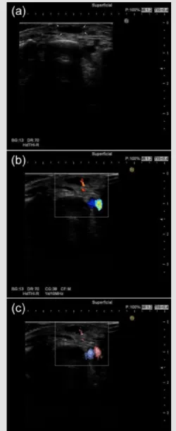
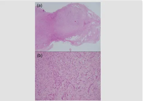
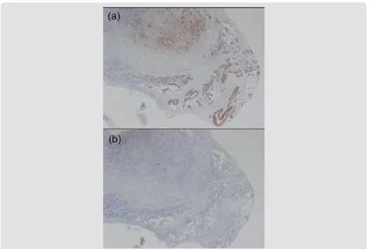
Characteristic Appearances of Nodular Fasciitis on High-Resolution Ultrasonography: With Vasculature Status from A Lesion-Located Perspective
Full text
Figure
Related documents
In addition to Canada, other dominions of the British Empire immediately.. entered the war to
Administration, the Norfolk State University Board of Visitors approves the 2013-2014 legislative priorities as presented and outlined in the attached exhibits with the authority
For the optimum blade configuration there would be two parameters, cord length (c) of each aerofoil and a local twist angle (β) of each aerofoil; which will be varied to
In general terms, the two lower stocking rate groups, especially that of beef cattle, have less intermediate consumption, so that they would be more sustainable in regard to
It does, but these are derivative of the duties applying to institutional agents, and grounded in the natural duty of justice to (i) comply with just institutions, (ii)
In this study, we used birth data during 2014–2016 in Ningbo, Zhejiang Province, China, and conducted a time-series study to investigate the association between exposure to ambient
The MOHLTC ‘ PDA initiative ’ enabled access to three core electronic resources for mobile devices: drug and medical reference materials, best prac- tice guidelines from the
( F , G ) Late phase FA and mid-phase ICG 12 months post-treatment demonstrating the area of grid laser photocoagulation (white arrows), and the obstruction of the arterial
