Article
1
Membrane-permeable octanoyloxybenzyl-masked
2
cNMPs as novel tools for non-invasive cell assays
3
Alexandra Ruthenbeck 1, Elisa Marangoni 1,2, Björn-Ph. Diercks 3, Aileen Krüger 3, Viacheslav O.
4
Nikolaev 3, Andreas H. Guse 3 and Chris Meier 1,*
5
1 Organic Chemistry, Department of Chemistry, Faculty of Sciences, University of Hamburg,
6
Martin-Luther-King-Platz 6, D-20146 Hamburg, Germany; [email protected]
7
2 School of Pharmacy, Medicinal Chemistry Unit, University of Camerino, via S. Agostino 1, 62032 Camerino,
8
Italy,
9
3 University Medical Center Hamburg-Eppendorf, Martinistraße 52, 20246 Hamburg, Germany;
10
11
* Correspondence: [email protected]; Tel.: +49-40-42838-4324
12
13
Abstract: Adenine nucleotide (AN) 2nd messengers such as 3’,5’-cyclic adenosine monophosphate
14
(cAMP) are central elements of intracellular signaling, but many details of underlying processes
15
remain still elusive. Like all nucleotides, cyclic nucleotide monophosphates (cNMPs) are
16
net-negatively charged at physiologic pH which limits their applicability in cell-based settings.
17
Thus, many cellular assays rely on sophisticated techniques like microinjection or electroporation.
18
This setup is not feasible for medium- to high-throughput formats, and the mechanic stress that
19
cells are exposed to raises the probability of interfering artefacts or false-positives.
20
Here, we present a short and flexible chemical route yielding membrane-permeable, bio-reversibly
21
masked cNMPs for which we employed the octanoyloxybenzyl (OB) group. We further show
22
hydrolysis studies on chemical stability and enzymatic activation, and present results of real-time
23
assays, where we used cAMP and Ca2+ live cell imaging to demonstrate high permeability and
24
prompt intracellular conversion of some selected masked cNMPs. Consequently, our novel
25
OB-masked cNMPs constitute valuable precursor-tools for non-invasive studies on intracellular
26
signaling.
27
Keywords: cyclic nucleotide monophosphate; bio-reversible protection; acyloxybenzyl phosphate
28
ester.
29
30
1. Introduction
31
Among cNMPs, the ubiquitous second messengers cAMP and cGMP constitute the most
32
prominent examples but also ‘non-canonical’ cNMPs such as 3’,5’-cyclic uridine monophosphate
33
(cUMP) have been reported and related to signaling processes.[1] For example, cAMP plays an
34
important role in many biological processes, in addition to its implication in Ca2+ mobilization. This
35
cyclic mononucleotide is generated from ATP through G protein-coupled receptor (GPCR) based
36
activation of adenylyl cyclase (AC). The signaling by cNMPs proceeds via two general pathways.
37
The first relies on direct binding and regulation of distinctive cyclic nucleotide-gated (CNG) ion
38
channels. The second mechanism is based on the activation of protein kinases A (PKA) or G to
39
further transduce the signal.[1–4]
40
Activated PKA/G promotes phosphorylation of a variety of proteins, which can be involved in
41
the regulation of metabolic processes, muscle contraction and gene transcription.[4] In contrast,
42
signaling through CNG channels allows a faster processing and implementation of increased cNMP
43
levels. The ion channels are generally non-selective for cations. However, the entry of sodium ions
44
(Na+) depolarizes the membrane which promotes the combined influx of Ca2+. Additionally,
45
voltage-gated Ca2+ channels open in response to membrane depolarization and thus enhance the
46
Ca2+ signal further.[2,3] Signals of cAMP and cGMP are ceased through their degradation by
47
phosphodiesterase to AMP and GMP, respectively.[2,3]
48
Due to the ubiquitous involvement in cellular processes, the role of cNMP signaling in the
49
context of inflammation and regulation of immune response constitutes a research field of rising
50
interest. It was for example found that regulatory T cells (Treg) exert their suppressive effect on
51
effector T cells (Teff) through increasing concentrations of cAMP. The rise of cAMP levels in Teff could
52
either be induced via a paracrine mechanism, or by a direct transfer of cAMP via gap junctions
53
between Treg and Teff.[5,6] However, the transfer of cAMP between a pair of T cells could not be
54
visualized directly yet, and also the underlying mechanisms allowing Treg cells to produce such
55
significantly higher cAMP levels than Teff cells are not elucidated to date. A loss of this
56
immuno-suppressive mechanism could contribute to the generation of autoimmune reactions.
57
Understanding the role of cNMPs in inflammation and T cell regulation and identifying the
58
associated molecular pathways thus could enable the identification of novel targets in the treatment
59
of autoimmune disease.
60
Cell-based studies on 2nd messengers are generally difficult to perform as application of the
61
highly polar compounds is carried out via effortful, single-cell preparative methods like
62
electroporation, microinjection or patch clamp. These methods require highly trained staff, careful
63
preparation and significant amounts of time in advance of each experiment while at the same time
64
the invasive application raises the potential for interfering artefacts or false-positives.[7] Membrane
65
permeable, bio-reversibly modified chemical derivatives of second messengers are highly desirable
66
to circumvent this drawback.
67
First reports on protected cNMPs by Engels et al. included cAMP and cGMP derivatives
68
carrying different benzyl groups at the phosphate.[8,9] Hughes et al. reported the synthesis and
69
biologic evaluation of N6,O2-dibutyryl cAMP-acetoxymethyl (AM) ester.[10] The synthesis started
70
from N6,O2’-dibutyryl cAMP, which was converted with AM bromide (AM-Br) under DIEA-basic
71
conditions in CH3CN over 4 days at room temperature.[10] The obtained masked nucleotide was
72
used in whole-cell incubation studies where an activation of PKA proceeded, but only 15 min after
73
the addition of di(Bu)cAMP-AM (10 µM) to the cell medium.[10] In a further study, Schultz et al.
74
synthesized cAMP-AM via a transient protection of the 2’-OH function with the trimethylsilyl (TMS)
75
group and successive esterification of the phosphate under the before described conditions.[11]
76
Despite a PKA activating effect of cAMP-AM the researchers found that cAMP-AM was less potent
77
than di(Bu)cAMP-AM and metabolized so rapidly that it gave only transient signals.[11] The
78
synthesis of AM-esters of cCMP and cUMP have also been reported.[12] However, cNMP-AMs are
79
not applicable for all cell types, seemingly due to insufficient chemical stability, and flexibility of the
80
synthesis approach is limited by availability of the respective cNMP.
81
The acyloxybenzyl (AB)-masking system was introduced originally for nucleoside
82
monophosphate (NMP) prodrugs and successfully transferred on nucleoside diphosphates (NDPs)
83
and nucleoside triphosphates (NTPs).[13–17] The respective prodrugs were shown to efficiently
84
diffuse across cell membranes and release the corresponding nucleotide intracellularly upon
85
enzymatic activation.[14] Further, the concept was expanded on compound classes like sugar
86
nucleotides.[18] The broad synthetic applicability of the AB masking group is complimented by
87
meeting the essential features of a prodrug concept: i) high chemical stability, ii) efficient enzymatic
88
activation and iii) sufficient lipophilicity to enable the passage through cell membranes. Moreover,
89
the two-part composition of the AB-masks allows variation of e.g lipophilicity or enzymatic
90
cleavability which adds further versatility to the concept.[13–15,19–21] Consequently, the
91
AB-concept was investigated for expansion on cNMPs to provide tools for studies on cellular effects
92
like e.g. calcium signaling in bulk settings on a variety of cells and without the need for example of
93
microinjection. As a first example, an octanoyloxybenzyl (OB)-mask was used here.
94
2. Results and Discussion
96
Inspiration for the synthesis approach towards OB-masked cNMPs was drawn from own
97
studies on the synthesis of non-symmetric phosphoramidites (PAs) that included an unprotected
98
nucleoside moiety. In these cases, side reactions were repeatedly observed amongst which the
99
formation of phosphites was prominent. These phosphites were concluded to result either from an
100
intermolecular or an intramolecular substitution of the remaining N-diisopropyl group by a second
101
nucleosidic hydroxy-group. In the latter case, a cyclic nucleoside phosphite would constitute the
102
reaction product, and further oxidation would result in a cyclic nucleoside monophosphate
103
derivative.
104
Based on this hypothesis, we set up a synthesis involving different nucleosides and an
105
OB-masked phosphordiamidite (OBPA2).
106
2.1. Preparation of AB-masked cNMPs
107
2.1.1. Synthesis of starting materials
108
The synthesis of bis(N,N-diisopropylamino)-4-octanoyloxybenzyl phosphordiamidite (OBPA2)
109
was adapted from Weinschenk et al. who used the building block for the synthesis of non-symmetric
110
PAs.[22] The reaction was performed as described in the literature starting from
111
bis(N,N-diisopropylamino)chlorophosphine 1 and 4-(hydroxymethyl)phenyloctanoate 2, and
112
yielded OBPA2 3 in 71% (Scheme 1).
113
114
Scheme 1. Synthesis of OBPA2 3: (i.) 1 equiv. 1, 1 equiv. 2, 1.3 equiv. TEA, THF, rt, 18 h, 3: 71%.
115
The nucleosides adenosine (A, 4), 2’-deoxy adenosine (dA, 5) and guanosine (G, 6) were
116
mono-N-butyrylated to add further lipophilicity to the envisaged OB-cNMPs. N6-Butanoyl-
117
adenosine 7 was synthesized starting from adenosine 4 via transient silylation of all hydroxy-groups
118
to selectively introduce the butyryl moiety at the N6-position.[23] The desired protected nucleoside 7
119
was obtained in 61% yield after automated RP flash column chromatography (Figure 2). The yield
120
was limited by the formation of N,N-diacylated adenosine and partial cleavage of the glycosidic
121
bond during concentration of the crude reaction mixture. Analogously, N6-butanoyl-dA 8 and
122
N2-butanoyl-G 9 were synthesized starting from the respective nucleosides and obtained in yields of
123
32% and 48%, respectively (Scheme 2).
124
2.1.2. Syntheses of OB-cNMPs
125
First, a model system, as which the route towards octanoyloxybenzyl-masked cUMP
126
(OB-cUMP, 10) served, was set up to study course and outcome of the phosphitylation in more
127
detail.
128
The starting conditions for the synthesis of OB-cUMP 10 were adapted from the approaches
129
towards non-symmetric PAs.[22] 4,5-Dicyanoimidazole (DCI, 0.25 M in CH3CN) was used as
130
activator for the PA-coupling, and tBuOOH (5.5 M in n-decane) served as oxidizing agent. The
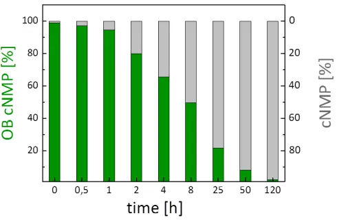
131
reaction was carried out in a mixture of acetonitrile and DMF due to the limited solubility of uridine
132
134
Scheme 2. Synthesis of N-butyryl nucleosides 7-9: (ii.) first 2.1 - 9.0 equiv. TMSCl, 0 °C to rt, 18 h,
135
second 1.1 equiv. butanoyl chloride, pyridine/THF or pyridine/CH2Cl2, rt, 5 - 18 h, third 1 M aq. HCl
136
or CH3OH, rt, 5 min or 18 h, 7: 61%, 8: 32%, 9: 48%.
137
In the first attempt, a solution of nucleoside 11 and OBPA2 3 was treated with 2.2 equivalents of
138
DCI that were added dropwise at 0 °C. The reaction mixture was allowed to warm to rt and stirred
139
30 min more before tBuOOH was added for oxidation. Lastly, the crude reaction mixture was
140
purified by automated RP flash column chromatography, and the masked cyclic nucleotide 10 was
141
obtained as a mixture of two diastereomers in a yield of only 13 % (Scheme 3).
142
Prompted by the surprisingly low yield, the reaction course was studied 31P-NMR
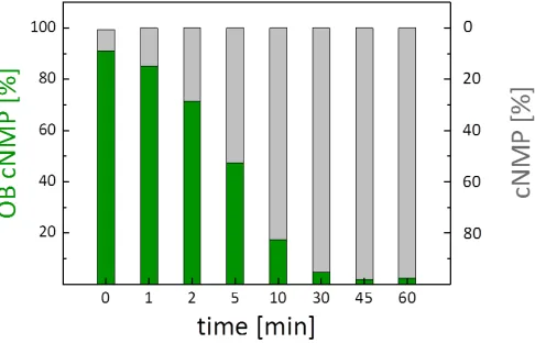
143
spectroscopically (Fig. 1). Interestingly, upon just mixing uridine 11 and OBPA2 3, the diamidite was
144
converted almost quantitatively and a signal at 13.2 ppm formed (Fig. 1). Comparison with
145
literature-data indicated that this signal likely corresponded to an activated amidite.[24]
146
Furthermore, signals in the range of PAs and phosphites were formed already as well (Fig. 1, top).
147
The addition of DCI then promoted the formation of the intermediate PA, but formation of the
148
anticipated cyclic phosphite seemed to occur only at low proportion. Steric hindrance in the attack of
149
the 3’-hydroxy group on the phosphorous atom or an insufficient nucleophilicity may be the reasons
150
that led to an insufficient formation of the cyclic phosphite.
151
152
Scheme 3: (iii.) 1.1 equiv. 3, 2.2 equiv. DCI (0.25 M in CH3CN), 1.5 equiv. tBuOOH (5.5 M in
153
n-decane), CH3CN/DMF 5:4, 0 °C to rt, 60 min, 10: 13%. (iv.) 1.1 equiv. 3, first portion of 1.3 equiv.
154
DCI (0.25 M in CH3CN), second portion of 1.3 equiv. DCI (0.25 M in CH3CN), 1.5 equiv. tBuOOH (5.5
155
M in n-decane), CH3CN/DMF 5:1, rt, 60 min, 10: 15%. (v.) 1.1 equiv. 3, first portion of 1.3 equiv. DCI
156
(0.25 M in CH3CN), second portion of 1.3 equiv. BTT (0.3 M in CH3CN), 1.5 equiv. tBuOOH (5.5 M in
157
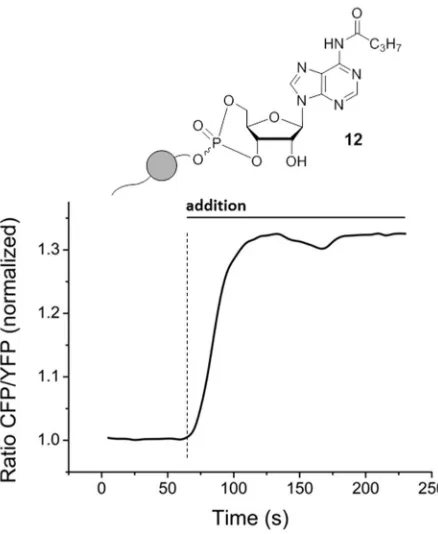
n-decane), CH3CN/DMF 5:1, rt, 60 min, 10: 13 – 19%. (vi.) 1 – 1.2 equiv. 3, first portion of 1 – 1.2 equiv.
158
saccharin and 1 – 1.2 equiv. 1-methylimidazole, second portion of 1 – 1.2 equiv. saccharin and 1 – 1.2
159
equiv. 1-methylimidazole, 1.5 equiv. tBuOOH (5.5 M in n-decane), CH3CN/DMF 5:1, rt, 60 min to 72
160
162
Figure 1: 31P-NMR spectra (CH3CN-d3, 162 MHz, 25 °C, shifts δ in [ppm]) of the reaction monitoring
163
between uridine 11 and OBPA2 3.
164
The synthesis of OB-cUMP 10 consequently was repeated with several changes to the protocol
165
in order to evaluate the influence of the reaction conditions and in particular the impact of the
166
activator (Scheme 3).
167
In a second approach, the ratio between CH3CN and DMF was altered to 5:1 and the mode of
168
addition of reagents was changed. This time, the nucleoside was placed in the reaction flask and
169
dissolved in CH3CN/DMF 5:1. A separate solution of OBPA2 3 in CH3CN and one equivalent of DCI
170
were added slowly and dropwise to nucleoside 11 at rt. Once the addition was completed, a second
171
equivalent of DCI was added, and the reaction mixture stirred at rt for 60 min. After the successive
172
oxidation and final purification, OB-cUMP 10 was obtained in 15% yield. Consequently, the outcome
173
of the reaction was almost identical to that of the attempt before.
174
Next, the activator was changed to 5-(benzylthio)-1H-tetrazole (BTT) which displays a higher
175
acidity and lower nucleophilicity than DCI, and the reaction protocol was varied as follows: uridine
176
11 was dissolved in CH3CN/DMF 5:1 and successively treated with a solution of OBPA2 3 in CH3CN
177
and one equivalent DCI. Both reagents were added in small portions over a period of 30 min. Upon
178
completed addition, the reaction mixture was stirred another 30 to 120 min at rt. Then, one
179
equivalent BTT was added slowly and dropwise over 10 min, and the reaction was kept stirring for
180
further 15 to 60 min. After the subsequent oxidation and final purification, OB-cUMP 10 was isolated
181
in yields between 13 – 19% which again constituted no significant improvement of the reaction
182
outcome. Lastly, an alternative activator-system composed of saccharine and 1-methylimidazole
183
was tested as it was reported to efficiently mediate also reactions between PAs and poorly
184
nucleophilic alcohols like tertiary alcohols.[25]
185
Saccharine and 1-methylimidazole were dissolved in a 1:1 ratio in CH3CN prior to the reaction
186
added to a solution of uridine 11 in CH3CN/DMF 5:1. Upon completion of the first addition, the
188
reaction mixture was stirred for 30 min, then treated with a second equivalent of the activator
189
solution and successively stirred for another 60 min to 72 h. After oxidation and automated RP
190
column chromatography, the desired product 10 was obtained in yields between 16 – 19%.
191
Monitoring of the reaction course via 31P-NMR spectroscopy indicated an incomplete activation of
192
OBPA2 3 even after 72 h. Further, the activated intermediates were again not converted efficiently to
193
the desired phosphite as concluded from the persistence of the respectively attributed phosphorous
194
signals.[25] This alternative approach constituted thus no improvement in comparison to the
195
previous protocols neither. In summary, the isolation of OB-cUMP 10 succeeded repeatedly despite
196
low yields, and thus the reaction protocols were transferred on the N-butyrylated nucleosides 7 – 9
197
(Scheme 4).
198
The N-butyrylated OB-cNMPs 12–14 were prepared following the synthesis protocols iv. and v.
199
described above. The yields obtained for OB-N(Bu)-cAMP 12 and OB-N(Bu)-cdAMP 13 were in a
200
similar range as found for OB-cUMP 10 with 14% and 13%, respectively (Scheme 4). The reaction
201
towards OB-N(Bu)-cGMP 14 proceeded to a lower extent and accordingly, the desired product was
202
isolated in a comparably low yield of 4% (Scheme 4). Interestingly, in the case of 14 the formation of
203
only one of the two possible diastereomers seemed favored as crude 31P-NMR spectra indicated.
204
Here, only one of the possible two phosphate signals was prominent, and consequently,
205
OB-N(Bu)-cGMP 14 was isolated as a single diastereomer. Additionally to the N-acylated derivative
206
12, the NH2-unmodified OB-cAMP 15 was prepared. The synthesis was performed analogously to
207
OB-cUMP 10 starting from nucleoside 4 and following synthesis variant iii. which made use of a
208
stepwise addition of the in total applied 2.4 equivalents DCI (Scheme 5).
209
N
NHR
O
OH/H OH HO
iv./v. O
OH/H O O
P O O
O O
C7H15
N
NHR
N(Bu)-A7
N(Bu)-dA8
N(Bu)-G 9
OB-N(Bu)-cAMP 12
OB-N(Bu)-cdAMP 13
OB-N(Bu)-cGMP 14
R = N-butyryl
210
Scheme 4: Syntheses of and N-butyrylated OB-cNMPs 12–14: to 12: (v.) 1.1 equiv. 3, first portion of
211
1.3 equiv. DCI (0.25 M in CH3CN), second portion of 1.3 equiv. BTT (0.3 M in CH3CN), 1.5 equiv.
212
tBuOOH (5.5 M in n-decane), CH3CN/DMF 3:1, rt, 60 min, 12: 14%. To 13: (iv.) 1.1 equiv. 3, first
213
portion of 1.3 equiv. DCI (0.25 M in CH3CN), second portion of 1.3 equiv. DCI (0.25 M in CH3CN),
214
1.5 equiv. tBuOOH (5.5 M in n-decane), CH3CN/DMF 1:1, rt, 60 min, 13: 13%. To 14: (iv.) 1.1 equiv. 3,
215
first portion of 1.5 equiv. DCI (0.25 M in CH3CN), second portion of 1.5 equiv. DCI (0.25 M in
216
CH3CN), 1.5 equiv. tBuOOH (5.5 M in n-decane), CH3CN/DMF 1:1, rt, 60 min, 14: 4%.
217
In summary, five different OB-cNMPs were successfully synthesized. With regard to the overall
218
similar outcomes of the various preparations, the scope of the reaction can likely be expanded on
219
further nucleosides and/or differently masked PA2s. This makes the chosen approach individually
220
adaptable and very flexible, particularly in contrast to the previously reported procedures involving
221
AM esters.
222
224
Scheme 5: Preparation of OB-cAMP 15: iii. 1.1 equiv. 3, 2.4 equiv. DCI (0.25 M in CH3CN) added in
225
two portions, 1.5 equiv. tBuOOH (5.5 M in n-decane), CH3CN/DMF 1:1, rt, 60 min, 15: 12%.
226
2.2. Functional evaluation of selected OB-cNMPs
227
Ideally, masked precursors of bio-active compounds are biologically inactive and display a
228
stability in physiologic media that on the one hand exceeds their specific activation significantly, and
229
on the other hand guarantees sufficient time for their approximation to their respective target
230
structure. Once activated, the previously inactive compound regains its biologic activity back and
231
should accordingly displays respective effects from target interaction. These demands applied for
232
the prepared OB-cNMPs equally as e.g. for nucleotide prodrugs. Consequently, the chemical
233
stability of the OB-cNMPs under physiologic conditions as well as the efficiency of the enzymatic
234
activation of the OB-mask by an exemplary esterase was evaluated. In complementation, the masked
235
nucleotides were applied in various cell assays to analyze their biologic effects e.g. in the context of
236
Ca2+ mobilization and cell activation.
237
2.2.1. Investigation of chemical stability and enzymatic activation by PLE
238
Stability determinations for OB-cNMPs 10,12 and 13 (as 5 mM stock sol. in DMSO) were
239
performed in PBS (50 mM, pH 7.3) as physiologic pH mimic. The compounds (2 mM, final conc. in
240
PBS/DMSO) were incubated over 120 to 200 h at 37 °C. Hydrolysis samples were taken at distinctive
241
times and analyzed via HPLC/MS (Fig. 2 & 3). The recorded HPLC chromatograms and ESI mass
242
spectra showed that the OB-cNMPs released only their respective parent cNMP from cleavage of the
243
OB-mask without the formation of further cleavage byproducts (Fig. 3).
244
245
Figure 2: Course of the chemical hydrolysis of OB-N(Bu)-cAMP 12 to N(Bu)-cAMP 15 given as
246
normalized values for each time point analyzed. Similar hydrolysis courses were measured for
247
249
250
Figure 3: HPLC chromatograms and ESI mass spectra of the chemical hydrolysis of OB-N(Bu)-cAMP
251
12 in PBS (50 mM, pH 7.3) at 0 h, 1 h, 8 h & 50 h. The two signals of the diastereomers vanished over
252
time while the signal for N(Bu)-cAMP 15 increased. The compounds were assigned from the mass
253
spectra recorded at the retention times coinciding with the signals found in the HPLC
254
chromatograms.
255
It was found that after approximately 8 h 50% of the OB-cNMPs were hydrolyzed (10: t½ = 8.6 h,
256
12: t½ = 7.4 h, 13: t½ = 7.5 h) (Fig. 2). This half-life implied a stability of the OB-cNMPs that should
257
facilitate convenient setups of cell-based assays and allow for satisfactory time to run, for example,
258
even (pre-) incubation experiments with the masked nucleotides.
259
The hydrolysis behavior of 10,12 and 13 was assessed further, and the relative areas of signals for
260
OB-cNMP and cNMP were determined (in percent). These values were normalized and then
261
averaged for all three hydrolyses under determination of their standard deviation. By this
262
hydrolyses was expected to be largely similar as the dissociation of the OB-mask should constitute
264
the determinant process.
265
The average deviation of the hydrolysis progress for OB-cNMPs 10,12 and 13 at the time points
266
measured was 5% with a single value maximum of 12%. From these calculations, it was deduced
267
that the hydrolysis course was indeed analog for all OB-cNMPs tested and subject to the pace of the
268
cleavage of the masking group and was almost independent of the nucleotide employed.
269
After successful probing the chemical stability of the OB-cNMPs satisfying, their enzymatic
270
activation by pig liver esterase (PLE) as an exemplary esterase was evaluated. The incubations of
271
OB-cNMPs 10,12 and 13 (2 mM final conc. in PBS/DMSO) with PLE were carried out with 0.05 u PLE
272
per hydrolysis sample (V = 20 µL) which enabled good traceability of the enzymatic conversion (Fig.
273
4). Again, no further cleavage products apart from the respective cNMPs were determined. The
274
N-butyryl group of 12 and 13 was not cleaved by PLE, even at longer incubation times (up to
275
60 min), as expected. The acquired chromatograms were processed as described for the chemical
276
hydrolysis to compare the progresses of the individual incubations with PLE (Fig. 5). The half-lives
277
of the studied OB-cNMPs were around 5 min under the applied conditions, which, however
278
depended significantly on the amount of esterase present. More importantly in this context, the
279
enzymatic activation of OB-cNMPs proceeded even at low PLE concentrations significantly faster
280
than their decomposition in PBS by a factor of approximately 100. In addition, the enzymatic
281
hydrolyses showed analog progression as indicated by a mean deviation of normalized signal areas
282
for OB-cNMPs and cNMPs of 4% with a maximum deviation of 9% for single time point values (Fig.
283
5). This permitted again the conclusion that the enzymatic reaction was almost independent of the
284
type of nucleotide and relied on the OB-mask applied.
285
286
Figure 4: HPLC chromatograms and ESI mass spectra of the enzymatic hydrolysis of
287
OB-N(Bu)-cAMP 12 in with PLE (0.05 u/hydrol. sol.) in PBS (50 mM, pH 7.3) at 1, 5 & 10 min. The two
288
signals of the diastereomers vanished over time while the signal for N(Bu)-cAMP 15 increased. The
289
compounds were assigned from mass spectra recorded at the retention times coinciding with the
290
292
Figure 5: Course of the enzymatic hydrolyses of of OB-N(Bu)-cAMP 12 to N(Bu)-cAMP 15 given as
293
normalized values for each time point analyzed. Similar hydrolysis courses were measured for
294
OB-cNMPs 10 and 13.
295
In summary, the results of both hydrolysis studies, chemical and enzymatic, went well along
296
with the initial criteria as they showed that the stability of the prepared OB-cNMPs was significantly
297
higher than the rate of enzymatic activation. Further, the masked nucleotides proved to be
298
satisfactory stable for application in cell-based assays as their stabilities allow for incubations even
299
over several hours.
300
Encouraged by the promising hydrolysis properties, the ability of the OB-cNMPs to cross
301
cellular membranes as well as their potential to induce cellular processes was studied successively.
302
2.2.2. Performance of selected OB-cNMPs in cell-based settings
303
Primary mouse cardiomyocytes carrying a FRET-sensor with a cAMP binding site were used to
304
examine the membrane-permeability of OB-N(Bu)-cAMP 12 in particular. The binding of
305
intracellular cAMP to the FRET sensor is indicated by a decreasing FRET-signal and an increasing
306
fluorescence ratio between cyan-fluorescent protein (CFP) and yellow-fluorescent protein (YFP).[26]
307
FRET-sensor carrying mouse cardiomyocytes were incubated with OB-N(Bu)-cAMP 12 (20 mM, at t
308
≈ 60 s) (Fig. 6). Immediately after addition of 12 to the extracellular medium, the ratio between CFP
309
and YFP started to increase and reached a steady maximum state at circa 110 s. These results imply
310
that OB-N(Bu)-cAMP 12 instantaneously crossed the cell membrane and was also rapidly activated
311
by intracellular esterases. Further, the product of the activation process was successfully recognized
312
by the cAMP binding site of the FRET sensor. For the studied substrate, OB-N(Bu)-cAMP 12, this
313
implicated that the N6-butyryl group was either removed by enzymatic hydrolysis, or that its
314
presence had no detrimental effect on receptor interaction.
315
In a second setup, Jurkat T cells were loaded with the Ca2+-sensitive fluorescent dye Fura-2.
316
Upon intracellular elevation of Ca2+, the absorption ratio between the two excitation wavelengths of
317
Fura-2 at 340 nm and 380 nm increases. This effect correlates directly with the amount of free
318
cytosolic Ca2+. Jurkat T cells loaded with Fura-2 were stimulated with OB-cNMPs 10, 12 and 13
319
(20 µM in DMSO, at t ≈ 120 s) added to their extracellular medium (Fig. 7). The Fura-2 ratio rose
320
rapidly almost immediately after addition of OB-N(Bu)-cAMP 12, and reached its maximum after
321
approximately 200 s. Then, the Ca2+ signal slowly decreased as indicated by the degression of the
322
signal. A similar trend was observed for OB-cUMP 10 but the induced Ca2+ signal was significantly
323
reduced compared to 12 (Fig. 7). In the case of OB-N(Bu)-cdAMP 13, no initial increase of the
324
intracellular Ca2+ concentration was measured. However, the ratio seemed to increase slightly over
325
327
Figure 6: Normalized FRET ratio (between CFP and YFP) over the course of OB-N(Bu)-cAMP 12
328
addition to mouse cardiomyocytes Epac1-camps biosensor for intracellular cAMP. An instantaneous
329
increase of FRET ratio after addition of 12 to the extracellular medium indicated intracellular release
330
of cAMP and its binding to the FRET biosensor. Representative experiments (n=5).
331
332
333
Figure 7: Stimulation of the of Jurkat T cells with OB-cNMPs 10,12 and 13. Left: Jurkat T cells were
334
stimulated after 120 s with the respective OB-cNMPs (20 µM) or DMSO (as negative control).
335
Furthermore, as positive control Thapsigagarin (1.67 µM) was added after 900 s. Mean signal ratio
336
between 340 nm and 380 nm from single cells are shown (DMSO n=26; OB cAMP n=77; OB cUMP
337
n=37; OB cdAMP n=14). The addition of 12 and 10 resulted in a transient increase of the Ca2+
338
concentration, while no transient increase is visible for 13 or DMSO. Right: Statistical analysis of the
339
mean delta peak for the OB-cNMPs and DMSO (data represent mean ± SEM). The most pronounced
340
effect is measured for 12 and statistically significant differences between are marked by asterisks (* p
341
< 0.05, ** p<0.01, *** p<0.001, Kruskal-Wallis Test).
342
The results confirmed again that the OB-masked cNMPs were able to cross the cell membrane
343
de-masked cNMPs promoted the observed effects based on the results of the previous hydrolysis
345
studies and the substrate specify Ca2+ signaling events display.
346
The hydrolysis product of 12 acted as it would be expected for cAMP supporting the
347
assumption that the N6-butyryl group was either removed enzymatically, too, or that its presence
348
did not impede receptor activation.
349
Comparison of the measured effects with those evoked by NH2-unmodified OB-cAMP 15 and
350
e.g. further nucleobase derivatives of adenosine or uridine in combination with incubation studies in
351
cell homogenate could help to finalize the analysis and clarify whether the N6-butyryl group is
352
cleaved or the interacting receptors and binding sites lack selectivity in the corresponding region.
353
Studies along these lines are currently performed in our laboratories.
354
In summary, the performed cell assays confirmed excellent membrane-permeability of the
355
selected OB-cNMPs. Further, the cellular effects observed allow the conclusion that the
356
bio-reversible protection at the phosphate was removed rapidly and efficiently. A fast enzymatic
357
activation of the prepared OB-cNMPs was shown analogously in hydrolysis studies using pig liver
358
esterase. The esterase cleaved the OB-mask even at low concentration within very short time, so that
359
similar effects can be expected to proceed in cells. Finally, the observed FRET-sensor binding site
360
interaction and induced Ca2+ mobilization proved that biologically active compounds were released
361
out of the masked cNMPs. Moreover, the masked nucleotides triggered processes like they are
362
attributed to their parent cNMPs (if existent in nature/identified yet).
363
3. Materials and Methods
364
All reactions involving water-sensitive reagents were conducted under anhydrous conditions
365
and a dry atmosphere of nitrogen.
366
Reagents were used as purchased from commercial suppliers.
367
Anhydrous N,N-dimethylformamide (DMF) was purchased and stored over 4 Å molecular sieves.
368
All other anhydrous solvents were purified and dried using a solvent purification system (MB SPS-800
369
from Braun) and stored over appropriate molecular sieves.
370
Solvents for normal phase (NP) chromatography were distilled prior to use. Acetonitrile was
371
purchased in HPLC grade for reversed phase (RP) chromatography and HPLC.
372
Evaporation of solvents was performed under reduced pressure on a rotary evaporator or using a
373
high vacuum pump.
374
Reactions were monitored via thin layer chromatography (TLC) carried out on pre-coated
375
Macherey-Nagel TLC plates Alugram® Xtra SIL G/UV254, and compounds stained with Vanillin
376
(Vanillin (5 g), 1000 mL MeOH/AcOH 9:1, 35 mL H2SO4) under heating.
377
For automated NP or RP chromatography two flash systems (Interchim Puriflash 430 or Sepacore®
378
Flash System, combined with Chromabond® Flash RS 80 SiOH (NP) or RS40 C18 ec (RP) columns)
379
were used. For purifications of phosphordiamidites, a chromatotron (Harrison Research 7924T) with
380
glass plates coated with 2 or 4 mm layers of VWR60 PF254 silica gel containing a fluorescent indicator
381
(VWR no. 7749) was used.
382
Analytical RP-High Performance Liquid Chromatography-Mass Spectrometry (RP-HPLC/MS) was
383
performed with an Agilent 1260 Infinity instrument (pump G1311B, autosampler G1329B, column
384
compartment G1316A, diode array detector G4221B, column Agilent Poroshell 120 EC-C18, 2.7 mm,
385
4.6x50 mm) coupled with single-quad MS (Advion expressionL CMS).
386
Ultrapure water was generated by a Sartorius Aurium® pro unit (Sartopore 0.2 µm, UV). As elution
387
buffer served a tetra-n-butylammonium acetate solution (10 mM, pH 7.2). HPLC/MS runs were
388
performed according to the following method: 0 – 15 min: water/acetonitrile gradient (2% – 98% B)
389
with a flow of 0.5 mL/min, 20 °C column temperature and UV detection at 259 nm and 270 nm, MS
390
scans from 150 to 1100 m/z.
391
Nuclear magnetic resonance (NMR) spectra were recorded at room temperature on Bruker Fourier 300
392
(300 MHz for 1H acquisitions), Bruker AMX 400 (400 MHz for 1H 101 MHz for 13C and 152 MHz for
393
31P acquisitions) or Bruker AVIII 600 (600 MHz for 1H and 151 MHz for 13C acquisitions)
394
the solvent resonance as internal standard. Coupling constants J are given in Hertz (Hz).
396
Two-dimensional NMR experiments (HSQC, HMBC) were used for the assignment of quaternary
397
carbons.
398
For mass spectrometric (MS) analytic, spectra were acquired on an Agilent 6224 ESI-TOF spectrometer
399
in positive and negative mode as required.
400
Infrared spectroscopy (IR) was carried out with a Bruker Alpha P FT-IR in attenuated total reflection
401
(ATR) mode at room temperature ranging from 400 cm-1 to 4000 cm-1.
402
For FRET measurements, primary mouse ventricular cardiomyocytes were isolated from
403
Epac1-camps biosensor expressing transgenic mice[27] as described[26] and plated onto laminin
404
coated glass cover slides. Measurements were performed 1 – 2 h after plating using a Nikon Ti
405
microscope based FRET imaging system containing pE-100 440 nm light source (CoolLED), DV2
406
Dual View and ORCA-03G charge-coupled device camera (Hamamatsu), and analyzed as
407
described[26]. Cells were kept in a buffer containing 144 mM NaCl, 5.4 mM KCl, 1 mM MgSO4, 1
408
mM CaCl2, 10 mM Hepes (pH 7.3) and stimulated with OB-cNMPs dissolved 1:1000 in the same
409
buffer from a freshly made 20 mM DMSO stock solution.
410
For Ca2+ mobilization assays, Jurkat T cells were incubated with the membrane-permeable AM ester of
411
the Ca2+ dye Fura-2 (4 µM, Calbiochem). Therefore, about 2 x 10⁶ cellswere centrifuged at 500 g for
412
5 min and resuspended in 1 mL of freshly supplemented RPMI medium containing Fura-2 AM.
413
Cells were incubated for 30 min at 37 °C. After centrifugation, cells were washed and resuspended in
414
Ca2+ buffer [140 mM NaCl, 5 mM KCl, 1 mM MgSO4, 1 mM CaCl2, 20 mM Hepes (pH 7.4), 1 mM
415
NaH2PO4, 5 mM glucose] and kept for 20 min at room-temperature (RT) for de-esterification. Cells
416
were added on prepared coverslips and allowed to adhere before measurement. Slides were
417
mounted onto a Leica IRBE microscope (100-fold magnification) and after 120s the respective
418
OB-cNMPs (20 µM) or DMSO (as control) were added. As positive control, Thapsigagarin (1.67 µM,
419
Calbiochem) was added after 900 s. A Sutter DG-4 was used as a light source, and frames were
420
acquired with an electron-multiplying charge-coupled device camera (C9100-13, Hamamatsu).
421
Images (512 × 512 pixels) were acquired in 16-bit mode with the following filter sets (AHF
422
Analysentechnik) [excitation (ex), beam splitter (bs), and emission (em), all in nanometers]: Fura-2
423
(ex, HC 340/26, HC 387/11; bs, 400DCLP; em, 510/84).
424
425
General Procedures:
426
GP I: N-butanoylation of nucleosides via transient TMS-protection:
427
The respective nucleoside (adenosine, guanosine or 2’-deoxyadenosine) was co-evaporated
428
three times and then dissolved in anhydrous pyridine (2 – 5 mL/mmol), and diluted either with the
429
same volume of THF or double the volume of CH2Cl2. At 0 °C, TMSCl (2.1 – 9.0 equiv.) was added.
430
The reaction mixture was allowed to warm up to rt and stirred for 5 – 18 h. Successively, butyryl
431
chloride (1.1 equiv.) was added slowly and the reaction mixture stirred for another 6 h at rt.
432
Cleavage of TMS ethers was promoted by the addition of either 1 M HCl (0.5 mL/mmol) under
433
vigorous stirring for 5 min, or methanol (2 - 5 mL/mmol) and stirring at rt for further 12 h. The
434
reaction was terminated by removal of all volatile components under high vacuum. The crude
435
residue was co-evaporated with toluene and CH2Cl2 several times, and then taken up in
436
acetonitrile/demin. water. Purification was performed by means of automated RP flash column
437
chromatography on C18 modified silica gel with an acetonitrile gradient in water (0% to 100%).
438
439
GP II: 3’,5’-Phosphorylation of nucleosides to their OB-masked cNMP analogues:
440
Under an atmosphere of nitrogen, the respective nucleoside was dissolved in DMF/CH3CN
441
(25 mL/mmol). In a separate flask, bis(N,N-diisopropylamino)-4-octanoyloxybenzyl
phosphor-442
amidite (1 equiv.) was dissolved in acetonitrile (25 mL/mmol total volume). The phosphor diamidite
443
solution and DCI (0.25 M in CH3CN, 1.3 – 1.5 equiv.) were added slowly and dropwise in five to ten
444
portions to the nucleoside solution. The addition of more DCI (0.25 M in CH3CN, 1.2 – 1.5 equiv.) or
445
5-(benzylthio)-1H-tetrazole (BTT, 0.3 M in CH3CN, 1.3 equiv.) followed, and the reaction mixture
446
stirred for 10 min more. Successively, all volatile components were removed in vacuum, and the
448
obtained residue was taken up in CH3CN/demin. water and purified by means of an automated RP
449
flash column chromatography on C18 modified silica gel with an CH3CN gradient in water (0% to
450
100%).
451
452
Syntheses:
453
Synthesis of bis(N,N-diisopropylamino)-4-octanoyloxybenzyl phosphordiamidite 3:
454
1.00 g (3.75 mmol) bis(N,N-diisopropylamino)chlorophosphine 1 were dissolved in 15 mL
455
anhydrous THF. In a separate flask, 0.68 mL (4.87 mmol, 1.3 equiv.) NEt3 and 0.94 g (3.75 mmol, 1
456
equiv.) 4-(hydroxymethyl)phenyloctanoate 2 were mixed with 7 mL anhydrous THF, and the
457
mixture was added dropwise to the chlorophosphine. The reaction mixture was stirred at rt for 20 h,
458
then filtrated and the filtrate concentrated to dryness in vacuum. The remaining residue was
459
purified by NP chromatography on silica gel with PE/TEA 98:2 as eluents, and the desired product
460
obtained as colorless syrup.
461
Yield: 1.28 g (2.67 mmol, 71%).
462
1H-NMR (600 MHz, chloroform-d): δ [ppm] = 7.36 (d, 2JH,H = 8.2 Hz, 2 H), 7.14 – 6.95 (m, 2 H), 4.63
463
(d, 2JH,H = 7.2 Hz, 2 H), 3.66 – 3.51 (m, 4 H), 2.54 (t, 2,3JH,H = 7.5 Hz, 2 H), 1.75 (p, 2,3JH,H = 7.4 Hz, 2 H),
464
1.51 – 1.23 (m, 8 H), 1.21 (d, 2JH,H = 2.7 Hz, 12 H), 1.20 (d, 2JH,H = 2.8 Hz, 12 H) 0.88 (t, 2,3JH,H = 7.3 Hz,
465
3 H).
466
13C{1H}-NMR (151 MHz, chloroform-d): δ [ppm] = 172.6, 149.7, 138.2, 127.9, 121.5, 65.8, 44.69,
467
44.56, 34.6, 31.8, 29.2, 29.1, 25.1, 24.8, 24.7, 24.1, 24.0, 22.75, 14.22.
468
31P{1H}-NMR (162 MHz, chloroform-d): δ [ppm] = 123.5.
469
IR (ATR): ṽ in [cm-1] = 2963.3, 2927.8, 2861.4, 2079.0, 2025.5, 1761.3, 1607.8, 1507.4, 1457.5, 1416.5,
470
1390.1, 1361.6, 1300.3, 1194.2, 1184.8, 1162.9, 1140.3, 1116.2, 1045.2, 1016.9, 952.7, 916.5, 866.3, 779.6,
471
748.7, 706.7, 642.7, 566.1, 527.6.
472
MS (MALDI): m/z [M-H] calc. for C27H48N2O3P-: 479.340, found: 479.245.
473
474
Synthesis of 6-N-butanoyl-adenosine 7:
475
In accordance with GP I, 1.40 g (5.26 mmol) adenosine 4 were dissolve in 34 mL pyridine/THF
476
1:1 and converted with 2.11 mL (16.5 mmol, 3.2 equiv.) TMSCl and 0.60 mL (5.78 mmol, 1.1 equiv.)
477
butyryl chloride. After 6 h stirring at rt, 2.5 mL 1 M HCl (aq.) was added to cleave the TMS ethers,
478
and after 5 min, all volatile components were removed under vacuum. Upon final purification of the
479
crude product via automated RP flash column chromatography on C18 modified silica gel with an
480
CH3CN gradient in water (0% to 100%), the product was obtained as colorless powder.
481
Yield: 1.09 g (3.22 mmol, 61%).
482
1H-NMR (500 MHz, DMSO-d6): δ [ppm] = 10.63 (s, 1 H), 8.69 (s, 1 H), 8.65 (s, 1 H), 6.01 (d,
483
3JH,H = 5.8 Hz, 1 H), 5.53 (d, 3JH,H = 5.8 Hz, 1 H), 5.23 (d, 3JH,H = 4.8 Hz, 1 H), 5.12 (t, 3JH,H = 5.6 Hz, 1 H),
484
4.63 (q, 3JH,H = 5.4 Hz, 1 H), 4.19 (q, 3JH,H = 4.3 Hz, 1 H), 3.98 (q, 3JH,H = 3.9 Hz, 1 H), 3.76 – 3.64 (m, 1 H),
485
3.58 (ddd, 2JH,H = 11.9 Hz, 3JH,H = 6.1 Hz, 3JH,H = 4.0 Hz, 1 H), 2.55 (t, 2,3JH,H = 7.3 Hz, 2 H), 1.63 (h,
486
2,3JH,H = 7.4 Hz, 2 H), 0.94 (t, 2,3JH,H = 7.4 Hz, 3 H).
487
13C-NMR (126 MHz, DMSO-d6): δ [ppm] = 171.5, 151.7, 151.6, 149.7, 142.7, 123.9, 87.6, 85.7, 73.7,
488
70.3, 61.3, 38.0, 18.2, 13.5.
489
IR (ATR): ṽ in [cm-1] = 3273.2, 2963.3, 2934.0, 2875.0, 1716.3, 1685.0, 1613.3, 1587.0, 1521.9, 1460.3,
490
1409.0, 1356.6, 1327.0, 1224.3, 1124.1, 1082.2, 1056.7, 984.6, 902.4, 866.3, 799.6, 745.0, 704.7, 643.7, 547.3.
491
MS (ESI-HR): m/z [M+H]+ calc. for C14H20N5O5+: 338.1459, found: 338.1469.
492
493
Synthesis of 6-N-butanoyl-2’-deoxyadenosine 8:
494
Following GP I, 1.14 g (4.24 mmol) 2’-deoxyadenosine 5 were dissolve in 24 mL
495
pyridine/CH2Cl2 1:2. At 0 °C, 1.13 mL (8.91 mmol, 2.1 equiv.) TMSCl were added, and the reaction
496
mixture was stirred for 18 h at rt. Successively, 0.48 mL (4.67 mmol, 1.1 equiv.) butyryl chloride were
497
added. After further 3 h stirring at rt, the TMS ethers were removed by addition of 8 mL CH3OH at
498
vacuum. The crude product was taken up in water containing little amount of CH3CN and purified
500
via automated RP flash column chromatography on C18 modified silica gel with an CH3CN gradient
501
in water (0% to 100%) to afford the desired product as colorless powder.
502
Yield: 0.43 g (1.35 mmol, 32%).
503
1H-NMR (400 MHz, methanol-d4): δ [ppm] = 8.66 (s, 1 H), 8.62 (s, 1 H), 6.61 – 6.52 (m, 1 H), 4.64
504
(dt, 3JH,H = 6.1 Hz, 3JH,H = 3.1 Hz, 1 H), 4.10 (q, 3JH,H = 3.4 Hz, 1 H), 3.88 (dd, 2JH,H = 12.2 Hz, 3JH,H = 3.4
505
Hz, 1 H), 3.79 (dd, 2JH,H = 12.2 Hz, 3JH,H = 3.9 Hz, 1 H), 2.88 (ddd, 2JH,H = 13.4 Hz, 3JH,H = 7.4 Hz,
506
3JH,H = 6.0 Hz, 1 H), 2.67 (t, 2,3JH,H = 7.4 Hz, 2 H), 2.51 (ddd, 2JH,H = 13.5 Hz, 3JH,H = 6.2 Hz, 3JH,H = 3.3 Hz, 1
507
H), 1.82 (h, 2,3JH,H = 7.4 Hz, 2 H), 1.08 (t, 2,3JH,H = 7.4 Hz, 3 H).
508
13C{1H}-NMR (101 MHz, methanol-d4): δ [ppm] = 174.4, 152.9, 150.7, 144.3, 123.2, 89.7, 86.7, 72.7,
509
63.4, 41.5, 39.9, 19.6, 14.0.
510
IR (ATR): ṽ in [cm-1] = 3336.8, 2964.6, 2933.1, 2875.3, 2592.1, 2330.9, 1682.2, 1612.5.1585.9, 1522.5,
511
1459.6, 1402.8, 1354.8, 1329.2, 1223.8, 1093.3, 1058.5, 993.8, 941.1, 867.1, 799.6, 749.6, 984.8, 644.4, 585.3,
512
561.2, 542.4, 527.4, 509.6, 464.7.
513
MS (ESI-HR): m/z [M+H]+ calc. for C14H20N5O4+: 322.1510, found: 322.1521.
514
515
Synthesis of 2-N-butanoyl-guanosine 9:
516
According to GP I, 1.55 g (5.48 mmol) guanosine 6 were co-evaporated with pyridine three
517
times and then dissolved in 81 mL pyridine/CH2Cl2 1:2. At 0 °C, 6.28 mL (49.4 mmol, 9 equiv.)
518
TMSCl were added, and the reaction mixture was stirred for 4 h at rt. Successively, 0.62 mL
519
(6.03 mmol, 1.1 equiv.) butyryl chloride were added. After 3 h stirring at rt, the cleavage of the TMS
520
ethers was induced by addition of 27 mL CH3OH, and after further 12 h at rt, the reaction was
521
terminated and all volatile components were removed under vacuum. The crude residue was taken
522
up in water containing little amount of CH3CN and finally purified via automated RP flash column
523
chromatography on C18 modified silica gel with an CH3CN gradient in water (0% to 100%) to afford
524
the desired product as colorless powder.
525
Yield: 0.92 g (2.61 mmol, 48%).
526
1H-NMR (400 MHz, DMSO-d6): δ [ppm] = 12.07 (bs, 1 H), 11.71 (bs, 1 H), 8.26 (s, 1 H), 5.80 (d,
527
3JH,H = 5.7 Hz, 1 H), 5.47 (d, 3JH,H = 5.7 Hz, 1 H), 5.17 (d, 3JH,H = 4.5 Hz, 1 H), 5.03 (t, 3JH,H = 5.4 Hz, 1 H),
528
4.43 (d, 3JH,H = 5.2 Hz, 1 H), 4.20 – 4.05 (m, 1 H), 3.90 (q, J = 3.9 Hz, 1 H), 3.64 (dt, 2JH,H = 11.9 Hz,
529
3JH,H = 4.8 Hz, 1 H), 3.55 (dt, 2JH,H = 11.9 Hz, 3JH,H = 4.7 Hz, 1 H), 2.45 (t, 2,3JH,H = 7.3 Hz, 2 H), 1.62 (h,
530
2,3JH,H = 7.4 Hz, 2 H), 0.92 (t, 2,3JH,H = 7.5 Hz, 3 H).
531
13C{1H}-NMR (101 MHz, DMSO-d6): δ [ppm] = 176.2, 154.8, 148.8, 148.0, 137.6, 120.1, 86.6, 85.3,
532
73.9, 61.1, 37.8, 17.9, 13.4.
533
IR (ATR): ṽ in [cm-1] = 3364.4, 3279.5, 2968.3, 2941.1, 1680.2, 1608.8, 1564.3, 1554.1, 1536.0, 1481.4,
534
1469.2, 1449.9, 1402.5, 1251.7, 1204.2, 1179.4, 1127.9, 1089.0, 1060.2, 993.3, 976.4, 901.1, 863.3, 818.6,
535
801.0, 762.7, 737.4, 717.0, 680.4, 643.7, 607.0, 589.1, 562.3, 510.2, 484.2.
536
MS (ESI-HR): m/z [M+H]+ calc. for C14H19N5O6+: 354.1408, found: 354.1400.
537
538
Synthesis of uridine-3’,5‘-(4-octanoyloxybenzyl)cyclophosphate 10:
539
According to GP II, 26 mg (0.11 mmol) uridine 10 were dissolved in 2 mL DMF and reacted
540
with 57 mg (0.12 mmol, 1.1 equiv.) OB-PA2 3, dissolved in 2.5 mL CH3CN, in the presence of 0.54 mL
541
(0.13 mmol, 1.3 equiv.) DCI (0.25 M in CH3CN) and 0.45 mL (0.13 mmol, 1.3 equiv.) BTT (0.3 M in
542
CH3CN). The addition of 32 µL (0.16 mmol, 1.5 equiv.) tBuOOH (5.5 M in n-decane) followed
543
successively. After purification by automated RP flash column chromatography on C18 modified
544
silica gel with a CH3CN gradient in water (0% to 100%) and lyophilization, the product was obtained
545
as colorless cotton and in two fractions containing each of the stereoisomers.
546
Yield: 11 mg (0.02 mmol, 19%).
547
1H-NMR (400 MHz, methanol-d4): δ [ppm] = 7.66 – 7.51 (m, 2 H), 7.47 (d, 3JH,H = 8.1 Hz, 1 H), 7.22 –
548
7.08 (m, 2 H), 5.69 (d, 3JH,H = 8.0 Hz, 1 H), 5.64 (d, 3JH,H = 0.9 Hz, 1 H), 5.24 – 5.14 (m, 2 H), 4.64 (ddd,
549
4.35 – 4.25 (m, 2 H), 4.25 – 4.10 (m, 1 H), 2.58 (t, 2,3JH,H = 7.4 Hz, 2 H), 1.73 (p, 2,3JH,H = 7.4 Hz, 2 H), 1.53 –
551
1.22 (m, 8 H), 1.05 – 0.82 (m, 3 H)
552
and
553
7.63 (d, 3JH,H = 8.1 Hz, 1 H), 7.53 – 7.47 (m, 2 H), 7.19 – 7.10 (m, 2 H), 5.73 (d, 3JH,H = 1.2 Hz, 1 H), 5.71 (d,
554
3JH,H = 8.1 Hz, 1 H), 5.19 (d, 2JH,H = 8.7 Hz, 2 H), 4.81 (ddd, 3JH,P = 9.8 Hz, 3JH,H = 5.1 Hz, 3JH,H = 1.0 Hz, 1
555
H), 4.67 (ddd, 3JH,P = 14.2 Hz, 2JH,H = 9.2 Hz, 3JH,H = 5.5 Hz, 1 H), 4.62 – 4.47 (m, 2 H), 4.42 (td,
556
2JH,H = 10.1, 3JH,H = 5.5 Hz, 1 H), 2.60 (t, 2,3JH,H 7.4 Hz, 2 H), 1.74 (p, 2,3JH,H 7.4 Hz, 2 H), 1.50 – 1.26 (m, 8
557
H), 1.02 – 0.84 (m, 3 H).
558
13C{1H}-NMR (101 MHz, methanol-d4): δ [ppm] = . 173.8, 165.9, 151.5, 150.1, 143.7, 134.3, 131.2,
559
123.3, 103.0, 97.5, 80.1, 72.4, 71.5, 71.2, 70.3, 35.0, 32.9, 30.2, 30.1, 25.0, 23.7, 14.4
560
and
561
172.8, 164.9, 151.7, 150.5, 143.5, 133.0, 130.2, 122.8, 103.1, 97.7, 79.3, 72.4, 71.2, 71.1, 70.6, 35.0, 32.8,
562
30.2, 30.1, 26.0, 23.7, 14.4.
563
31P{1H}-NMR (162 MHz, methanol-d4): δ [ppm] = --3.93, -5.01.
564
MS (ESI-HR): m/z [M+Na]+ calc. for C24H31N2O10PNa+: 561.1609, found: 561.1445.
565
566
Synthesis of 6-N-butanoyl-adenosine-3’,5‘-(4-octanoyloxybenzyl)cyclophosphate 12:
567
According to GP II, 48 mg (0.14 mmol) 6-N-butanoyl-adenosine 7 were dissolved in 4 mL DMF
568
and treated with a solution of 76 mg (0.16 mmol, 1.1 equiv.) OB-PA2 3 in 4 mL CH3CN and 0.97 mL
569
(0.24 mmol, 1.7 equiv.) DCI (0.25 M in CH3CN) as well as 0.62 mL (0.19 mmol, 1.3 equiv.) BTT (0.3 M
570
in CH3CN). Then, 43 µL (0.21 mmol, 1.5 equiv.) tBuOOH (5.5 M in n-decane) were added. After
571
purification by automated RP flash chromatography on C18 modified silica with a CH3CN gradient
572
in water (0% to 100%), the product was obtained as colorless cotton and mixture of two
573
diastereomers.
574
Yield: 13 g (0.02 mmol, 14%).
575
1H-NMR (400 MHz, methanol-d4): δ [ppm] = 8.68, 8.58 (2 x s, 2 H), 8.49, 8.38 ( 2x s, 2 H),
576
7.62 – 7.56, 7.55 – 7.49 (2 x m, 4 H), 7.17 – 7.12, 7.13 – 7.06 (2 x m, 4 H), 6.20, 6.12 (2 x s, 2 H), 5.47 (dd,
577
3JH,P = 9.1 Hz, 3JH,H = 5.1 Hz, 1 H) 5.23 (2 x d, 2JH,H = 10.4 Hz & 8.1 Hz, 4 H), 5.05 (ddd, 3JH,P = 9.6 Hz,
578
3JH,H = 5.1 Hz, 3JH,H = 1.6 Hz, 1 H), 4.89 – 4.86 (m, 1 H), 4.73 – 4.34 (m, 7 H), 2.71 – 2.55 (m, 6 H,), 2.51 (t,
579
2,3JH,H = 7.4 Hz, 2 H), 1.86 – 1.70 (m, 6 H), 1.68 – 1.57 (m, 2 H), 1.53 – 1.25 (m, 16 H), 1.05 (2 x t,
580
2,3JH,H = 7.4 Hz, 6 H), 0.98 – 0.95 (m, 6 H).
581
13C{1H}-NMR (151 MHz, methanol-d4): δ [ppm] = 173.8, 173.6, 152.8, 152.4, 150.8, 143.1, 133.3,
582
133.2, 130.7, 130.6, 124.5, 123.1, 123.0, 88.8, 81.7, 80.4, 69.3, 69.2, 68.5, 59.8, 39.9, 34.5, 31.8, 29.2, 29.1,
583
25.0, 22.8, 18.5,14.2, 13.9.
584
31P{1H}-NMR (162 MHz, methanol-d4): δ [ppm] = -3.80, -4.90.
585
MS (ESI-HR): m/z [M+H]+ calc. for C29H39N5O9P+: 632.2848, found: 632.2848.
586
587
Synthesis of 6-N-butanoyl-2’-deoxyadenosine-3’,5‘-(4-octanoyloxybenzyl)cyclophosphate 13:
588
According to GP II, 50 mg (0.15 mmol) 6-N-butanoyl-2’-deoxyadenosine 8 were dissolved in
589
4 mL DMF and reacted with 82 mg (0.17 mmol, 1.1 equiv.) OB-PA2 3, dissolved in 4.3 mL CH3CN, in
590
the presence of 1.60 mL (0.40 mmol, 2.6 equiv.) DCI (0.25 M in CH3CN) in total and 50 µL
591
(0.25 mmol, 1.6 equiv.) tBuOOH (5.5 M in n-decane). After purification by automated RP flash
592
column chromatography on C18 modified silica gel with a CH3CN gradient in water (0% to 100%)
593
and lyophilization, the product was obtained as colorless cotton and mixture of two diastereomers.
594
Yield: 12 mg (0.02 mmol, 13%).
595
1H-NMR (400 MHz, methanol-d4): δ [ppm] = 8.69 (s, 1 H), 8.61 (s, 1 H), 8.47 (s, 1 H), 8.37 (s, 1 H),
596
7.65 – 7.57, 7.55 – 7.48 (2 x m, 2 H), 7.19 – 7.10 (m, 4 H), 6.59 (dd, 3JH,H = 9.0 Hz, 3JH,H = 2.0 Hz, 1 H), 6.55
597
(dd, 3JH,H = 6.7 Hz, 3JH,H = 4.0 Hz, 1 H), 5.64 (q, 3JH,H/P = 9.2 Hz, 1 H), 5.33 (q, 3JH,H/P = 9.2 Hz, 1 H), 5.23 (2
598
x d, 2JH,H = 13.1 Hz & 3JH,H = 12.9 Hz, 4 H), 4.70 – 4.40 (m, 4 H), 4.22 (td, 3JH,H = 9.9 Hz, 3JH,H = 5.6 Hz, 1
599
H), 4.08 (td, 3JH,H = 9.9 Hz, 3JH,H = 4.7 Hz, 1 H), 2.98 (ddd, 2JH,H = 13.1 Hz, 3JH,H = 7.7 Hz, 3JH,H = 1.9 Hz, 1
600
H), 2.83 – 2.70 (m, 3 H), 2.69 – 2.57 (m, 6 H), 2.54 (t, 2,3JH,H = 7.4 Hz, 2 H), 1.90 – 1.70 (m, 6 H), 1.66 (q,
![Figure 1: 31P-NMR spectra (CH3CN-d3, 162 MHz, 25 °C, shifts δ in [ppm]) of the reaction monitoring between uridine 11 and OBPA2 3](https://thumb-us.123doks.com/thumbv2/123dok_us/7902780.1311968/5.595.97.525.71.406/figure-nmr-spectra-shifts-reaction-monitoring-uridine-obpa.webp)