Scholarship@Western
Scholarship@Western
Electronic Thesis and Dissertation Repository
7-25-2017 12:00 AM
Genetic Engineering Studies of Escherichia coli and Microalgae
Genetic Engineering Studies of Escherichia coli and Microalgae
for Expression of Hydrolytic Enzymes and Development of High
for Expression of Hydrolytic Enzymes and Development of High
Throughput Screening Technique
Throughput Screening Technique
Shreyas S. Yedahalli
The University of Western Ontario
Supervisor
Dr. Amarjeet Bassi
The University of Western Ontario Joint Supervisor Dr. Lars Rehmann
The University of Western Ontario
Graduate Program in Chemical and Biochemical Engineering
A thesis submitted in partial fulfillment of the requirements for the degree in Doctor of Philosophy
© Shreyas S. Yedahalli 2017
Follow this and additional works at: https://ir.lib.uwo.ca/etd
Part of the Biochemical and Biomolecular Engineering Commons, Biotechnology Commons, Molecular Biology Commons, and the Other Biomedical Engineering and Bioengineering Commons
Recommended Citation Recommended Citation
Yedahalli, Shreyas S., "Genetic Engineering Studies of Escherichia coli and Microalgae for Expression of Hydrolytic Enzymes and Development of High Throughput Screening Technique" (2017). Electronic Thesis and Dissertation Repository. 4706.
https://ir.lib.uwo.ca/etd/4706
This Dissertation/Thesis is brought to you for free and open access by Scholarship@Western. It has been accepted for inclusion in Electronic Thesis and Dissertation Repository by an authorized administrator of
i
The field of biochemical engineering has made substantial progress through major advances
in genetic and metabolic engineering with applications in various sectors such as energy,
food science, pharmaceuticals, etc. The hosts used for this work are constantly broadening. A
host particularly important for energy applications are microalgae. The potential to enhance
microalgae genetically for energy applications is not well explored and was therefore
investigated in this thesis. Non-photosynthetic micro-organisms and photosynthetic
microalgae offer a potential approach to enhance sustainable biochemical production. In this
study expression vectors for Escherichia coli (E. coli) and Chlorella vulgaris (C. vulgaris) were constructed for the expression of two enzymes: exo-inulinase enzyme and
β-glucuronidase (GUS reporter) reporter protein. The expressed enzymes were characterized
for their activity. The study involved three phases.
In the first phase of the research, the inulin hydrolyzing enzyme exo-inulinase was expressed
in E. coli strain Rosetta-gami B(DE3). For this purpose, first the exo-inulinase gene from Aspergillus niger 12 (A. niger 12) was isolated by reverse transcription polymerase chain reaction (RT-PCR) and further an expression vector pET32a+EX-INU was constructed. The
recombinant exo-inulinase was then expressed in E. coli strain Rosetta-gami B(DE3). The recombinant exo-inulinase was purified, and characterized for its activity. The molecular
weight of the recombinant exo-inulinase was 81 Da (similar to native exo-inulinase). The Km and Vmax toward inulin were 5.3 ± 1.1 mM and 402.1 ± 53.1 µmol min-1 mg-1 protein respectively and the optimum temperature and pH for maximum enzyme activity were 55 0C
and 5.0 respectively.
In the second phase of the research, the 4-Methylumbelliferyl-β-D-glucuronide (4-MUG)
hydrolyzing enzyme β-glucuronidase was expressed in the photosynthetic microalgae
ii
1168 by expression of the GUS reporter enzyme. In a lysed cell study, enzyme kinetic
analysis for the expressed phenotype was also carried out. The values of Km and Vmax of the
recombinant GUS enzyme were 0.1304 ± 0.0101 mM and 0.35 ± 0.004 pmol 4-MU/min/ml
of crude cell lysate respectively.
In the third and final phase of the research, the proposed HTS method developed in the
previous study was applied to a second microalgal system. First, 4-MUG hydrolyzing GUS
enzyme was expressed in Chlamydomonas reinhardtii CC1690 (C. reinhardtii). The transgenic C. reinhardtii expressing GUS enzyme was developed by A. tumefaciens transformation techniques. The expression vector used in this work was
pBIN-Hyg-Tx–GUS-INT. High throughput screening of transgenic colonies expressing β-glucuronidase (GUS
activity) was carried out directly from agar plates. 1 out of 126 transgenic C. reinhardtii colony, showed the highest fluorescence intensity 1,113. This study confirmed the
application of the HTS method to microalgal systems. This is a new tool which can be
applied for fast screening of genetic transformations and expression in microalgal systems.
iii
Co-Authorship Statement
This thesis was completed under the supervision of Dr. Amarjeet Bassi and Dr. Lars
Rehmann. Four articles were written and co-authored. For all four articles the first author is
Shreyas Yedahalli. The co-author and corresponding author are Dr. Lars Rehmann and Dr.
iv
Acknowledgments
First, I would like to thank my advisors Dr. Amarjeet Bassi and Dr. Lars Rehmann, for their
guidance, encouragement, patience, and full support during my entire graduate study. I am
sincerely thankful and grateful for all their help academically and financially throughout my
Ph.D. study. It has always been a great honor to have them as my advisor. I have truly
learned and benefited a lot from them.
I would also like to thank all the former and current laboratory members in our research
group and office colleagues especially Dibakar mondal, Junwoo Kim, Tahereh Sarchami and
Dr. Erin Johnson. Also, I would like to thank friends from back home Vinod, Sreenivas,
Sangamantha, Sriram and Prodip.
Finally, my warmest thanks must be to my family. I would like to thank my sisters Dr.
Ashwini, and Dr. Shalini. Thanks to my parents, Mr. Somegowda, and Mrs. Navamani, who
v
Dedicated to
To my parents Some Gowda Yedahalli Namadaru and Navamani Chandagal
vi
Table of Contents
Co-Authorship Statement... iii
Acknowledgments... iv
Table of Contents ... vi
List of Tables ... xi
List of Figures ... xiii
List of Appendices ... xviii
Abbreviations ... xix
Chapter 1 ... 1
1 Introduction ... 1
1.1 Research structure ... 3
1.2 Objectives ... 5
1.2.1 Overall objective ... 5
1.2.2 Specific objectives ... 5
1.3 Outline of the Thesis ... 7
1.4 References ... 9
Chapter 2 ... 10
2 Molecular Biology Tools and Approaches for Genetic Modification of Microalgae – A Review of Recent Developments ... 10
2.1 Introduction ... 11
2.1.1 Genome sequence ... 13
2.1.2 Gene transformation techniques ... 20
2.1.3 Selectable markers ... 29
2.1.4 Photosynthetic markers ... 35
vii
2.1.7 Inducible system ... 52
2.1.8 Affinity tags ... 60
2.1.9 Export signal ... 62
2.2 Conclusions ... 66
2.3 References ... 70
Chapter 3 ... 83
3 General materials and methods ... 83
3.1 Strains, growth, and transformation ... 83
3.1.1 Escherichia coli ... 83
3.1.2 Agrobacterium tumefaciens LBA 4404 ... 84
3.1.3 Chlorella vulgaris (UTEX 2714) ... 85
3.2 Polymerase chain reaction ... 86
3.2.1 Colony PCR ... 87
3.3 Agarose gel preparation and gel electrophoresis ... 89
3.4 Glycerol stock ... 89
3.5 Composition of Lysogeny broth ... 89
3.6 Agar plate preparation... 92
3.7 Plasmid preparation and storage ... 92
Chapter 4 ... 93
4 Expression of exo-inulinase gene from Aspergillus niger 12 in E. coli strain Rosetta-gami B (DE3) and its characterization ... 93
4.1 Introduction ... 94
4.2 Materials and methods ... 96
4.2.1 Strains, plasmids and reagents ... 96
viii
4.2.4 Recombinant exo-inulinase purification ... 99
4.2.5 Protein quantification ... 100
4.2.6 Western blot and SDS-PAGE of recombinant exo-inulinase ... 100
4.2.7 Measurement of expressed exo-inulinase activity ... 100
4.2.8 pH Optimum and Stability studies ... 101
4.2.9 Temperature optimum and thermostability ... 102
4.2.10 Effect of metal ion on recombinant exo-inulinase and invertase activity102 4.3 Results ... 103
4.3.1 Expression in Rosetta-gami B(DE3) ... 103
4.3.2 Purification of over expressed recombinant exo-inulinase ... 104
4.3.3 Characterization of exo-inulinase ... 111
4.4 Discussion ... 118
4.5 Conclusions ... 119
4.6 References ... 120
Chapter 5 ... 124
5 High throughput screening of β-glucuronidase (GUS) reporter in transgenic microalgae transformed by Agrobacterium tumefaciens ... 124
5.1 Introduction ... 125
5.2 Materials and methods ... 128
5.2.1 Strains, plasmids, and culture condition ... 129
5.2.2 Construction of expression vector ... 129
5.2.3 Electroporation of A. tumefaciens and transformation of C. vulgaris .... 130
5.2.4 Quantitative Measurement of GUS Activity... 131
5.2.5 Screening for GUS activity in recombinant culture ... 131
ix
5.2.8 Quantification of GUS activity in lysed cell ... 133
5.2.9 Effect of different concentration of Triton X-100 on GUS activity ... 133
5.2.10 GUS enzyme kinetics ... 134
5.3 Results ... 135
5.3.1 Construction of Expression Vector ... 135
5.3.2 Antibiotic resistance study of C. vulgaris ... 136
5.3.3 Transformation and reporter gene expression ... 137
5.3.4 Direct screening of intact recombinant C. vulgaris ... 137
5.3.5 GUS activity in culture supernatant ... 139
5.3.6 Detection of GUS activity in intact recombinant C. vulgaris ... 139
5.3.7 Detection of GUS activity in lysed recombinant C. vulgaris ... 140
5.3.8 Effect of Triton X-100 on GUS activity in intact cell and its supernatant ... 142
5.3.9 GUS enzyme activity ... 145
5.4 Discussion ... 146
5.5 Conclusion ... 148
5.6 References ... 150
Chapter 6 ... 153
6 Application of high throughput screening technique for transgenic Chlamydomonas reinhardtii colonies on agar plate ... 153
6.1 Introduction ... 154
6.2 Materials and methods ... 155
6.2.1 Antibiotic test ... 155
6.2.2 Expression vector ... 155
6.2.3 Isolation and growth of single colony ... 155
x
6.3.1 Transformation of E. coli ... 156
6.3.2 Restriction digest analysis of expression vector ... 158
6.3.3 Gene transformation of Agrobacterium tumefaciens ... 159
6.3.4 Culturing and antibiotic resistance study of C. reinhardtii ... 160
6.3.5 Co-cultivation of A. tumefaciens and C. reinhardtii ... 161
6.3.6 Re - streaking of transformed colonies ... 162
6.3.7 Direct screening of intact recombinant C. reinhardtii ... 163
6.4 Discussion ... 164
6.5 Conclusion ... 165
6.6 References ... 166
Chapter 7 ... 168
7 Conclusions and Recommendations ... 168
7.1 Conclusions ... 168
7.2 Recommendations ... 170
Appendices ... 172
xi
List of Tables
Table 2.1 - Sequenced genes in microalgae ... 17
Table 2.2 – Gene transformation in microalgae ... 25
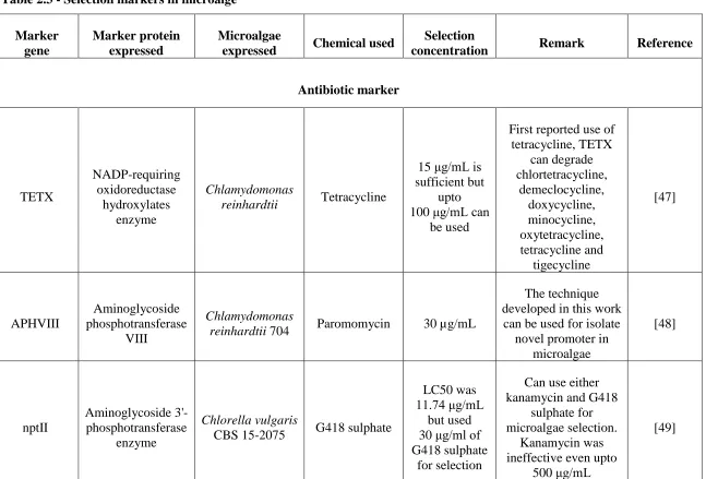
Table 2.3 - Selection markers in microalga... 31
Table 2.4 - Photosynthetic markers in microalgae ... 37
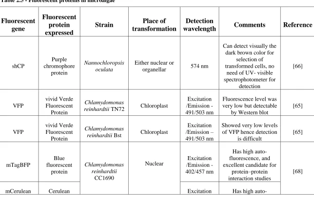
Table 2.5 - Fluorescent proteins in microalgae ... 42
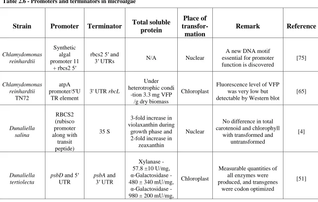
Table 2.6 - Promoters and terminators in microalgae ... 48
Table 2.7 - Inducible system in microalgae ... 56
Table 2.8 - Affinity tags in microalgae ... 61
Table 2.9 - Export signal in microalgae ... 64
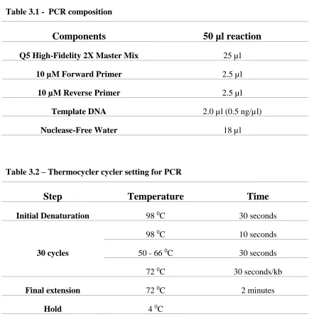
Table 3.1 - PCR composition ... 87
Table 3.2 – Thermocycler cycler setting for PCR ... 87
Table 3.3 – Colony PCR composition ... 88
Table 3.4 – Colony PCR thermocycler setting ... 88
Table 3.5 - Composition of Lysogeny broth ... 90
Table 3.6 - Composition of YEB medium ... 90
Table 3.7 - Composition of TAP medium ... 90
Table 3.8 - SOC media ... 91
xii
organism and recombinant exo-inulinase and invertase activity from current study. (*) E. coli
from current study; n=2 and average ± SE. ... 110
Table 4.3 - Effect of metal ions on recombinant exo-inulinase and invertase activity. The
purified enzyme was pre-incubated for 1 hr with 5 mM of metal ion in pH 5, 50 mM
Citrate-Phosphate buffer at 50 0C. The control was used to calculate the relative activity. n=2 and
xiii
List of Figures
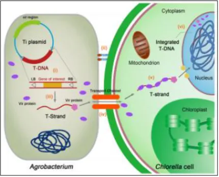
Figure 1.1 - Schematic representation of Chlorella vulgaris transformation by Agrobacterium
tumefaciens [1]... 2
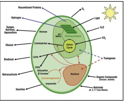
Figure 2.1 – Schematic representation of microalgae. The figure shows the expression of
transgene in nucleus and chloroplast. The potential enhancement of commercially important
bioproducts [103] ... 12
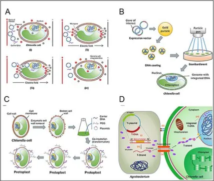
Figure 2.2 - Different transformation techniques of Chlorella sp. (A) Electroporation
transformation. (B) particle bombardment transformation. (C) PEG-mediated transformation.
(D) Agrobacterium-mediated genetic transformation [104]. ... 21
Figure 4.1 - pET-32a+EX-INU expression plasmid construct ... 98
Figure 4.2 - SDS-PAGE analysis of exo-inulinase enzyme expressed in Rosetta-gami
B(DE3). M- Marker, Lane 1 – 0.5 µg of purified Histidine tagged exo-inulinase protein;
Lane 2 – 0.5 µg flow through fraction from Ni2+-NTA purification. The gel was run at 100 V
for 90 minutes. The gel was stained in GelCode Blue Stain reagent according to
manufactures recommendation. ... 105
Figure 4.3 - Western blot analysis of Histidine tag exo-inulinase expressed in Rosetta-gami
B(DE3). M – Marker; Lane 1 – 0.5 µg of purified Histidine tagged exo-inulinase protein;
Lane 2 – 0.5 µg flow through fraction from Ni2+-NTA purification. The gel was run at 100 V
for 90 minutes. The gel was stained with InVision His-tag In-gel stain according to
manufactures recommendation. ... 106
Figure 4.4 - Michaelis–Menten kinetics plot for recombinant exo-inulinase activity. The
inulinase activity was carried out at 40 0C, pH 5.0 and 50 mM acetate buffer. The inulin
concentration ranging from 1-50 mg/mL was used for assay. Micro moles of fructose
liberated are shown by Michaelis–Menten kinetics. n=2, average ± SE and R2 – 0.98. ... 108
Figure 4.5 - Michaelis–Menten kinetics plot for recombinant invertase activity of
xiv
Micro moles of fructose liberated are shown by Michaelis–Menten kinetics. n=2, average ±
SE and R2 – 0.96. ... 109
Figure 4.6 - Effect of pH on recombinant exo-inulinase and invertase activity and their
stability. (A) Effect of pH on recombinant exo-inulinase and invertase activity; (B) Effect of
pH on recombinant exo-inulinase and invertase stability. I – Inulin; S – Sucrose. n=2,
average ± SE. Correlation coefficient for effect of pH on exo-inulinase – 0.98 and invertase –
0.98. Correlation coefficient for effect of pH stability on exo-inulinase – 0.72 and invertase –
0.94... 113
Figure 4.7 - Effect of temperature on recombinant exo-inulinase and invertase activity and
their stability. (A) Effect of temperature on recombinant exo-inulinase and invertase activity;
(B) Effect of temperature on recombinant exo-inulinase and invertase stability. I – Inulin; S –
Sucrose. n=2 and average ± SE. Correlation coefficient for effect of temperature on
exo-inulinase – 0.86 and invertase – 0.85. Correlation coefficient for effect of temperature
stability on exo-inulinase – 0.96 and invertase – 0.98. ... 115
Figure 5.1 - Schematic representation of expression vector pBIN+TetR-TetO constructed.
... 135
Figure 5.2 – DNA Flash gel run at 50 V. M – Generuler 1 kb Plus DNA Ladder, 1 –
pUC19+TX-GUS-INT double digest with EcoRI and HindIII (expected gene size of 2.6Kb
and 2.9 Kb). 2 – pBin+TetR+TetO double digested with HindIII and BspTI (expected gene
size of 12,299 bp and 3815 bp) ... 136
Figure 5.3 - Direct screening of intact recombinant C. vulgaris. 31 transgenic c. vulgaris and
1 control sample were screened in 96 well microplate. Sample number 1 being control.
Sample number 18 showed highest GUS activity. 150 µl of cells were pelleted and washed
with PBS buffer. The pelleted cells were suspended in assay buffer and incubated for 2 hours
and the fluorescence intensity was measured by excitation/emission wavelength of 365/455
nm. ... 138
Figure 5.4 – GUS activity in transgenic cell culture supernatant. 75 µl of transgenic cell
xv
2 3
3 hours, the fluorescence intensity recorded was 1,107.33 ± 3.78. ... 139
Figure 5.5 – Cell lysate GUS activity with mixing. 150 µl of pelleted cells was suspended in
assay buffer in 96 well microplate and incubated at 37 0C and 150 rpm in shaker. The
reaction was stopped by adding 50 µl of 1 M Na2CO3. At the end of 7 hours, the 4-MU
formed was 172 ± 8 µM/ OD600. ... 140
Figure 5.6 – Intact cell GUS activity with mixing. 75 µl of cell lysate was mixed with 75 µl
of assay buffer in 96 well microplate and incubated at 37 0C and 150 rpm in shaker. The
reaction was stopped by adding 50 µl of 1 M Na2CO3. At the end of 8 hours, the 4-MU
recorded was 4,303 ± 55 µM/mg of crude cell lysate. ... 141
Figure 5.7 – Effect of Triton X-100 on intact cell GUS activity. 150 µl of pelleted cells was
suspended in assay buffer containing different concentration of Triton X-100 (0 %, 0.01 %,
0.1 % and 1 %) in 96 well microplate and incubated at 37 0C. The reaction was stopped by
adding 50 µl of 1 M Na2CO3. At the end of 4 hours, the highest fluorescence intensity
recorded was 8,994 ± 357 for 0.1 % triton X-100. ... 143
Figure 5.8 – Effect of Triton X-100 on intact cell supernatant GUS activity. 150 µl of
pelleted cells was suspended in assay buffer containing different concentration of Triton
X-100 (0 %, 0.01 %, 0.1 % and 1 %) in 96 well microplate and incubated at 37 0C. The reaction
was stopped by adding 50 µl of 1M Na2CO3. At the end of 4 hours, the highest fluorescence
intensity recorded was 28,397 ± 787 for 0.1 % triton X-100. ... 144
Figure 5.9 – GUS enzyme Michaelis–Menten kinetics plot. 75 µl of cell lysate was mixed
with 75 µl of assay buffer containing different concentration of 4-MUG (0.06 mM to 5.0
mM) in 96 well PCR microplate and incubated at 37 0C in water bath for 10 minutes. The
reaction was stopped by adding 50 µl of 1 M Na2CO3. The values of kinetic constants Km
and Vmax for GUS were found to be 0.1304 ± 0.0101 mM and 0.35 ± 0.004 pmol
4-MU/min/ml of crude cell lysate respectively. ... 145
Figure 6.1 – GUS expression vector pBIN-Hyg-Tx-GUS-INT used for transformation of
xvi
transformed into E. coli strain DH5α. 50 µl of cells and 1 µl of 100 ng of vector were mixed
and kept on ice for 30 minutes. The cells were transformed by keeping in water bath (42 0C)
for 30 seconds and 950 µl of SOC was added and grew the cells by keeping at 37 0C for 60
minutes. 50 µl of cells were streaked on the Lb agar plate containing Kn antibiotic. The
recombinant cells were visible after 24 hr incubation at 37 0C. ... 158
Figure 6.3 - Agarose gel electrophoresis of vector restriction digestion. M: 1 kb Plus DNA
Ladder; Lane 2: Double digest of pBIN-HygTX-GUS-INT with EcoRI and HindIII (2858 bp
and 11986 bp). The flash gel was run at 50 V for 90 minutes. ... 159
Figure 6.4 - Electroporation transformation of Agrobacterium tumefaciens. The vector
pBIN-HygTX-GUS-INT was transformed into Agrobacterium tumefaciens strain LBA4404.
20 µl of electrocompetent cells and 2 µl of total 100 ng of vector were mixed and kept on ice.
The mixture was transferred into a chilled 0.1 cm cuvette and electroporated. The
electroporated cells were grown in 1 ml of SOC media at 30 0C for 60 minutes. 50 µl of cells
were streaked on the YEB agar plate containing Kn + St + Rf antibiotic. The recombinant
cells were visible after 48 hr incubation at 30 0C. ... 160
Figure 6.5 - Hygromycin B resistance of C. reinhardtii. The C. reinhardtii cells were
inoculated in 5 ml of TAP media and different concentration of Hygromycin B was added to
the media. The cell growth was monitored by OD600 absorbance. The cells were unable to
grow between 5 µg/ml - 30 µg/ml of antibiotic. ... 161
Figure 6.6 - Transformed C. reinhardtii – (A) Untransformed C. reinhardtii (control) grown
on TAP agar plate containing 10 µg/ml of Hygromycin B. (B) C. reinhardtii transformed
with binary vector pBIN-HygTX-GUS-INT grown on TAP agar plate containing 10 µg/ml of
Hygromycin B. The untransformed cells did not grow and transformed cells grew as
expected. ... 162
Figure 6.7 – Re - streaking of transformed colonies. Single colonies were streaked in a single
box and grew at 25 °C, 150 RPM, 90 % humid atmosphere and under 140 µmol m-2 s-1 white
light illumination of 16 h on/8 h off. The streaked cells were visible after 2 week of growth.
xvii
reinhardtii and 1 control sample were screened in 96 well microplates. Sample number 1
being control. Sample number 50 showed highest GUS activity. The scraped cells from the
TAP agar plate were suspended in 150 µl of assay buffer and incubated for 2 hours and
reaction was stopped by adding 50 µl of stop solution and the fluorescence intensity was
measured by excitation/emission wavelength of 365/455 nm. At the end of 2 hours, the
xviii
List of Appendices
Appendix A: Specification of the UV-Star 96 well microplate ... 172
Appendix B: Schematic representation of intermediate vector pUC19+Tx-GUS-INT ... 174
Appendix C: Blue white screening ... 175
Appendix D: A. tumefaciens LBA 4404 ... 176
Appendix E: Transformed Chlorella vulgaris ... 177
Appendix F: GUS Histochemical analysis ... 178
Appendix G: Intact cell GUS activity without mixing ... 179
Appendix H: Cell lysate GUS activity without mixing... 180
Appendix I: GUS Histochemical analysis ... 181
Appendix J: Protein calibration curve ... 182
xix
Abbreviations
Chlamydomonas reinhardtii C. reinhardtii
Aspergillus niger A. niger
Agrobacterium tumefaciens A. tumefaciens
Parachlorella kessleri P. kessleri
Chlorella variabilis C. variabilis
Chlorella protothecoides C. protothecoides
Botryococcus braunii B. braunii
Coccomyxa subellipsoidea C. subellipsoidea
Monoraphidium neglectum M. neglectum
Thalassiosira pseudonana T. pseudonana
Fistulifera solaris F. solaris
Nannochloropsis oceanica N. oceanica
Nannochloropsis gaditana N. gaditana
Nannochloropsis granulate N. granulate
xx
Ostreococcus tauri O. tauri
Cyclotella cryptica C. cryptica
Dunaliella salina D. salina
Pleurochrysis carterae P. carterae
Pseudochoricystis ellipsoidea P. ellipsoidea
Nannochloropsis salina N. salina
Lobosphaera incisa L. incisa
Amphora coffeaeformis A. coffeaeformis
Chlorella zofingiensis C. zofingiensis
Phaeodactylum tricornutum P. tricornutum
Dunaliella tertiolecta D. tertiolecta
Tetraselmis chuii T. chuii
Thalassiosira pseudonana T. pseudonana
Isochrysis galbana I. galbana
Parietochloris incisa P. incisa
xxi
Symbiodinium microadriaticum S. microadriaticum
Haematococcus pluvialis H. pluvialis
Ankistrodesmus convolutus A. convolutes
Arabidopsis thaliana A. thaliana
Neochloris oleoabundans N. oleoabundans
Chlorella vulgaris C. vulgaris
Escherichia coli E. coli
New england biolabs NEB
Super Optimal broth with Catabolite repression SOC
Yeast Extract Broth YEB
Yeast Mannitol Broth YM broth
Polymerase chain reaction PCR
Reverse transcription polymerase chain reaction RT-PCR
Messenger RNA mRNA
Deoxyribonucleic acid DNA
xxii
Phosphate-buffered saline PBS
Isopropyl β-D-1-thiogalactopyranoside IPTG
Dinitrosalicylic acid DNS
Bicinchoninic acid assay BCA
Sodium dodecyl sulfate polyacrylamide gel electrophoresis SDS-PAGE
Laemmli Tris-HCl gels TGX gels
Nominal molecular weight limit NMWL
transfer RNA tRNA
Kilo dalton kDA
Kanamycin Kn
Hygromycin B HygB
Ampicillin Ap
Streptomycin St
Rifampicin Rf
Geneticin selective antibiotic G418 sulfate
xxiii
Maximum enzyme activity Vmax
β-glucuronidase GUS
University of Texas UTEX
Optical density OD
Regression coefficient R2
4-Methylumbelliferyl-β-D-glucuronide 4-MUG
4-Methylumbelliferone 4-MU
5-bromo-4-chloro-3-indolyl-beta-D-glucuronic acid X-Gluc
Exo-inulinase EX-INU
Chapter 1
1
Introduction
Bio-chemicals from microbes are an attractive source of sustainable production. They are
produced by photosynthetic and photosynthetic microbes. However, for
non-photosynthetic heterotrophic microbes, carbohydrates are essential for their growth and
biochemical production. The development of new tools and techniques in biotechnology
has been the main driving force for many advances. Many prokaryotic and eukaryotic
systems are being applied for a myriad of biotechnological applications including biofuel
production.
Even though Escherichia coli is one of the most well studied microbes, the expression of
eukaryotic proteins in E. coli is still difficult due to factors like codon bias, and the environment inside the cytoplasm is reducing, which does not aid in di-sulphide bond
formation. Over the past decade, microalgae have emerged as microbes of increasing
interest due to their ability to produces compounds ranging from renewable fuels to
monoclonal antibodies. To this day, very few tools are available for the transformation of
microalgae. Most of the transformation techniques are based on heterologous
transformation. The transgenic mRNA and recombinant protein expression varies from
strain to strain. Thus, challenges still exist in finding new tools and systems for the
transformation of eukaryotic genes.
In this study first, the exo-inulinase gene from Aspergillus niger 12 was expressed in E.
were carried out. Next, the transformation of Chlorella vulgaris by Agrobacterium
tumefaciens for expression of GUS (β-glucuronidase) reporter was demonstrated (Figure 1.1). A new method of isolating high level of GUS enzyme, i.e., high-throughput
screening, was developed based on the recombinant system’s ability to hydrolyze 4-MUG
to 4-MU. The transformation and high through screening of a second microalgal species
was then demonstrated using the methods developed in the previous study.
Chlamydomonas reinhardtii transformed by A. tumefaciens were able to express the GUS reporter.
Figure 1.1 - Schematic representation of Chlorella vulgaris transformation by
engineering and expression of heterologous proteins: Progress, challenge and
perspective”, by B. Yang, J. Liu, Y. Jiang, and F. Chen. Biotechnol. J., vol. 11, no. 10, pp. 1244–1261, Oct. 2016. Adapted with permission.
1.1
Research structure
The research study was divided into three main phases. The first phase involved an
investigation on expression of exo-inulinase and its characterization. The second phase
involved the expression of GUS enzyme in Chlorella vulgaris, development of a high
throughput screening technique for intact transgenic C. vulgaris expressing GUS enzyme and its characterization. The third phase was the expression of GUS enzyme in
Chlamydomonas reinhardtii and development of high throughput screening technique for
transgenic C. reinhardtii expressing GUS enzyme using colonies directly from agar plates.
In the first phase, the exo-inulinase cDNA from Aspergillus niger 12 was isolated by RT-PCR. The isolated exo-inulinase gene was ligated into Escherichia coli expression vector pET 32a(+) to get final expression vector pET 32a+EX-INU. The expression vector was
transformed into E. coli strain Rosetta-gami B (DE3). The recombinant exo-inulinase expressed has a N-terminal histidine tag. Exo-inulinase was purified by affinity
chromatography. The purified enzyme was quantified. The purity and molecular weight
of recombinant exo-inulinase was analyzed by SDS-PAGE and Western blot. This was
followed by an exo-inulinase activity study using inulin and sucrose. The exo-inulinase
characteristics like stability, optimum pH, optimum temperature, and effect of metal ions
In the second phase, the binary vectors pBIN-HYG-TX-GUS-INT and pBIN-Tet R were
used for the construction of expression vector pBIN+TetR-TetO. The expression vector
was transformed into Agrobacterium tumefaciens LBA 4404 by electroporation. C.
vulgaris was transformed by A. tumefaciens LBA 4404 carrying expression vector pBIN+TetR-TetO. The transgenic Chlorella vulgaris were grown on G418 antibiotic containing agar plates and each colony were subsequently grown in liquid culture
containing G418 sulfate antibiotic. Screening was carried out using a GUS histochemical
assay. The same strains were subjected to GUS high throughput screening. In the GUS
activity assay, 4-MUG was hydrolyzed to 4-MU. The 4-MU has an excitation/emission
wavelength 365/455 nm. The GUS activity study was carried out for cell lysate and
intact cells. Further, for cell lysate, enzyme kinetics were further assessed. Also, the
effect of Triton X-100 on release of 4-MU into the buffer was carried out. Overall, this
work led to development of a high throughput screening technique for intact transgenic
C. vulgaris.
In the third phase, the binary vector pBIN-HYG-TX-GUS-INT was transformed into A.
tumefaciens LBA 4404 by electroporation. C. reinhardtii was transformed by A. tumefaciens LBA 4404 carrying expression vector pBIN-HYG-TX-GUS-INT. The transgenic C. reinhardtii were grown on Hygromycin B antibiotic containing agar plates
and single colonies were re-streaked on fresh agar plate containing Hygromycin B. The
re-streaked colonies were directly used for high throughput screening in a 96 well black
1.2
Objectives
Towards the completion of this study, one overall objective and several sub-objectives
were proposed.
1.2.1
Overall objective
The overall objective of this study was to demonstrate the expression of recombinant
proteins in E. coli, C. vulgaris and C. reinhardtii. The recombinant proteins expressed
were characterized and activity was measured. For microalgae, a new high throughput
screening was developed.
1.2.2
Specific objectives
The following were specific sub-objectives or milestones of this study.
Objective 1: Investigation of exo-inulinase enzyme expression in E. coli strain
Rosetta-gami B (DE3)
The expression vector pET 32a+EX-INU was constructed by molecular cloning
techniques. The expression vector was transformed into E. coli strain Rosetta-gami B
(DE3). The recombinant inulinase was successfully expressed. The recombinant
exo-inulinase was analyzed for molecular weight and purity by western blot and SDS-PAGE.
The exo-inulinase was of expected size and the enzyme was soluble and active.
Objective 2: Transformation of C. vulgaris and C. reinhardtii by A. tumefaciens LBA
4404
tumefaciens LBA 4404by electroporation. Transformation of C. vulgaris was carried out
by co-cultivation of A. tumefaciens LBA 4404 carrying pBIN+TetR-TetO with C. vulgaris. Transgenic C. vulgaris grew on G418 antibiotic TAP agar plate.
For C. reinhardtii, expression vector pBIN-HYG-TX-GUS-INT was used. The expression vector pBIN-HYG-TX-GUS-INT was transformed into A. tumefaciens LBA 4404 by electroporation. Transformation of C. reinhardtii was carried out by
co-cultivation of A. tumefaciens LBA 4404 carrying pBIN-HYG-TX-GUS-INT with C. reinhardtii. Transgenic C. reinhardtii grew on Hygromycin B antibiotic TAP agar plate.
Objective 3: Development and evaluation of a new high throughput screening
technique for transgenic C. vulgaris and C. reinhardtii expressing GUS gene
In the C. vulgaris study, transgenic C. vulgaris colonies from G418 sulfate antibiotic TAP agar plates were grown in G418 antibiotic TAP liquid culture. The screening was
carried out with small volumes of transgenic (150 µl) C. vulgaris. The screening was
carried out in 96 well black microplates. 32 samples in triplicate were screened at once.
From this study, the highest GUS expressing strain was isolated without cell lysis (intact
cell). Stability over long period of incubation was carried out. The optimum
concentration of Triton X-100 necessary in the assay buffer for intact cell study and its
stability over long duration was carried out.
In the C. reinhardtii study, transgenic C. reinhardtii from G418 antibiotic agar plates were directly added into assay buffer containing 4-MUG. From this study, transgenic C.
Objective 4: Characterization and enzyme kinetic study of recombinant
exo-inulinase from E. coli and recombinant β-glucuronidase (GUS) enzyme from C.
vulgaris
The recombinant exo-inulinase expressed in E. coli was purified and characterized. Enzyme kinetics were carried out to find the Km and Vmax towards inulin and sucrose. This was followed by optimum pH, optimum temperature, thermal stability, effect of
metal ion was carried out for exo-inulinase and invertase activity.
For C. vulgaris, lysed cell study was carried out on crude GUS cell lysate to find the Km
and Vmax towards 4-MUG. Small volume (75 µl) of cell lysate was sufficient to carry out the kinetics for each point. Stability of crude cell lysate over long period of incubation
was carried out. Finally, effect of different concentration of Triton X-100 and incubation
time on crude GUS cell lysate was carried out.
1.3
Outline of the Thesis
This thesis is structured as follows:
Chapter 1 – Introduction
Introduces the thesis, including an overview of genetic engineering,
transformation techniques, recombinant protein expression systems used in E. coli, C. vulgaris, C. reinhardtii.
Reviews the progress on development of recombinant microalgae. The review
looks into the different components involved in genetic engineering of microalgae. The
components are genome sequence, gene transformation techniques, selectable markers,
photosynthetic markers, fluorescent proteins expressed in microalgae, promoters and
terminators, inducible systems, affinity tags, and export signals.
Chapter 3 – Materials and Methods
This chapter provides details of the materials and methods that are common to all
the following chapters (chapter 4, chapter 5, and chapter 6). Its gives details of molecular
biology techniques, strains, plasmids, and growth media. Materials and methods pertinent
to individual chapters are presented accordingly in those chapters.
Chapter 4 - Expression of exo-inulinase gene from Aspergillus niger 12 in E.
coli
This chapter investigates the expression of recombinant exo-inulinase enzyme in
E. coli strain Rosetta-gami B (DE3). The prokaryotic gene isolated from Aspergillus niger 12 was expressed successfully. The Rosetta-gami B (DE3) is a transgenic E. coli
which aids in expression of active eukaryotic proteins. Also, the recombinant
exo-inulinase was purified, enzyme kinetics, and characterization were carried out.
Chapter 5 - Expression of β-glucuronidase in C. vulgaris and development of
novel high throughput screening from liquid culture
This chapter investigates the expression of β-glucuronidase reporter enzyme in C.
screening transgenic C. vulgaris expressing β-glucuronidase reporter enzyme without cell
lysis was developed. The expression of β-glucuronidase reporter enzyme can be
quantified without the cell lysis.
Chapter 6 - Application of high throughput screening technique for
transgenic Chlamydomonas reinhardtii colonies on agar plate
This chapter investigates the expression of β-glucuronidase reporter enzyme in C.
reinhardtii by using A. tumefaciens as a transformation tool. Also, a new method of screening transgenic C. reinhardtii expressing β-glucuronidase reporter enzyme without
cell lysis was developed. The expression of β-glucuronidase reporter enzyme can be
quantified without the cell lysis.
Chapter 7 - Concluding remarks and recommendations
Based on the experimental results acquired in this thesis, final concluding remarks and
pertinent recommendations are provided.
1.4
References
[1] B. Yang, J. Liu, Y. Jiang, and F. Chen, “Chlorella species as hosts for genetic
engineering and expression of heterologous proteins: Progress, challenge and
Chapter 2
2
Molecular Biology Tools and Approaches for Genetic
Modification of Microalgae – A Review of Recent
Developments
Shreyas Yedahalli, Amarjeet Bassi
*, Lars Rehmann
Department of Chemical and Biochemical Engineering,
Faculty of Engineering, Western University, London ON N6A 5B9, Canada
Abstract
Microalgae are micro-organisms which thrive in diverse environmental conditions by
utilizing sunlight, CO2, and inorganic nutrients. Microalgae produce various
bio-chemicals which can be converted to biofuel, biopharmaceuticals, animal feed, cosmetics,
health, specialty chemicals that have a potential impact on many biotechnological
industries. However, such applications are currently hampered by high costs of
production due to low yield of bio-chemicals. Many of these issues can be addressed by
genetic and metabolic engineering of microalgae. A few genetic engineering tools have
been developed successfully for a few commercially important microalgae. But there are
many microalgae for which genetic engineering tools need to be developed. Many factors
contribute to the successful gene transformation required for transgenic microalgae
engineering approaches and techniques that have been carried out in microalgae. The
objectives of this study are to assist researchers in this field by providing state-of-the art
developments in genetic engineering over the past decade of trangenic microalgae.
Keywords –
microalgae, nuclear transformation, chloroplast transformation,promoters, selection marker, fluorescent protein, inducible system, export signal.
2.1
Introduction
Microalgae are a diverse group of oxygenic, photosynthetic eukaryotic microorganisms.
The number of species of eukaryotic microalgae can range from 30,000 to 70,000 species
in the world [1]. Microalgae thrive in a wide range of environments like fresh water, sea
water, land, plants, acidic ponds, cold environment, hot environment, and volcanic
locations. Microalgae can grow in low concentration of nitrates, phosphates, and carbon
dioxide. Most of these nutrients are produced as waste from industry, farms, and fertilizer
run offs. This makes microalgae ideal candidates for biotechnological and industrial
applications. Many microalgae species produce unique bioactive compounds of
commercial interest [2] like a few microalgae e.g., Dunaliella, and Monoraphidium, can
grow in more extreme environments such as highly saline and highly caustic condition
[3] [4]. Trebouxiophyceae sp. MX-AZ01 is a fresh water microalga but it can grow in extremely low pH of 2.3 and in high concentration of toxic heavy metals [5]. These
characteristics make certain species of microalgae ideal for outdoor cultivation in diverse
environments.
Many high value bio-chemicals like carotenoids, astaxanthin, β-Carotene, lutein, and
and cosmetic industry. Also, microalgae have application in low value high volume
bioproducts in biofuel, animal feed, fish feed, bio-remediation, and waste water
treatment. Microalgae can also be used to produce many biopharmaceutical products like
monoclonal antibodies [6]. Microalgae have potential for production of the
above-mentioned bio-products but further improvements in microalgae are necessary for them
to be industrially feasible. Genetic engineering offers many tools to realize the full
potential of microalgae.
To date, only one commercial gene expression kit is reported for C. reinhardtii
(
https://www.thermofisher.com/ca/en/home/life-science/protein-biology/protein-expression/algal-protein-expression/chlamydomonas-protein-expression-kit.html). This
indicates that more work on genetic engineering of microalgae is necessary. Several
commercially important microalgae genome sequences are currently available. In this
review, the latest developments in this field over the past decade are discussed. The
important components of genetic engineering like genome sequence, transformation
methods, selection markers, marker free selection system, fluorescent proteins, promoters
and terminators, inducible systems, affinity tags, and export signals have been reviewed.
2.1.1
Genome sequence
Due to the diverse applications of microalgae in biotechnology, genome sequences play
an important role in understanding genome wide expression, identification and genetic
engineering of microalgae for further exploitation. Several microalgae genes which have
been sequenced are maintained by different websites. A comprehensive list of all the
microalgae that have been isolated is maintained by AlgaeBase
(http://www.algaebase.org/). The first major genomic effort was directed towards human
genome and human pathogen genomes; however, the cost of sequencing was very high.
With the development of new technologies like high-throughput sequencing the cost of
has been substantially reduced. In microalgae, genes are in three organelles. These are the
nucleus, chloroplast, and in mitochondria. Table 2.1 shows the gene sequencing that has
been carried out over the past decade for various microalgae systems. These are discussed
C. reinhardtii, a fresh water species was the first microalgae to be sequenced and is
considered a model for genetic engineering work in microalgae. The nuclear genome size
for this species is 121 Mbp and genetic engineering techniques have been developed for
mithochondria, chloroplast, and nucleus. C. reinhardtii has main application for recombinant protein production like cellulose, therapeutic proteins and monoclonal
antibodies [7]. C. reinhardtii genome sequence is available
(http://genome.jgi.doe.gov/Chlre3/Chlre3.home.html). Cyclotella cryptica (C. cryptica), a
brackish water microalgae and is promising microalgae for large scale omega-3 fatty acid
production. The size of the nuclear genome is 161.7 Mbp [8]. C. cryptica genome sequence is available (http://genomes.mcdb.ucla.edu/Cyclotella/download.html).
Thalassiosira pseudonana (T. pseudonana) is a marine microalge and it has shown promising application as drug delivery vehicle. The nuclear gene size is 34 Mbp,
chloroplast is 1290 Kbp and mitochondria is 440 Kbp [9][10]. T. pseudonana genome
sequence is available (http://genome.jgi.doe.gov/Thaps3/Thaps3.home.html).
Parachlorella kessleri, is a fresh water microalga that has shown promising application
for biofuel production. The whole transcriptome and genome sequence for this system is
available and the nuclear geneome size is 62.5 Mbp [11]. Another species, Chlorella variabilis NC64A is a fresh water microalgae and it is a suitable model microalgae for
studying virus and algal interactions. The genome sequence is available and the nuclear
genome size is 46 Mbp, while chloroplast genome size is 124,793 bp and in the
mitochondria genome size is 78,500 bp [12][13]. Picochlorum SENEW3 is a marine microalge and it is developed as model system for studying the salinity stress in marine
is a marine microalge and has application in biofuel production. The nuclear genome size
is 49.7 Mbp, while the chloroplast genome size is 134.9 Kbp and in the mitochondria the
genome size is 38.6 Kbp [15][16].
Chlorella protothecoides sp. 0710 is a fresh water microalge and has application in biofuel production. The nuclear genome size is 22.9 Mbp, while the chloroplast is 84.5
Kbp and in the mitochondria the genome size is 57.2 Kbp [17] [18]. Nannochloropsis
oceanica IMET1 is a marine microalge and has application in biofuel production. The nuclear gene size is 31.36 Mbp, chloroplast is 117.5 Kbp and mitochondria is 38 Kbp
[19].
Nannochloropsis gaditana CCMP526 is a marine microalga and has application in
biofuel production. The nuclear gene size is 29 Mbp, chloroplast is 114.9 Kbp and
mitochondria is 38.9 Kbp [20]. Nannochloropsis oceanica CCMP531 is a marine microalga and has application in biofuel production. The nuclear gene size is 35.5 Mbp,
mitochondria is 38 Kbp, and chloroplast is 117.6 Kbp [19]. Nannochloropsis granulate CCMP529 is a marine microalga and has application in biofuel production. The nuclear
gene size is 30.1 Mbp, mitochondria is 38.7 Kbp, and chloroplast is 117.6 Kbp [19].
Nannochloropsis oculata CCMP525 is a marine microalga and has application in biofuel production. The nuclear gene size is 34.5 Mbp, mitochondria is 38.4 Kbp, and chloroplast
is 117.4 Kbp [19]. Nannochloropsis salina CCMP537 is a marine microalga and has application in biofuel production. The nuclear gene size is 26.9 Mbp, mitochondria is
Phaeodactylum tricornutum is a marine microalga and has application in biofuel
production. It is a model system for studying diatoms. The nuclear gene size is 27.4 Mbp,
and chloroplast is 117.4 Kbp [21] [22] [23]. Ostreococcus tauri is a marine microalga
and it is an interesting microalge as they are the smallest free living photosynthetic
eukaryotes. The gene sequence is available and the nuclear gene size is 13 Mbp [24] [25].
Coccomyxa subellipsoidea C-169 is fresh water psychrotolerant microalgae. It is used as
a model system to study the chromosome repair from irradiation. The nuclear gene size is
48.8 Mbp [12].
Trebouxiophyceae sp. MX-AZ01 is a fresh water microalgae which grows in extremely low pH of 2.3 and in high concentration of toxic heavy metal. The nuclear gene is not
available while the mitochondria gene size is 74.4 Kbp and chloroplast is 149.7 Kbp. The
study of genes may unveil some of the proteins which codes for the microalgae to grow
under extreme condition [5]. Monoraphidium neglectum is unique microalgae which
grows in fresh water, marine water and in the pH range of 5-10. The nuclear gene size is
68 Mbp, mitochondria is 94 Kbp, and chloroplast is 135.3 Kbp [3]. Botryococcus
braunii race B (Showa) is unique microalgae which secretes triterpene (C30H48) and has application in biofuel production. The nuclear gene size is 166.2 Mbp, mitochondria is
Table 2.1 - Sequenced genes in microalgae (Nuclear – a, Mitochondria – b, and Chloroplast – c)
Species
Genome size
(Mbp)
Genetically
engineered
part
Growth
condition
Application
Reference
Chlamydomonas reinhardtii
121 a Nuclear,
chloroplast and mitochondria
Fresh water Recombinant proteins
production [7]
Parachlorella kessleri 62.5 Mbp a Nuclear Fresh water Biofuel [11]
Chlorella variabilis NC64A 46 Mbp
a, 78.5 Kbp
b and 124.7 Kbp c N/A Fresh water
Model system for studying virus/algal
interactions
[12] and [13]
Chlorella protothecoides sp. 0710
22.9 Mbp a, 57.2
Kbp b, and 84.5
Kbp c
Nuclear Fresh water Biofuel [17] and
[18]
Trebouxiophyceae sp. MX-AZ01
74.4 Kbp b, and
149.7 Kbp c N/A
Fresh water and pH 2.3
Grows in extremely low pH, and toxic heavy metal concentrations
[5]
Botryococcus braunii race B (Showa)
166.2 Mbp a,
129.3 Kbp b, and
156.4. Kbp c
Nuclear Fresh water Biofuel [26], [27],
and [28]
Coccomyxa subellipsoidea
C-169 48.8 Mbp
a N/A Fresh water
Psychrotolerant
Model to study
irradiation
Monoraphidium neglectum 68 Mbp
a, 94 Kbp b,
and 135.3 Kbp c N/A
Fresh water, Marine water, and
pH 5-10
Grows in wide pH range and salt concentrations of
up to 1.0% NaCl
[3]
Thalassiosira pseudonana 34 Mbp
a, 1.3 Kbp
b, and 440 Kbp c Nuclear Marine water Drug delivery vehicle
[9]
[10]
Picochlorum SENEW3 13.5 Mbp a N/A Marine water
Model system for studying salinity stress in
microalgae
[14]
Fistulifera solaris JPCC DA0580
49.7 Mb a, 38.6 Kbp
b, and 134.9 Kbp c Nuclear Marine water Biofuel
[15], [16], and [29]
Nannochloropsis oceanica IMET1
31.36 Mbp a, 38
Kbp b, and 117.5
Kbp c
N/A Marine water Biofuel [19]
Nannochloropsis gaditana CCMP526
29.0 Mbp a, 38.9
Kbp b, and114.9
Kbp c
Nuclear Marine water Biofuel [20]
Nannochloropsis oceanica CCMP531
35.5 Mbp a, 38 Kbp
b, and 117.6 Kbp c N/A Marine water Biofuel [19]
Nannochloropsis granulate CCMP529
30.1 Mbp a, 38.7
Kbp c
Nannochloropsis oculata CCMP525
34.5 Mbp a, 38.4
Kbp b, and 117.4
Kbp c
N/A Marine water Biofuel [19]
Nannochloropsis salina CCMP537
26.9 Mbp a, 41.9
Kbp b, and 114.8
Kbp c
N/A Marine water Biofuel [19]
Phaeodactylum tricornutum 27.4 Mbp
a, and
117.4 Kbp c Nuclear Marine water
Model systems for studying diatoms
[21], [22], and [23]
Ostreococcus tauri 13 Mbp a N/A Marine water Smallest free-living
photosynthetic eukaryote
[30] and [25]
2.1.2
Gene transformation techniques
Gene transformation is a method where new genes are incorporated into the genome of a
cell. The introduction of a foreign gene into a host cell is an important step in genetic
engineering. In biotechnology, genetic engineering has been exploited for improving host
cell to produce a desired product or to contain particular traits. Gene transfer can be
carried out by natural methods or physical methods. Natural methods include the use of
Agrobacterium and conjugation method. Physical methods of gene transfer include electroporation, gene gun and glass bead methods. Table 2.2 shows the gene
transformation techniques that have been developed over the past decade. As mentioned
earlier, C. reinhardtii is a model microalgae for genetic engineering work and one of the most extensively studied microalgae. It is also called “green yeast” due to its relative ease
of handling and gene manipulation. Several gene transformation techniques have been
developed in C. reinhardtii. In this section, new developments in C. reinhardtii and other microalgae are discussed. Kim et al. [31] developed a non-invasive method of nuclear
transformation. They used positively charged amicoclay nanoparticles which coat
negatively charged DNA. The aminoclay coated DNA mixture was plated on selective
plates containg C. reinhardtii by high friction force applied during plating. The transformation efficieny was 5.03 x 10² transformants/µg DNA. The method is simple,
efficient, safe, and reproducible technique [31].
Díaz-Santos et al. [32] carried out a glass bead method for nuclear transformation in C.
reinhardtii and expressed ble and flocculin gene. The ble gene express bleomycin binding protein which inhibits in vitro DNA cleavage by the antibiotic bleomycin
flocculins, which is responsible for the flocculation process in yeasts. The transformation
efficiency was 2 × 10⁻⁷ transformants/μg DNA. The efficiency is low when compared to
the non-invasive method recently developed by Kim et al [31]. Wannathong et al. [33]
used a glass bead method for chloroplast transformation in C. reinhardtii. They have developed a marker free transformation system by using cell wall deficient and psbH deficient C. reinhardtii to express human growth hormone protein.
Figure 2.2 - Different transformation techniques of Chlorella sp. (A) Electroporation transformation. (B) Particle bombardment transformation. (C) PEG-mediated
transformation. (D) Agrobacterium-mediated transformation [104]. Adapted from
Loera-Quezada et al. [34] used electroporation for nuclear transformation of C.
reinhardtii. They expressed phosphite oxidoreductase which allows it to use phosphite for its growth. Since, most microbes lack phosphite digesting enzymes, transgenic C.
reinhardtii can be grown outdoor without fear of being outcompeted by other microbes. The transformation efficiency was 156 transformants/μg DNA. Bae et al. [35] and their
research group have come up with a new method for transformation. They developed a
high throughput nanowire incorporated microdevice for nuclear transformation of C. reinhardtii. The transformation efficiency was 6.52 × 10⁻³ transformants/μg DNA, 6.52 ×
104 -fold higher than conventional glass bead method, and 9.66 × 10⁴-fold higher than
electroporation. Below is transformation of few commercially important microalgae.
Dunaliella salina, is marine microalgae was transformed by A. tumefaciens. The transformation was carried out successfully however, it is interesting since, A. tumefaciens is a fresh water microbe which do not grow in saline condition. Simon et al.
could carry out transformation by co-cultivation of D. salina with A. tumefaciens in 1 M NaCl. Since, A. tumefaciens growth in high saline water has not been reported till date.
The transformation frequency was 40 ± 5 colonies/106D. salina cells [4]. Fariba Akbari et al. [36] carried out a novel method of transformation of marine microalgae D. salina by using quaternary ammonium salt containing soybean oil. The transformation
efficiency of 2.8 x 10⁵ transformants/μg DNA was achieved which is quite high. Also,
this new method is low cost, fast and can be used for marine microalgae.
Endo et al. [37] have carried out nuclear transformation Pleurochrysis carterae by using polyethylene glycol (PEG)-mediated transfer. This is the first reported transformation
transformant/μg DNA). Kasai et al. [38] have carried out nuclear transformation of P.
ellipsoidea by particle bombardment. The transformation efficiency was 57.3 ± 6.9 transformants/μg DNA. The P. ellipsoidea used for transformation was a uracil mutant
and used uridine monophosphate synthase gene (PeUMPS) for selection. The technique
developed here is useful for working with “self-cloning” systems in microalgae.
Kang et al. [39] carried out stable nuclear transformation of Nannochloropsis salina by
particle bombardment. The transformation efficiency was 5.9 ± 1.6 and 4.7 ± 2.0
transformants/μg DNA for TUB and UEP promoters respectively. Transformation
efficiency varied depending on the type of promoter used. The transformation efficiency
was similar to that of electroporation. Zorin et al. [40] carried out nuclear transformation
of Lobosphaera incisa by electroporation. Transformation was 1 transformant / µg DNA. The transformation efficiency was very low for electroporation and further development
is needed for L. incisa.
Amphora coffeaeformis is a marine diatom. Buhmann et al. [41] has carried out nuclear transformation by particle bombardment. The transformation efficiency was 800
transformants/ 108 cells which is very low. The author have suggested using smaller
particle size and acceleration pressure to improve efficiency. Liu et al. [42] have carried
out nuclear transformation of Chlorella zofingiensis by electroporation and particle
bombardment. The transformation efficiency was 14 × 10−6 transformants/μg DNA for
electroporation and 3.7 × 10⁻⁶ transformants/μg DNA for particle bombardment. The
electroporation transformation showed 3.7-fold higher transformation compared to
particle bombardment. Also, it is shown the promoters influence transformation
Neochloris oleoabundans is a candidate for biofuel production. Chungjatupornchai et al.
[43] have carried out nuclear transformation by electroporation. The transformation
efficiency was highest with pAR-Hyg4 (codon optimized) vector. The transformation
efficiency was 2.0 × 10⁻⁷ transformants/µg DNA. Tetraselmis chuii was nuclear
transformed by using A. tumefaciens. The transformation frequency was 1.50 ± 0.93 × 10⁻⁴. Also, A. tumefaciens is shown to transform T. chuii successfully for
the first time [44].
Karas et al. [45] transformed P. tricornutum and T. pseudonana by E. coli. The transformation was carried out by conjugation into the nucleus of microalgae. The
transformation efficiency of both the transformation was 4.0 × 10⁻⁴ diatom cells. This is
the first reported nuclear episomal vector for diatoms and stable episome replication of
plasmid even in absence of antibiotic. The diatom P. tricornutum was plastid
transformed by electroporation. The transformation efficiency was
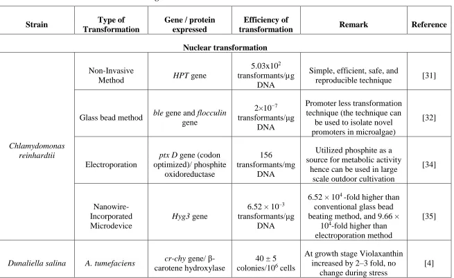
Table 2.2 – Gene transformation in microalgae
Strain Type of
Transformation
Gene / protein expressed
Efficiency of
transformation Remark Reference
Nuclear transformation
Chlamydomonas reinhardtii
Non-Invasive
Method HPT gene
5.03x10² transformants/µg
DNA
Simple, efficient, safe, and
reproducible technique [31]
Glass bead method ble gene and flocculin gene
2×10⁻⁷ transformants/μg
DNA
Promoter less transformation technique (the technique can
be used to isolate novel promoters in microalgae)
[32]
Electroporation
ptx D gene (codon optimized)/ phosphite
oxidoreductase
156 transformants/mg
DNA
Utilized phosphite as a source for metabolic activity
hence can be used in large scale outdoor cultivation
[34]
Nanowire-Incorporated Microdevice
Hyg3 gene
6.52 × 10⁻³ transformants/μg
DNA
6.52 × 10⁴ -fold higher than conventional glass bead beating method, and 9.66 ×
104-fold higher than electroporation method
[35]
Dunaliella salina A. tumefaciens cr-chy gene/ β-carotene hydroxylase
40 ± 5 colonies/106 cells
At growth stage Violaxanthin increased by 2–3 fold, no
change during stress
Quaternary ammonium salt containing soybean
oil
GFP gene
2.8 x 10⁵ transformants/μg
DNA
Low cost, fast, and can be used for transformation of
Halophile microalgae [36] Pleurochrysis carterae Polyethylene glycol (PEG)
PyGUS gene and GFP gene
1
transformants/μg DNA
First stable genetic transformation of a coccolithophore algae by
PEG [37] Pseudochoricystis ellipsoidea Particle bombardment Uridine monophosphate
synthase gene (PeUMPS)
57.3 ± 6.9 transformants/μg
DNA
Useful for researchers working on self-cloning
systems in microalgae
[38] Nannochloropsis salina Particle bombardment sfCherry fluorescent protein
5.9 ± 1.6 and 4.7 ± 2.0 transformants/μg
DNA for TUB and UEP promoters
Similar transformation efficiency as electroporation technique and transformation efficiency varies by promoter
used
[39]
Lobosphaera incisa Electroporation ble gene and Δ5 desaturase gene
1transformants / µg DNA
Technique can be used to further develop endogenous
markers, inducible and constitutive promoters [40] Amphora coffeaeformis Particle bombardment
AC3362 gene (cell surface protein) and
800 transformants/ 108
cells
Particle size and acceleration pressure are important factors
for transformation efficiency
Yellow fluorescent protein
by particle bombardment
Chlorella
zofingiensis Electroporation
PDS-L516F/ norflurazon-resistant marker and phytoene desaturase protein
14 × 10−6 transformants/μg
DNA
3.7-fold higher transformation compared to
particle bombardment, and promoter’s effects on transformation efficiency
(RBCS > PDS & NIT) (different promoters tested)
[42]
Particle bombardment
PDS-L516F/ norflurazon-resistant marker and phytoene desaturase protein
3.7 × 10⁻⁶ transformants/μg
DNA
3.7-fold lower transformation compared to electroporation,
promoter’s effects on transformation efficiency
(RBCS > PDS & NIT) (different promoters tested)
[42]
Neochloris oleoabundans
Electroporation Hyg3, Hyg4, and ChGfp gene
5.2 × 10⁻⁴, 2.0 × 10⁻⁴, and
3.0 × 10⁻⁴ transformants/mg
DNA
Development of
transformation and co-
transformation system in
N. oleoabundans
[43]
Phaeodactylum tricornutum
Conjugation of P. tricornutum by
E. coli
p0521 and p0521s vector
4.0 × 10⁻⁴ diatom cells
First reported nuclear episomal vector for diatoms
and stable episome replication of plasmid even
in absence of antibiotic
[45]