1071-412X/05/$08.00
⫹
0
doi:10.1128/CDLI.12.4.537–541.2005
Copyright © 2005, American Society for Microbiology. All Rights Reserved.
Real-Time Reverse Transcription-PCR Quantitation of Substance P
Receptor (NK-1R) mRNA
Jian-Ping Lai, Steven D. Douglas, Yan-Jian Wang, and Wen-Zhe Ho*
Division of Allergy and Immunology, Joseph Stokes Jr. Research Institute at the Children’s Hospital of Philadelphia,
Department of Pediatrics, University of Pennsylvania Medical School, Philadelphia, Pennsylvania 19104
Received 9 August 2004/Returned for modification 21 December 2004/Accepted 31 January 2005
The substance P (SP)-preferring receptor, neurokinin-1 receptor (NK-1R), has an important role in
inflam-mation, immune regulation, and viral infection. We applied a newly developed real-time reverse transcription
(RT)-PCR assay to quantify NK-1R mRNA in human neuronal cell line (NT-2N), a human B-cell line (IM9),
monocyte-derived macrophages (MDM), peripheral blood lymphocytes (PBL), and human astroglioma cells
(U87 MG). The NK-1R real-time RT-PCR assay has a sensitivity of 100 mRNA copies, with a dynamic range
of detection between 10
2and 10
7copies of NK-1R gene transcripts per reaction. This assay is highly
repro-ducible, with an intraassay coefficient variation of threshold cycle (C
t
) of less than 1.9%. The NK-1R real-time
RT-PCR is highly sensitive for quantitative determination of NK-1R mRNA in human immune cells (MDM
and PBL) that express low levels of NK-1R mRNA. In addition, the assay has the ability to accurately
quantitate the dynamic changes in NK-1R mRNA expression in interleukin-1

-stimulated U87 MG. These data
indicate that the NK-1R real-time RT-PCR has potential for a wide application in investigation of NK-1R
expression at the mRNA level under physiological and pathological conditions in both the central nervous
system and the immune system.
Substance P (SP), the most extensively studied and potent
member of the tachykinin family, is a modulator of
neuroim-munoregulation, in particular, the immune functions of
mono-nuclear phagocytes (10). SP specifically activates NF-
B, a
transcription factor involved in the control of cytokine
expres-sion (22, 26), and stimulates human peripheral blood
mono-cytes to produce inflammatory cytokines including
interleu-kin-1 (IL-1), IL-6, IL-12, and tumor necrosis factor alpha
(TNF-
␣
) (11, 19, 24). SP was initially considered a peptide of
neural origin (28, 31). SP has also been identified in
nonneu-ronal cell types, including murine macrophages (28, 31),
hu-man endothelial cells (23, 27), eosinophils (1), Leydig cells in
human and mouse testis (4), and human lymphocytes and
monocytes/macrophages (10, 12, 18). SP is secreted by human
immune cells, participates in immunoregulation in an
auto-crine fashion (3, 30), and has a role in the pathogenesis of
immune-mediated diseases, including neuroimmunologic
dis-eases and AIDS (9, 16, 21).
The biologic responses to SP are mediated by the
neuroki-nin-1 receptor (NK-1R), the SP preferring receptor, which is a
G-protein-coupled receptor bearing seven transmembrane
do-mains (33). NK-1R is present on T cells (36), including CD4
⫹and CD8
⫹T cells, B lymphocytes (36), monocyte/macrophages
(25), and mast cells (35). Using a competitive PCR technique,
NK-1R mRNA has been detected in lipopolysaccharide
(LPS)-activated murine macrophages (2). In our study of the role of
SP and NK-1R in immunoregulation of the immune cells, we
demonstrated that monocyte/macrophages, T lymphocytes,
and microglia express NK-1R mRNA as determined by reverse
transcription (RT)-PCR (10, 12, 18). The RT-PCR assay,
how-ever, is not only laborious and time-consuming, but it also has
potential variation and contamination due to post-PCR
ma-nipulation. Furthermore, the RT-PCR assay lacks the
capabil-ity to accurately quantitate NK-1R mRNA. In the present
study, we developed a simple, sensitive, rapid, and
reproduc-ible real-time RT-PCR assay in order to quantitate NK-1R
mRNA in human neuronal and immune cells.
MATERIALS AND METHODS
Cells and treatment.Peripheral blood was obtained from three healthy normal adult donors. The Institutional Research Board of our institution approved this investigation. The blood samples were identified as human immunodeficiency virus type 1 (HIV-1) antibody negative by anonymous testing with the enzyme-linked immunosorbent assay (ELISA) method (Coulter Immunology, Hialeah, FL). Informed consent was obtained from these subjects. Monocytes were puri-fied according to our previously described technique (6, 7). Freshly isolated monocytes were plated in 24-well plates at a density of 106cells/well in
Dulbec-co’s modified Eagle medium (DMEM) containing 10% fetal calf serum (FCS). The total length of time in culture for monocyte-derived macrophages (MDM) was 7 to 10 days. The viability of MDM was monitored by trypan blue exclusion and cell adherence to the wells. Nonadherent peripheral blood lymphocytes (PBL) were collected from gelatin-coated flasks and washed three times with phosphate-buffered saline (PBS). PBL viability was measured by a cell prolifer-ation assay. IM9 (human B lymphoblasts), which expresses NK-1R (29), was obtained from the American Type Culture Collection (ATCC) (Rockville, MD). Human neuronal (NT-2N) cells were derived from Ntera2/cl.D1 (NT2) cells, a human teratocarcinoma cell line (32). Both IM9 and NT-2N were used as positive controls for NK-1R mRNA (20). Human astroglioma cells (U87 MG) were obtained from ATCC and maintained in DMEM with 2 mML-glutamine, 100 U/ml penicillin, 100g/ml streptomycin, and 10% heat inactivated fetal bovine serum. In order to quantitate NK-1R mRNA expression changes, UG87 MG cultured in 24-well plates (105
cells/well) were incubated with or without IL-1(4 ng/ml) as we previously described (5).
RNA extraction.Total RNA was extracted from MDM, PBL, IM9, NT-2N (1 ⫻106cells) and U87 MG (4⫻105cells) and using Tri-Reagent (Molecular
Research Center, Cincinnati, OH) as instructed by the manufacturer. In brief, total RNA was extracted by a single step, guanidinium thiocyanate-phenol-chloroform extraction. After centrifugation at 13,000⫻gfor 15 min, RNA-containing aqueous phase was precipitated in isopropanol. RNA precipitates
* Corresponding author. Mailing address: Division of Allergy and
Immunology, The Children’s Hospital of Philadelphia, 34th & Civic
Center Blvd., Philadelphia, PA 19104. Phone: (215) 590-4462. Fax:
(215) 590-2025. E-mail: HO@EMAIL.CHOP.EDU.
537
on August 17, 2020 by guest
http://cvi.asm.org/
were then washed once in 75% ethanol and resuspended in 50l of RNase-free water.
Cloning of NK-1R cDNA fragment.The NK-1R mRNA fragment was cloned and identified with the human NK-1R primer pairs (HSPR3/HSPR4) from IM9 cells as reported earlier (14, 15). Briefly, the PCR products amplified by these primers were separated on a 2% agarose gel and then purified with Wizard PCR Preps DNA Purification System (Promega, Madison, WI). The purified NK-1R cDNA fragment was then cloned into a plasmid using the Eukaryotic TA Cloning Kit (Invitrogen Corporation, San Diego, CA). The cloned plasmid containing the NK-1R cDNA fragment was purified with Wizard Plus Minipreps DNA Purifi-cation System (Promega, Madison, WI). The presence and orientation of the NK-1R cDNA insert was determined by restriction analysis using EcoRV diges-tion and DNA sequencing. The purified plasmid was linearized by EcoRI re-striction enzyme digestion and purified by phenol-chloroform extraction and alcohol precipitation. This plasmid containing the NK-1R cDNA fragment was used as a template to synthesize mRNA in vitro in order to evaluate the sensi-tivity and the reproducibility of the real-time RT-PCR assay.
In vitro mRNA synthesis.NK-1R mRNA transcripts were obtained by tran-scribing the linearized plasmid containing the NK-1R cDNA insert with ME-GAshortscript kit (Ambion, Austin, TX). After digestion with RNase-free DNase (Promega), the resulting RNA transcripts were purified with phenol-chloroform extraction and alcohol precipitation as previously reported (14, 15). The purified RNA transcript was used to construct a standard curve in order to quantitatively measure NK-1R mRNA levels in MDM, PBL, and U87 MG by real-time RT-PCR with the primer pair of NK-1R.
Design of TaqMan probe and primers.The PCR primers and TaqMan probe used were designed using Primer Express software (PE Biosystems). The primer pair of NK-1R sense and antisense (sense: 5⬘-CACACTATGGGCCAGTGAG ATC-3⬘; antisense: 5⬘-GCACACCACGACAATCATCATT-3⬘) was specific for a 109-bp fragment of NK-1R transcripts. The TaqMan probe sequence was 5⬘-TCTCTGCCAAG-CGCAAGGTGGTC-3⬘. The length of the TaqMan probe for NK-1R was designed such that the annealing temperature was 10°C higher than that needed for NK-1R primers. The probe was labeled at the 5⬘end with 6-carboxyfluorescein (6-FAM) and at the 3⬘end with black hole quencher-1. The
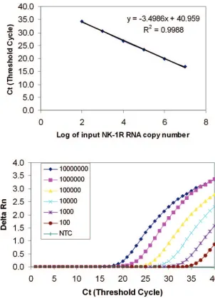
FIG. 1. Sensitivity and linearity analysis of the NK-1R real-time RT-PCR. A reading of change in fluorescence (Rn) as a function of cycle
numbers is demonstrated for a range of known input copy numbers of the NK-1R RNA transcript derived from the plasmid containing NK-1R
cDNA fragment. Tenfold serial dilutions of the NK-1R RNA starting from 10
2to 10
7molecules per reaction were amplified by the real-time
RT-PCR. (A) The standard curve of the serial dilutions of the NK-1R RNA with a correlation coefficient (
R
2) of 0.9988. (B) Amplification plot
of the serial dilutions of NK-1R RNA. The dynamic detection range is 5 orders of magnitude from 10
2to 10
7molecules, and the detection
sensitivity is 100 NK-1R mRNA copies per reaction. NC, negative control which lacked PCR-amplified product when reverse transcriptase was
omitted from the RT reaction using 10
7molecules of NK-1R RNA standard.
on August 17, 2020 by guest
http://cvi.asm.org/
sequence of the primer pair for glyceraldehyde-3-phosphate dehydrogenase (GAPDH) was 5⬘-GGTGGTCTCCTCTGACTTCAACA-3⬘(sense); 5⬘-GTTGC TGTAGCCA-AATTCGTTGT-3⬘ (antisense). The primers and probe resus-pended in Tris-EDTA (TE) buffer were synthesized by Integrated DNA Tech-nologies, Inc. (Coralville, IA), and stored at⫺30°C.
Reverse transcription.Total RNA (1g) and NK-1R RNA standard were subjected to reverse transcription. Both the random primers and the specific NK-1R primer (antisense) were used in the same reaction. The random primers were used to prime GAPDH. The final reaction mixture (20l) contained the following elements: 5 mM MgCl2, 1⫻RT buffer, 500M each deoxynucleoside
triphosphates (dNTPs), 1 unit/l recombinant RNasin, 10 to 15 units of AMV reverse transcriptase (Promega), 50 ng random primers, and 0.1M NK-1R-specific antisense primer. The RT was performed at 42°C for 1 h. The reaction was terminated by holding the reaction mixture at 99°C for 5 min. One-tenth (2l) of the resulting cDNA was used as a template for real-time PCR ampli-fication.
Real-time PCR assay.The ABI Prism 7000 Sequence Detection System (ABI 7000 SDS) was used for real-time PCR analysis. Thermal cycling conditions were designed as follows: initial denaturation at 95°C for 10 min, followed by 40 cycles of 95°C for 15 s and 60°C for 1 min. Fluorescent measurements were recorded during each annealing step. At the end of each PCR run, data were automatically analyzed by the system and amplification plots were obtained. For each PCR, 2 l of cDNA template was added to 48l of PCR Master mixture (5l of 1⫻ PCR buffer II, 5 mM MgCl2, 250M dNTPs, 400 nM of each primer, 1.5 u of
AmpliTaq Gold DNA polymerase, 400 nM of TaqMan probe, and 24.7l of water). The PCR buffer contained 5-carboxy-X-rhodamine (5-ROX) (500 nM) as the reference dye for normalization of the reactions. Any possible fluctuations in 5-ROX signals were used to correct the sample signal. The master mixture was prepared freshly for each real-time PCR amplification. In order to generate a NK-1R RNA standard curve to quantify NK-1R mRNA in human immune cells, known amounts of the NK-1R RNA standard were serially diluted 10-fold and amplified in the same plate under the identical conditions. The quantity of NK-1R mRNA in the samples was automatically calculated by the ABI 7000 SDS based on the data obtained from the standard curve. All amplification reactions were performed in duplicate, and average copy numbers of the duplicates were presented in this report, unless otherwise specified. In order to control the integrity of RNA and normalize NK-1R mRNA levels in MDM, PBL, and U87 MG, a GAPDH mRNA fragment in these cells was also amplified using our established real-time RT-PCR with Brilliant SYBR green QPCR Master Mix (Stratagene, La Jolla, CA) as previous reported (17). In order to normalize the NK-1R mRNA levels, the NK-1R mRNA copy numbers in MDM, PBL, and U87
MG samples were divided by the total RNA (ng) determined by the GAPDH real-time RT-PCR in the same sample and then multiplied by 1,000 in order to convert the unit to NK-1R mRNA copy numbers per microgram (g) of total RNA. The levels of NK-1R mRNA in these cells are expressed as the mean copy number of NK-1R mRNA perg of total RNA.
RESULTS
Sensitivity of the real-time RT-PCR.
The analytical
sensitiv-ity of the real-time RT-PCR was determined using a serial
dilution of NK-1R RNA transcripts containing 10, 10
2, 10
3,
10
4, 10
5, 10
6, and 10
7copies of the transcripts and tested four
times, each in duplicate. The real-time RT-PCR detected
NK-1R mRNA copy numbers as low as 10 molecules, with the
detection rate of 37.5% (3 out of 8 replicates) (data not
shown). The detection rate, however, was 100% with NK-1R
mRNA copy numbers of 100 or higher (8 out of 8 replicates).
The detection limit, therefore, was set at 100 RNA molecules
per reaction. A representative result is shown in Fig. 1.
Linearity, range of quantification, and precision.
Amplifi-cation of NK-1R RNA transcripts at different concentrations
showed the linearity over a range of 5 orders of magnitude
(Fig. 1) with the correlation coefficient
R
2⫽
0.99. In order to
determine the variation of repetitive measurements of
real-time PCR between different runs, 10-fold serial dilutions of
NK-1R cDNA (ranging from 10
2to 10
7copies per reaction)
were examined by the real-time PCR in four different
experi-ments. The coefficient of variation (CV) of threshold cycle (C
t
)
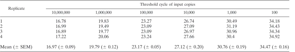
values within an assay was 1.9% (Table 1), and the interassay
variation of C
t
is comparable to that of the intraassay variation
(Table 2).
Real-time RT-PCR quantification of NK-1R mRNA.
Human
monocytes, macrophages, lymphocytes, and U87 MG express
NK-1R mRNA as demonstrated using conventional RT-PCR
TABLE 1. Intra-assay
aaccuracy of NK-1R real-time RT-PCR
Replicate
Threshold cycle of input copies
10,000,000 1,000,000 100,000 10,000 1,000 100
1
16.9
19.7
23.4
26.8
30.2
33.9
2
16.9
20.1
23.3
26.6
30.4
33.5
3
16.8
19.6
23.1
27.0
30.4
34.4
4
16.4
20.0
23.2
27.0
31.0
35
Mean (
⫾
SD)
16.7 (
⫾
0.2)
19.8 (
⫾
0.2)
23.3 (
⫾
0.1)
26.7 (
⫾
0.2)
30.5 (
⫾
0.4)
34.2 (
⫾
0.7)
CV
b(%)
1.38
1.11
0.56
0.75
1.18
1.9
aFour replicate samples from each dilution were amplified in the same plate. bCV, coefficient of variation.
TABLE 2. Inter-assay
aaccuracy of NK-1R real-time RT-PCR
Replicate Threshold cycle of input copies
10,000,000 1,000,000 100,000 10,000 1,000 100
1
16.78
19.83
23.27
26.74
30.49
34.18
2
16.99
19.49
23.09
27.09
31.19
34.43
3
16.89
19.77
23.09
26.97
30.96
34.34
4
17.22
20.06
23.24
27.66
30.4
34.92
Mean (
⫾
SEM)
16.97 (
⫾
0.09)
19.79 (
⫾
0.12)
23.17 (
⫾
0.05)
27.12 (
⫾
0.20)
30.76 (
⫾
0.19)
34.47 (
⫾
0.16)
aThe data are generated from four separate assays performed on different days.
on August 17, 2020 by guest
http://cvi.asm.org/
(5, 10, 12). We first examined the feasibility of the real-time
RT-PCR for quantification of NK-1R mRNA in MDM and
PBL isolated from three subjects. NK-1R mRNA levels in
NT-2N cells and IM9 cells were also quantitated by the
real-time RT-PCR. The reproducibility of the assay with four
rep-etitions was excellent, with a variation of less than 15% for all
the samples tested except IM9 (23.69%) (Table 3). NK-1R
mRNA levels varied in immune cells isolated from three
dif-ferent donors. As expected, the neuronal cells (NT-2N) and
IM9 expressed significantly higher levels of NK-1R mRNA
than human immune cells (Table 3). In order to further
deter-mine the accuracy and specificity of the assay, we exadeter-mined the
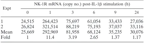
dynamic changes in NK-1R mRNA in IL-1

-treated U87 MG.
IL-1

(4 ng/ml) significantly induced NK-1R mRNA
expres-sion in U87 MG (Table 4). The highest levels of NK-1R
mRNA were observed in U87 MG cells one hour after IL-1

stimulation (Table 4). The changes in the levels of NK-1R
mRNA were quantitated accurately and reproducibly by the
real-time RT-PCR (Table 4).
DISCUSSION
NK-1R has been identified not only on neuronal cells, but
also on the immune cells, including monocytes, macrophages,
and T-cells (10, 12). NK-1R has important roles in the
immu-noregulation of immune cells and in inflammatory diseases.
Using a conventional RT/nested PCR assay, we showed that
human immune cells express NK-1R (10, 12). Because of the
low levels of NK-1R expression in the immune cells, it is
difficult to quantitate NK-1R mRNA in these cells. Thus, the
establishment of a highly quantitative and sensitive assay for
NK-1R mRNA becomes imperative.
Real-time RT-PCR analysis has been employed successfully
for both basic research and clinical applications (8, 37). Since
PCR amplification is an exponential assay, a very small change
in the amplification efficiency produces a dramatic difference
in the amount of final products (34). The monitoring of the
entire process of PCR (real time), rather than merely the end
product, permits precise quantitation. More importantly,
real-time PCR, which uses both primers and a probe, significantly
increases the specificity of the assay. Since the NK-1R
real-time RT-PCR has a wide dynamic detection range (10
2to 10
7copies per reaction), sample dilution or concentration is not
required, which is one of the problems encountered in
com-petitive RT-PCR (13). It is not necessary to run the
PCR-amplified products on agarose gel after real-time PCR, which
not only eliminates post-PCR procedure, but also avoids
vari-ation and contaminvari-ation caused by post-PCR manipulvari-ation.
In the present study, we successfully utilized real-time PCR
for quantification of NK-1R mRNA levels in human immune
cells (MDM, PBL, and IM9) and neuronal cells (NT-2N). We
also examined the accuracy and reproducibility of the real-time
RT-PCR for quantifying the dynamic changes in NK-1R
mRNA in IL-1

-treated human astroglioma cells (U87 MG).
Using conventional RT-PCR we previously showed that IL-1

induced the expression of NK-1R mRNA in U87 MG (5). In
the current study, we demonstrated that the real-time RT-PCR
assay is capable of demonstrating the changes in NK-1R
mRNA levels in U87 MG with high accuracy and specificity. In
addition, the real-time RT-PCR is more sensitive than
conven-tional RT-PCR, since the latter requires a nested PCR
ampli-fication (85 cycles) (10, 12). In conclusion, the NK-1R
real-time RT-PCR is highly sensitive, precise, and reproducible.
The assay is particularly useful for quantitation of NK-1R
mRNA in nonneuronal cells that express low levels of NK-1R
mRNA. Thus, the real-time RT-PCR assay is an important
tool for the investigation of the role of NK-1R in inflammation,
immune regulation and viral infections.
ACKNOWLEDGMENTS
This work was supported by MH 49981 to S.D.D. and
NIH-DA112815 to W.Z.H.
REFERENCES
1.Aliakbari, J., S. P. Sreedharan, C. W. Turck, and E. J. Goetzl.1987. Selective localization of vasoactive intestinal peptide and substance P in human eo-sinophils. Biochem. Biophys. Res. Commun.148:1440–1445.
2.Bost, K. L., S. A. Breeding, and D. W. Pascual.1992. Modulation of the mRNAs encoding substance P and its receptor in rat macrophages by LPS. Reg. Immunol.4:105–112.
TABLE 3. Intra-assay accuracy of NK-1R real-time RT-PCR for the specimens
aReplicate
NK-1R mRNA (copy no.)
D1 D2 D3 Cell line
PBL MDM PBL MDM PBL MDM IM9 NT-2N
1 3,090 1,865 9,362 1,846 20,136 3,241 171,472 1,717,887
2 2,421 1,640 8,448 1,522 16,607 3,139 106,608 1,435,145
3 3,012 1,361 9,856 1,858 18,763 3,478 110,087 1,363,270
4 2,843 1,424 9,606 1,419 16,607 3,733 120,444 1,425,958
Mean (⫾SD) 2,842 (⫾299) 1,573 (⫾229) 9,318 (⫾614) 1,661 (⫾224) 18,022 (⫾1,730) 3,398 (⫾265) 127,153 (⫾3,0125) 1,485,565 (⫾158,40) CVb
(%) 10.52 14.55 6.58 13.48 9.60 7.80 23.69 1.10
aD, donor; healthy peripheral blood. Abbreviations: PBL, peripheral blood lymphocytes; MDM, monocyte-derived macrophages; IM9, human B lymphoblasts;
NT-2N, human neuronal cells derived from Ntera2/cl.D1 (NT2) cells.
bCV, coefficient of variation.
TABLE 4. Quantitation of NK-1R mRNA in IL-1

-treated
U87 MG
Expt NK-1R mRNA (copy no.) post-IL-1stimulation (h)
0 1 3 6 9 24
1
24,515
264,423
75,697
61,054
33,433
27,036
2
26,824
321,514
88,219
75,193
37,037
33,116
Mean
25,669
292,969
81,958
68,124
35,235
30,076
Fold
1
11.4
3.19
2.65
1.37
1.17
on August 17, 2020 by guest
http://cvi.asm.org/
3.Castagliuolo, I., M. Riegler, A. Pasha, S. Nikulasson, B. Lu, C. Gerard, N. P. Gerard, and C. Pothoulakis.1998. Neurokinin-1 (NK-1) receptor is required in Clostridium difficile-induced enteritis. J. Clin. Investig.101:1547–1550. 4.Chiwakata, C., B. Brackmann, N. Hunt, M. Davidoff, W. Schulze, and R.
Ivell.1991. Tachykinin (substance-P) gene expression in Leydig cells of the human and mouse testis. Endocrinology128:2441–2448.
5.Guo, C. J., S. D. Douglas, Z. Gao, B. A. Wolf, J. Grinspan, J. P. Lai, E. Riedel, and W. Z. Ho.2004. Interleukin-1beta upregulates functional expres-sion of neurokinin-1 receptor (NK-1R) via NF-kappaB in astrocytes. Glia
48:259–266.
6.Hassan, N. F., D. E. Campbell, and S. D. Douglas.1986. Purification of human monocytes on gelatin-coated surfaces. J. Immunol. Methods95:273– 276.
7.Hassan, N. F., J. R. Cutilli, and S. D. Douglas.1990. Isolation of highly purified human blood monocytes for in vitro HIV-1 infection studies of monocyte/macrophages. J. Immunol. Methods130:283–285.
8.Heid, C. A., J. Stevens, K. J. Livak, and P. M. Williams.1996. Real time quantitative PCR. Genome Res.6:986–994.
9.Ho, W. Z., J. P. Lai, Y. Li, and S. D. Douglas.2002. HIV enhances substance P expression in human immune cells. FASEB J.16:616–618.
10.Ho, W. Z., J. P. Lai, X. H. Zhu, M. Uvaydova, and S. D. Douglas.1997. Human monocytes and macrophages express substance P and neurokinin-1 receptor. J. Immunol.159:5654–5660.
11.Kincy-Cain, T., and K. L. Bost.1997. Substance P-induced IL-12 production by murine macrophages. J. Immunol.158:2334–2339.
12.Lai, J. P., S. D. Douglas, and W. Z. Ho.1998. Human lymphocytes express substance P and its receptor. J. Neuroimmunol.86:80–86.
13.Lai, J. P., S. D. Douglas, and W. Z. Ho.2002. Mimic-based RT-PCR quan-titation of substance P mRNA in human mononuclear phagocytes and lym-phocytes. Methods Mol. Biol.193:129–147.
14.Lai, J. P., S. D. Douglas, E. Rappaport, J. M. Wu, and W. Z. Ho.1998. Identification of a delta isoform of preprotachykinin mRNA in human mononuclear phagocytes and lymphocytes. J. Neuroimmunol.91:121–128. 15.Lai, J. P., S. D. Douglas, F. Shaheen, D. E. Pleasure, and W. Z. Ho.2002.
Quantification of substance P mRNA in human immune cells by real-time reverse transcriptase PCR assay. Clin. Diagn. Lab. Immunol.9:138–143. 16.Lai, J. P., W. Z. Ho, G. X. Zhan, Y. Yi, R. G. Collman, and S. D. Douglas.
2001. Substance P antagonist (CP-96,345) inhibits HIV-1 replication in hu-man mononuclear phagocytes. Proc. Natl. Acad. Sci. USA98:3970–3975. 17.Lai, J. P., J. H. Yang, S. D. Douglas, X. Wang, E. Riedel, and W. Z. Ho.2003.
Quantification of CCR5 mRNA in human lymphocytes and macrophages by real-time reverse transcriptase PCR assay. Clin. Diagn. Lab. Immunol.10:
1123–1128.
18.Lai, J. P., G. X. Zhan, D. E. Campbell, S. D. Douglas, and W. Z. Ho.2000. Detection of substance P and its receptor in human fetal microglia. Neuro-science101:1137–1144.
19.Laurenzi, M. A., M. A. Persson, C. J. Dalsgaard, and A. Haegerstrand.1990. The neuropeptide substance P stimulates production of interleukin 1 in human blood monocytes: activated cells are preferentially influenced by the neuropeptide. Scand. J. Immunol.31:529–533.
20.Li, Y., S. D. Douglas, D. E. Pleasure, J. Lai, C. Guo, P. Bannerman, M. Williams, and W. Ho.2003. Human neuronal cells (NT2-N) express
func-tional substance P and neurokinin-1 receptor coupled to MIP-1 beta expres-sion. J. Neurosci. Res.71:559–566.
21.Li, Y., S. D. Douglas, L. Song, S. Sun, and W. Z. Ho.2001. Substance P enhances HIV-1 replication in latently infected human immune cells. J. Neu-roimmunol.121:67–75.
22.Lieb, K., B. L. Fiebich, M. Berger, J. Bauer, and K. Schulze-Osthoff.1997. The neuropeptide substance P activates transcription factor NF-kappa B and kappa B-dependent gene expression in human astrocytoma cells. J. Immu-nol.159:4952–4958.
23.Linnik, M. D., and M. A. Moskowitz.1989. Identification of immunoreactive substance P in human and other mammalian endothelial cells. Peptides
10:957–962.
24.Lotz, M., J. H. Vaughan, and D. A. Carson.1988. Effect of neuropeptides on production of inflammatory cytokines by human monocytes. Science241:
1218–1221.
25.Lucey, D. R., J. M. Novak, V. R. Polonis, Y. Liu, and S. Gartner.1994. Characterization of substance P binding to human monocytes/macrophages. Clin. Diagn. Lab. Immunol.1:330–335.
26.Marriott, I., M. J. Mason, A. Elhofy, and K. L. Bost.2000. Substance P activates NF-kappaB independent of elevations in intracellular calcium in murine macrophages and dendritic cells. J. Neuroimmunol.102:163–171. 27.Milner, P., V. Ralevic, A. M. Hopwood, E. Feher, J. Lincoln, K. A.
Kirk-patrick, and G. Burnstock.1989. Ultrastructural localisation of substance P and choline acetyltransferase in endothelial cells of rat coronary artery and release of substance P and acetylcholine during hypoxia. Experientia45:121– 125.
28.Nakanishi, S.1987. Substance P precursor and kininogen: their structures, gene organizations, and regulation. Physiol. Rev.67:1117–1142.
29.Parnet, P., M. Mitsuhashi, C. W. Turck, B. Kerdelhue, and D. G. Payan.
1991. Tachykinin receptor cross-talk. Immunological cross-reactivity be-tween the external domains of the substance K and substance P receptors. Brain Behav. Immun.5:73–83.
30.Pascual, D. W., and K. L. Bost.1990. Substance P production by P388D1 macrophages: a possible autocrine function for this neuropeptide. Immunol-ogy71:52–56.
31.Pernow, B.1983. Substance P. Pharmacol. Rev.35:85–141.
32.Pleasure, S. J., C. Page, and V. M. Lee.1992. Pure, postmitotic, polarized human neurons derived from NTera 2 cells provide a system for expressing exogenous proteins in terminally differentiated neurons. J. Neurosci. 12:
1802–1815.
33.Quartara, L., and C. A. Maggi.1997. The tachykinin NK1 receptor. Part I: ligands and mechanisms of cellular activation. Neuropeptides31:537–563. 34.Raeymaekers, L.1993. Quantitative PCR: theoretical considerations with
practical implications. Anal. Biochem.214:582–585.
35.Shanahan, F., J. A. Denburg, J. Fox, J. Bienenstock, and D. Befus.1985. Mast cell heterogeneity: effects of neuroenteric peptides on histamine re-lease. J. Immunol.135:1331–1337.
36.Stanisz, A. M., R. Scicchitano, P. Dazin, J. Bienenstock, and D. G. Payan.
1987. Distribution of substance P receptors on murine spleen and Peyer’s patch T and B cells. J. Immunol.139:749–754.
37.Tyagi, S., D. P. Bratu, and F. R. Kramer.1998. Multicolor molecular beacons for allele discrimination. Nat. Biotechnol.16:49–53.