Available online:
https://edupediapublications.org/journals/index.php/IJR/
P a g e | 131Identification of Deleterious Nssnps of Interleukin-2
(IL-2) Gene and Its Structural Stability Using
Computational Methods
Tayyaba Sadaf
1*, Fatima Darakhshan
11
Atta-ur-Rahman School of Applied Biosciences, National University of Sciences and
Technology. Islamabad, Pakistan.
*
Corresponding author email:
[email protected]
Second author email: [email protected]
Abstract
Non-synonymous SNPs (nsSNP) found in the
coding regions of a gene cause variations in amino acid residues and are known to influence the
function as well as structure of the encoded proteins. Interleukin 2 (IL-2) is described as a
proinflammatory cytokine which plays a significant part to govern the response of immune system. It
viably contributes in the pathogenesis of several
types of cancers and autoimmune diseases. Taking into account the significance of IL-2, a functional
examination by various in silico approaches was performed to investigate the probable effect on
phenotypic variation due to genetic mutations. In the current study, out of 1071 SNPs, 42 nsSNPs
were collected and scrutinized to categorize the deleterious variants. The functional impact of
diverse set of mutations was first annotated and
protein instability changes were evaluated. Additionally, the protein movement analysis and
flexibility of protein at residual level was identified. Secondly, in order to gain insight in protein
structural analysis, the IL-2 protein structure was generated; refined modelled structure was then
minimized and further assessed by different tools.
The conserved regions, solvent accessibility of native protein and structural divergence of mutant
models were also estimated. As a result, our study helped to screen the most pathogenic and highly
deleterious mutations and suggested that these substitutions may alter the structure–function
relationship. It was inferred that the selected
nsSNPs can be a vital candidate for diseases
caused by the IL-2 gene and other pathological
conditions.
Keywords
IL-2; single nucleotide polymorphisms; protein modelling; computational analysis; deleterious variations
1.
Introduction
Cytokines represent diverse set of proteins and polypeptides that act as signalling molecules performing various functions in the immune system. Cytokines comprise interleukins, interferons, colony-stimulating factors, and a variety of growth factors. The imbalance in the secretion of these cytokines and their resulting signalling systems is an essential factor of the pathogenesis of autoimmune diseases.
Available online:
https://edupediapublications.org/journals/index.php/IJR/
P a g e | 132negative immune activation (D’Souza &
Lefrançois, 2003).
The human IL-2 gene is found on chromosome 4q26-q28 consisting of four exons separated by three introns (Fujita et al., 1983). Several genetic variations in IL-2 are known and biomedical scientists have discussed their association with susceptibility to a range of conditions including breast cancer (Hu et al., 2013), gastric atrophy (Togawa et al., 2005), rheumatoid arthritis (Fedetz et al., 2003), hepatocellular carcinoma (HCC), oesophageal squamous cell carcinoma and multiple sclerosis (Sayad & Movafagh, 2014). The detailed investigation of polymorphisms linked to IL-2 gene expose their function in regulating the rate of inducible expression and secretion of IL-2 (Williams et al., 1988).
IL-2 polymorphisms recognized in the promoter region (-330T/G, rs2069762) and exon 1 region (+114T/G, rs2069763) have been found to be associated with chronic neuroinflammatory and autoimmune disorder i.e. multiple sclerosis in Japanese and Iranian population (Sayad
&Movafagh, 2014). Similarly, these two mutations have also been assessed in breast cancer patients suggesting a positive association with increased susceptibility to breast cancer (Hu et al., 2013). In Taiwan, IL-2 gene polymorphism (G/T, rs2069763) has also been related with a multisystemic disorder of autoimmune disease known as systemic lupus erythematosus (SLE) (Lin et al., 2008).
The investigation of these variations i.e. single nucleotide polymorphisms (SNPs) and their association with human diseases provide new insights for the identification of genetic indicators for diagnosis and prognosis and perhaps novel medicinal approaches for therapeutic purpose. In the light of importance of SNPs to pathogenesis of autoimmune diseases mentioned above, this study is designed to evaluate major disease associated genetic variations using computational approach. For this purpose, different computational tools are applied to investigate SNPs in human IL-2 gene and their consequence on the structure, function and stability of its respective protein.
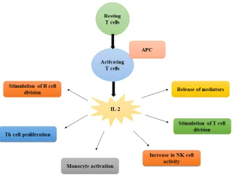
Figure 1. Schematic diagram of Immunoregulatory behaviour of interleukin-2.
2.
Material and Methods
2.1.
Data collection
Computational analysis was carried out for expatiated sequence and structure based assessment for IL-2 non-synonymous SNPs (nsSNPs). For this
Available online:
https://edupediapublications.org/journals/index.php/IJR/
P a g e | 133format was collected from NCBI protein.
Ensembl(Aken et al., 2016), Online Mendelian Inheritance in Man (OMIM: 605384) (McKusick, 2016) and RCSB Protein Data Bank (Berman et al., 2006) were used to obtain the associated data for our computational study.
2.2.
Softwares used for SNPs annotations
The functional impact of nsSNPs were assessed using different computational tools including: SIFT (http://sift.jcvi.org) (Kumar et al., 2009; Ng & Henikoff, 2006), PolyPhen-2 (http://genetics.bwh.harvard.edu/pph2/) (Adzhubei
et al., 2013), SNAP-2
(https://rostlab.org/services/snap2web/) (Bromberg et al., 2008) and PROVEAN (http:// provean.jcvi.org/index.php) (Choi & Chan, 2015). The program SIFT (Sorting Intolerant From Tolerant) was used to characterize intolerant from tolerant amino acid substitutions and to predict the effect of amino acid substitution on the function of protein. The SIFT scores vary from 0 to 1. The scores less than 0.05 indicates amino acid substitutions are ‘damaging’ or ‘not tolerated’, while scores higher than 0.05 are thought to be ‘tolerated’ (Ng & Henikoff, 2003).
PolyPhen-2 was used for further screening of detrimental SNPs from benign. This server uses a naïve Bayesian classifier to evaluate the effect of amino acid substitution on the structure and function based characteristics of protein. The tool estimates the position-specific independent count (PSIC) for each variant and computes the score difference between variants. The PSIC score ranges from 0-1 and categorises the mutations into three groups as benign, possibly damaging or probably damaging. The nsSNPs with score closer to one, is ranked as damaging (Adzhubei et al., 2013). SNAP-2, a neural network based program, was then implemented to estimate the functional effects of nsSNPs. SNAP-2 employs the information of automatically generated multiple sequence alignment and various structural features of protein i.e. solvent accessibility and predicted secondary structure etc. to make predictions if any variation is expected to change protein function. This tool takes the input as FASTA format of protein sequences. The output provides a score ranging from −100 (strong neutral prediction) to +100 (strong effect prediction) and expected accuracy, thus classifying the nsSNPs as ‘Effect’ or ‘neutral’ (Bromberg et al., 2008).
Biological functional changes of a protein due to a variant were then processed by a web based tool PROVEAN (Protein Variant Effect Analyzer) that
operates on sequence clustering and alignment based scoring. The score is calculated based on the variation in sequence similarity of a query sequence to a protein sequence homolog between without and with an amino acid variation of the query sequence. The protein variant is categorized as deleterious if the prediction score is not as much as the cutoff value i.e. - 2.5 (Choi et al., 2012).
2.3.
Predicting effect of nsSNPs on protein
stability
nsSNPs leading to protein instability upon single amino acid substitution was evaluated by a support vector machine (SVM) based I-Mutant 2.0
(http://folding.biofold.org/i-mutant/i-mutant2.0.html)) tool. The free energy change value (DDG) is calculated by subtracting the unfolding Gibbs free energy value of mutated protein from wild protein (kcal/mol).The output indicates the decreased stability if DDG value is less than 0, conversely, DDG value greater than 0 shows an increase in protein stability i.e. a positive DDG value indicates that the mutated protein possesses high stability and vice versa (Capriotti et al., 2005).
In addition, Mupro server (http://mupro. proteomics.ics.uci.edu/) established on SVM and neural network algorithms was used to find out the effect of nsSNPs on protein stability. The server uses the primary amino acid and substituted amino acid and the protein sequences with mutation location as the input. The output includes the change in energy value and a confidence score between -1 and 1 indicating the confidence of the predicted value. The output score less than 0 shows decrease in protein stability due to the mutation; contrariwise, a score greater than 0 refers an increase in protein stability (Cheng et al., 2006).
2.4.
Modelling and Validation of protein
structure
The amino acid substitutions can remarkably alter protein structure and function. In this context, acquaintance of a protein's 3D structure is crucial for better insight of protein functionality. Therefore, IL-2 structure was modelled using I-TASSER (Yang et al., 2015) available at:
https://zhanglab.ccmb.med.umich.edu/I-TASSER/
Available online:
https://edupediapublications.org/journals/index.php/IJR/
P a g e | 1342000) was then applied for the energy minimization
of refine modelled structure. Further, the structure was validated through MolProbity (http://molprobity.biochem.duke.edu/) (Chen et al., 2010; Davis et al., 2007; Davis et al., 2004) and ERRAT (https://services.mbi.ucla.edu/SAVES/). The Swiss-PDBviewer(Guex et al., 2000) was utilized to perform amino acid substitutions, followed by the energy minimization of mutant modelled structures. GROMOS96 43B1 implementation of Swiss-PdbViewer 4.1.0 (Guex et al., 2000) was used to compute the force field energy of wild model including all bonds, torsions, angles, improper, electrostatic and non-bonded atoms (Johansson et al., 2012).
2.5.
Exploring the flexibility at residual
level
The protein movement analysis, its binding efficiency and several types of interactions can be studied by identifying the flexibility of certain amino acids in protein. Therefore, the potential structural changes based on the B-factor distribution of the residues were estimated by ResQserver
(https://zhanglab.ccmb.med.umich.edu/ResQ/). ResQ is a method for approximating residue specific quality of protein structure prediction and the inherent B-factor profile of all residues along the chain by combining local structure assembly variations with sequence- and structure-based profiling(Yang et al., 2016).
2.6.
Detection of functional SNPs in
conserved Regions
The evolutionary conservation of amino acid substitution in proteins based on the phylogenetic relations between homologous sequences was analysed by ConSurf (http://consurf.tau.ac.il/2016/) web tool that uses an empirical Bayesian inference. The conserved regions were assessed with the help of conservation scores and colouring grading system by submitting the FASTA sequence of IL-2 gene as an input option.
2.7.
Analysis of protein structural
divergence
In order to compute changes resulting from the nsSNPs, the wild and mutated predicted structures were then evaluated using a range of structure assessment tools.
The divergence of the mutant structure from the native structure is expected due to the amino acid substitution. This difference between two structures can change the functional activity with respect to binding efficiency of the molecules. Structural divergence due to missense mutations in IL-2 protein was scrutinized by computing root mean square deviation (RMSD) values of native and mutant structures by SuperPose (http://wishart.biology.ualberta.ca/SuperPose/). The output of SuperPose tool gives a diversity of RMSD values for heavy atoms, alpha carbons, backbone atoms and all atoms (average and pairwise) to find individual residue shifts (Maiti et al., 2004).
2.8.
Analysis of secondary structure and
solvent accessibility
The estimation of secondary structure and solvent accessibility of structure was achieved by RaptorX property prediction (Wang et al., 2016a; Wang et al., 2016b), PredictProtein(Rost et
al., 2004)) and artificial neural network based program NetSurfP(Petersen et al., 2009). The output of RaptorX gives information about the tertiary structure, 3-state and 8-state secondary structure, disordered regions and solvent accessibility for a protein (Källberg et al., 2012). Additionally, the output of NetSurP server provides two subclasses i.e. buried and exposed; based on the z-score, defined for solvent accessibility of amino acids for both mutant and wild structures of IL-2 Protein.
2.9.
Prediction of phylogeny-based protein
function
The molecular functions of IL-2 protein sequence was predicted using a statistical method SIFTER (Statistical Inference of Function Through Evolutionary Relationships) that utilizes a protein family's phylogenetic tree, as the natural assembly for expressing protein relationships (Sahraeian et al., 2015).
2.10.
Active site identification
Available online:
https://edupediapublications.org/journals/index.php/IJR/
P a g e | 135region or not. A residue of the protein is considered
to be a part of binding site if the residue is less than 4 Å from any atom of the probe from probe clusters (Ngan et al., 2011). Additionally, COACH (http://zhanglab.ccmb.med.umich.edu/COACH/), an algorithm-based web server, was applied for further evaluation of the ligand binding sites and active site residues of IL-2 protein (Yang et al., 2013).
2.11.
Analysis of protein-protein
Interactions
The functional interaction of IL-2 protein was also analyzed using the online database STRING (Search Tool for the Retrieval of Interacting Genes/Proteins) version 10.5 (Szklarczyk et al., 2017). Both the physical (direct) and functional (indirect) associations were identified depending on the known experimental interactions, predicted genomic interactions, text mining, co-expression and protein homology.
3.
Results and Discussion
The Single nucleotide polymorphisms (SNPs) are assumed to perform a vital part in various human diseases. Generally, more than 4 million distinctive human SNPs are described by several SNP databanks including; human genome variation database (HGVBase) and the NCBI database dbSNP, providing insights to future research. It is believed that structural modelling of a protein and impact of SNPs on protein is imperative in understanding biological processes of associated diseases at molecular level. Furthermore, nsSNPslead to change in the protein sequence which may modify the protein function. Recently, several in silico studies (Akhoundi et al., 2016; Ali Mohamoud et al., 2014; Chakrapani et al., 2017; D RASAL et al., 2016; Dabhi & Mistry, 2014; Goswami, 2015; Kamaraj & Purohit, 2013; Nailwal
& Chauhan, 2017; Naveed et al., 2016; Raghav et al., 2013; Rajasekaran et al., 2008)predicted the importance of nsSNPs associated with genes and provided a platform to ascertain the molecular mechanisms of diseases and influence of variations on the structure and function of protein.
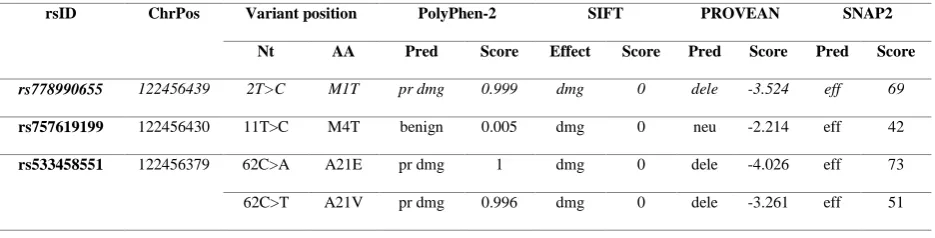
In our study, nonsynonymous SNPs were chosen from coding region for the computational analysis. A total of 42 SNPs of IL-2 gene from NCBI dbSNPwere first functionally investigated using different computational programs, namely, SIFT, PolyPhen-2, PROVEAN and SNAP-2 as summarized in Table 1. These insilico tools estimated whether the SNPs have detrimental or neutral effect on protein function. The computational tools used as a part of this investigation have already been tried and endorsed by several other biomedical scientists as well (Masoodi et al., 2013; Kumar et al., 2015).
SIFT predicted 22 (52.3%) out of 42 nsSNPs as ‘damaging’ based on the tolerance index score. Among these nsSNPs predicted as damaging, 16 (72.7%) were with score 0.00, 5 (22.7%) were with score 0.01 and only one with 0.03 score. The remaining 20 (47.6%) were categorized as ‘tolerated’ with score ranging 0.05-0.57 (Table 1 and Figure 2A). The PSIC score calculated by PolyPhen-2 filtered a total of 26 (61.9%) variants as damaging. Among them, 15 (57.6%) nsSNPs were classified as probably damaging and 11 (42.3%) were predicted as possibly damaging. The rest of 16 (38.1%) variants were ranked benign according to PolyPhen-2 as described in Table 1 and Figure 2A. However, 37 (88.1%) nsSNPs were predicted as effective and 05 (11.9%) nsSNPs as neutral based on the score calculated by SNAP-2 as summarized in Table 1 and Figure 2A. The effect of nsSNPs on protein function was further validated by PROVEAN program which classified 26 (61.9%) nsSNPs as deleterious with score < -2.5 whereas 16 (38.1%) nsSNPs were predicted as neutral (Table 1 and Figure 2A).
Table 1. Screening and prediction of deleterious nsSNPs of IL-2 gene by different computational tools
rsID ChrPos Variant position PolyPhen-2 SIFT PROVEAN SNAP2
Nt AA Pred Score Effect Score Pred Score Pred Score
rs778990655 122456439 2T>C M1T pr dmg 0.999 dmg 0 dele -3.524 eff 69
rs757619199 122456430 11T>C M4T benign 0.005 dmg 0 neu -2.214 eff 42
rs533458551 122456379 62C>A A21E pr dmg 1 dmg 0 dele -4.026 eff 73
Available online:
https://edupediapublications.org/journals/index.php/IJR/
P a g e | 136rs752534234 122456380 61G>A A21T pr dmg 1.000 dmg 0 dele -3.203 eff 55
rs754421965 122456376 65C>T P22L pr dmg 1 dmg 0 dele -8.096 eff 75
rs758928770 122456367 74G>T S25I pos dmg 0.956 dmg 0 dele -4.25 eff 42
rs77806995 122456361 80C>A T27K pr dmg 1 Tol 0.22 dele -2.994 eff 30
rs765882548 122456346 95T>C L32P pos dmg 0.9 dmg 0.01 neu -1.431 eff 23
rs762536978 122456344 97C>A Q33K pos dmg 0.82 tol 0.05 dele -2.62 eff 25
rs3087209 122456328 113T>G L38R pr dmg 0.998 dmg 0 dele -4.882 eff 68
rs146566026 122456312 129G>T M43I benign 0.011 tol 0.17 neu -1.445 eff 12
rs988966007 122456308 133T>G L45V pos dmg 0.891 dmg 0.01 neu -2.291 eff 49
rs751468439 122456202 149A>G N50S pos dmg 0.903 dmg 0.01 dele -3.718 eff 27
rs765829053 122456196 155A>G K52R pos dmg 0.527 tol 0.22 neu -1.406 neu -2
rs202027273 122456191 160C>T P54S benign 0.002 tol 0.34 dele -3.208 eff 2
rs760934382 122456178 173G>T R58M benign 0.114 tol 0.24 dele -3.845 neu -10
rs767776092 122456158 193T>C Y65H pos dmg 0.955 tol 0.07 dele -2.908 eff 70
rs994708825 122456152 199C>G P67A pr dmg 1 dmg 0 dele -7.188 eff 67
rs771093163 122456146 205A>G K69E benign 0.000 dmg 0.03 neu -2.164 eff 37
rs753445824 122453853 208G>A A70T pos dmg 0.818 dmg 0.01 dele -2.668 eff 29
rs767872192 122453852 209C>T A70V benign 0.025 tol 0.14 neu -2.484 neu 0
rs774269792 122453849 212C>T T71I pr dmg 0.96 dmg 0 dele -4.843 eff 52
rs759849798 122453843 218T>C L73P pr dmg 1.000 dmg 0 dele -6.237 eff 79
rs752080371 122453841 220A>C K74Q pr dmg 0.999 tol 0.08 neu -2.46 neu -3
rs146270985 122453835 226C>T L76F pr dmg 1.000 dmg 0 dele -3.738 eff 70
rs866077447 122453799 262G>A E88K pos dmg 0.526 tol 0.06 dele -2.751 eff 32
rs773490003 122453786 275T>C L92S pr dmg 0.994 dmg 0 dele -4.245 eff 67
rs997605621 122453765 296A>C H99P benign 0.356 tol 0.23 dele -4.003 eff 10
rs749017354 122453746 315A>C L105F benign 0.069 tol 0.42 neu -0.607 eff 19
315A>T L105F benign 0.069 tol 0.42 neu -0.607 eff 19
rs746832307 122453745 316A>G I106V benign 0.028 tol 0.23 neu -0.679 eff 22
rs772064424 122453738 323A>G N108S benign 0.149 dmg 0 dele -4.752 eff 42
rs756737575 122453730 331G>A V111I benign 0.001 tol 0.57 neu -0.703 eff 29
331G>T V111L benign 0 tol 0.32 neu -2.199 eff 47
rs778851120 122451852 362C>T T121I pr dmg 1 dmg 0 dele -4.469 eff 52
rs1000418138 122451843 371T>C M124T benign 0 tol 1 neu -0.688 neu -2
Available online:
https://edupediapublications.org/journals/index.php/IJR/
P a g e | 137rs865810716 122451819 395C>A A132E pr dmg 0.984 tol 0.08 neu -2.468 eff 30
rs777464920 122451810 404T>C V135A benign 0.007 tol 0.08 neu -2.159 eff 23
rs543296992 122451796 418A>G R140G benign 0.000 dmg 0.01 dele -3.142 eff 56
rs374465594 122451789 425T>C I142T pos dmg 0.709 tol 0.06 dele -4.055 eff 69
Chrpos= chromosome position; Nt= nucleotide; Pred= prediction; AA= amino acid; Pr dmg= probably damaging; pos dmg= possibly damaging; tol= tolerated; neu= neutral; dele= deleterious; eff= effect.
Figure 2. Bar chart representation of annotated IL-2 SNPs. A) Number of deleterious and tolerated nsSNPs predicted by various softwares. B) Number of screened nsSNPs based on stability of protein predicted through I- mutant and
Mupro.
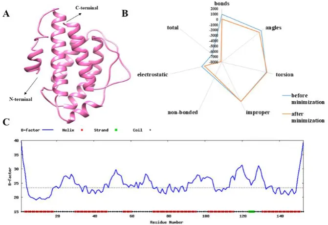
Figure 3. A) The 3D modelled structure of IL-2 protein predicted by I-TASSER. B) Energy parametric profile of IL-2 protein before and after minimization. C) B-factor profile for IL-2 protein sequence predicted by ResQ.
Table 2: Prediction of effect of nsSNPs on IL-2 protein stability through I-Mutant 2 and MuPro
Available online:
https://edupediapublications.org/journals/index.php/IJR/
P a g e | 138SVM Neural Network
Effect DDG Effect Confidence
Score
Effect Confidence
Score
Met1Thr decrease -0.85 decrease -1 decrease -0.9
Met4Thr decrease -1.08 decrease -0.7 decrease -0.8
Ala21Glu decrease -0.22 decrease -1 decrease -0.9
Ala21Val increase 0.26 decrease -0.8 decrease -0.9
Ala21Thr decrease -0.52 decrease -1 decrease -0.9
Pro22Leu increase -1.07 decrease -0.1 increase 0.6
Ser25Ile increase 0.68 increase 0.1 increase 0.7
Thr27Lys decrease -1.5 decrease -1 decrease -0.9
Leu32Pro increase 0.79 decrease -0.9 decrease -0.9
Gln33Lys increase 0.17 increase 0 decrease -0.6
Leu38Arg decrease -0.59 decrease -0.1 decrease -0.7
Met43Ile decrease -0.37 decrease -0.1 increase 0.5
Leu45Val decrease -0.8 decrease -1 decrease -0.9
Asn50Ser decrease -0.08 decrease -1 decrease -0.9
Lys52Arg decrease -0.09 decrease -0.3 decrease -0.6
Pro54Ser decrease -1.17 decrease -1 decrease -0.9
Arg58Met decrease -1.31 increase 0.4 increase 0.8
Tyr65His decrease -0.98 decrease -1 decrease -0.9
Pro67Ala decrease -0.147 decrease -1 decrease -0.9
Lys69Glu increase 0.42 decrease -0.2 decrease -0.6
Ala70Thr decrease -0.59 increase 0.7 decrease -0.5
Ala70Val decrease -0.2 increase 1 increase 0.7
Thr71Ile decrease -0.6 decrease -0.3 increase 0.5
Available online:
https://edupediapublications.org/journals/index.php/IJR/
P a g e | 139Lys74Gln decrease -0.38 decrease -0.8 decrease -0.9
Leu76Phe increase 0.63 decrease -0.9 decrease -0.9
Glu88Lys decrease -0.49 decrease -0.4 increase 0.5
Leu92Ser decrease -2.33 decrease -1 decrease -0.9
His99Pro decrease -0.07 increase 0.03 increase 0.5
Leu105Phe decrease -0.29 decrease -1 decrease -0.8
Ile106Val decrease -0.54 decrease -0.9 decrease -0.8
Asn108Ser increase 0.35 increase 0.09 decrease -0.6
Val111Ile decrease -0.52 decrease -0.7 decrease -0.8
Val111Leu decrease -1.4 decrease -0.4 decrease -0.8
Thr121Ile increase 0.46 increase 0.1 decrease -0.5
Met124Thr decrease -0.59 decrease -0.2 decrease -0.7
Asp129His increase 1.27 decrease -0.4 decrease -0.5
Ala132Glu decrease -1.25 increase 0.5 increase 0.5
Val135Ala decrease -1.29 decrease -1 decrease -0.9
Arg140Gly decrease -0.62 decrease -1 decrease -0.8
Available online:
https://edupediapublications.org/journals/index.php/IJR/
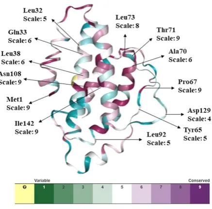
P a g e | 140Figure 4. Conservation analysis of IL-2 protein by ConSurf. The color coding bar shows conservation score.
Interestingly, it was noted that 18 nsSNPs estimated as damaging by SIFT, were also ranked as deleterious by PolyPhen-2. This observation provides an obvious sign of a strong relation between evolutionary based and the structural based approaches i.e. SIFT and PolyPhen-2 respectively. Furthermore, in this analysis, we have identified and predicted 13 nsSNPs that were found to be common and functionally significant (deleterious) by all four computational tools.
3.1.
Predicting effect of nsSNPs on protein
stability
The influence of single amino acid substitution on the stability of protein structure and function was evaluated by I-Mutant 2.0 and MuPro servers. The results of stability changes of 42 nsSNPs are demonstrated in Table 2 and Figure 2B which are projected to be either amplify or reduce the free energy change upon amino acid substitutions. I-Mutant 2.0 revealed that 32 (76.2%) out of 42 variants causes decrease in protein stability with DDG value less than zero. Similarly, MuPro predicted protein stability through two algorithms: SVM and Neural network. Both algorithms predicted 33 (78.5%) variants with the confidence score less than zero, thus revealing decrease in the IL-2 protein stability (Table 2 and Figure 2B). This destabilizing effect in majority of the deleterious mutations gives an indication about the disturbance
in the structure and function of IL-2 protein because of nsSNPs.
3.2.
Modelling and Validation of protein
Structure
Available online:
https://edupediapublications.org/journals/index.php/IJR/
P a g e | 141Thepromising energy minimization techniques can
be employed in the domain of crystallography, structure prediction and molecular dynamics simulation for the refinement of the protein structure (Moult et al., 2014). In this context, we performed energy minimization for the selected model of IL-2 protein. Minimized model of IL-2 had lower optimized energy as indicating the higher stability (Figure 3A).
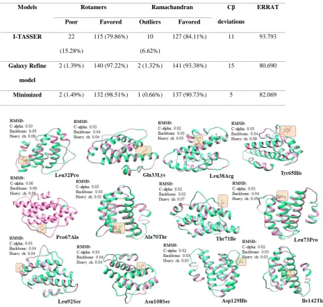
The model was then validated after energy minimization by submitting to the verification tools ERRAT and Molprobity. The Ramachandran analysis using MolProbity justified that most of the residues (84-93%) exist in favourable regions (Table 4). Subsequent outlier removal and poor rotamer correction improved the predicted models. The output values obtained from ERRAT for I-TASSER, Galaxy refined and minimized model were 93.7%, 80.7% and 82%, respectively, revealing the overall good quality of these models.
3.3.
Exploring the flexibility at residual
level
The flexibility in IL-2 protein model can also be evaluated through the analysis of the factor. B-factor demonstrates the inherent thermal mobility of amino acids or atoms in proteins. Therefore, we estimated B-factor by ResQ server. The output (Figure 3C) showed that the areas at the N- and C-terminals and majority of the loop regions have higher B-factor values demonstrating that these regions are structurally more flexible than other regions. Besides, the B-factors calculated for alpha regions are lower, proposing these regions are structurally more stable.
3.4.
Detection of functional SNPs in
conserved Regions
The selected single nucleotide polymorphisms were examined for conservation analysis to predict the conservation rate. Figure 4. represents the ConSurf results that comprises the color scale based on the conservation scores (9 - conserved, 1 - variable), demonstrating the evolutionary relationships among sequence homologs. The output of the ConSurf tool also gives the score that is the normalized conservation scores. It is noticed from data that among the selected residual positions, the positions 1, 67, 71, 73, 108 and 142 were found to be highly conserved and functional with conservation scale value ranging 8-9 (Figure 4).
From these results, we can infer that the mutations at the 1, 67, 71, 73, 108 and 142 positions can have great functional and structural impact on the IL-2 protein due to their high conservation frequency.
3.5.
Analysis of protein structural
divergence
The accurate RMSD score for all the variants were determined by SuperPose tool as mentioned in Figure 5. The greater the RMSD value is, the higher the variation between the two structures is, which thus modifies their functional performance (Reva et al., 1998). It is noted that the overall RMSD values of alpha carbons, backbone and heavy atoms of modelled structures are all resembling suggesting that they are structurally very similar and might cause a slight change in the mutant structures.
3.6.
Analysis of secondary structure and
solvent accessibility
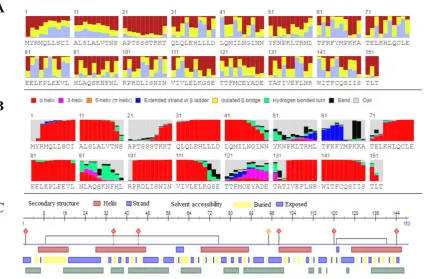
The biophysical analysis including secondary structure and solvent accessibility of IL-2 protein was first predicted using RaptorX tool as shown in Figure 6A-B. The 8-class secondary structure of IL-2 precited by RaptorX consist of: 57% helix, 3% extended strand and 39% coil region. The solvent accessed is expected to contain 24% Buried, 51% Medium and 23% Exposed regions.
The solvent accessibility of IL-2 protein when evaluated by PredictProteinFigure 6C, the results revealed the presence of alpha-helices predominantly which is in consonance with the results predicted by RaptorX.
Moreover, the biophysical analysis including surface and solvent accessibility of IL-2 protein achieved through NetSurfP suggested that the stability of the protein can be disturbed due to the position and the nature of a mutated residue. It has been reported that as the solvent accessibility of a residue lessens, it destabilizes the protein. The output generated by NetSurP (Table 5) demonstrates that there is no change in the class assignment for any of 13 SNPs due to mutation. 07 out of 13 of mutant and native residues were found in buried regions that infer their low accessibility to surface and solvent. The mutant residue exhibited the changes in relative surface accessibility (RSA),absolute surface accessibility and Z-fit score for RSA prediction (Table 5).
Available online:
https://edupediapublications.org/journals/index.php/IJR/
P a g e | 142Models GDT-HA RMSD MolProbity Clash Score Poor
rotamers
Ramachandran
favoured
I-TASSER 1.0000 0.000 3.159 13.3 13.2 84.1
Galaxy Refine 0.9542 0.425 2.342 26.5 0.7 93.4
RMSD= root mean square deviation
Table 4. IL-2 protein structure validation through Molprobity and ERRAT
Models Rotamers Ramachandran Cβ
deviations
ERRAT
Poor Favored Outliers Favored
I-TASSER 22
(15.28%)
115 (79.86%) 10
(6.62%)
127 (84.11%) 11 93.793
Galaxy Refine
model
2 (1.39%) 140 (97.22%) 2 (1.32%) 141 (93.38%) 15 80.690
Minimized 2 (1.49%) 132 (98.51%) 1 (0.66%) 137 (90.73%) 5 82.069
Figure 5. RMSD values of IL-2 mutant model protein calculated by SuperPose.
Table 5. Surface accessibility of wild type and mutants of IL-2 protein.
Available online:
https://edupediapublications.org/journals/index.php/IJR/
P a g e | 143Relative Surface
Accessibility (RSA)
Absolute Surface
Accessibility (ASA)
Z-fit score for RSA
prediction
Met1Thr Exposed
Exposed 0.784 0.860 156.958 119.227 -0.108 0.693
Leu32Pro Exposed
Exposed 0.548 0.576 100.339 81.777 1.141 1.199
Gln33Lys Exposed
Exposed 0.311 0.335 55.455 68.930 0.887 1.019
Leu38Arg Buried
Buried 0.120 0.150 22.027 34.396 0.282 0.527
Tyr65His Buried
Buried 0.267 0.273 57.143 49.677 -0.377 -0.364
Pro67Ala Buried
Buried 0.286 0.295 40.555 32.465 -0.519 -0.504
Ala70Thr Buried
Buried 0.166 0.182 18.271 25.243 -0.933 -0.785
Thr71Ile Exposed
Exposed 0.428 0.399 59.294 73.852 -0.424 -0.442
Leu73Pro Buried
Buried 0.136 0.162 24.938 23.045 0.667 0.139
Leu92Ser Buried
Buried 0.114 0.123 20.928 14.439 0.463 0.266
Asn108Ser Exposed
Exposed 0.321 0.318 46.965 37.328 -0.460 -0.418
Asp129His Exposed
Exposed 0.600 0.600 86.417 109.213 -0.199 -0.255
Ile142Thr Buried
Available online:
https://edupediapublications.org/journals/index.php/IJR/
P a g e | 144Figure 6. Secondary structure and solvent accessibility of native IL-2 protein: A) Solvent accessibility prediction of IL-2 protein by RaptorX B) 8-class secondary structure prediction by RaptorX; C) Secondary structure
prediction by PredictProtein
Table 6. Molecular function predictions for the entire human IL-2 protein by SIFTER.
GO term Protein Prediction Confidence score
GO:0005125 Cytokine activity 0.81
GO: 0005134 Interleukin-2 receptor binding 0.81
GO: 0019209 Kinase activator activity 0.75
GO: 0008083 Growth factor activity 0.75
Table 7. Residues at ligand binding sites of IL-2 protein predicted by FTSite.
Binding Sites AminoAcid Residues
FTSite 1: Met66, Pro67, Lys68, Lys69, Ala70, Arg140, Trp141, Thr143, Phe144, Ser147
Available online:
https://edupediapublications.org/journals/index.php/IJR/
P a g e | 145Available online:
https://edupediapublications.org/journals/index.php/IJR/
P a g e | 1463.7.
Prediction of phylogeny-based protein
function
The IL-2 protein sequence subjected for the phylogeny-based protein function prediction reveals the presence of different domains in IL-2 protein structure, highlighting the multiple functions of IL-2 protein that includes Cytokine activity, binding to Interleukin-2 receptor, Kinase activator activity and Growth factor activity as stated in Table 6. All together, we can say that human IL-2 act as a growth factor and help to regulate different signalling systems that are essential to understand the pathogenesis of associated diseases.
3.8.
Active site identification
The identification of active sites is very important, any mutation in these regions may disrupt the binding capacity of the ligand to its protein. Thus, change in ligand binding site affects the function of protein by changing the stability of protein (Ng & Henikoff, 2006; Yang et al., 2012).
In our analysis, FTSite detected 3 ligand binding sites on IL-2 Protein. The amino acid residues observed in these 3 sites of IL-2 protein are presented in Table 7. It is observed that 6 amino acid residues i.e. Leu32, Gln33, Leu38, Pro67, Ala70 and Asn108 out of 13 selected deleterious nsSNPs are identified among these sites.
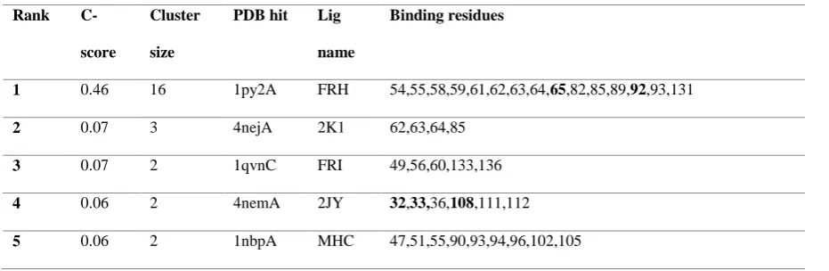
Moreover, ligand binding residues were also identified by COACH algorithm that employs various ligands to examine their binding sites. The ligands with greater C-score (confidence score) signify an authentic result. Table 8 indicates that the ligand FRH has better C-score with 0.46 value. Only two of these sites i.e. Tyr65His and Leu92Ser matches the deleterious SNPs selected for IL-2 protein. However, three sites Leu32Pro, Gln33Lys and Asn108Ser that also act as the deleterious SNPs, were found having the binding affinity for 2JY ligand but with low C-score.
3.9.
Analysis of protein-protein Interactions
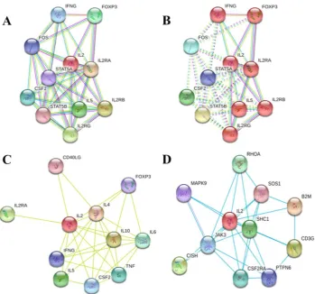
The analysis of protein–protein interaction is crucial to reveal all functional interactions among cellular proteins. The STRING database exhibited 10 functional partners of IL-2 (Figure 7) with highest interaction confidence score > 0.90 indicating strong association with IL-2 protein. Predicted interaction network with different parameters and filters observed with IL-2 protein has demonstrated IL2R as the strongest interaction partner (Figure 7).
All the study reflects that the selected nsSNPs of IL-2 gene might influence the function of protein and might be potent drug target for the treatment of diseases associated with IL-2 gene polymorphisms.
Table 8. Function prediction of IL-2 modelled protein using COACH server.
Rank
C-score
Cluster
size
PDB hit Lig
name
Binding residues
1 0.46 16 1py2A FRH 54,55,58,59,61,62,63,64,65,82,85,89,92,93,131
2 0.07 3 4nejA 2K1 62,63,64,85
3 0.07 2 1qvnC FRI 49,56,60,133,136
4 0.06 2 4nemA 2JY 32,33,36,108,111,112
Available online:
https://edupediapublications.org/journals/index.php/IJR/
P a g e | 147Figure 7. Protein–protein interaction network of IL-2 using STRING 10.5 server: A) top 10 interactor with highest confidence score without clustering, B) K means clustering with 5 number of clusters, C) active interaction source is
Available online:
https://edupediapublications.org/journals/index.php/IJR/
P a g e | 1484.
Conclusion
The study was focused to thoroughly investigate the plausible effects of variations on the structure and function of IL-2 protein. Therefore, the complete 3D structure of IL-2 was primarily modelled and validated. Furthermore, both sequence based and structure based methods were implemented for evaluation of non-synonymous single nucleotide polymorphisms (nsSNPs). Out of 42 nsSNPs, 12 nsSNPs namely p.Leu32Pro, p.Gln33Lys, p.Leu38Arg, p.Tyr65His, p.Pro67Ala, p.Ala70Thr, p.Thr71Ile, p.Leu73Pro, p.Leu92Ser, p.Asn108Ser, p.Asp129His and p.Ile142Thr were found pathogenic and highly deleterious and might induce alterations in protein structure, stability, solvent accessibility and binding affinity. The computational analysis of free energy change indicates the decrease in IL-2 protein stability due to mutations. Consequently, our study recommends that the selected nsSNPs of IL-2 gene can be viewed as significant candidates in developing diseases related to IL-2 gene polymorphisms.
Conflict of interest
None.References
[1] Adzhubei, I., Jordan, D. M., & Sunyaev, S. R. (2013). Predicting functional effect of human missense mutations using PolyPhen‐2. Current protocols in human genetics, 7-20.
[2] Aken, B. L., Achuthan, P., Akanni, W., Amode, M. R., Bernsdorff, F., Bhai, J., et al. (2016). Ensembl 2017. Nucleic acids research, 45(D1), D635-D642.
[3] Akhoundi, F., Parvaneh, N., & Modjtaba, E.-B. (2016). In silico analysis of deleterious single nucleotide polymorphisms in human BUB1 mitotic checkpoint serine/threonine kinase B gene. Meta gene, 9, 142-150.
[4] Ali Mohamoud, H. S., Manwar Hussain, M. R., El-Harouni, A. A., Shaik, N. A., Qasmi, Z. U., Merican, A. F., et al. (2014). First comprehensive in silico analysis of the functional and structural consequences of SNPs in human GalNAc-T1 gene. Computational and mathematical methods in medicine, 2014.
[5] Berman, H. M., Westbrook, J., Feng, Z., Gilliland, G., Bhat, T. N., Weissig, H., et al. (2006). The Protein Data Bank, 1999– International Tables for Crystallography Volume F: Crystallography of biological
macromolecules (pp. 675-684): Springer.
[6] Bromberg, Y., Yachdav, G., & Rost, B. (2008). SNAP predicts effect of mutations on protein function. Bioinformatics, 24(20), 2397-2398.
[7] Capriotti, E., Fariselli, P., Calabrese, R., & Casadio, R. (2005). Predicting protein stability changes from sequences using support vector machines. Bioinformatics, 21(suppl_2), ii54-ii58.
[8] Chakrapani, V., Rasal, K. D., Kumar, S., Mohapatra, S. D., Sundaray, J. K., Jayasankar, P., et al. (2017). In Silico Analysis of nsSNPs of Carp TLR22 Gene Affecting its Binding Ability with Poly I: C. Interdisciplinary Sciences: Computational Life Sciences, 1-12.
[9] Chen, V. B., Arendall, W. B., Headd, J. J., Keedy, D. A., Immormino, R. M., Kapral, G. J., et al. (2010). MolProbity: all-atom structure validation for macromolecular crystallography. Acta Crystallographica Section D: Biological Crystallography, 66(1), 12-21.
[10]Cheng, J., Randall, A., & Baldi, P. (2006). Prediction of protein stability changes for single‐site mutations using support vector machines. Proteins: Structure, Function, and Bioinformatics, 62(4), 1125-1132.
[11]Choi, Y., & Chan, A. P. (2015). PROVEAN web server: a tool to predict the functional effect of amino acid substitutions and indels. Bioinformatics, 31(16), 2745-2747.
[12]Choi, Y., Sims, G. E., Murphy, S., Miller, J. R., & Chan, A. P. (2012). Predicting the functional effect of amino acid substitutions and indels. PloS one, 7(10), e46688.
[13]D RASAL, K., Chakrapani, V., PATRA, S. K., Jena, S., D MOHAPATRA, S., Nayak, S., et al. (2016). Identification and prediction of the consequences of nonsynonymous SNPs in glyceraldehyde 3-phosphate dehydrogenase (GAPDH) gene of zebrafish Danio rerio. Turkish Journal of Biology, 40(1), 43-54. [14]D’Souza, W. N., & Lefrançois, L. (2003). IL-2
is not required for the initiation of CD8 T cell cycling but sustains expansion. The Journal of Immunology, 171(11), 5727-5735.
[15]Dabhi, B., & Mistry, K. N. (2014). In silico analysis of single nucleotide polymorphism (SNP) in human TNF-α gene. Meta gene, 2, 586-595.
Available online:
https://edupediapublications.org/journals/index.php/IJR/
P a g e | 149acids. Nucleic acids research, 35(suppl_2),
W375-W383.
[17]Davis, I. W., Murray, L. W., Richardson, J. S., & Richardson, D. C. (2004). MOLPROBITY: structure validation and all-atom contact analysis for nucleic acids and their complexes. Nucleic acids research, 32(suppl_2), W615-W619.
[18]Fedetz, M., Matesanz, F., Cáliz, R., Ferrer, M. A., Collado, M. D., Alcina, A., et al. (2003). Lack of association between-384 and 114 IL-2 gene polymorphisms and rheumatoid arthritis. The Journal of rheumatology, 30(3), 435-437. [19]Fujita, T., Takaoka, C., Matsui, H., &
Taniguchi, T. (1983). Structure of the human interleukin 2 gene. Proceedings of the National Academy of Sciences, 80(24), 7437-7441.
[20]Gaffen, S. L., & Liu, K. D. (2004). Overview of interleukin-2 function, production and clinical applications. Cytokine, 28(3), 109-123.
[21]Goswami, A. M. (2015). Structural modeling and in silico analysis of non-synonymous single nucleotide polymorphisms of human 3β-hydroxysteroid dehydrogenase type 2. Meta gene, 5, 162-172.
[22]Guex, N., Diemand, A., Peitsch, M., & Schwede, T. (2000). SwissPDBViewer program. Glaxo Smith Kline R. &D.
[23]Heo, L., Park, H., & Seok, C. (2013). GalaxyRefine: protein structure refinement driven by side-chain repacking. Nucleic acids research, 41(W1), W384-W388.
[24]Hu, X.-B., Ouyang, L.-Z., & Tang, L.-L. (2013). Interleukin-2 gene polymorphisms and prognosis of breast cancer. Genetic testing and molecular biomarkers, 17(6), 453-457. [25]Johansson, M. U., Zoete, V., Michielin, O., &
Guex, N. (2012). Defining and searching for structural motifs using DeepView/Swiss-PdbViewer. BMC bioinformatics, 13(1), 173. [26]Källberg, M., Wang, H., Wang, S., Peng, J.,
Wang, Z., Lu, H., et al. (2012). Template-based protein structure modeling using the RaptorX web server. Nature protocols, 7(8), 1511-1522.
[27]Kamaraj, B., & Purohit, R. (2013). In silico screening and molecular dynamics simulation of disease-associated nsSNP in TYRP1 gene and its structural consequences in OCA3. BioMed research international, 2013.
[28]Ko, J., Park, H., Heo, L., & Seok, C. (2012). GalaxyWEB server for protein structure prediction and refinement. Nucleic acids research, 40(W1), W294-W297.
[29]Kumar, P., Henikoff, S., & Ng, P. C. (2009). Predicting the effects of coding non-synonymous variants on protein function using the SIFT algorithm. Nature protocols, 4(7), 1073-1081.
[30]Kumar, P., Singh, R. K., & Mahalingam, K. (2015). In silico analysis of fat mass obesity associated (FTO) gene using computational algorithms. Int. J. Pharm. Bio Sci, 6, 589-599.
[31]Lenardo, M. J. (1991). lnterleukin-2 programs mouse αβ T lymphocytes for apoptosis. Nature, 353(6347), 858-861.
[32]Lin, Y.-J., Wan, L., Sheu, J. J.-C., Huang, C.-M., Lin, C.-W., Lan, Y.-C., et al. (2008). G/T polymorphism in the interleukin-2 exon 1 region among Han Chinese systemic lupus erythematosus patients in Taiwan. Clinical Immunology, 129(1), 36-39.
[33]Maiti, R., Van Domselaar, G. H., Zhang, H., & Wishart, D. S. (2004). SuperPose: a simple server for sophisticated structural superposition. Nucleic acids research, 32(suppl_2), W590-W594.
[34]Masoodi, T. A., Al Shammari, S. A., Al-Muammar, M. N., Alhamdan, A. A., & Talluri, V. R. (2013). Exploration of deleterious single nucleotide polymorphisms in late-onset Alzheimer disease susceptibility genes. Gene, 512(2), 429-437.
[35]McKusick, V. (2016). Online Mendelian Inheritance in Man, OMIM™. Johns Hopkins University: Baltimore. MIM Number:# 193700.
[36]Moult, J., Fidelis, K., Kryshtafovych, A., Schwede, T., & Tramontano, A. (2014). Critical assessment of methods of protein structure prediction (CASP)—round x. Proteins: Structure, Function, and Bioinformatics, 82(S2), 1-6.
[37]Nailwal, M., & Chauhan, J. B. (2017). Analysis of consequences of non-synonymous SNPs of USP9Y gene in human using bioinformatics tools. Meta Gene, 12, 13-17.
[38]Naveed, M., Tehreem, S., Mubeen, S., Nadeem, F., Zafar, F., & Irshad, M. (2016). In-silico analysis of non-synonymous-SNPs of STEAP2: To provoke the progression of prostate cancer. Open Life Sciences, 11(1), 402-416.
Available online:
https://edupediapublications.org/journals/index.php/IJR/
P a g e | 150[40]Ng, P. C., & Henikoff, S. (2006). Predicting
the effects of amino acid substitutions on protein function. Annu. Rev. Genomics Hum. Genet., 7, 61-80.
[41]Ngan, C.-H., Hall, D. R., Zerbe, B., Grove, L. E., Kozakov, D., & Vajda, S. (2011). FTSite: high accuracy detection of ligand binding sites on unbound protein structures. Bioinformatics, 28(2), 286-287.
[42]Petersen, B., Petersen, T. N., Andersen, P., Nielsen, M., & Lundegaard, C. (2009). A generic method for assignment of reliability scores applied to solvent accessibility predictions. BMC structural biology, 9(1), 51. [43]Raghav, D., Sharma, V., & Agarwal, S. M.
(2013). Structural investigation of deleterious non-synonymous SNPs of EGFR gene. Interdisciplinary sciences, computational life sciences, 5(1), 60.
[44]Rajasekaran, R., Doss, C. G. P., Sudandiradoss, C., Ramanathan, K., Rituraj, P., & Rao, S. (2008). Computational and structural investigation of deleterious functional SNPs in breast cancer BRCA2 gene. Chinese Journal of Biotechnology, 24(5), 851-856.
[45]Reva, B. A., Finkelstein, A. V., & Skolnick, J. (1998). What is the probability of a chance prediction of a protein structure with an rmsd of 6 Å? Folding and Design, 3(2), 141-147.
[46]Rost, B., Yachdav, G., & Liu, J. (2004). The predictprotein server. Nucleic acids research, 32(suppl_2), W321-W326.
[47]Roy, A., Kucukural, A., & Zhang, Y. (2010). I-TASSER: a unified platform for automated protein structure and function prediction. Nature protocols, 5(4), 725-738.
[48]Roy, A., Yang, J., & Zhang, Y. (2012). COFACTOR: an accurate comparative algorithm for structure-based protein function annotation. Nucleic acids research, 40(W1), W471-W477.
[49]Sahraeian, S. M., Luo, K. R., & Brenner, S. E. (2015). SIFTER search: a web server for accurate phylogeny-based protein function prediction. Nucleic acids research, 43(W1), W141-W147.
[50]Sakaguchi, S., Yamaguchi, T., Nomura, T., & Ono, M. (2008). Regulatory T cells and immune tolerance. Cell, 133(5), 775-787.
[51]Sayad, A., & Movafagh, A. (2014). The Association of− 330 Interleukin-2 Gene Polymorphism with Its Plasma Concentration
in Iranian Multiple Sclerosis Patients. Scientifica, 2014.
[52]Shen, Y., Liu, Y., Liu, S., & Zhang, A. (2012). The association between-330T/G polymorphism of interleukin 2 gene and bladder cancer. DNA and cell biology, 31(6), 983-987.
[53]Szklarczyk, D., Morris, J. H., Cook, H., Kuhn, M., Wyder, S., Simonovic, M., et al. (2017). The STRING database in 2017: quality-controlled protein–protein association networks, made broadly accessible. Nucleic acids research, 45(D1), D362-D368.
[54]Togawa, S., Joh, T., Itoh, M., Katsuda, N., Ito, H., Matsuo, K., et al. (2005). Interleukin‐2 gene polymorphisms associated with increased risk of gastric atrophy from Helicobacter pylori infection. Helicobacter, 10(3), 172-178. [55]Wang, S., Li, W., Liu, S., & Xu, J. (2016).
RaptorX-Property: a web server for protein structure property prediction. Nucleic acids research, 44(W1), W430-W435.
[56]Wang, S., Peng, J., Ma, J., & Xu, J. (2016). Protein secondary structure prediction using deep convolutional neural fields. Scientific reports, 6.
[57]Williams, T., Eisenberg, L., Burlein, J., Norris, C., Pancer, S., Yao, D., et al. (1988). Two regions within the human IL-2 gene promoter are important for inducible IL-2 expression. The Journal of Immunology, 141(2), 662-666. [58]Yang, J., Roy, A., & Zhang, Y. (2012).
BioLiP: a semi-manually curated database for biologically relevant ligand–protein interactions. Nucleic acids research, 41(D1), D1096-D1103.
[59]Yang, J., Roy, A., & Zhang, Y. (2013). Protein–ligand binding site recognition using complementary binding-specific substructure comparison and sequence profile alignment. Bioinformatics, 29(20), 2588-2595.
[60]Yang, J., Wang, Y., & Zhang, Y. (2016). ResQ: an approach to unified estimation of B-factor and residue-specific error in protein structure prediction. Journal of molecular biology, 428(4), 693-701.