Dissertation zur Erlangung des Doktorgrades
der Fakultät für Chemie und Pharmazie
der Ludwig-Maximilians-Universität München
Role of the CNGB1a Subunit of the Rod Cyclic
Nucleotide-Gated Channel in Channel Gating and Pathogenesis of
Retinitis Pigmentosa
Elvir Becirovic
aus Sarajevo
Erklärung
Diese Dissertation wurde im Sinne von § 13 Abs. 3 bzw. 4 der Promotionsordnung vom 29. Januar 1998 von Prof. Dr. Martin Biel betreut.
Ehrenwörtliche Versicherung
Hiermit versichere ich ehrenwörtlich, dass die vorgelegte Arbeit selbstständig und ohne unerlaubte Hilfe verfasst wurde. Es wurden keine anderen Hilfsmittel außer den angegebenen verwendet.
München, den ... ……... (Elvir Becirovic)
Table of contents
1 Introduction ... 1
1.1 Anatomy of the retina ... 1
1.2 Anatomy of rods... 2
1.3 Signalling transduction in rods ... 3
1.4 CNG channels ... 4
1.4.1 Topology and structural features of CNG channels ... 5
1.4.2 Gating of CNG channels ... 6
1.5 Role of CNGB1 in rod CNG channels ... 7
1.5.1 Characteristics of the CNGB1 locus ... 7
1.5.2 Retinitis pigmentosa mutations in the CNGB1 gene ... 7
1.6 Role of GARP in rods... 8
1.7 Goals of this study ... 9
2 Materials and methods ...10
2.1 Molecular biology ...10
2.1.1 Plasmids ...10
2.1.2 Polymerase chain reaction (PCR) ...11
2.1.3 Purification of DNA fragments ...11
2.1.4 Restriction analysis and preparation of samples for cloning ...12
2.1.5 Ligation and dephosphorylation ...12
2.1.6 Transformation ...13
2.1.7 Inoculation of bacterial cells and isolation of plasmid DNA (alkaline lysis) ...13
2.1.8 TOPO cloning...14
2.1.9 In-Fusion cloning ...15
2.1.10 Introduction of mutations in DNA constructs ...15
2.1.11 Reverse transcription (RT) ...16
2.1.12 Cloning of CNG channels ...16
2.2 Cell culture ...16
2.2.1 Cultivation and transfection of mammalian cell lines...16
2.3 Protein biochemistry ...17
2.3.1 Isolation and quantification of proteins ...17
2.3.2 Membrane preparations ...18
2.3.3 Western blotting ...19
2.3.4 Co-immunoprecipitation ...19
2.3.5 Biotinylation assay ...20
2.5 Electrophysiological recordings ...21
2.6 Statistics ...22
3 Results ...23
3.1 Splicing analysis of the c.3444+1G>A mutation in CNGB1 ...23
3.1.1 In silico splicing analysis of c.3444+1G>A ...23
3.1.2 Creation of wild type and mutant minigene constructs ...24
3.1.3 Exon trapping experiments in HEK293T cells ...25
3.1.4 In vitro expression of wild type and mutant rod CNG channels ...26
3.2 Functional analysis of the G993V mutation in CNGB1 ...28
3.2.1 In silico analysis ...28
3.2.2 Expression of CNGA1GV ...30
3.2.3 Electrophysiological measurements of heteromeric CNGA1/CNGB1aGV channels ...31
3.2.4 Coassembly and cell surface expression of CNGA1/CNGB1aGV heteromers.32 3.2.5 Identification of inhibitory domains in CNGB1a ...34
3.2.6 Coexpression of GARP as soluble protein ...37
3.2.7 Role of a functional CNBD of CNGB1 for CNG channel activation ...39
3.2.8 Opening probability of channels containing or lacking the GARP domain ...40
4 Discussion ...42
4.1 Splicing analysis of the c.3444+1G>A mutation in CNGB1 ...42
4.2 Functional analysis of the G993V mutation in CNGB1 ...43
5 Summary ...46
Zusammenfassung ...47
6 Literature ...49
6.1 Cited publications ...49
6.2 Own publications ...52
Accepted publications ...52
Publications under review or under revision ...52
7 Appendix ...53
7.1 Supplementary tables and figures ...53
7.2 Abbreviations ...59
7.3 Curriculum Vitae ...61
Lebenslauf ...63
1 Introduction
1.1 Anatomy of the retina
The retina represents the light sensing part of the eye. It lines the back of the eye overlying the choroid layer. One characteristic of the vertrebrate inverse retina is the fact that light has to pass the nerve cell layer until it reaches the photoreceptor cells. The latter consist of rods and cones and represent the light detecting part of the retina. The outer segments of photoreceptors are embedded in the light collecting pigment epithelium. In the adjacent outer nuclear layer (ONL) the cell bodies of rods and cones are located. Next to the ONL, in the outer plexiform layer (OPL) the synapses of rods and cones as well as of bipolar and horizontal cells are arranged. They are followed by the inner nuclear layer (INL) which is composed of cell bodies of bipolar, horizontal, and amacrine cells. Synaptic connections of bipolar cells to ganglion cells are situated in the inner plexiform layer (IPL). The cell bodies of the ganglion cells form the ganglion cell layer (GCL). The axons of the ganglion cells converge to the optic nerve and transmit the final output of the percepted light to the brain (Fig. 1).
Fig. 1 Schematic representation of the retinal structure. Adapted from
1.2 Anatomy of rods
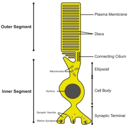
Rods and cones are the primary light sensitive cells of the retina. In contrast to cones which are specialized to the perception of daylight and colours (photopic system), rods are responsible for dim light vision (scotopic system). The schematic structure of a rod photoreceptor cell is shown in Fig. 2. A rod cell consists of an outer segment, an inner segment, the cell body and the synapse. The outer and inner segments are connected with a cilium that represents the bottleneck road for the transport of cargo from the cell body to the outer segments. Light detection and the downstream signalling transduction take place in the outer segments. Their interior space is filled with stacks of membranes called discs. The outer membrane encloses the outer segments and is the place of generation of the rod membrane potential.
Fig. 2 Schematic view of a rod photoreceptor. Rods are composed of an outer segment that is
1.3 Signalling transduction in rods
Rods are able to detect even a single photon. This extremely high light sensitivity requires a massive signal amplification within the phototransduction cascade.
In dark, constitutively active gyanylate cyclases (GCs) produce high cGMP levels. High cGMP levels in turn keep the cGMP sensitive cyclic nucleotide-gated (CNG) channel in its open state and give rise to the influx of Na+ and Ca2+, respectively. This depolarises the rod cell up to -40 mV resulting in a “dark current”. Finally, this depolarisation triggers a sustained transmitter release at the synapse.
Photons are absorbed by retinal, a chromophore that is covalently attached to the G protein coupled receptor rhodopsin [1]. After absorption, retinal isomerizes from the 11-cis form to the all-trans form. This results in conformational changes of rhodopsin (bleaching). One of the intermediates of this bleaching process called metarhodopsin II (Rh*) activates the G protein transducin which stimulates the cGMP phosphodiesterase (PDE6) to hydrolyse cGMP to GMP. The decrease in cGMP concentration in turn leads to the closure of the CNG channel. As a consequence, the outer membrane hyperpolarises to -70 mV [2] and gives rise to the switch-off of transmitter release to bipolar cells. Thus, the light signal information finally sent to the brain is generated by the termination (and not by the generation) of transmitter release.
In order to regenerate the dark current, the rod cell has to restore the cGMP concentration to the dark level. This process is regulated by the Ca2+ concentration and by the action of gyanylate cyclases (GC-E/F). The latter are controlled by gyanylate cyclase activating proteins (GCAPs). In their inactive form, GCAPs bind Ca2+ molecules. During light response, the CNG channel is closed and the Ca2+ levels are low [3]. The decrease in Ca2+ levels converts GCAP to the active form which then stimulates GCs to produce cGMP. High cGMP levels finally lead to opening of CNG channels and to the restoration of the dark current (Fig. 3 A).
Fig. 3 Signalling transduction in rods. A) Molecular mechanisms occurring in the rod outer
segments upon light induced rhodopsin activation. B) Phosphorylation of Rh* by rhodopsin kinase (GRK1) gives rise to binding of arrestin which prevents rhodopsin from sustained activation of transducin. For details, see text. NCKX, Na+/Ca2+/K+ exchanger; Gt, transducin; CaM, calmodulin.
1.4 CNG channels
Fig. 4 Subunit composition of CNG channels.
1.4.1 Topology and structural features of CNG channels
CNG channels belong to the superfamily of voltage-gated ion channels. The members of this channel group consist of an intracellular N-terminus, six transmembrane domains (S1-S6) and an intracellular C-terminus. Transmembrane segments of CNG channels are connected to each other by short loops. The last loop located between S5 and S6 forms the pore region. The C-terminal domain is subdivided into three functional domains: C-linker, cyclic nucleotide-binding domain (CNBD) and the distal C-terminus [11, 12].
CNG channels are structurally and evolutionary related to hyperpolarisation activated cyclic nucleotide-gated (HCN) channels. Recently, the crystal structure of the C-terminus of HCN channels consisting of the C-linker and the CNBD was determined (Fig. 5, [13]). The comparison of the CNBD of HCN channels to that of other cyclic nucleotide-binding proteins like the gene activator protein (CAP, [14]) and cAMP dependent protein kinase (PKG1, [15]) reveals that these proteins share very similar folding properties. The C-linker comprises six α-helices (A’-F’) followed by an α-helix of the CNBD and a β-roll consisting of eight β-strands (1-8). Another short α-helix (called P-helix) is located between β-strands 6 and 7. The last β -strand is followed by two additional α-helices (B- and C-helix, respectively).
Fig. 5 Topology of CNG channels. Left: representative topology of a CNG subunit consisting of the
intracellular N- and terminus and six transmembrane domains (1-6). Right: 3D model of the C-terminus of CNGB1a based on the molecular dynamics (MD) simulation performed by the group of Prof. Dr. Klaus T. Wanner, Department of Chemistry, Ludwig Maximilians Universität München. MD simulation was based on the recently identified crystal structure of the C-terminus of HCN2 [13]. For details, see text.
1.4.2 Gating of CNG channels
1.5 Role of CNGB1 in rod CNG channels
1.5.1 Characteristics of the CNGB1 locus
The CNGB1 locus is a complex gene giving rise to the transcription of at least four different variants due to alternative splicing. In rod photoreceptors, three transcripts deriving from the CNGB1 gene and encoding CNGB1a, GARP1 and GARP2 could be identified [22-24]. In
olfactory sensory neurons, only CNGB1b is expressed [25]. This splice isoform lacks the most part of the long amino terminus which is present in CNGB1a.
In contrast to CNGA1, CNGB1a subunit is not able to form functional channels when expressed alone in heterologous expression systems. However, when coexpressed with CNGA1 the CNGB1a subunit confers several characteristic properties to the heteromeric channel [24]. For example, heteromeric CNGA1/CNGB1a channels show an increased sensitivity to cAMP and are efficiently blocked by L-cis-diltiazem. Furthermore, CNGA1/CNGB1a heteromers show a typical single channel flickering behaviour, an increased inhibition by calmodulin (CaM), and a decreased block by extracellular Ca2+ [11, 12, 26, 27].
The involvement of CNGB1 in the transport of heteromeric channels to their final destination represents another important function of this subunit. In the CNGB1 KO model, the transport of CNGA2 and CNGA4 to the olfactory cilia as well as the transport of CNGA1 to the outer segments is strongly impaired [28, 29]. Recently, some aspects of the molecular mechanisms regarding the transport of CNG channels to olfactory cilia and outer segments of rod phororeceptors could be clarified. It has been shown that the distal C-terminus of CNGB1a plays a crucial role in this process [17, 30].
1.5.2 Retinitis pigmentosa mutations in the CNGB1 gene
in the CNGB1 locus to recessive form of RP [31, 32]. One of these mutations (c.3444+1G>A) represents a splice site mutation positioned on the donor site of Exon 32, the other one (c.2978G>T; p.G993V) represents a point mutation located in the CNBD of CNGB1. However, so far no functional analysis of these two mutations was performed in order to verify their pathogenicity or to decipher the molecular mechanism leading to the RP phenotype.
1.6 Role of GARP in rods
The exact molecular functions of the glutamic acid rich proteins, GARP1 and GARP2 (herein referred to as GARPs) have not been well characterised so far. GARPs are soluble proteins and represent alternatively spliced isoforms of the CNGB1 gene (see chapter 1.5.1). The protein sequence of GARPs is almost completely identical to the respective region of the N-terminus of CNGB1a. In contrast to GARP1, GARP2 lacks the negatively charged glutamic acid rich region and is expressed at high levels in rod outer segments [23, 24, 33].
Disc rims are regions of the rod disc membranes adjacent to the rod plasma membrane. It has been reported that GARP2 interacts with different disc rim associated proteins, like peripherin-1 or PDE6 [33-35]. Some of the proposed functions of GARPs arising from these studies are the fine-tuning of the cGMP signalling and scaffolding functions, like the maintenance of the disc rim integrity and tethering of CNG channels to disc rims. Most probably because of their high content of charged glutamic acids, GARPs were also shown to have features of natively unfolded proteins [33].
1.7 Goals of this study
This study was set out to address two important questions:
1) How do specific mutations in the CNGB1 gene result in RP? 2) What is the role of CNGB1a in gating of rod CNG channels?
The main focus of this study was to answer these questions by functional analysis of the two RP causing mutations in the CNGB1 gene.
As described in chapter 1.5.2, the first mutation (c.3444+1G>A) represents a splice site mutation positioned on the donor site of Exon 32. This mutation could impact CNGB1a on mRNA and on protein level and was therefore tested for splicing and expression in HEK293T cells.
The second mutation (c.2978G>T; p.G993V) is a point mutation located in the CNBD of CNGB1a. Since this domain is crucial for proper gating of the CNG channel, the p.G993V mutation harbours the potential to impair the gating of these channels. The effects of this mutation on channel gating were analysed by different computational, electrophysiological, and protein biochemical experiments.
Fig. 6 RP mutations in CNGB1a. Blue box marks the CNBD, green circle shows the position of the
2 Materials and methods
2.1 Molecular biology
2.1.1 Plasmids
pcDNA3.1 vector
pcDNA3.1 (Invitrogen) represents a commonly used mammalian expression vector. It consists of following elements:
- Cytomegalovirus (CMV) promoter, capable of driving heterologous gene expression in mammalian cell lines.
- Simian virus (SV40) origin of replication (ori), allowing for replication in mammalian cells.
- Colicinogenic factor E1 (ColE1) ori, responsible for replication in prokaryotes. - Filamentous phage (f1) ori which allows recovery of single stranded plasmids in
prokaryotes.
- Multiple cloning site (MCS) for introduction of genes or gene fragments of interest. - Polyadenylation signal (pA) of the bovine growth hormone (BGH).
- Neomycin resistance (NeoR) under the control of SV40 promotor for selection of stable mammalian cell lines.
- Ampicillin resistance (AmpR) for selection of recombinant bacterial cells.
pCRII®-TOPO® vector
This vector was used for subcloning of PCR products (see chapter 2.1.8). It contains a lacZ promoter followed by the MCS and lacZ reporter gene. Thus, insertion of constructs into the MCS disrupts the expression of β-galactosidase and can be used for selection of recombinant bacterial clones on X-gal containing plates. Additionally, this vector contains a f1 ori and pUC ori for plasmid replication in prokaryotes. For selection of recombinant bacteria, kanamycin and ampicillin resistance (AmpR) are included in this vector.
pIRESeGFP vector
CMV promoter, SV40 pA, SV40 promotor, SV40 ori, f1 ori, KanR, NeoR, pUC ori and the herpes simplex virus thymidine kinase (HSV-TK) pA.
2.1.2 Polymerase chain reaction (PCR)
The conditions of each PCR reaction were adjusted to the respective application. Table 1 shows an overview about the standard PCR conditions of different polymerases used in this study:
Table 1 Standard PCR conditions for different polymerases.
Polymerase Taq Pfu (Stratagene) Herculase
(Stratagene)
Phusion
(Finnzymes)
Initial denaturation 95°C 1 min 95°C 1 min 95°C 1 min 98°C 30 sec
Denaturation 95°C 30 sec 95°C 45 sec 95°C 20 sec 98°C 10 sec
Annealing X °C 30 sec X °C 45 sec X °C 20 sec X °C 30 sec
Elongation 72°C 30 sec/kb 72°C 1 min/kb 72°C 30 sec/kb 72°C 30 sec/kb
Final elongation 72°C 5 min 72°C 5 min 72°C 5 min 72°C 5 min
Storage 10°C ∞ 10°C ∞ 10°C ∞ 10°C ∞
The pipetting scheme of the PCR was adapted from the manual of the respective manufacturer.
2.1.3 Purification of DNA fragments
After PCR amplification, the PCR products may be purified for further applications. The purification was performed using the PureLinkTM Quick Gel Extraction Kit (Invitrogen) accoriding to manufacturar’s protocol. All solutions needed for this procedure were provided with the kit. Briefly, fivefold amount of solubilization buffer was added to the PCR product and the solution was loaded on the column. The latter was placed in 2 mL Eppendorf tubes and was centrifuged at max. speed for 45 sec at room temperature. The flow through was discarded and 700 µL wash buffer was added to the column following by an additional centrifugation at identical conditions. After discarding the flow through, the column was spun at max. speed for 2 min at room temperature in order to remove all ethanol present in the wash buffer. Now, 30 µL ddH2O or elution buffer was added on the center of the column which was allowed to incubate 10 min at room temperature. Finally, the DNA was eluted into a fresh 1.5 mL Eppendorf tube by centrifugation at max. speed for 45 sec at room
temperature. To check for proper purification, 5 µL of the purified DNA were loaded on agarose gel.
2.1.4 Restriction analysis and preparation of samples for cloning
Restriction enzymes were purchased from New England BioLabs (NEB) or from Fermentas. Restriction analysis conditions were performed following the manufacturer’s instructions. For cloning applications, the amount of DNA in the restriction reaction was 3-5 µg. After the incubation time, the cut DNA was loaded on the appropriate agarose gel and was allowed to run until sharp bands could be cut off from the gel. Then, the cut piece of gel was weighed and the threefold volume of gel solubilization buffer (PureLinkTM Quick Gel Extraction Kit, see chapter 2.1.3) was added (i. e. if the gel piece weighed 200 mg, the amount of gel solubilization buffer was 600 µL). Now, the sample was incubated at 55°C for 5-10 min or until the gel was completely solved. This solution was loaded on the columns from the Quick Gel Extraction Kit and the the same procedure as described in chapter 2.1.3 was performed.
2.1.5 Ligation and dephosphorylation
Ligation reaction was performed using following pipetting scheme:
x µL vector DNA y µL insert DNA
2 µL T4 ligase buffer (NEB) 1 µL T4 ligase (NEB) 20 µL total volume
This reaction was incubated for 30 min - 2 h at room temperature. The vector-to-insert ratio was variable and was determined empirically for each ligation considering the amount of the respective DNA observed on the check agarose gel after the DNA purification. After the ligation vector DNA was dephosphorylated by following protocol:
2.1.6 Transformation
For transformation different chemically competent E.coli strains were used depending on the application. First, 100 µL aliquots of competent cells (stored at -80°C) were thawed on ice. Then, 1-3 µL DNA (i. e. ligation reaction) were added to the cell suspension which was gently swirled and was allowed to incubate on ice for 5 min. Now, a heat pulse was applied by incubating the cells for 30 sec at 42°C in a water bath. Immediately after this, the cell suspension was placed on ice for 2 min. If the resistance for selection of the plasmid DNA was ampicillin, the cells were directly plated on the appropriate agar plates. In cases of chloramphenicol or kanamycin resistance, 900 µL prewarmed LB(+) medium was added to the cells. The latter were incubated for 1 h at 37°C with shaking followed by an centrifugation at 3500 rpm for 5 min at room temperature. Approx. 800 µL of the supernatant was removed and the cells were resuspended in the remaining part of the LB(+) medium. Finally, the cells were plated on the respective agar plates and incubated over night at 37°C.
LB(+) medium LB(+) Agar
Pepton 10 g Agar 15 g
Yeast extract 5 g LB(+) medium ad 1 L NaCl 5 g autoclave
Glucose 1 g for preparing resistant agar plates, cool the LB(+) agar ddH2O ad 1 L medium to 55°C and add the appropri ate resistance in adjust pH to 7.2 - 7.5 concentrations described in chapter 0.
autoclave
2.1.7 Inoculation of bacterial cells and isolation of plasmid DNA (alkaline lysis)
containing the plasmid DNA was transferred into fresh 1.5 mL Eppendorf tubes. To precipitate DNA, 520 µL 100 % isopropanol was added to the mix. After vortexing, the mix was spun at rpm max. for 15 min at 4°C. Subsequentl y, the pellet was washed with 70 % ethanol and the solution was centrifuged at rpm max. for 5 min at 4°C. Then, the supernatant was discarded and the pellet was dried in a vacuum centrifuge at room temperature for 5 min. Now, the pellet was suspended in 30 µL of ddH2O and 1 µL of this plasmid DNA solution was used for control restriction analysis. If the expected results were obtained from this restriction analysis, the corresponding plasmid DNA was send for sequencing without further purification.
To yield plasmid DNA in larger amounts and in higher purity, PureYieldTM Plasmid Midiprep System (Promega) was used. For that purpose, colonies were inoculated in 100-200 mL LB(+) medium and similar procedure as described above was performed following the manufacturer’s instructions.
2.1.8 TOPO cloning
TOPO cloning (TOPO TA Cloning® Kit Dual Promotor, Invitrogen) represents a rapid and convenient cloning method for PCR products. Topoisomerase I from Vaccinia virus [37] attached to the pCRII®-TOPO® vector allows for covalent bonding of PCR products to this vector. However, this reaction only takes place if the respective PCR product posesses a deoxyadenosine (A) on it’s 3’ end. In contrast to most polymerases with proof reading activity (i.e. Pfu-polymerase), conventional Taq polymerase has terminal transferase activity that adds a single “A” to the 3´ ends of the PCR products.
First, PCR products which were amplified using a polymerase without terminal transferase activity were purified by standard procedures as described in chapters 2.1.3 and 2.1.4. Then, following components were added to 8 µL of the purified PCR product:
0.5 µL dNTP (10 mM each)
1 µL Taq buffer (containing 2.5 mM MgCl2) 0.5 µL Taq-Polymerase
This reaction was incubated for 30 min at 72°C and was placed on ice. TOPO cloning reaction was performed using following pipetting scheme:
4.5 µL of the Taq polymerase reaction product 1 µL salt solution (provided with the kit)
0.5 µL TOPO vector (provided with the kit)
2.1.9 In-Fusion cloning
For some cloning applications, as a alternative to mutagenesis PCR and/or overlap-PCR, In-FusionTM Advantage PCR Cloning Kit (Clontech) was used. PCR products sharing a 15 bp homology with the sequence at the ends of the linearized vector can be covalently attached to the vector giving rise to circular plasmid DNA. The In-Fusion enzyme provided with the kit catalyzes this covalent bounding.
The In-Fusion reaction was set up as follows:
≈ 100 ng purified PCR product
≈ 200 ng purified vector
2 µL 5 x In-Fusion reaction buffer 1 µL In-Fusion enzyme
Ad 10 µL ddH2O
If larger volumes of vector or insert were used, the volume of buffer, of the enzyme and the total volume were doubled. The In-Fusion mix was now incubated at 37°C for 15 min following by an incubation at 50°C for 15 min. Then , the reaction was placed on ice and diluted in TE buffer (pH 8.0) up to a volume of 50 µL. Finally, 3-5 µL of this solution were used for a standard transformation protocol.
TE Buffer
10 mM Tris-HCl pH 8.0 3 mM EDTA
2.1.10 Introduction of mutations in DNA constructs
2.1.11 Reverse transcription (RT)
Reverse transcription was performed using the ThermoScriptTM RT-PCR System Kit (Invitrogen) following the instructions of the manufacturer. During the cDNA synthesis, random hexamers as well as oligo(dT) primer were added to the reaction. After the cDNA synthesis, herculase (Stratagene) was used for the subsequent PCR.
2.1.12 Cloning of CNG channels
Cloning of the rat CNGB1a/CNGB1b and CNGA2/CNGA4 subunits was described previously [25, 38, 39]. Bovine CNGA1 was a gift of Dr. U. Benjamin Kaupp (Caesar Bonn). As far as not otherwise mentioned, all experiments from the electrophysiological and biochemical recordings described in chapter 3.2 were obtained using rat CNGB1 an bovine CNGA1 channels. Human CNG channels were PCR amplified from retinal cDNA, were subcloned into the pcDNA3.1 and/or pIRESeGFP vector and sequenced.
2.2 Cell culture
2.2.1 Cultivation and transfection of mammalian cell lines
For most in vitro transfections in this study, HEK293T cells were used. They were cultivated in DMEM + GlutaMAXTM-I medium (+ 4,5 g/L glucose, - pyruvate + 10 % FBS + 1 % penicillin/streptomycin) at 37°C with 10 % CO2. COS7 cells were cultivated under same conditions in DMEM + GlutaMAXTM-I medium (+ 4,5 g/L glucose, + pyruvate + 10 % FBS + 1 % penicillin/streptomycin).
Transient transfections of HEK293T cells or COS7 cells were performed using the calcium phosphate technique [40] or with FuGENE® (Promega). The calcium phosphate based transfection was performed by adding following solutions to a 15 mL Falcon tube:
10 µg DNA, 50 µL 2,5 M CaCl2 ad 500 µL ddH2O
correspond to ∅ 10 cm plates. For ∅ 15 cm plates, the volume of all solutions was doubled and the amount of DNA was increased to 25-30 µg.
Transfection with FuGENE® (Roche) was performed for subsequent immunocytochemical and electrophysiological applications. For 16-well plates 30 ng DNA was used per well, for
∅ 3.5 cm plates 3 µg DNA was used. First, fresh GlutaMAXTM-I medium (without FBS and without penicillin/streptomycin) was added to cryo tubes followed by the addition of DNA. Thereby, tenfold volume of medium related to the total volume on DNA was added (i. e. 1 µL DNA corresponds to 10 µL medium). After the 5 min incubation, FuGENE was added directly to the reaction which was mixed subsequently by pipetting up and down. In this case, threefold volume of FuGENE in µL was used related to the amount of DNA in µg (i. e. 1 µg DNA corresponds to 3 µL FuGENE). This reaction was allowed to incubate for 30 min at room temperature and was then added directly to the medium of the cells. The latter were incubated at 37°C with 10 % CO2 until proceeding wi th the respective application.
2 x BBS Solution
10,65 g BES 16,35 g NaCl
0,21 g Na2HPO4 · 2H2O ad 950 mL H2O
adjust to pH 6,95 with NaOH ad 1L ddH2O
steril filtrate
2.3 Protein biochemistry
2.3.1 Isolation and quantification of proteins
Proteins were isolated from cultured mammalian cells by the following protocol:
48 h after transfection, medium was removed and the cells were harvested in a 1.5 mL Eppendorf tube. Then, the suspension was centrifuged at 4°C for 5 min at 1000 x g. Hereafter, the pellet was resuspended in lysis buffer and was rotated at 4°C for 30 min. Subsequently, the cells were spun at 4°C for 10 min at rpm max. and the supernatant was transferred into a fresh 1.5 mL Eppendorf tube.
centrifuged at 4°C at rpm max. and the supernatant was transferred into fresh 1.5 mL Eppendorf tubes.
To determine the concentration of isolated proteins, Bradford assay was performed [41]. Thereby, 5 µL of the protein solution (5 µL lysis buffer were used as blank control) were transferred into 1 mL plastic cuvettes followed by an addition of 95 µL 0.15 M NaCl solution. Then, 1 mL coomassie blue solution was added and was allowed to incubate for 2 min at room temperature. Immediately after the incubation time, protein concentration was measured using the Bradford assay program on the BioPhotometer (Eppendorf).
1 x Lysis Buffer Coomassie Blue Solution
2.5 mL Triton X-100 50 mg coomassie brilliant blue G250
15 mL 5 M NaCl 25 mL 95 % ethanol
400 µL 2.5 M CaCl2 50 mL 85 % phosphoric acid (H3PO4) ad 500 mL ddH2O
2.3.2 Membrane preparations
For enrichment of membrane proteins, preparations of cell membranes were performed. For this purpose, 1-2 mL of the 1 x membrane preparation buffer containing the proteinase inhibitor cocktail mix was added to the harvested HEK293T cells or mouse tissue and the breakup of cell or of the tissue was performed using the Potter S homogenizer. Then, the suspension was centrifuged at 4°C for 10 min at 500 0 x g. The supernatant was transferred into 6 mL ultracentrifuge tubes and 1 x membrane preparation buffer was added to a final volume of 4 mL. Ultracentrifugation was performed at 4°C for 45 min at 30000 rpm (Beckman 45 Ti rotor). Pellet was suspended in 50-100 µL 1 x membrane preparation buffer and 5 µL were used for determination of the protein concentration as described in chapter 2.3.1.
3 x Membrane Preparation Buffer:
3,15 g MOPS 77 g Sucrose
2.3.3 Western blotting
Western blotting was performed by standard procedures. After the protein transfer to the PVDF membrane, the latter was shortly equilibrated with methanol and was blocked in 5 % milk powder for 1 h at room temperature with shaking. Then, the incubation of the membrane with the appropriate primary antibody was performed. The optimal incubation time and the optimal antibody concentration were determined empirically. Hereafter, the membrane was washed three times in TBST for 5 min followed by an 1-2 h incubation with the secondary antibody at room temperature with rotation. Now, the membrane was washed 3-4 times with TBST for 5 min and once in ddH2O. After the incubation with the luminol reagent according to manufacturer’s protocol (Millipore or Santa Cruz), the membrane was put into a film cassette and was exposed to a x-ray film (Fuji). The optimal exposure time was determined empirically.
10 x TBS 1 x TBST
12.1 g Tris 100 mL TBS
80.2 g NaCl 0.1 % Tween
ad 1 L ddH2O protect from light
2.3.4 Co-immunoprecipitation
To analyse protein-protein interactions, co-immunoprecipitation experiments using protein G dynabeads (Invitrogen) were performed. First, approx. 5 µg antibody and PBS were added to 30 µL dynabeads up to a final volume of 500 µL. This solution was rotated for 30 min at 4°C and subsequently, the supernatant was removed on the magnet followed by a wash step with 200 µL PBS. Then, 1 mg of the protein lysate was added to the beads and the reaction was filled up with PBS to a final volume of 500 µL. Now, the suspension was rotated for 30 min at 4°C followed by three washing steps with PBS. After the last wash, the suspension was transferred into fresh 1.5 mL Eppendorf tubes and the supernatant was removed completely. Beads were resuspended in 6 x Lämmli buffer (with or without DTT, depending on the application) and were incubated at 70°C for 15 min. Finally, the supernatant was loaded on the appropriate SDS PAGE gel.
quenched subsequently by adding Tris-HCl pH 7.5 (final concentration 50 mM) and incubating the reaction for 15 min at room temperature. Then, protein lysate was added and the reaction was processed as described above.
6 x Lämmli 6 x Lämmli + DTT
7 mL 4 x Tris-HCl/SDS pH 6.8 = 6 x Lämmli
3 mL glycerol + 930 mg DTT
1 g SDS
1.2 mg bromphenolblue ad 10 mL ddH2O
2.3.5 Biotinylation assay
Biotinylation represents a commonly used method for detection of cell surface proteins [42]. 48 days after transfection, medium was removed from HEK293T cells which then were washed with PBS. Cell surface biotinylation was performed using 0.8 mM of non-cell permeable sulfo-NHS-SS-biotin (Pierce, Rockford) in PBS for 30 minutes at 4°C. The reaction was stopped with 10 mM glycine and the cells were transferred into fresh 50 mL Falcon tubes. After 2 wash steps with PBS (each time pelleting the cells by centrifugation at 4°C for 5 min at 1500 x g), the cell pellet was sol ubilised in 1 x lysis buffer with the proteinase inhibitor cocktail mix (see chapter 2.3.1). Then, biotinylated proteins were precipitated on Neutravidin agarose resin (Pierce) using 80 µL of neutravidin beads per reaction. First, beads were washed with 200 µL PBS and centrifuged after each wash step at 4°C for 1 min at 2500 x g. Biotinylated protein lysate (400 µg/reaction) was added to the washed beads and was rotated for 2 h at 4°C. Next, the suspensio n was washed 4 additional times with PBS containing 1 % NP-40 under the same centrifugation conditions as described above. Finally, elution was performed by adding 40 µL of elution buffer to the beads. Samples were incubated 10 min at 70°C before loading them on the SDS PAGE gel.
Elution Buffer
280 µL PBS
40 µL 10 % NP-40
2.4 Förster resonace energy transfer (FRET)
FRET represents a commonly used tool for the analysis of molecular dynamics such like protein-protein interactions [43]. This method is based on the energy transfer between two fluorophores located in a defined spatial proximity to each other (Typically, less then 10 nm). Thereby, the emission spectrum of the first fluorophore (donor) is partially absorbed by the second fluorophore (acceptor) giving rise to a decrease in the emmision spectrum of the donor and an increase of the acceptor emission spectrum. This spectral shift is finally measured as FRET signal. However, a prerequisite for a FRET signal is a spectral overlap of the donor and the acceptor. Based on this, for biological use the most commonly used pair of fluorophors is the combination of CFP as donor and YFP as acceptor. Interaction of proteins of interest can be studied by coupling them to these fluorophores, by coexpressing them in an appropriate cell line and by subsequent measurement of the FRET signal. In this study, FRET was used to analyse the assembly of CNGB1a subunits carrying the GV mutation with the CNGA1 subunit using following protocol:
HEK293 cells were transfected with CNGA1-CFP or soluble CFP (negative control) and wildtype or mutant YFP-CNGB1a. A CFP-YFP tandem construct served as FRET positive control [44]. Fluorescent images were captured 48-72 h after transfection on an Axioplan 2 microscope (Zeiss, Oberkochen, Germany) equipped with a Plan Neofluar 40 x objective (numeric aperture 0.75), a HMRc ccd camera and an AttoArc HBO100 mercury lamp illumination unit. Three fluorescent images per cell were collected using the CFP HC filter, YFP HC filter and FRET CFP-YFP HC filter sets (Semrock, Rochester). Fluorescence images were processed using the FRET plus macro (Zeiss) and the FRET efficiency was calculated as N-FRET using the method described by Xia and Liu [45].
2.5 Electrophysiological recordings
2.6 Statistics
3 Results
3.1 Splicing analysis of the c.3444+1G>A mutation in CNGB1
As described in chapter 1.5.2 the c.3444+1G>A splice site mutation in CNGB1 has been associated with RP [32]. This mutation is located at the donor site of exon 32 and has been proposed to result in a frameshift and truncation of the last 28 amino acids (aa) of the corresponding protein. However, this ambiguous conclusion was not verified by experimental data. Therefore, the effects of c.3444+1G>A on splicing were reexamined by in silico and in vitro experiments.
3.1.1 In silico splicing analysis of c.3444+1G>A
In order to reconstruct the mechanism by which c.3444+1G>A could lead to truncation of the last 28 aa of CNGB1a, in silico analysis was performed using the NNSplice 0.9 splice site prediction software (http://www.fruitfly.org/seq_tools/splice.html) (Fig. 7). The DNA sequence used for this analysis starts with exon 32 and ends with the stop codon of CNGB1. A plausible explanation for the splicing scenario proposed by the original study would be the use of cryptic donor sites.
Fig. 7 Donor site prediction of a CNGB1 genomic DNA fragment encompassing exon 32-33 (3911 bp in length) using the NNSplice 0.9 splice site prediction software (http://www.fruitfly.org/seq_tools/splice.html). The respective positions of the predicted donor sites
As shown in Fig. 7, the DNA sequence analysed herein harbours seven potential cryptic donor sites. Use of the cryptic donor site in exon 33 would indeed delete the sequence that encodes the last 28 aa. However, it would also lead to retention of intron 32. In this case, due to an intronic stop codon 171 bp after exon 32, the corresponding protein would lack all 97 aa encoded by exon 33.
3.1.2 Creation of wild type and mutant minigene constructs
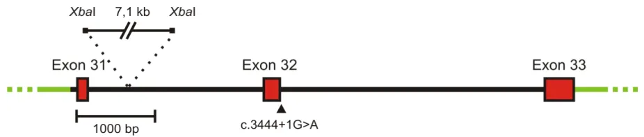
In conclusion, no possible splice scenario could be reconstructed that would give rise to the deletion of only the last 28 aa of the CNGB1 protein. Therefore, the splicing of the construct containing the c.3444+1G>A mutation was analysed experimentally. For this purpose, a DNA fragment starting from the last 55 bp of intron 30 and ending with the last 42 bp after the stop codon within exon 33 of CNGB1 was amplified by PCR from human genomic DNA and was sequenced. For cloning convenience, a 7.1 kb fragment of intron 31 flanked by XbaI sites was deleted. The final 6.4 kb minigene construct was subcloned into the pcDNA3 vector. The c.3444+1G>A mutation was inserted using standard site directed mutagenesis (Fig. 8).
Fig. 8 Schematic representation of the minigene construct used for the exon trapping experiment showing the position of the deleted intronic XbaI-fragment. pcDNA3 vector backbone
3.1.3 Exon trapping experiments in HEK293T cells
Wild type and mutant minigene constructs were transfected in HEK293T cells and after 48 h, cells were harvested followed by total RNA isolation. After cDNA synthesis and PCR amplification with vector specific primers the splicing products derived from the minigenes were sequenced (Fig. 9).
Fig. 9 Exon trapping experiment from transfected HEK293T cells. On the left, revese transcriptase
PCR from HEK293T cells transfected with mutant and wild type minigene constructs is depicted. The electropherogram for the c.3444+1G>A mutant shows the skipping of exon 32. Wild type construct was spliced correctly (data not shown). On the right, schematic representation of the splice products is shown. The length of the respective PCR products is indicated by double arrows. WT: wild type, Mut: c.3444+1G>A mutation.
Fig. 10 Schematic comparison of the WT and mutant protein demonstrating the lack of the entire distal C-terminus and the last 10 aa of the αααC helix in the context of the c.3444G>A α mutation. Skipping of exon 32 causes a frameshift which results in addition of 68 unrelated amino
acids after aa position 1075 of the CNGB1a protein (highlighted in grey). The numbers represent the length of the respective proteins (1245 aa for WT and 1143 for the mutant). S1-S6: transmembrane segments.
3.1.4 In vitro expression of wild type and mutant rod CNG channels
Fig. 11 Western blot of membranes isolated from HEK293T cells transfected with CNGA1 and wild type or mutant CNGB1a probed with anti-B1 (top panel) or anti-ATPase (bottom panel). The
weaker expression of the mutant protein was normalized in the presence of the proteasome inhibitors MG-132 and ALLN. The blot was probed with an antibody directed against the N-terminus of CNGB1a (PPc6N, [23]). As loading control anti-ATPase antibody (1:1000, clone α6F, developed by D.M. Fambrough, obtained from the Developmental Studies Hybridoma Bank, Iowa) was used. In proteasome inhibition experiments MG-132 and ALLN (25 µM each, Calbiochem) were added directly to the cells sixteen hours prior to harvesting.
As shown in Fig. 11, in the western blot analysis using an antibody directed against the N-terminus of CNGB1a the expected 240 kDa band for the wild type CNGB1a could be detected. As anticipated, the mutant CNGB1a protein was smaller than the wild type counterpart. Furthermore, the expression level of the mutant CNGB1a was considerably reduced compared to the wild type CNGB1a. Since this difference in expression could be reversed by the addition of the proteasome inhibitors MG-132 and ALLN, respectively, the conclusion can be drawn that the mutant protein is partially degraded by the proteasome. In summary, the c.3444+1G>A mutation was shown to result in skipping of exon 32 on mRNA level thereby leading to a frameshift after exon 31 and a premature stop codon. On protein level this mutation gives rise to deletion of the last 170 aa of the mutant protein including 10 aa of the αC helix within the CNBD. Finally, in vitro results from HEK293T cells after coexpression of the mutant CNGB1a with CNGA1 have shown that the mutant CNGB1a is prone to proteasomal degradation.
3.2 Functional analysis of the G993V mutation in CNGB1
As described in chapter 1.7, this part of the study addressed the role of the CNGB1a subunit on the activation process of the heteromeric rod CNG channel. For this purpose, a naturally occurring point mutation in the human CNGB1 gene that leads to a glycine-valine exchange at position 993 in the CNBD of CNGB1a (CNGB1aGV) was analysed. This mutation was identified in a French family suffering from retinitis pigmentosa (RP) [31].
3.2.1 In silico analysis
Fig. 12 In silico analysis of the G993V mutation. A) Sequence alignment of the β2-αC region of the CNBD of human CNGB1a, human CNGA1, E. coli CAP, sea urchin (sp) HCN1 and human PKG1. Invariant residues are highlighted in red. The invariant glycine residue within the β2-β3 loop that is mutated to valine (GV mutation) in the CNGB1a subunit of RP patients is marked with an asterisk. Residues identified in the crystal structure of HCN channel CNBDs to participate in cyclic nucleotide-binding [51] are marked with arrowheads. B) Predicted structure of the cGMP nucleotide-binding pocket of CNGB1a (left) and CNGB1aGV (right). The sequences were threaded onto the crystal structure of the CNBD of spHCN [51] by using Modeller [52] and analysed using molecular dynamics simulations. The glycine and the valine residue in the β2-β3 loop of CNGB1a and CNGB1aGV, respectively, are represented as space filling spheres. Residues in the β6 and β7 strands as well as in the C helix participating in cGMP-binding are shown as sticks. Only one monomer is shown for clarity.
3.2.2 Expression of CNGA1GV
Based on the findings from the in silico analysis, the GV mutation could have the potential to impair cGMP binding and, hence, channel activation. To test this hypothesis the GV mutation was introduced into the CNBD of CNGA1 and the expression as well as the electrophysiological properties of the mutant CNGA1 were analysed (Fig. 13).
Fig. 13 CNGA1GV is expressed on the plasma membrane of transfected HEK293 cells. A) Top
panel: Western blot of membranes from HEK293 cells transfected with CNGA1-CFP or CNGA1GV-CFP, respectively. The blot was probed with anti-GFP antibody. Bottom panel: membranes probed against the ATPase α subunit. (B) Representative current traces elicited by 1 mM cGMP and voltage steps (2 s each) to -80 mV and +80 mV in excised patches from HEK293 cells expressing CNGA1 (A1), or CNGA1GV (A1GV), CNGA1GV/CNGB1a (A1/B1a) and CNGA1GV/CNGB1b (A1/B1b).
3.2.3 Electrophysiological measurements of heteromeric CNGA1/CNGB1aGV channels
Next step was to analyse the effects of the GV mutation in CNGB1a on the heteromeric channel complex. To this end, CNGA1 was coexpressed together with either wild type or mutant CNGB1a and the properties of the resulting currents were compared to currents induced by CNGA1 alone (Fig. 14).
Fig. 14 Electrophysiological characterisation of heteromeric CNGA1/CNGB1GV channels.
Surprisingly, currents obtained after coexpression of CNGA1 and CNGB1aGV did not exhibit typical CNGB1a-mediated features but were indistinguishable from currents of homomeric CNGA1 channels (Fig. 14A-D). While single channel currents of heteromeric CNG channels are characterised by very brief open-closed transitions (single channel flicker), single channel currents obtained after coexpression of CNGA1 and CNGB1aGV displayed long openings which is typical of homomeric CNGA1 channels (Fig. 14 A). In addition, the strong outwardly-rectifying current-voltage relation of channels obtained after coexpression of CNGA1 and CNGB1aGV in the presence of extracellular divalent ions was virtually identical to that of the homomeric CNGA1 channel, but clearly differed from that of weakly rectifying wild type heteromeric channels (Fig. 14 B). In contrast to wild type CNGB1a, CNGB1aGV did not increase Ca2+/CaM sensitivity if coexpressed with CNGA1 (Fig. 14 C). Probably the most indicative feature resulting from the presence of the CNGB1 subunit is the generation of a high affinity site for block by L-cis-diltiazem [26]. Indeed, unlike homomeric CNGA1 channels, heteromeric CNGA1/CNGB1a channels were efficiently blocked by this drug. By contrast, currents obtained after coexpression with CNGB1aGV were not sensitive to L-cis-diltiazem (Fig. 14 D).
3.2.4 Coassembly and cell surface expression of CNGA1/CNGB1aGV heteromers
Fig. 15 Cell-surface expression and assembly of CNGA1/CNGB1GV heteromers. A) Biotinylation
of non-transfected (ø), CNGA1/CNGB1a (WT) and CNGA1/CNGB1aGV (GV) transfected HEK293 cells probed with anti-B1 (top panel) or anti-tubulin (tub, bottom panel) antibodies. B) Co-immunoprecipitation (co-IP) from lysates of HEK293 cells co-transfected with myc-tagged CNGA1 and wild type CNGB1a or CNGB1aGV, respectively. Top panel: immunoprecipitation (IP) using anti-myc for pulldown and anti-B1 for detection. Bottom panel: Western blots showing the starting material for the co-IP probed with anti-myc (left) or anti-B1 antibodies (right). C) N-FRET ratios calculated from HEK293 cells co-transfected with CNGA1-CFP and YFP-CNGB1a (WT) or YFP-CNGB1aGV, respectively. HEK293 cells transfected with CFP and YFP-CNGB1a or a CFP-YFP tandem were used for the calculation of negative (neg) and positive (pos) control N-FRET values, respectively. Number of experiments is given in brackets.
3.2.5 Identification of inhibitory domains in CNGB1a
Another explanation for the selective detection of homomeric CNGA1 currents in HEK293 cells transfected with CNGA1 and CNGB1aGV may be that homomeric and mutant heteromeric CNG channels do coexist in the plasma membrane but in contrast to homomeric CNGA1 channels mutant heteromers cannot be activated by cGMP and, thus, do not contribute to any measurable current. In an attempt to identify structural determinants conferring an inhibitory action of CNGB1aGV the GV mutation was introduced into the olfactory CNGB1b subunit that differs from CNGB1a by lacking the GARP domain. This new mutant was coexpressed with CNGA1 and patch clamp recordings were performed (Fig. 16).
Fig. 16 Identification of N-terminus of CNGB1a (herein referred to as “GARP domain”) as the inhibitory domain. The GV mutation does not impair channel function in the context of the olfactory
CNGB1b isoform lacking the GARP domain. Top panel: Schematic representation of CNGB1a and CNGB1b subunits. The position of the GV mutation is highlighted by an asterisk. Amino acid position 555 is highlighted since starting from this position CNGB1a and CNGB1b are identical. Bottom panel: Summary graph showing the L-cis-diltiazem sensitivity of various homo- or heteromeric channels. Currents were elicited by 300 µM cGMP at +80 mV. L-cis-diltiazem was used at 50 µM for A2, A2/A4, A2/A4/B1b and A2/A4/B1bGV and at 10 µM for all other channels.
Surprisingly, as shown in Fig. 16, L-cis-diltiazem sensitive currents were now obtained with both wild type and mutant CNGB1b subunits. Sensitivity to L-cis-diltiazem was also consistently observed when the GV mutation was introduced into the native olfactory CNG channel complex consisting of CNGA2/CNGA4 and CNGB1b. Like for CNGA1 the wild type but not the mutant CNGB1a subunit induced L-cis-diltiazem sensitivity when coexpressed together with CNGA2. These results indicated that the GARP domain mediates the dominant negative effect of CNGB1aGV. To identify the inhibitory determinants within the GARP domain, a systematic mutation screening was performed by deleting specific regions of this domain. The respective constructs were coexpressed with CNGA1 in HEK293 cells followed by electrophysiological measurements of L-cis-diltiazem mediated CNG current inhibition. Fig. 17 shows a summary of the most important mutation constructs analysed in this approach.
Fig. 17 Mutagenesis screen within the GARP domain. Top panel: cartoon showing the CNGB1a
As is evident from Fig. 17, study of mutants with deletions in the GARP domain identified two regions (residues 214-253 and 364-481) of CNGB1a that confer most of the inhibitory effect. Fusion of an unrelated peptide (YFP) to the N-terminus of CNGB1bGV did not result in channel inhibition excluding unspecific effects which could be mediated merely by the length of the N-terminal domain.
As described in chapter 2.1.12, the experiments shown so far were obtained using rat CNGB1a and bovine CNGA1 channels. To examine if inhibitory effects of the GARP domain can be obtained using CNG channels from other species, the GV mutation was introduced into the human CNGB1a (hCNGB1a) subunit containing and lacking the GARP domain. After coexpression with human CNGA1 (hCNGA1), L-cis-diltiazem sensitivity was measured for these channels. The results are shown in Fig. 18.
Fig. 18 Effects of the GV mutation and of the GARP domain on human rod CNG channels.
Percent inhibition by 10 µM L-cis-diltiazem of currents obtained after cotransfection of human CNGA1 with wild type or mutant human CNGB1a lacking or containing the GARP domain in presence of 300 µM cGMP. hB1a#452-1245 corresponds to the rat CNGB1a construct B1a#556-1339 (see i.e.Fig. 17 andFig. 19). Number of experiments is shown in brackets.
3.2.6 Coexpression of GARP as soluble protein
To test whether the GARP domain can act inhibitory as an autonomous protein it was coexpressed as separate protein (B1a#1-555) together with CNGA1 and a CNGB1a truncation construct (B1a#556-1339, see cartoon in Fig. 19 A).
Fig. 19 GARP acts as an autonomous unit when coexpressed as soluble protein. A) Schematic
representation of the truncated B1a#556-1339 GV channel and the protein corresponding to the GARP domain. B) Percent inhibition by 10 µM L-cis-diltiazem of currents obtained after cotransfection of CNGA1 with various CNGB1a-derived constructs as indicated. Number of experiments is given in brackets. ** = p < 0.01
To test if the inhibitory effect of GARP is mediated by direct binding of this domain to the CNGB1a or CNGA1 subunit, co-imunoprecipitation experiments were performed (Fig. 20).
Fig. 20Co-immunoprecipitation of the GARP domain (B1a#1-555) with CNGA1 (myc-tagged) or CNGB1a#556-1339 WT or GV. A) Schematic representation of the truncated B1a#556-1339 GV
channel and the protein corresponding to the GARP domain (B1a#1-555) and binding sites of the B1a-specific antibodies used for experiments shown in B and C, respectively. B) Co-IP using the anti-myc antibody capable of the immunoprecipitation of myc tagged CNGA1 C) Co-IP using anti CNGB1 directed against the C-terminus of this protein.
1, coexpression of A1 + B1a#1-555; 2, A1 + B1a#1-555 + B1a#556-1339; 3, A1 + B1a#1-555 + B1a#556-1339GV; 4, B1a#1-555 + B1a#556-1339. Anti-B1, rabbit polyclonal antibody directed against the C-terminus of CNGB1a. PPc6N, rabbit polyclonal antibody directed against the N-terminus of CNGB1a. IP, immunoprecipitation.
3.2.7 Role of a functional CNBD of CNGB1 for CNG channel activation
Results shown in Fig. 20 indicate that the GARP domain binds to both CNGA1 and CNGB1 subunits in the heteromeric CNG channel complex. In wild type heteromers the identified GARP-channel interaction does not interfere with cGMP-dependent activation. By contrast, in the presence of the GV mutation the GARP domain completely shuts down channel activity. Based on MD simulations (Fig. 12) the conclusion can be drawn that the inhibitory effect of GARP may be causally linked to the inability of CNGB1aGV to bind cGMP. To test this hypothesis the effects of GARP in the presence of a double mutation in the β7 strand of the CNBD (R1042E and T1043A, herein after referred to as RT>EA) that is well known to abolish cyclic nucleotide-binding in CNG and HCN channels [13, 51] were analysed (Fig. 21).
Fig. 21 Summary graph comparing the L-cis-diltiazem sensitivity of heteromeric channels containing wild type and cyclic nucleotide-binding deficient (RT>EA) B1a or B1b subunits.
RT>EA: substitution of arginine and threonine within the β7-sheet of the CNBD by glutamate and alanine, respectively. Number of experiments is given in brackets.
3.2.8 Opening probability of channels containing or lacking the GARP domain
The experiments shown so far indicated that the GARP domain blocks gating of heteromeric CNG channels which contain a CNGB1 subunit deficient for cGMP binding. The next question was whether the GARP domain could also interfere with the gating of wild type heteromeric channels containing a functional CNBD. If so, differences in some electrophysiological parameters should also be detectable when comparing wild type CNGA1/CNGB1a with CNGA1/CNGB1a#556-1339 channels lacking GARP. To test one of these parameters, cGMP sensitivity for both channel types was measured but no apparent differences were detectable in this experiment (data not shown). However, the analysis of the maximal open probability for cGMP (Pmax) showed that heteromeric channels containing the GARP domain had a significantly lower Pmax compared to channels lacking this domain (Fig. 22).
Fig. 22 Summary graph comparing the maximal open probability for cGMP (Pmax) of wild type
homomeric A1, heteromeric A1/B1a and A1/B1a#556-1339 channels. (Pmax(cGMP): CNGA1/CNGB1a = 0.86 ± 0.02 (n=7), CNGA1/CNGB1a#556-1339 = 0.93 ± 0.02 (n=11), p < 0.05).
Based on the results described in chapters 3.2.1-3.2.8, a final model was worked out in order to explain the observed effects (Fig. 23).
Fig. 23 Model of the action of the GARP domain in wild type and mutant heteromeric CNG channels. A) In wild type rod channels, GARP domain serves as a gating inhibitor. Binding of cGMP
to CNGB1a subunit releases the tonic inhibition mediated by the GARP domain giving rise to channel opening. For reasons of clarity, the contempable intermediates (i. e. if only one or two cGMP are bound to CNGA1) are not included in this model. B) GV mutation in CNGB1a probably prevents cGMP binding to this subunit. Therefore, the tonic inhibition of GARP may not be released and the channels remain in the closed state. C) If the GARP domain is removed from the mutant CNGB1 subunit the heteromeric channels are able to open in the presence of cGMP for two reasons. The first reason is the fact that binding of two cGMP was shown to be sufficient for maximal opening of olfactory CNG channels (see chapter 1.4.2). The second reason is the absence of the GARP domain as gating inhibitor.
4 Discussion
4.1 Splicing analysis of the c.3444+1G>A mutation in CNGB1
In this part of the study, the pathogenic effect of a previously reported splice site mutation in CNGB1 could be verified experimentally. Initial in silico analysis suggested that no splicing
scenario would lead to “a frameshift and truncation of the last 28 aa” of CNGB1a as postulated by the original study [32]. Using in vitro exon trapping experiments it could be shown that this mutation gives rise to skipping of exon 32. However, due to the limitation of exon trapping experiments, the possibility that in photoreceptors the mutation may have other effects on splicing cannot be completely excluded.
Based on the results obtained in HEK293T cells in this study, the c.3444+1G>A mutation may lead to the RP phenotype by negatively affecting the
i) Channel expression. The expression of the mutant CNGB1a is compromised by the action of the proteasome. This may also be the case in rod photoreceptors resulting in reduction or in complete loss of channel. Mutations that result in premature stop codons are known to trigger nonsense mediated mRNA decay (NMD) [54]. Since skipping of exon 32 gives rise to a premature stop codon, it seems possible that c.3444+1G>A mutant transcripts are affected by NMD in vivo, which would also negatively affect channel expression.
(ii) Channel targeting. Recently, it has been shown that the distal C-terminus of CNGB1a contains an ankyrin G binding motif responsible for the proper targeting of the channel to rod outer segments [17]. This domain is located within the deleted sequence in the mutant CNGB1a. Thus, if the channel is expressed, its targeting to rod outer segments may be affected by the mutation.
(iii) Channel function. It has been shown that the structural integrity of the αC helix of the CNBD is crucial for proper channel gating [48, 55, 56]. Since the c.3444+1G>A mutation results in loss of the last 10 aa of the αC helix, the mutant channel, even if expressed at normal levels in rod outer segments, would be most probably non-functional. Which of these parameters (and to which extent) contributes to the disease in affected patients remains to be determined.
A comparison of the clinical investigations performed on patients which are affected by the c.3444+1G>A mutation with those affected by the c.2978G>T (p.G993V) mutation would be helpful in order to gain some insights into functional consequences of these two mutations in vivo. However, since clinically relevant parameters (i. e. electroretinogram) were examined
4.2 Functional analysis of the G993V mutation in CNGB1
the absence of the GARP domain there is no longer a constraint on channel gating. Now, binding of cGMP to the CNGA1 subunits is sufficient for full activation. The exact molecular mechanism underlying the action of the GARP domain remains to be determined. However, the co-immunoprecipitation experiments indicate that GARP can directly bind to both CNGA1 and CNGB1 suggesting that a direct protein-protein interaction underlies the effect of GARP. The GARP domain was shown to have a flexible, largely unordered structure in solution [61]. It seems possible that like other unstructured proteins such as bacterial FlgM [62] GARP can gain structure in the presence of specific binding partners. The identification of two sequences within the GARP domain that are responsible for most of its inhibitory effect supports the notion that GARP specifically interacts with the CNG channel. However, deletion of one of these regions alone was not sufficient to prevent the inhibitory effect (chapter 3.2.5). Since in this study GARP was shown to interact with both CNGB1 and CNGA1 it seems plausible that one of these regions is involved in binding to CNGB1, the second one for binding to CNGA1. Therefore, both interactions have to be disrupted to completely remove the inhibitory effect mediated by the GARP domain.
One common feature of the two inhibitory regions is the fact that they posses a unusually high percentage of negatively charged amino acids, especially glutamate. This part of the GARP domain may interfere with the N-terminus of CNGA1 that contains a positively charged lysine-rich region. The αC helix of CNGB1 also harbours a high amount of positively charged residues making it a good candidate for the interaction with the GARP domain. However, the exact mechanism how the negative charge interferes with channel gating is beyond the scope of this study and remains to be determined.
In this study it could also be shown that GARP acts inhibitory if expressed as separate protein indicating that it forms an autonomous folding unit. The latter finding is important since it indicates that soluble GARP1 and GARP2 that are coexpressed together with CNGB1a in rod outer segments also can inhibit channel gating. It was proposed that the GARP domain of CNGB1a tethers the heteromeric CNG channel to the disc rim of outer segments by interacting with peripherin-2 [35, 61]. It is not known whether or not the GARP-peripherin-2 interaction competes with the interaction between GARP and the channel complex. However, even if this is the case, free soluble GARPs could still bind to the channel. This is particularly relevant for the major GARP isoform, GARP2, that contains one of the sequences conferring channel inhibition and is present in about 25 fold molar excess with respect to CNGB1a [61].
in the absence of cGMP to the lowest possible levels. In rod outer segments only approximately 1 % of CNG channels are open in the dark and this percentage even decreases upon light-induced cGMP hydrolysis [65]. Thus, it is tempting to speculate that GARP domains to some extent contribute to the low percentage of open CNG channels and, hence, increase the sensitivity and precision of rod phototransduction. In agreement with this hypothesis, GARP domains are not present in cone photoreceptors that are much less sensitive to light.
In CNGB1a/CNGB1b KO mice [28, 29] GARP1 and GARP2 remain intact and the morphology of the rod photoreceptors is not affected by the lack of the CNGB1a subunit alone. In contrast, CNGB1 KO mice deficient for both CNGB1a and GARPs show an impaired structure of outer segments and a spoiled discs morphogenesis [36]. This supports the assumption that GARPs also function as scaffolding proteins. However, since no specific GARP1 or GARP2 KO mice are available so far, synergistic effects contributing to this phenotype cannot be excluded. Potential mutations within the GARP region in the CNGB1 locus have been identified in some patients suffering from autosomal recessive RP [36]. However, the affected families were too small to establish a causative relationship between the mutation and the disease.
5 Summary
Rod CNG (cyclic nucleotide-gated) channels are heterotetrameric ion channels consisting of three CNGA1 and one CNGB1a subunit. To analyse the role of CNGB1a within the rod channel complex, functional consequences of two pathogenetically relevant mutations in the CNGB1 gene were characterised. Both mutations are associated with retinitis pigmentosa
(RP), a congenital progressive eye disease.
The first mutation (c.3999G>A) represents a donor site G>A transversion in exon 32 and has been proposed to result in a frameshift and truncation of the last 28 aa of the corresponding protein. However, no experimental evidence was provided for this assumption. Based on this, in a previous study it has been shown that last 28 aa of CNGB1a harbour a motif required for the proper targeting of this subunit to rod photoreceptor outer segments. This suggests defective targeting to be the major cause for the RP phenotype in affected patients. However, using exon trapping experiments, in this study it could be demonstrated that in contrast to the assumption described above the c.3999G>A mutation leads to skipping of exon 32 resulting in loss of the last 170 aa of CNGB1a. The 170 aa deletion covers the complete distal C-terminus including the last 10 aa of the important alpha αC helix within the CNBD. When expressed in HEK293T cells the corresponding mutant full-length CNGB1a subunit was susceptible to proteosomal degradation. Based on these findings, apart from the defective targeting other mechanisms may be responsible for the RP phenotype in affected individuals.
Zusammenfassung
Zyklonukleotid-aktivierte CNG (cyclic nucleotide-gated) Kanäle aus Stäbchen sind heterotetramere Ionenkanäle bestehend aus drei CNGA1 und einer CNGB1a Untereinheit. Zur Analyse der Rolle der CNGB1a Untereinheit im Stäbchen-Kanalkomplex wurden in dieser Arbeit die funktionellen Konsequenzen von zwei pathogenetisch relevanten Mutationen im CNGB1 Gen untersucht. Beide Mutationen sind mit Retinitis Pigmentosa (RP), einer erblichen degenerativen Augenerkrankung, assoziiert.