IDENTIFICATION OF
A
GENETIC ELEMENT THAT CONTROLS THE ORGAN-SPECIFIC EXPRESSION OF Adhl IN MAIZEJAMES C. WOODMAN1.2 AND MICHAEL FREELING3
Department of Genetics, University of California, Berkeley, California 94720
Manuscript received March 10, 1980 Revised copy received April 3, 1981
ABSTRACT
Allozyme balances serve as markers of quantitative behavior of electro- phoretically distinguishable alleles. By the use of ADH Set I allozyme bal- ances, it is demonstrated that all Adhl-S/Adhl-F individuals from more than
20 diverse S / F families exhibit a reciprocal correlation between Adhl quanti- tative behavior i n two maize organs: the scutellum and primary root. Within an electrophoretic mobility class, the Adhl allele that is relatively underex- pressed in the scutellum is relatively overexpressed in the primary root, and vice versa. Segregation tests prove that this “reciprocal effect” is the property
of a cis-acting site that is closely linked to or within the Adhi structural gene, and it is not affected by diverse genetic backgrounds. Immunological and [3H]-
leucine incorporation experiments establish that Adhl quantitative variants differ in ADHl .ADH1 synthetic rates in the anaerobic primary root. The reciprocal-effect phenomenon suggests that the cis-acting loci controlling Adhl
quantitative expression in each respective organ are at least in close proximity, or may share common DNA sequences. We discuss the possibility that the reciprocal-effect locus is a regulatory component of the Adhl cistron.
AMONG the strategies used to investigate differential gene expression in higher organisms, genetical and biochemical studies on naturally occurring regula- tory variants have proven particularly informative. Of special significance has been the identification of genetic elements that determine the relative expression of structural genes in a particular organ or at a specific developmental time. These genetic elements have been called temporal genes (PAIGEN and GANSCHOW
1965) and act either in cis or trans to the structural gene whose expression they affect (SCHWARTZ 1962, 1971; EFRON 1970; BOUBELIK et al. 1975; DICKINSON 1975,1980; DICKINSON and CARSON 1979; PAIGEN et al. 1975; BREEN, LUSIS and
PAIGEN
1977; ABRAHAM and DOANE 1978; LUSIS andWEST
1978). Two com- m,on features of these temporal variants are (1) their altered function seems to be specific to a particular developmental stage or organ, and (2) all were found in natural populations or laboratory lines, rather than following mutagenesis ( FREELING and WOODMAN 1979).The data are taken from a dissertation submitted in partial fulfillment of the requirements for the Ph.D. degree at
Present address: Depament of Agronomy and Plant Genetics, University of Minnesota, St. Paul, Minnesota. the University of California, Berkeley.
3 T o whom reprint requests should be addressed.
358
J. C. W O O D M A N A N D M. FREELINGNaturally occurring maize alcohol dehydrogenase-I (Adhl locus; ADH en- zyme, E.C. 1 .I .I .I) variants that specify electrophoretically distinguishable pro- ducts have provided data on organ-specificity of allele action. Previous investiga- tions on two Adhl electrophoretic alleles, Adhl-IS and Adhl-IF (abbrev. I S and
I F ) have shown that, in l F / l S hybrids, the Adhl-IF product predominates in the primary root, mesocotyl, pollen (SCHWARTZ 1971) and anaerobically induced primary roots
(FREELING
1975) ; whereas, both are more equally expressed in the embryo (SCHWARTZ 1971). These two allelic variants specify clear differences in organ-specific expression.Following the lead of SCHWARTZ (1971), we have quantified the balance be- tween electrophoretically distinguishable allozymes specified by various natu- rally occurring Adhl alleles. ADH allozyme balances in S / F individuals were determined for two organs: the scutellum (embryonic storage organ of the kernel) and the anaerobically induced primary root. Since a scutellar slice suitable for a n allozyme balance determination can be removed from a kernel without impairing germination, our allozyme balance studies can be extended to other organs of these same individuals.
Our results were quite unexpected. Quantitative differences among naturally occurring Ad& alleles map to the Adhl structural gene and act cis to it. More- over, the Adhl allele that is relatively underexpressed in the scutellum is recipro- cally overexpressed in the primary root, and vice versa. We do not find any relationship between ADHI. ADHI protein differences and this Adhl organ- specific reciprocal expression. With these data, we argue that the quantitative
Adhl variation measured involves regulatory components of the Adhl cistron.
MATERIALS A N D METHODS
There are two unlinked genes specifying alcohol dehydrogenase enzymes in maize (SCHWARTZ 1966; FREELING and SCHWARTZ 1973) :Adhl (on chromosome l L , SCHWARTZ 1971) and Adh2 (on chromosome 4s; DLOUHY and FREELING, unpublished). When both genes are expressed, three electrophoretically separable ADH dimers are produced: ADHI .ADH1 (Set I), ADHl -ADH2 (Set 11) and ADHZ.ADH2 (Set 111). Set I is the major ADH (> 95'%) in the scutellum and pollen. Subjecting seedlings t o anaerobiosis results in the de novo synthesis
of ADH enzymes in the primary root (FREELING 1973; SACHS and FREELING 1978). Thus, three ADH Set I allozymes can be visualized in electrophoretograms of scutellar extracts from S / F
heterozygotes (Figure 1); whereas, six types of ADH enzymes will appear i n anaerobic root electrophoretograms (Figure 2).
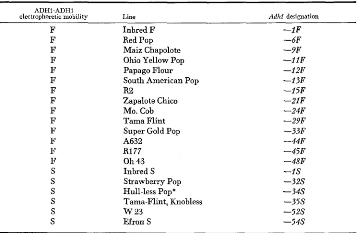
Lines and nomenclature: The lines used i n these studies are listed in Table 1 according to the anodal migration rate of their ADHl .ADHI dimers. The Adhl allele of each lines is desig- nated by a number denoting the family and a letter denoting the electrophoretic genotype.
Adhl-F isoalleles confer electrophoretically faster products than do Adhl-S isoalleles. The inbred F and inbred S lines developed by SCHWARTZ (1971) carry Adh-1-IF and Adhl-IS, respectively. T h e Adhl-54S allele was derived from EFRON'S AdhC, Adhl-S line (EFRON 1970). Inbred F,
inbred S and Efron S are available from this laboratory. The remaining lines were generously supplied by the Maize Genetics Cooperative.
ORGAN-SPECIFIC EXPRESSION O F Adhl IN M A I Z E
359
TABLE 1
Adhl genotypes of the maize lines used in this study
ADHl.ADH1
electrophoretic mobility Line Adhf designation
F Inbred F -1F
F F F F F F F F F F F F F S S S S S S Red Pcp Maiz Chapolote Ohio Yellow Pop Papago Flour South American Pop
R2
Zapalote Chico MO. Cob Tama Flint Super Gold Pop A632
R177 Oh 43 Inbred S
Strawberry Pop Hull-less Pop* Tama-Flint, Knobless W 2 3
Efron S
-6F -9F - 1 l F -12F -13F -15F - 2 l F -24F -29F -3327 -44F -45F -48F -1s -32s - 3 4 s - 3 5 s - 5 2 s - 5 4 s
* Established from a line having both Adhl-S and Adhl-F alleles.
then placed into the chamber until saturated, blotted, inserted into a starch gel and subjected to electrophoresis. Each kernel was numbered and used in subsequent experiments.
Germination: Numbered kernels were soaked for 18 hr, spaced evenly on moist paper towels in covered glass trays and germinated without light a t 27" and 85% humidity. After 2.5 to 3.5
days, seedlings with primary roots between 5.0 and 7.0 cm long were immersed completely under induction buffer: 5 mM Tris-HC1, p H 8.0 with 75 pg/ml chloramphenicol (FREELING and SCHWARTZ 1973). Anaerobiosis proceeded without light at 27", using 25 ml of induction buffer per seedling.
Individual roots: Following anaerobic induction, the distal 5 cm portion of each primary root was removed, individually macerated in 1 x 1 cm cylindrical chambers containing 35 gl of
extraction buffer. Again, a Whatman # 3 filter paper was placed into each chamber, blotted and subjected to starch gel electrophoresis.
[$HI-leucine incorporation experiments: Anaerobic induction and root extraction procedures were performed according to SACHS, FREELINC and OKIMOTO (1980).
Pooled scutella: Scutellar slivers from dry kernels were macerated with mortar and pestle in
appropriate volumes of extraction buffer. The brei was poured through miracloth and centrifuged at least twice to remove the lipid layer.
Pooled roots: Ten to 20 5-cm primary roots from 2.5 to 3.5-day-old seedlings were homogenized in extraction buffer (1 pg/mg root) with a mortar and pestle. The homogenate was centrifuged a t 30,000 x g for 15 min and the resulting supernatant saved.
All tissue extractions took place at 4" with precooled buffers and equipment.
360 J. C. WOODMAN A N D M. FREELING
Starch gel electrophoresis and gel staining: Previously described ( SCHWARTZ and ENDO 1966; FREELING 1973) electrophoretic and ADH staining methods have been further modified for the densitometric quantification of ADH Set I allozyme balances. The 11% w/v starch gels were stored for 4 to 10 h r at 4" before use. Three Whatman # 3 filter papers (5 x 6 mm) saturated with tissue extract were inserted into each gel at a position 2 cm from the cathodal wick. Electro- phoresis was carried out for 3 hr at 4" and a constant 250 volts. The gels were sliced horizontally i n half in a cutting mold, which insured uniform thickness. The bottom halves were immersed in ADH-specific stain (SCHWARTZ and ENDO 1966) for two h r in the dark. After the stain was re- moved, the gels were stored in water overnight before densitometric inspection.
Native-SDS PAGE gels and fluorography: [ 3H] -leucine-labeled anaerobic primary root ex- tracts were subjected to native-SDS two-dimensional polyacrylamide gel electrophoresis by the methods of SACHS, FREELINC and OKIMOTO (1980). Fluorography of dried gels was by the method of BONNER and LASKEY (1974), using Kodak SB5 film.
Two-dimensional immunoelectrophoresis: Two-dimensional immunoelectrophoresis followed the method of SCHWARTZ (1972). Referring to Figures 4 and 5, a Whatman # 3 filter paper (2.2 x 0.5 cm) soaked i n crude scutellar o r anaerobic root extract was inserted 2 cm from the cathodal wick into slot x-y of an 11 % starch gel (18 x 13 x 0.5 cm). The first electrophorectic dimension was run for 3 h r at 4" and constant 250 volts. A 5
x
0.5 cm Whatman # I filter paper saturated with unfractionated anti-ADH antiserum and a 5 x 0.5 cm Whatman #3 filter paper soaked inAdhl-Ct crude scutellar extract were inserted into slots a-b and c-d, respectively. With the a-b slot parallel to the anodal wick, the starch gel was subjected to electrophoresis in the second dimension for 3% h r at 4" and constant 250 volts. The starch gel portion flanked by the a-b and c-d slots was sliced in half and stained for ADH activity. The ADH immunoprecipitate stains blue. ADH antiserum was collected from New Zealand white rabbits that were immunized with purified ADH1-1S-ADHl-lS enzyme. A D H I . ADHI protein was purified from dry kernels by the procedure of KELLEY and FREELING (1980).
Densitometry: The stain intensities of the ADH Set I allozymes were determined by scanning gels in a Transidyne General 2970 integrating densitometer. The overlapping curves of the Set I
profile were separated and their area established by electronic integration. The relative stain intensity of a n allozyme is reported as its percent contribution to the Set I stain intensity. In
statistical analysis of allozyme patterns, the relative staining intensities of a given allozyme (e.g.,
percent S.S) were transformed into angles according to the relation: angle = arcsine vpercentage
.
Among S / F siblings, the mean percent contribution (upper 95% confidence interval) of an allo- zyme was calculated from ten or more samples. Since the asymmetrical confidence limits for any mean percent contribution varied less than 5 % , only the upper confidence limit ( L , ) is reported.
RESULTS A N D CONCLUSIONS
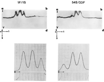
ADH allozyme balance variants: W e searched maize lines for Adhl relative- activity variants by using the following rationale. Adhl/Adhl-F individuals produce three Set I allozymes: the S.S and F . F homodimers and the S. F hetero- dimers (Figure 1). If both Adhl electrophoretic alleles are equally expressed and their products have equal specific activities, then a 1:2: 1 (S*S:S,F:F.F) ratio of ADH allozymes will result. If, for some reason, the expression of the
ORGAN-SPECIFIC EXPRESSION O F Adhl I N M A I Z E 36 1
r IGURE I .-Electrophoretograms and corresponding densitometnc traces of scutellar extracts prepared separately from three F, S / F families; from left to right: I F / I S , IS/33F and 54S/33F. The starch gel was specifically stained for ADH activity. The 0 denotes the origin and the
+
denotes the anode.
Before using this rationale, we needed to determine whether the densitometric quantification of ADH Set I allozyme balances was reproducible. The signifi- cance of genotype, sample preparation and gel preparation upon the variation in ADH allozyme belance was assessed in an analysis of variance experiment. Scutellar slices from inbred F/inbred S ( Z F / I S ) siblings were chosen in order to minimize variation due to genotype. ADH was extracted from pooled slices and 36 individual slices. Six gel sets (six gels per set) were prepared separately. Variation between pooled and individual scutellar extracts may be attributed to both genotype and experimental error. Variation among the gel sets and within gel sets will be due to experimental error only. The results were that genotype, sample preparation and gel preparation did not have significant effects on total variation.
362 J. C. WOODMAN A N D M. FREELING
and 1800-3500 units ADH/ml, respectively. Starch gels were left in ADH ac- tivity stain for a period of 100-180 minutes. Within this sample activity range and staining period,
ADH
SetI
allozyme balances of scutellar or anaerobic root extracts from I F / l S individuals were not significantly different ( p>
0.05) in a t-test.ADH
allozyme balances are considered to be similar when the mean relative staining intensities of electrophoretically identical homodimers are not significantly different, t-test ( p>
0.05).The scutellar allozyme balances of various
F1,
S/F hybrids are listed in Table 2. Three general types of balances were found (Figure 1): either the S.S homo- dimer stain intensity was greater than, equal to or less thanF.F
homodimer stain intensity. The variance of the relative staining intensities ofF-F
homodimers among theS/F
families were not significantly different ( p>
0.05) i n F tests when compared to the percentF.F
variance from the I F / I S family. This sug- gests that each family is homogeneous for a particularADH
allozyme pattern. This is not surprising considering that these S / F families are single-cross hy- brids derived from inbred lines.Based on these scutellar allozyme balances, there appear to be F alleles whose quantitative expression is less than, equal to or greater than that of S alleles, and
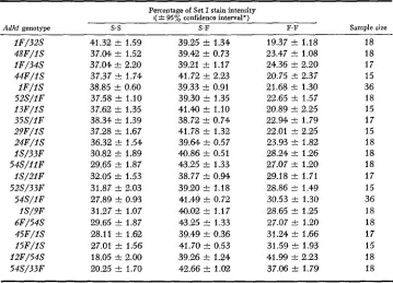
TABLE 2
ADH Set I allozyme balances in the scutellum of various Adhl-S/Adhl-F F , Hybrids
Percentage of Set I stain intensity I(
*
95% confidence interval*)Sample size
Adhl genotype s.s S.F F.F
I F / 3 2 S 4 8 F / I S I F / 3 4 S 4 4 F / I S I F / I S 52S/I F I 3 F / I S 35S/IF 2 9 F / I S 2 4 F / I S I S / 3 3 F 5 4 S / I I F I S / 2 I F 52S/33F 54S/IF IS/9F 6F/54S 4 5 F / I S I 5 F / I S 12F/54S 54S/33F
41.32 f 1.59 37.M f 1.52 37.04 f 2.20 37.37 2 1.74 38.85 f 0.60 37.58 2 1.10 37.62 f 1.35 38.34 +. 1.39 37.28 +. 1.67 36.32 f 1.54 30.82 f 1.89 29.65 f 1.87 32.05 2 1.53 31.87 f 2.03 27.89 f 0.93 31.27 2 1.07 29.65 f 1.87 28.11 f 1.62 27.01 f 1.56 18.05 f 2.00 20.25 f 1.70
39.25 2 1.34 39.42 2 0.73 39.21 +. 1.17 41.72 2 2.23 39.33 f 0.91 39.30 f 1.35 41.40 2 1.10 38.72 f 0.74 41.78 f 1.32 39.64 2 0.57 40.86 f 0.51 43.25 f 1.33 38.77 +. 0.94 39.20 2 1.18 41.49 f 0.72 40.02 2 1.17 43.25 f 1.33 39.49 f 0.36 41.70 +. 0.53 39.26 +. 1.24 42.66 2 1.02
19.37 k 1.18 23.47 5~ 1.08 24.36 k 2.20 20.75 31 2.37 21.68 f 1.30 22.65 f 1.57 20.89 2 2.25 22.94 +- 1.79 22.01 2 2.25 23.93 f 1.82 28.24 f 1.26 27.07 2 1.20 29.18 f 1.71 28.86 i- 1.49 30.53 f 1.30 28.65 f 1.25 27.07 f 1.20 31.24 4 1.66 31.59 +- 1.93 41.99 2 2.23 37.06 st: 1.79
18 18 17 15 36 18 15 17 15 18 18 18 1 7 15 36 18 18 17 15 18 18
ORGAN-SPECIFIC E X P R E S S I O N O F
Adhl
I N MAIZE 363vice versa. Thus, the relative expression of an
Adhl
quantitative variant is not associated with the electrophoretic mobility of theAdhl
gene product. The relative-activity relationships among Adhl alleles are shown in Table 3. In all pairwise combinations of S and F alleles constructed to date (18 S / F families, in addition to those in Table 2), the Adhl activity relationship has not been vio- lated, irrespective of genetic background.Reciprocal efJect: After the S / F individuals had their scutellar allozyme balances quantified, they were germinated and subjected to a 24-hour anerobic induction treatment. Set I ADH allozyme balances from these individuals are compared in Table 4. Even though every allozyme balance was skewed towards the ADH enzymes containing ADHI-F subunits, there were at least three types of balances (Figure 2). Moreover, comparisons of anaerobic root and scutellar balances (see Table
4)
revealed that the S/F family that showed the smallest amount of a homodimer in one organ concomitantly exhibited the greatest amount of this homodimer in the other organ, and vice versa. In order to define this “reciprocal effect” quantitatively, we determined the relationship between the relative number of A D H l - S subunit molecules (percent ADHI-S) in each organ of every S / F family. By assuming that any ADHI-S subunit equals anyADHl
-F
subunit in specific enzyme under our reaction conditions-an assump-tion we will soon prove correct-we calculated percent ADHl-S directly as percent S.S activity
4-
1/2
percent S.F activity. Percent ADHI-S from an- aerobic primary roots calculated from Set I allozyme balances was equal to percent ADHI-S in Set I1 allozymes for every S/F family in Table 4 (data not shown). Thus, in anaerobic primary roots, Adh2 expression does not influenceADH Set I allozyme balances. As shown in Figure 3, every S / F family obeys a common relationship, r =
-
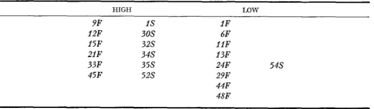
0.89, between ADH allozyme balances in anaero- bic roots and scutella. Among the hypotheses that can be postulated to explain this “reciprocal effect”, several can be eliminated by the following studies char- acterizing Adhl quantitative behavior in both organs.TABLE 3
Relatiue scutellar expression of Adhl alleles of diuerse origin*
HIGH LQW
9F I S 1F
12F 30s 6F
15F 3 2 s 1 l F
21F 3 4 s 13F
33 F 3 5 s 24F 5 4 s
45F 5 2 s 29F
44F 48F
* The relative expression of an Adhl allele was determined from scutellar ADH Set I allozyme balances of various F, S / F families (Table 2 ) . Within each group, the Adhl alleles are ordered according to the electrophoretic mobility of their product ADH subunits, “S’ (slow) or “F’
364 J. C . W O O D M A N A N D M. FREEIJNG
2.2 Set
IU
-+!m!!k
2:;
SetII
FIGURE 2.-Electrophoretograms and corresponding densitometric traces of anaerobic primar?. root extracts prepared separtely from three F, S / F families; from left to right: I F / I S , I S / 3 3 F and 54S/33F. The starch gel was specifically stained fro ADH activity. The 0 denotes the origin and the f denotes the anode.
Segregation of Adhl quantitative expression: Since ADH Set I allozy” ratios can be measured in S/F heterozygotes, our test for linkage of Adhl quantitative expression with the ADHl electrophoretic mobility site (Le., the Adhl struc- tural gene) is somewhat indirect. If Adhl quantitative expression is controlled by genetic factors tightly linked to the Adhl structural gene, then the S/F prog- eny generated from a cross involving a n
F, S/F
individual should express simi- lar ADH allozyme patterns. If, on the other hand, Adhl quantitative expression is controlled by loci unlinked or loosely linked to the Adhl structural gene, then several ADH allozyme patterns may appear among the S/F progeny. To dis- tinguish between these and other possibilities, we determined the segregation of ADH allozyme patterns among S/F progeny generated from selected crosses (Tables5
and 6). It is clear that Adhl quantitative expression in scutella and anaerobic roots segregated with the Adhl electrophoretic mobility site. PainviseORGAN-SPECIFIC EXPRESSION O F Adhl I N M A I Z E TABLE 4
ADH Set I allozyme balances in 5-em primary roots of 24-hour anaerobically induced Adhl-S/Adhl-F F , seedlings
365
Percentage of Set I stain intensity
( 2 95% confidence interval*) Scutellar
- ~ _ _ _ _ - Sample allozyme
Adhl genotype s.s S.F F,F size balance (% S.S)+
I F / 3 2 S 4 8 F / I S I F / 3 4 S 4 4 F / I S I F / I S 52S/I F I 3 F / I S 3 5 S / I F 2 9 F / I S 2 4 F / I S I S / 3 3 F 5 4 S / i l F IS/21F 52S/33F 54S/I F I S / 9 F 6F/54S 4 5 F / I S I 5 F / I S 12F/54S 54S/33F
9.05 t 0.45 10.55 t 1.71 10.80 C 1.71 11.18 t 1.42 11.25 t 1.56 11.50 t 1.21 11.55 t 1.73 11.81 t 1.14 12.26 t 1.53 12.49 C 1.33 14.42 k 1.37 14.47 t 1.62 14.52 k 1.72 14.59 t 1.00 14.84 1: 0.94 15.65 t 1.87 15.71 f 1.21 16.76 t 1.20 16.78 t 2.29 22.08 t 2.41 22.52 1: 1.65
41.04 Ir 1.21 45.57 f 1.43 42.58 f 1.80 43.51 t- 1.50 45.00 +. 1.08 45.40 f 1.08 43.90 C 1.58 41.61 f 1.20 43.83 t- 1.47 41.47 Ir 1.50 4.5.32 f 2.08 47.10 Ir 1.40 46.22 f 1.65 45.39 f 1.05 48.40 f 1.10 46.48 k 2.00 46.44 k 1.16 46.25 f 1.05 46.20 Ir 2.12 44.74
*
2.16 42.88 +. 1.4049.91 t 2.07 43.38 t 1.34 46.62 f 2.08 46.31 t 1.81 43.76 F 1.34 43.10 f 1.82 44.55 t 1.73 46.58 F 0.83 43.91 t 1.47 46.04 t 1.93 40.24 C 2.21 38.43 C 1.53 39.29 t 2.06 40.02 t 1.18 36.74 t 1.32 37.67 t 2.20 37.85 F 1.31 37.99
*
1.10 37.02 t 2.33 33.18 t 2.33 34.60 k 1.6528 23 15 13 27 17 22 29 19 18 29 14 22 16 27 12 12 30 22 16 18 41.32 37.04 37.04 37.37 38.85 37.58 37.62 38.34 37.28 36.32 30.82 31.63 32.05 31.87 27.89 31.27 29.65 28.11 27.01 18.05 20.25
* The mean percent contribution t 95% confidence interval of the S.S and F.F hamodimers and S . F heterodimer of the Set I strain intensity was calculated from arcsine transformed percentages of individual primary roots, as detailed in MATERIALS AND METHODS.
1. From Table 2.
respectively, were not significantly different ( p
>
0.05) from those ofF,
S/Ffamilies as indicated by F tests. Further, the data in Table 6 demonstrate that ADH Set
I
allozyme balance variation is not associated with Adh2 variation, since Adh2 is unlinked to Adhl (FREELING and SCHWARTZ 1973). We conclude from these data that all four Adhl quantitative alleles tested ( l F , 33F, I S and54s) reflect a polymorphism at a cis-acting locus close to or within the Adhl
structural gene.
A more direct test of the cis-acting nature of Adhl quantitative expression in scutella and anaerobic roots comes from the evidence presented below. Siblings generated by the cross of l S / 3 3 F X Ct/Ct were analyzed for their ADH allozyme balances. Since Adhl-Ct produces a dimeric product with an electrophoretic mobility greater than S.S or F.F homodimers (SCHWARTZ 1966), both l S / C t
366 J. C. WOODMAN A N D M. FREELING
34 38 42 4 6 50 54 58 6 2
%s
(scutellum)
FIGURE 3.-% ADHI-S subunits in anaerobic primary roots us. % ADHI-S subunits in the scutellum of various S/F families. % ADHI-S was calculated directly from ADH Set I allozyme activity ratios (Tables 2 and 4), by the following relationship: % ADHI-S = % S . S $- '/z '% S . F .
Each point represents a different S / F family.
Adhl quantitative expression predicts the following relationship: the ratio of ADH1-33F subunits us. ADHl-Ct subunits in 3 3 F / C t organs divided by ADHI- 1 s subunits us. ADH1-Ct subunits in l S / C t organs will be equal to the ratio of ADH1-33F subunits us. ADH-1-1s subunits in the identical organs of 3 3 F / l S
individuals. As shown in Table 7, the predicted and observed ratios agree re- markably well for both the scutella (1.02 us. 1.05) and anaerobic root (1.77
ORGAN-SPECIFIC EXPRESSION OF
Adhl IN MAIZE
36 7M
2
8
x
U2 2
$
%
$ 5
$ 2
u) k 9H & $
g
% ? cZ v )
r+
skyzba
x
.s
U 2
E 2
i?
.s
w
%
9
k i $
.U -4
-
* UG
3
I- T 2 -4&
U 3 h Y .eW
$
~ c o c o * * * c o * * o * * * ~ * * w * c c Y .z
0)
**YW?;~??;~~;?~?~;~
.e~ 8 3 ~ 0 1 a C \ I m - r O a m a 0 1 m a - a ~
.-
m Gw 4-
U
c.
v)
01 " m w u ) c I 3 * 0 1 m * * 0 m - u ) c o M r O
75
h 9 q " c ? q ? c ? ? ? " . q h ? " q c ? W
- - a - - * + - O + - a W W a - o . A
t I $1 t I t i t i t I t I ti tI $1 t l $1 $1 t i t i t I ti tI
ppJUp&X~L-&5X$$$j$&& h n $ 2
m z
3 2
5 g
E L
~ g ~ g ~ % s X K s % : 2 % % s a
32
2-
w <$3 c m
.# z g F r $
.$$
uer s o . - A A d < A d 0 s d s o ~ 0 0 s
.z
s%
muk
;
242
$ 5 m m c c m g o r O h m a r O a a c c o a h m * % a
2 2 o?"."": ? 9 h Y h ? 0 T h 0 " + . h E,g;
8s M M * m M * * m * * * * * ~ 3 % ~ 3
u = g $
p
g z" 0 3
26"o
0g;
h t . c 3 - m h w ~ 5 o u ) c o - ~ u ) w a a al+ 2 4- e k
..
U 5 2 o A A . s A s & & s s & 4 & . - s u s &
$1 ti ti ti t I $1 t l tl tI $1 t l tI ti t l t I t I t l ti
3
z32j
q % ".? q q % 09 q q
+.
7 q 0" '4" ".-v)a
E
t. m u ) u ) w m w
-
h h o t. o m a co coco X h w q%e$;;;
$37
5
-t-
3;;;+
2
m
? 2 k.22
w w c o c o c o c o c o c o a l w c o c o m a a ~ ~ ~ &.3:z
3 3 % ;
&
3
:g2?Lqs 3 .z
5 2
3 2
8 gyI$ t t k k k k k k k k k @ k k @ k @ @ @ 3 a*$;
.a
&U$E
z z z z & & & & q % & % & & & % % %
+ Z t l @
2%
g 2 . zT T*;;B",
$ $ 2 & 2 - 2 5 $ w
% 5 0.2 8
@ @ k @ b T f : E 2
k @ k % ? @ 0, @ k
s s
Lq ?s
? @ @ 0,O Z $ E *"
5 %
2 s
3 %
2 %
5
: s %
2s
2 2
2 %
& & $ g . 2& ? g z
%
2
& ? & - & & & 3 2 %
a & & %
$ 5 5
&g
:U?2
2 i? %
b
s
2 % 2z
13
% gg
kz
z
%s
Y)u
t l $1 $1 $1 t I $1 t I $1 $1 $1 t I tl +I $1 tI $1 t I $1
h c o - c 3 & ~ a c o o - a a - o
I v)
s 1"
a"-
m a + h c o - a + - m m m o m a w - w
'"
m m M 0 1 O O h ~ h M c o w m m c c a * a M m m w 0 1 0 1 a M a 0 1 m a a - a M a a
w
~ m u ) u ) u ) m a a a m m m m m m u ) ME:
2
U m CO M M " m M a 01 a a a a a M a a
z
C . 5 ~ ~ W 0 0 0 0 L L L ~ 5 0 0 L ~ C . 53
sw
w 010.201 hD.M
-
h h h h M M "i "i h h w. h M w. "i h h h 0 \ \ \ \ M m M " i \ \ M \ M M M \ \ \h h h h M M h M v- M w . q h U.
Q c
"M
wl4.m v, W
k
w.- c m a
h h h \ \ \ h * h \ \ h \ \ \ h h h
\ \ \ @ k \ \ \ \ @ 0, \k @ k \ \ \ x x x x x x x x x x x x x x x x x x @ % t t W t t ttk tt k k Y k 4, k k @ @ @
g
i 2
.$
:;
??3??23????2
?????Y
h~ E T 3U
-cv)alu,u E
t + * g m
h h h M v - M M Y - v - v - M Q h w . M M M M M
368 J. C. WOODMAN A N D M. FREELING
E
2 .c,-_
P
s
-8 7 2&
$$
$9
-r kl@l2 2
6
rt:
(D 8 MEi
p
2i i
3 s
2
2.2
&.,$E
4:
U 02 3
$ 8
- 4 - 3
x hl
T
2
-3 4g
1 Pd R I -
z
2
+-3
g
$
~
z
~
k
g
U~
~
~
~
~
~
~
~
>
E 3 ;
s
q
2
.* %?.,.
cn-
a
UJ
al2
' E % n
v)
!
o w v1
.E
5
z z
4- $ 2
E ? ~ % Z 3 ! 3 S ? % % 8 8 3 ? $ i ?
z
.q
$
z g
2-
2 g a . 2
p z
$4
.$j
o o ~ - o o o o - o o o o a8
.om3 ;% - m m * - - m C . l - m u m . + * d39:
;i
$
tl $1 $1 $1 tl $1 41 ti $1 $1 31$1 $1 .tl2
.P 2- " J c w 0 0 m * o M m - W O h m c o .s z2m"8 8 9 " m T h ? q T a q $ \ r ! ? T 8.3 d
2:
$ $ $ % % g ! $ $ % P $ $ P ! $
2i
%%Er+ Z E E
c a
s
W O2-
X t l m
:q
!A + m h c % c n w * l n - w a a e * 8
"FE
.;
9
$2
c q G ? h 9 c ? ? 9 c ? m h 9 ? q T
-
2s$T
. + . + w * c - i - - - - , + m a
%
. g w G4 g d . 5
;+
a%% F- U)32
" " b
2
%%E&
c ? " " G ? t T t t W . " c ? " " \ r ! yUJ
2.5;
s - . & - w - * - * - * - m a a&S
3 8 %. 3 s a
0;
kZ
azz
.-q
;?d
& O h E 2 E &a $ 0 2% h h h h ~ % % ~ ~6=$$A %0
"$12
~ ~ ~ ! $v\ m in
azx
3.5
Q
F
'F,
On.$ i-%C<-& a
4":
S W . 2 E-
k W . -2
ga3"'SZ 8"
WIG3 L4-k "e.-+ a
vI s-g.2 * a
$&+#mFF%?FF
.-?a c 07
8,
E 8 3%
2 2
x
%2
%
2
tx
z
5
2
x
z
2.22
a *a g 3
Uo ~.9~2y&~kL~~~~~
2;:7Q
h h $ h @ W k @ W h @ G 3 @ W " * r h +, h h q w n U - v v m LA83
2.-
m In v \ " ci2Tsb:
8
.eo - w m ~ m + m - h m C ~ ~ ~ - h % s
*
'f o!
".
6! v o! --! v 9 9 In G?a
\r!m m - w - - m a a a - a - ~
z
+I ti$1 $1 $1 $1 $1 $1 tI $1 $1 $1 $I $1o d ~ d ~ o i m o & ~ o ~ * &
2
+ * * - P m m m * w m t m m m
.*.- '9 q \c!
+
cq c? 0. 0 c? a. k 9 W. l.q+ a + w a m a m - a o & m o
2
-h-c
. s ~ G G S < & . s & & & & & & G , m at1 ti $1 $1 tI tI tI +I tI $1 tI +I $1 tl
2
+ e - c o h m w ~ W m * O m m ~-
a O m ~ m M M m ~ W m m O 0
a . w
i
8 m u J m " m a a a * - P * " m %bl z - - - P * * * * d - * m m m
2
8
2y & & % 9 z z % % % % % %
\ \ \ \ m \ \ L " \ m "g i
z$j
@ @ @ @ \ k r % k \ \ @ ~ . \ \ " + -*
k
\ \ \ @ k \ \ \ @ @)\Et @ b
g
x x x x x x x x x x x x x x
- ? & - e l1 1 9 \ m m m m ' \ \ m m m \
k W \ W \ \ \ \ % 14 \ \ \ W QUDYm E
* m-i-++m$
ORGAN-SPECIFIC EXPRESSION OF
Adhl
IN MAIZE 3693 7
3 70 J. C . WOODMAN AND M. F R E E L I N G
quantitative locus controlling anaerobic-root quantitative expression is no farther than 0.45 map units away from the Adhl structural gene and might be much closer.
Estimating the location of the AdhZ quantitative site(s) that control Adhl
expression in the scutellum proved to be more difficult. The three types of scutel- lar allozyme balances (Figure 2) are not skewed greatly towards any particular homodimer. Thus, to prove that an exceptional allozyme balance among
F2
S/F progeny was a result of a previous recombinational event would require a strict test, such as flanking-marker exchange. Unfortunately, the lines used in this study did not have genetic markers flanking Adhl. Construction of such stocks might well have disrupted the Adhl chromosome regions that we hoped to assess.Biochemical studies on ADH allozyme balances: The cis-acting nature of
Adhl quantitative variation emphasizes the possibility that Adhl structural gene variation underlies the polymorphism in
ADH
allozyme balances in either the scutellum or anaerobic primary root. One might expect that the balancesl F / l S 54s I33 F
a b
a
- -
bORGAN-SPECIFIC EXPRESSION OF
Adhf
IN MAIZE 371 result from specific activity or in vivo degradation rate differences between ADHl *ADH1 enzymes. The experiments testing these hypotheses were per- formed on twoS/F
hybrids, ( I F / I S and 54S/33F), with clearly different al- lozyme balances.Since the precipitation peak heights in two-dimensional immunoelectropho- retic profiles correspond to the relative number of ADH enzyme molecules
(SCHWARTZ 1972), the specific activities of ADH allozymes can be examined indirectly by comparing immunoelectrophoretic profiles with the densitometric traces of ADH activity gels. Comparisons of ADH allozyme balances in the scutellum and anaerobic root of 1 F / f S and 54S/33F individuals, respectively, are shown in Figures 4 and 5. Because the immunoelectrophoretic and densi- tometric trances are superimposable, it can be concluded for these S/F hybrids that the ADH allozyme activity ratios in starch gels directly reflect the relative number of ADHl
*ADHl
molecules. Furthermore, these results suggest that the ADHl .ADHI enzymes containing the products of the I F , 33F, I S or 5 4 s alleles have identical or near identical specific activities in either scutellar or anaerobic root extracts. Thus, it seems likely that these four Adhl quantitative variants arise from variation in a mechanism(s) that controls ADHl .ADHI production. The relative number of S andF
subunits synthesized by S/F anaerobic pri- mary roots can be determined directly byrSH]
-leucine incorporation experi- ments. (SACHS andFREELING
1978;FERL,
DLOUHY and SCHWARTZ 1979). Pri- mary roots were exposed to [3H]-leucine for the last five hours of a 24-hour3
72
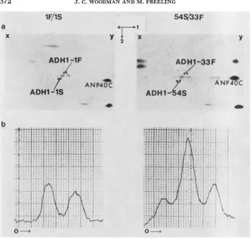
J. C. W O O D M A N A N D M. F R E E L I N GFIGURE 6.-(A) Fluorograms of native-SDS, two-dimensional gels of anaerobic root extracts prepared from either I F / I S or 54S/33F primary roots. The primary roots were exposed to 100
pCi [3H]- leucine for the last five hrs of a 24-hr anaerobic induction period. Only the region of
the Z D gel containing ADHl and ADH2 polypeptides is shown here. ADH subunits are indicated along with the landmark protein ANP40C (SACHS, FREELING and OKIMOTO 1980). x-y marks the axis where the first dimension, native polyacrylamide gel was layered on top of the SDS gel. The arrows denote the direction of the anode in dimensions 1 and 2; 0 denotes the origin. (B) Densitometric traces of the respective fluorograms. Only ADH Set I allozymes shown here. The 0 denotes the origin and the
+
indicaes the anode in dimension one.anaerobic induction period. Extracts from these roots were subjected to native- SDS, two-dimensional polyacrylamide gel electrophoresis. Since no other
[3H]
-
labeled polypeptides lie in the ADH region of the gel, the relative intensity of radioactively labeled ADHl polypeptides will correlate with the relative num- ber of
ADHl
subunits synthesized during the 5-hour pulse. This assumption is further supported by the evidence that ADHl-IS and ADH1-IF polypeptides have equal numbers of leucines(KELLEY
andFREELING
1980). As shown in Figure 6, there is a distinct difference between the relative intensity of[3H]-
ORGAN-SPECIFIC EXPRESSION O F A d h l I N M A I Z E 373
ADHI-S subunits was 34.3% for I F / I S primary roots and 46.2% for 5 4 S / 3 3 F
primary roots. Average percent ADHI-S data calculated from allozyme ac- tivity ratios in 24 hour anaerobically induced primary roots is 33.8% and 44.0%
for these respective hybrids. We conclude from these data and the immuno- electrophoretograms that, for the I F , 33F, I S and 5 4 s alleles, the variation in
A d h l quantitative expression in anaerobic primary roots is attributable to dif- ferences between ADHI. ADHI allozyme synthetic rates.
Because we measured ADH Set
I
allozyme balances in dry scutella, we can- not determine directly whether the allozyme balance variation in this region arises from differences between synthesis, degradation or dimerization rates ofADHI .ADHI enzymes. However, we do not find any correlation with the i n
vitro thermolabilities or dimerization properties of ADHI .ADHI homodimers and the scutellar ADH allozyme balances (unpublished results). We find that
F . F homodimers are more thermostable aEd reassociate in vitro more completely
than S .S homodimers. These experiments were performed on crude scutellar extracts from the l F , 33F, I S and 54s lines. S / F hybrids constructed from these lines do not always display a scutellar balance skewed towards F - F homodimers. (Table 2 ) . These data favor the differential ADHI. ADHI synthesis argument for scutellar ADH allozyme balance variation, but obviously do not rule out other mechanisms.
The thermolability behavior of IF.IF, 33F.33F, 1SelS and 54S.54S enzymes points to the possibility ADHI. ADHI degradation rate differences may account partially for differences in ADHI
.
ADHI enzyme accumulation in anaerobically induced primary roots. We find that ADH activity in 5-cm primary roots froml F / I S , l S / 3 3 F and 5 4 S / 3 3 F seedlings increases at a zero-order rate up to and beyond 48 hours of anaerobic induction. Before and during this induction period, the ADH Set
I
allozyme balances do not change. These data imply that each Set I allozyme is accumulating at a zero-order rate; that is, there appears to be- no detectable influence of a first-order degradation process on ADHI. ADHIallozyme production in primary roots.
In summary, allozyme balances in the scutellum and anaerobic root of I F / l S
and 5 4 S / 3 3 F individuals reflect differences between the relative number of ADH
Set 1 allozyme molecules. In the anaerobic roots, the relative level of ADHI.
ADHI allozyme molecules is attributable to differences between the zero-order
synthetic rates of ADHI. ADHI enzymes. The relative level of ADH Set 1 allo- zymes in the scutellum is not associated with the electrophoretic mobility, in vitro
dimerization property differences between ADHI
.
ADHI allozymes. Coupled with the segregation tests, these biochemical studies provide evidence that theA d h l quantitative variants, I F , 33F, 1s and 5 4 S , differ at a cis-acting locus that regulates the production of ADHl .ADHI molecules. Moreover, the reciprocally correlated ADH allozyme balances (i.e.,Adhl quantitative expression) in the scutellum and primary root suggest that the genetic elements coordinating organ- specific A d h l expression are at least tightly linked and may even share common
DNA sequences. Since we could not separate the reciprocal-effect site and A d h l
3 74 J. C. W O O D M A N A N D M. F R E E L I N G
Developmental stability of the reciprocal effect: Before considering the possible molecular mechanisms accounting for the reciprocal effect phenomenon, it is necessary to determine whether the reciprocal effect is developmentally stable within each S/F individual. If Adhl quantitative behavior in either the scutellum o r anaerobic primary root is the property of ADHl polypeptides, then the ADH
allozyme balance in each organ would be expected to be independent of one an- other; that is, there should be no association between the most extreme scutellar and anaerobic root allozyme balances within a n S / F family. By an extreme allo- zyme balance, we mean an ADH allozyme balance (represented as percent F.F) greater or less than one standard deviation away from the mean allozyme balance of an S / F family. Of the 221 S / F individuals generated from the crosses listed in Tables
5
and 6,22 express an extreme balance in both organs. As shown i n Table 8, there is a clear association between extreme allozyme balances in each organ; the highly significant x2, corrected for continuity, was equal to 8.41. Each of these22 S/F individuals have a unique genetic background. Thus, the inverse relation- ship between extreme ADH allozyme balances in the scutellum and anaerobic primary root implies that the reciprocal effect marks an extremely stable develop- mental process. Moreover, these data reaffirm that the organ-specific Adhl quan- titative programs are interrelated in a strictly quantitative manner. It seems likely that ADHl polypeptide differences do not account solely for the reciprocal-effect phenomenon.
DISCUSSION
In this report, we describe differences in the quantitative expression of Adhl
alleles from 21 maize inbred or exotic lines. By use of ADH allozyme balances in
S / F hybrids constructed from the inbred lines, we discovered an inverse relation- ship between Adhl quantitative behavior in the scutellum and the anaerobically induced primary root. Within the same electrophoretic mobility class. the allele with the lowest expression in the scutellum exhibits the greatest expression in the anaerobic root, and vice versa. We have termed this novel phenomenon the “reciprocal effect.”
In order to eliminate the several formal explanations that could account for the reciprocal effect, we performed genetical and biochemical experiments on four
Adhl quantitative variants: I F , 33F, I S and 54s. Segregation tests establish that
TABLE 8
Twenty-two S/F individuals with eztreme ADH allozyme balances* in both their
scutellum and anaerobic primary root
Extreme in
anaerobic pnmary root
high % F.F low % F,F Total
high % F.F 3 7 10
low ’% F.F 10 2 12
Total 13 9 22
Extreme in scutellum
ORGAN-SPECIFIC EXPRESSION O F Adhl I N MAIZE 375
A d h l quantitative behavior in the scutellum and anaerobic primary root is the property of a cis-acting site(s) within
0.45
map units of the Adhl structural gene. The possibility that organ-specific Adhl quantitative behavior is the property of ADHI .ADH1 polypeptides is not supported by our biochemical studies. First, ADH Set 1 allozyme activity ratios in both organs reflect ADH Set 1 allozyme protein ratios. Second, Adhl quantitative behavior in the scutellum is not asso- ciated with qualitative differences among ADH SetI
allozymes (i.e., electro- phoretic mobility,in uitro
thermolability andin vitro
dimerization property). Finally,[3H]
-leucine incorporation experiments indicate that A d h l quantitative expression in anaerobic primary roots is attributable to differences between the zero-order synthesis rates among ADH allozymes.Further proof that Adhl quantitative expression in anaerobic primary roots is not associated with mechanisms operating on ADHl polypeptides per se comes from
in vitro
translation studies(FERL,
BRENNAN and SCHWARTZ 1980; SACHS, FREELING and OKIMOTO 1980). Anaerobic primary root mRNA from l F / l Sseedlings produces, in a n in uitro translation system, 1 s and 1F monomers equal in molecular weight to those from dissociated ADHl .ADH1 and ADHl
.AD=
active enzymes. Further, the relative intensities of F and S radioactively labeled monomers were consistent with the relative
in uiuo
expression of these alleles inF / S anaerobic seedlings. These studies show that neither differential ADHI processing nor ADHl dimerization rates in vivo accounts for Adhl quantitative expression in F / S anaerobic primary roots.
Since all Adhl quantitative variants tested exhibit the reciprocal effect, it seems likely that the I F , 33F, 1s and 54s variants are representative of the four general types of Adhl quantitative variants. Therefore, we conclude that differences in ADHl polypeptide synthesis rates account for the polymorphism in Adhl ex- pression in anaerobic roots of the 21 lines listed in Table 1. I n the case of scutella, we can conclude only that Adhl quantitative behavior is most likely not due to qualitative differences between ADHI .ADH1 enzymes.
A speculation concerning the reciprocal-effect element: W e have shown that the quantitative behavior of every Adhl allele and allele combination tested fits a n organ-specific reciprocal relationship. The question of the positional relation- ship of Adhl quantitative site (s) in the scutellum and primary root is important in understanding the nature of the reciprocal effect. A reciprocal-effect mutant, Adhl-SlPfila, has been recovered from progeny of an Adhl mutant originally derived from material irradiated with accelerated neon ions (FREELING and CHENG 1978;
FREELING
and WOODMAN 1979). It is likely that a single mutational event can alter Adhl quantitative behavior simultaneously in the scutellum and primary root. However, Adhl-Sl951a expression is not proof that the reciprocal- effect site is a single locus, since it is the only recovered mutant of this nature. Nevertheless, these data provide added support to the hypothesis that the cis- acting loci coordinating organ-specific Adhl quantitative expression are, at the least, in close proximity.3 76 J. C. W O O D M A N A N D M. FREELING
of behavior (see reviews by
PAIGEN
1971; CALHOUN and HATFIELD 1975; FORGET 1978). However, we did not find a relationship between Adhl structural gene variation and Adhl quantitative polymorphism. We propose that ADHl be- havior is not responsible for the reciprocal effect. On the other hand, it is possible that DNA sequences encoding ADHI information might specify regulatory in- formation as well (see BOGENHAGEN, SAKONJU and BROWN 1980). Irrespective of the location of reciprocal- effect loci, our data establish that there is a regulatory genetic element of the Adhl cistron that coordinates organ-specific Adhl quan- titative expression. Moreover, the extreme allozyme balance relationship suggests that Adhl quantitative behavior (reciprocal effect) is programmed at a determi- native step prior to (or during) the establishment of scutellar and primary root cell lineages. W e propose that the determinative event involves a n unequal dis- tribution of Adhl quantitative potential. We advance no molecular models, but predict that these regulatory AdU variants will be profitably studied at the level of nucleotide sequences.The importance of measuring regulatory variation: During the past ten years, i t has been well documented by techniques that measure qualitative differences among enzymes (e.g., electrophoretic mobility and heat stability of kinetic prop- erties) that natural populations are highly polymorphic at structural gene loci. The evolutionary significance of most structural gene variation is the subject of much speculation, and it is still undetermined. One hypothesis is that regulatory rather than structural gene variation plays the predominant role in adaptive evo- lution (BRITTEN and DAVIDSON 1971 ; KING and WILSON 1975;
WILSON,
CARLSON and WHITE 1977). This theory has received support from evidence that regula- tory genes are adaptively significant in prokaryotes(HALL
1978, and references therein). However, before the importance of regulatory variation in adaptive evolution can be adequately assessed, the extent and nature of regulatory varia- tion within and among populations must be determined.Several studies indicate that regulatory gene variation may be quite common in natural populations (WARD and HERBERT 1972;
WARD
1975;PRAKASH
1977; MCDONALD and AYALA 1978; ABRAHAM and DOANE 1978; DICKINSON and CAR-SON 1979; DICKINSON 1980). This study illustrates that allozyme balances can be
a powerful tool for detecting regulatory variation among diverse individuals. W e have identified a genetic element that coordinates the organ specificity of Adhl quantitative expression. We have surveyed only six maize races: Northern Flint
( 3 0 S ) , Great Plains Flint (29F and 3 5 9 , Chapalote ( P F ) , Zapalote Chico (2127)
,
Papago Flour (128’) and corn-belt dents. Since there are at least 300 maize races (cf., BROWN and GOODMAN 1977), we most likely have not characterized the full extent of Adhl regulatory variation. Whether the quantitative polymorphism a t the Adhl cistron has any adaptive significance, such as providing a differential response to flooding stress (see
KRSHALL,
BROUE and PRYOR 1973; BROWN, MARSHALL and MUNDY 1976; DIEDENHOFEN 1977) remains to be determined.JAMES C. WOODMAN was a recipient of Public Health Service predoctoral training grant,
5T32-GM07127. This research was supported by Public Health Service grant GM21739 to M.
ORGAN-SPECIFIC EXPRESSION OF Adhl IN MAIZE 377 LITERATURE CITED
ABRAHAM, I. and W. W. DOANE, 1978 Genetic regulation of tissue-expression of Amylase struc- tural genes in Drosophila melanogaster. Proc. Natl. Acad. Sci. U.S. 75: 4446-4450. BOGENHAGEN, D. F., S . SAKONJU and D. D. BROWN, 1980 A control region in the center of the
5 s RNA gene directs specific initiation of transcription: 11. The 3’ border of the region. Cell 19: 27-35.
BONNER, W. M. and R. A. LASKEY, 1974 A film detection method for tritium labeled proteins and nucleic acid in polyacrylamide gels. Eur. J. Biochem. 46: 83-88.
BOUBEL~K, M., A. LENGEROVA, D. W. BAILEY and V. MATOUSEK, 1975 A model for genetic analysis of programmed expression as reflected in the development of membrane antigens. Develop. Biol. 47: 206-214.
BRADFORD, M. M., 1976 A rapid and sensitive method for the quactitation of microgram quan- tities of protein utilizing the principle of protein-dye binding. Ana:. Biochem. 72 : 248-254.
BREEN, G. A. M., A. J. LUSIS and K. PAIGEN, 1977 Linkage of genetic determinants for mouse
P-galactosidase electrophoresis and activity. Genetics 85 : 73-84.
BRITTEN, R. and E. DAVIDSEN, 1971 Repetitive and non-repetitive DNA sequences and a specu- lation on the origin of evolutionary novelty. Quart. Rev. Biol. 46: 11 1-138.
BROWN, A. H. D., D. R. MARSHALL and J. MUNDY, 1976 Adaptiveness of variants at an alcohol dehydrogenase locus in Bromus mollis L. (soft bromegrass). Aust. J. Biol. Sci. 29 : 389-396. BROWN, W. L. and M. M. GOODMAN, 1977 Races of corn. pp. 49-88. In: Corn and Corn Im-
provement. Edited by G. F. SPRAGUE. American Society of Agronomy, Inc., Madison, Wisconsin.
Autoregulation ef gene expression. Ann. Rev.
A genetic locus affecting the developmental expression of an enzyme in Drosophik melanogaster. Develop. Biol. 42: 131-140.
-
, 1980 Tissue specificity of enzyme expression regulated by diffusable factors: evidence i n Drosophila hybrids. Sci- ence 207: 995-997.Regulation of the tissue specificity of a n enzyme by a cis-acting genetic element: evidence from interspecific Drosophila hybrids. Proc. Natl. Acad. Sci. US. 76: 4559-4562.
Activities of three alcohol dehydrogenase genotypes and possible adap- tation to flood stress in cultivated sunflowers, Helianthus a-nnus. Ph.D. Thesis. University of Kansas.
EFRON, Y., 1970 Alcohol dehydrogenase in maize: genetic control of enzyme activity. Science
FERL, R. J., M. D. BRENNAN and D. SCHWARTZ, 1980 In vitro translation of maize ADH: Evi-
FERL, R. J., S . R. DLOUHY and D. SCHWARTZ, 1979 CALHOUN, D. H. and G. W. HATFIELD, 1975
DICKINSON, W. J., 1975 Microbiol. 2 9 : 275-299.
DICKINSON, W. J. and H. L. CARSON, 1979
DIEDENHOFEN, U., 1977
170: 751-753.
dence for the anaerobic induction of mRNA. Biochem. Genetics 18: 685-692.
Analysis of maize alcohol dehydrogenase by native-SDS two dimensional electrophoresis and autoradiography. Molec. Gen. Genet. 169 :
7-12.
FORGET, B. G., 1978
FREELING, M., 1973
Molecular lesions in thalassemia. Trends Biochcm. Sci. 3: 86-89.
Simultaneous induction by anaerobiosis or 2,4-D of multiple enzymes specified by two unlinked genes: differential Adhl-AdhZ expression in maize. Molec. Gen. Genet. 127: 215-227. __ , 1975 Further studies o n the balance between Adh, and Adh, in maize: gene competition programs. Genetics 81 : 641-6548.
Radiation-induced alcohol dehydrogenase mutants i n maize following allyl alcohol selection of pollen. Genet. Research 31 : 107-129.
3 78 J. C. WOODMAN A N D M. FREELING
FREELING, M. and D. SCHWARTZ, 1973 Genetic relationships between the multiple alcohol dehy- drogenase of maize. Biochem. Genetics 8: 27-36.
FREELING, M. and J. C. WOODMAN, 1979 Regulatory variant and mutant alleles in higher organisms and their possible origin via chromosomal breaks. pp. 85-111. In: T h e Plant Seed. Edited by I. RURENSTEIN, R. L. PHILLIPS and C. E. GREEN. Academic Press, New York.
HALL, B. G., 1978 Exeprimental evolution of a new enzymatic function. 11. Evolution of multiple functions for EBG enzyme in E . coli. Genetics 89: 453-465.
KELLEY, J. and M. FREELING, 1980 Purification of maize alcohol dehydrogenase-1 (E.C. 1.1.1.1.) allozymes and comparisons of their tryptic peptides. Biochim. Biophys. Acta 624: 102-1 IO. KING, M. C. and A. C. WILSON, 1975 Evolution at two levels in humans and chimpanzees.
Science 188: 107-116.
Lusrs, A. J. and J. D. WEST, 1978 X-linked and autosomal genes controlling mouse a- Galactosidase expression. Genetics 88 : 327-342.
MARSHALL, D. R., P. BROUE and A. J. PRYOR, 1973 Adaptive significance of alcohol dehydro- genase isozymes in maize. Nature (New Biology) 244: 16-17.
Genetic and biochemical basis of enzyme activity variation in natural populations. I. Alcohol dehydrogenase in Drosophila melanogaster.
Genetics 89: 371-388.
The genetics of enzyme realization. pp. 1-46. In: Enzyme Synthesis and Degradation in Mammalian Systems. Edited by M. RECHCIGL and S. KARGER. Basel Switzer- land.
PAIGEN, K. and R. S. GANSHOW, 1965 Genetic factors in enzyme realization. Brookhaven Symp.
PAIGEN, K., R. T. SWANK, S. TOMINO and R. E. GANSCHOW, 1975 The molecular genetics of
mammalian glucoronidase. J. Cell Physiology 85 : 379-392.
PRAKASH, S. 1977 Allelic variants at the xanthine dehydrogenase locus affecting enzyme activity
in Drosophila pseudoobscura. Genetics 87: 159-168.
SACHS, M. M. and M. FREELING, 1978 Selective synthesis of alcohol dehydrogenase during anaerobic treatment of maize. Molec. Gen. Genet. 161 : 11 1-1 16.
SACHS, M. M., M. FREELING and R. OKIMOTO, 1980 The anaerobic proteins of maize. Cell 20:
SCHWARTZ, D., 1962 Genetic studies on mutant enzymes in maize. 111. Control of gene action in the synthesis of pH 7.5 esterase. Genetics 47: 1609-1615. __ , 1966 The genetic control of alcohol dehydrogenase in maize: gene duplication and repression. Proc. Natl. Acad. Sci. U.S. 56: 1431-1436. --, 1971 Genetic control of alcohol dehydrogenase-A competition model for regulation of gene action. Genetics 67: 411-425.
-
, 1972 Amethod of high resolution immunoelectrophoresis for alcohol dehydrogenase isozymes. J. Chromatogr. 67: 385-388.
SCHWARTZ, D. and T. ENDO, 1966 Alcohol dehydrogenase polymorphisms in maize-simple and compound loci. Genetics 53: 709-715.
WARD, R. D., 1975 Alcohol dehydrogenase activity in Drosophila melanogaster: a quantitative character. Genet. Res. Camb. 26: 81-93.
WARD, R. D. and P. HERBERT, 1972 Variability of alcohol dehydrogenase activity in a natural
Biochemical evolution. Ann. Rev. Biochem.
Corresponding editor: R. L. PHILLIPS
MCDONALD, J. F. and F. J. AYALA, 1978
PAIGEN, K., 1971
Biol. 18: 99-115.
761-768.
population of Drosophila melanogaster. Nature (New Biology) 236 : 243-244. WILSON, A. D., S. S. CARLSON and T. J. WHITE, 1977