Pathway and biomarker discovery in a
posttraumatic stress disorder mouse model
Chi-Ya Kao
Dissertation der Graduate School of Systemic Neurosciences
der Ludwig-Maximilians-Universität München
Pathway and biomarker discovery in a
posttraumatic stress disorder mouse model
Dissertation of the
Graduate School of Systemic Neurosciences
at the Ludwig-Maximilians-University Munich
submitted by Chi-Ya Kao
First reviewer/supervisor: Prof. Dr. Christoph W. Turck
Second reviewer: Dr. Carsten T. Wotjak
Third reviewer: Prof. Dr. Anja Horn-Bochtler
Abstract
i
Abstract
Posttraumatic stress disorder (PTSD), a prevalent psychiatric disorder, is caused by exposure
to a traumatic event. Individuals diagnosed for PTSD not only experience significant
functional impairments but also have higher rates of physical morbidity and mortality.
Despite intense research efforts, the neurobiological pathways affecting fear circuit brain
regions in PTSD remain obscure and most of the previous studies were limited to
characterization of specific markers in periphery or defined brain regions. In my PhD study, I
employed proteomics, metabolomics and transcriptomcis technologies interrogating a foot
shock induced PTSD mouse model. In addition, I studied the effects of early intervention of
chronic fluoxetine treatment. By in silico analyses, altered cellular pathways associated with PTSD were identified in stress-vulnerable brain regions, including prelimbic cortex (PrL),
anterior cingulate cortex (ACC), basolateral amygdala (BLA), central nucleus of
amygdala(CeA), nucleus accumbens (NAc) and CA1 of the dorsal hippocampus. With RNA
sequencing, I compared the brain transcriptome between shocked and control mice, with and
without fluoxetine treatment. Differentially expressed genes were identified and clustered,
and I observed increased inflammation in ACC and decreased neurotransmitter signaling in
both ACC and CA1. I applied in vivo 15N metabolic labeling combined with mass spectrometry to study alterations at proteome level in the brain. By integrating proteomics
and metabolomics profiling analyses, I found decreased Citric Acid Cycle pathway in both
NAc and ACC, and dysregulated cytoskeleton assembly and myelination pathways in BLA,
CeA and CA1. In addition, chronic fluoxetine treatment 12 hours after foot shock prevented
altered inflammatory gene expression in ACC, and Citric Acid Cycle in NAc and ACC, and
Abstract
ii
immune response and energy metabolism in PTSD pathogenesis. Furthermore, I performed
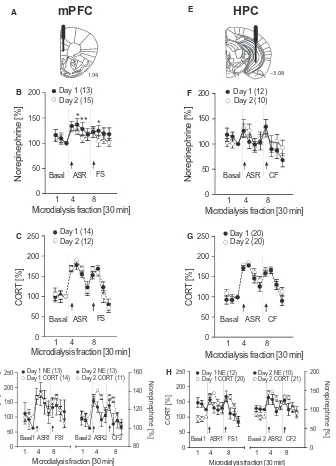
microdialysis in medial prefrontal cortex and hippocampus to measure the changes in
extracellular norepinephrine and free corticosterone (CORT) in the shocked mouse and
related them to PTSD-like symptoms, including hyperaroual and contextual fear response. I
found that increased free CORT was related to immediate stress response, whereas
norepinephrine level, in a brain region specific manner, predicted arousal and contextual fear
response one month after trauma. I also applied metabolomics analysis to investigate
molecular changes in prefrontal microdialysates of shocked mice. Citric Acid Cycle,
Glyoxylate and Dicarboxylate metabolism and Alanine, Aspartate and Glutamate metabolism
pathways were found to be involved in foot shock induced hyperarousal. Taken together, my
study provides novel insights into PTSD pathogenesis and suggests potential therapeutic
Table of contents
iii
Table of contents
Abstract……….. i
Table of contents……….. iii
Abbreviations……… v
1. INTRODUCTION ... 1
1.1. History, epidemiology and diagnosis of PTSD ... 1
1.2. Neurobiology of PTSD ... 3
1.2.1. Alterations in the neuroendocrine system ... 3
1.2.2. Neurotransmitter system in PTSD ... 6
1.2.3. Alterations of the immune system ... 9
1.2.4. Pathoanatomical alterations associated with PTSD... 10
1.2.4.1. Prefrontal cortex ... 12
1.2.4.2. Hippocampus ... 13
1.2.4.3. Amygdala... 14
1.2.4.4. Nucleus accumbens ... 15
1.3. Risk factors and biomarkers of PTSD ... 16
1.3.1. Risk factors ... 16
1.3.2. Biomarkers... 19
1.4. Pharmaco- and psychological therapy of PTSD ... 20
1.5. Animal models of PTSD ... 22
1.5.1. PTSD animal model... 23
1.6. Hypothesis-free –omics: quantitative proteomics and metabolomics, and RNA-seq transcriptomics ... 25
1.6.1. Quantitative proteomics ... 26
1.6.1.1. Mass spectrometry based proteomics ... 27
1.6.1.2. Quantitative proteomics using stable isotope labeling ... 28
1.6.2. Quantitative metabolomics ... 29
1.6.3. Quantitative transcriptomics ... 31
1.6.4. Computational biology/Systems biology... 32
Table of contents
iv
1.8. Aims of the thesis ... 35
2. RESEARCH ARTICLES... 37
2.1. Fluoxetine treatment rescues energy metabolism pathway and myelin sheath protein alterations in a posttraumatic stress disorder mouse model ... 37
2.2. Transcriptomic profiling reveals affected inflammatory pathway in a mouse model of posttraumatic stress disorder ... 79
2.3. Norepinephrine and corticosterone in the medial prefrontal cortex and hippocampus predict PTSD-like symptoms in mice ... 119
2.4. NextGen Brain Microdialysis – applying modern metabolomics technology to the analysis of extracellular fluids ... 130
3. DISCUSSION AND FUTURE PERSPECTIVES... 139
3.1. Energy metabolism ... 139
3.1.1. Altered energy metabolism in psychiatric diseases ... 139
3.1.2. Interaction between energy metabolism, inflammatory response and neuroendocrine function ... 141
3.1.3. Role of energy metabolism in different neurons ... 142
3.2. Systemic view of PTSD brain circuitry ... 143
3.3. Fluoxetine treatment ... 145
3.4. Future perspectives ... 146
Bibliography ... 148
Acknowledgements ... 183
Curriculum vitae and publications ... 185
List of author contributions ... 187
Abbreviations
v
Abbreviations
5-HTTLPR serotonin transporter
ACC anterior cingulate cortex
aCSF artificial cerebrospinal fluid
ACTH adrenocorticotropic hormone
APA American Psychiatric Association
API atmospheric pressure ionization
BLA basolateral amygdala
CA1 cornu ammonis 1
CAGE cap analysis of gene expression
CeA central nucleus of amygdala
CID collision-induced dissociation
CNS central nervous system
CORT corticosterone
CRH corticotropin-releasing hormone
CRHR1 CRH receptor 1
CR-PTSD combat-related PTSD
CSF cerebrospinal fluid
DSM Diagnostic and Statistical Manual of Mental Disorders
ESI electrospray ionization
ETC electron transport chain
ETD electron transfer dissociation
FKBP5 FK506 binding protein 5
FS foot shock
Abbreviations
vi
GC glucocorticoids
GC-MS gas chromatography mass spectrometry
GLT-1 glutamate transporter-1
GR glucocorticoid receptors
GWAS genome-wide association studies
HCD high energy collision dissociation
HPA hypothalamic-pituitary-adrenal
HPLC high performance liquid chromatography
ICAT isotope-coded affinity tag
ICPL isotope-coded protein label
IL-1 interleukin 1
IL infralimbic cortex
iTRAQ isobaric tag for relative and absolute quantitation
LC locus coeruleus
LC-MS liquid chromatography mass spectrometry
LTP long-term potentiation
LTQ linear ion trap
m/z mass-to-charge
MALDI matrix-assisted-laser-desorption ionization
mPFC medial prefrontal cortex
MPSS massively parallel signature sequencing
MR mineralocorticoid receptors
MRM multiple-reaction-monitoring
MS mass spectrometry
NAA N-acetylaspartate
NAc nucleus accumbens
Abbreviations
vii
NFκB nuclear factor κ B
NGFI-A nerve growth factor-induced protein A
NMDA N-methyl-D-aspartate
NMR nuclear magnetic resonance
NPY neuropeptide Y
PACAP pituitary adenylate cyclase-activating peptide
PGC Psychiatric Genomics Consortium
PrL prelimbic cortex
PTSD posttraumatic stress disorder
PVN paraventricular nucleus
Q quadrupole
QqQ triple-quadrupole
Q-TOF quadrupole-time-of-flight
RNA-seq RNA sequencing
ROS reactive oxidative species
SAGE serial analysis of gene expression
SAM sympathetic-adrenal-medullary
SCZ schizophrenia
SILAC stable isotope labeling using amino acids in cell culture
SILAM stable isotope labeling of mammals
SNP single nucleotide polymorphisms
SPS single prolonged stress
SRM selected-reaction-monitoring
SSRIs selective serotonin reuptake inhibitors
TNF-α tumor necrosis factor-α
TOF time-of-flight
Abbreviations
viii
1. INTRODUCTION
1
1.
INTRODUCTION
1.1.
History, epidemiology and diagnosis of PTSD
The term Trauma was originally used to describe physical wound or injury (Spiers and
Harrington, 2001). Later in medicine and psychiatry literature, trauma was referred to a
wound inflicted upon the mind rather than the body (Caruth, 1996). During the 19th century
and into the mid-20th century, there was an ongoing debate whether traumatic disorder was
psychological or physiological. Not until post-World War II was the concept of interaction
between psychology and neurophysiology for traumatic disorders identified and recognized
(Abram, 1970; Selye, 1974).
What we call PTSD nowadays was first classified as “Stress Response Syndrome” caused by
tremendous stress reaction under the category of transient situational personality disorder in
the first edition of Diagnostic and Statistical Manual of Mental Disorders (DSM-I) published by the American Psychiatric Association (APA, 1952). In 1980, PTSD was first officially
mentioned in DSM-III (APA, 1980). The revised DSM-III (APA, 1987) then separated the cause of PTSD from ordinary stressors. In the current DSM-5 (APA, 2013) PTSD was officially removed from anxiety disorder to a new category of trauma- and stress-related
disorder with more distinct diagnoses based on behavioral symptoms.
The epidemiology of PTSD was not recognized until 1980 in DSM-III (APA, 1980). Described by the National Comorbidity Survey (NCS), the life-time prevalence of PTSD was
10.4 % in females and 5.0 % in males of the U.S. population (Kessler et al, 1995), while in Germany was reported to be 2.2 % in females and less than 1 % in males (Perkonigg et al, 2000). The definition of PTSD starts with an exposure to trauma, a stressor that is not in the
range of normal human experience. Among those individuals who were exposed to severe
1. INTRODUCTION
2
stress dose and coping strategy of the affected individual. Other risk factors including
childhood stress/trauma, domestic violence, gender or pre-existing anxiety/depression also
impact the onset of PTSD (Foa et al, 2006; Yehuda et al, 2011a).
The development of PTSD is often preceded by acute stress disorder. The symptoms usually
begin within the first three months after encountering the trauma but may also initiate more
than six months after the stressor (Ballenger et al, 2000; Brunello et al, 2001; Kessler, 2000). To date, the diagnosis of PTSD relies much on self-reports, clinical expert interviews and
clinical behavioral monitoring. According to DSM-5, five criterions should be met for the
diagnosis of PTSD:
Criterion A – The patient was exposed to a traumatic event, such as death threat, serious
injury or sexual violence or has witnessed another person being exposed to such tragic events,
with the response of intense fear.
Criterion B – Intrusion (re-experiencing) symptoms which include intrusive memories,
flashbacks as well as nightmares of the traumatic event. Re-experiencing symptoms are
usually triggered by environmental cues that remind the patients of the tragic experience.
Criterion C – Symptoms which involve avoiding thoughts, feelings, places, activities or
people associated with the traumatic event.
Criterion D – Emotional numbing, decreased interest and symptoms of negative alterations in
cognition and mood related to the traumatic experience.
Criterion E – Hyperarousal symptoms, including sleep difficulties, hypervigilance,
exaggerated startle response, aggressiveness, loss of concentration or self-destructive
1. INTRODUCTION
3
1.2.
Neurobiology of PTSD
1.2.1. Alterations in the neuroendocrine system
Upon stressor stimulation, glucocorticoids (GC) and catecholamines play a major role in
regulating stress response and result in brain metabolism and behavior changes. Immediate
release of catecholamines induces an acute response to the stressors and is regulated by
sympathetic-adrenal-medullary (SAM) axis. GC secretion, on the other hand, affects
long-term adaption processes and is regulated by hypothalamic-pituitary-adrenal (HPA) axis.
HPA axis is composed of the paraventricular nucleus (PVN) of hypothalamus, the anterior
pituitary and the cortex of the adrenal gland. Stressor first simulates corticotropin-releasing
hormone (CRH) release from parvocellular secretory neurons which project from the
hypothalamic PVN to the median eminence of the hypothalamic-hypophyseal portal
circulation. CRH is then transported to anterior pituitary via the blood vessel system and
stimulates the secretion of adrenocorticotropic hormone (ACTH). Subsequently, ACTH is
transported to the adrenal cortex, where it rapidly stimulates biosynthesis and release of GC
(cortisol for human and corticosterone for rodents) (Papadimitriou and Priftis, 2009) as well
as the mineralocorticoids (Funder, 2010). Under physiological conditions, HPA axis
activation is regulated by two negative feedback loops, inhibitory effects of cortisol on the
ACTH-secreting cells in the anterior pituitary and the CRH-secreting neurons in the
hypothalamus. In addition, limbic brain regions involved in emotional response to stress
modulate HPA axis activity, with frontal cortex and hippocampus inhibiting while amygdala
increasing HPA axis activity (Herman et al, 2005).
In addition to HPA axis, stress exposure activates SAM axis immediately, which leads to
production and release of the catecholamines (norepinephrine, epinephrine) from adrenal
1. INTRODUCTION
4
neurons in the forebrain regions, including frontal cortex, hippocampus and amygdala
(Robertson et al, 2013). Increased GC and catecholamines evoke physical stress response, the “fight-or flight” response, by increasing heart rate, blood pressure and pupil dilation while
inhibiting digestion, urination or reproductive functions (Carrasco and Van de Kar, 2003) to
prepare the organism to survive and deal with stressors.
Many studies have indicated HPA and SAM axes activity dysregulations in PTSD patients.
However, in clinical studies, inconsistent findings of cortisol levels were observed in PTSD
patients. Increased cortisol reactivity in response to psychosocial stress has been observed in
sexual and physical childhood abuse patients (Heim et al, 2000). On the other hand, lower cortisol secretion has been reported in patients with PTSD (Morris et al, 2012; Yehuda and Seckl, 2011b). Yet, Wingenfeld et al. (Wingenfeld et al, 2015) found lower cortisol levels only in patients with lifetime PTSD, but not in patients with current PTSD. This phenomenon
may underlie the biological significance reflecting dose-dependent and time course effects of
cortisol release. In addition to PTSD, abnormalities in cortisol secretion and HPA axis
activity were also reported in other depressive and anxiety-related disorders.
Hypercortisolism was found in major depression patients, with hypersecretion of cortisol at
baseline and in the dexamethasone suppression test (Parker et al, 2003). Abelson and Curtis (Abelson and Curtis, 1996) observed that alterations in HPA system in panic disorder patients
are modulated by illness severity and treatment seeking behavior. The shared dysregulation
of HPA axis in major depression, PTSD and other stress-related disorders might in part
explain the high comorbidity of these psychiatric disorders.
Release of GC induces massive changes throughout the whole body, primarily activating
adaptation mechanisms. Chronic stimulation of GC release by stress may initiate differential
1. INTRODUCTION
5 actions in the brain. Mineralocorticoid receptors (MR) bind cortisol with nearly ten-fold
higher affinity than glucocorticoid receptors (GR) (Veldhuis et al, 1982). At basal or during mild stressful conditions cortisol mostly binds to MR to enhance synaptic strength (LTP).
During cortisol level elevation, GRs become fully occupied and LTP induction is impaired
(de Kloet et al, 2005; Joëls et al, 2006). This dual system may work in opposing directions regulating the behavioral responses (Pavlides et al, 1995). Chronic GC hypersecretion results in MR and GR downregulation. In a rat model of single-prolonged-stress PTSD, MR and GR
levels were found decreased (Zhe et al, 2008). In addition, GC sensitivity correlates with symptom changes in combat veteran PTSD patients and GC receptor hypersensitivity has
been shown to develop after childhood trauma and (later) PTSD, indicating the important
roles of MR and GR in the clinical state (McGowan et al, 2009; Yehuda et al, 2014). Moreover, GC regulates the HPA system via a negative feedback loop by inhibiting ACTH
and CRH secretions. Elevated CRH levels are associated with increased fear-potentiated
startle response (Keen-Rhinehart et al, 2009; Pelton et al, 1997) and enhanced fear conditioning (Roozendaal et al, 2002). Higher concentrations of CRH in the cerebrospinal fluid were observed in PTSD patients (Baker et al, 1999).
Catecholamine (norepinephrine) not only regulates sleep and arousal, but also attention,
learning and memory. Activation of the norepinephrinergic system mediates consolidation of
memories associated with emotional events (Barsegyan et al, 2014; Soeter and Kindt, 2011). Dysregulated norepinephrine signaling has been linked to PTSD symptom severity, with
chronic PTSD patients showing greater CNS norepinephrinergic activity than healthy
subjects (Geracioti et al, 2001). In another study, facilitation of norepinephrine signaling by using the α2 selective antagonist yohimbine resulted in increased anxiety in PTSD patients
1. INTRODUCTION
6
PTSD (Brunet et al, 2008; Cohen et al, 2011; Dębiec et al, 2011; Pitman et al, 2002; Stein et al, 2007). These studies indicate a dual role of norepinephrine, both adaptive and deteriorating, in the mediation of the post-stress response (Jett and Morilak, 2013; Lapiz and
Morilak, 2006). In addition to regulation of stress responses, norepinephrine and GC interact
with each other and mediate downstream events. Several studies indicated that GC affects
α1-adrenergic receptor function and modulates α- and β-α1-adrenergic receptor-coupled signaling
(Duman et al, 1989; Stone et al, 1987). Moreover, local administration of β-adrenergic receptor antagonist propranolol in the amygdala prevented GC-induced memory
enhancement from emotional arousal-induced norepinephrine activation (Quirarte et al, 1997; Roozendaal et al, 2006). In PTSD patients, interaction between norepinephrine and cortisol was shown to be predictive for negative intrusive memories in PTSD (Nicholson et al, 2014).
1.2.2. Neurotransmitter system in PTSD
Central neurotransmitter imbalance has been linked to the pathophysiological mechanisms of
PTSD. Traumatic experiences activate certain neuronal circuits and induce long-lasting
changes in neurotransmitter systems. In addition to the abnormalities in norepinephrine and
GC signaling described in chapter 1.2.1, monoaminergic neurotransmitters, including
serotonin and dopamine, were found dysregulated in PTSD. Clinical studies showed that
combat-related PTSD (CR-PTSD) is associated with enhanced norepinephrinergic activity
and diminished serotonin activity (Spivak et al, 1999). In addition, hypofunction of serotonergic system was associated with PTSD symptoms, i.e. increased startle response and
impulsive aggression in chronic CR-PTSD. Treatment of CR-PTSD patients with SSRIs
showed improvements in symptoms including arousal, intrusion, irritability and avoidance
1. INTRODUCTION
7 important role in stress adaptation in animal studies (Joseph and Kennett, 1983). A
significantly lower binding affinity of amygdala serotonin transporter was found in PTSD
patients compared to healthy subjects (Murrough et al, 2011). A meta-analysis revealed an association between SS genotype of the serotonin transporter (5-HTTLPR) polymorphism
and PTSD in high trauma-exposed patients, implicating the role of 5-HTTLPR polymorphism
as a risk factor for developing PTSD (Gressier et al, 2013). Taken together, serotonin deficit is an important factor for the development and persistence of PTSD symptoms.
The mesolimbic dopaminergic pathway also plays an important role in fear and anxiety. It
was shown that over-activation of dopamine transmission resulted in increased fear response
(Pezze and Feldon, 2004). Dysfunction of dopamine in frontal cortex is associated with
intrusive thoughts and insufficient extinction of trauma-related memory, which are core
features of PTSD (Morrow et al, 1999; Seamans and Yang, 2004). A clinical study showed that the decline of dopamine metabolite homovanillic acid levels was associated with
laboratory-induced symptoms in chronic PTSD patients (Geracioti et al, 2013). On the other hand, higher urinary excretions of dopamine, norepinephrine and epinephrine were observed
in PTSD outpatients, and dopamine and norepinephrine levels were correlated with the
severity of PTSD (Yehuda et al, 1992). Similar to serotonergic transporters, dopamine transporter single nucleotide polymorphisms are associated with the occurrence of PTSD
among trauma survivors (Segman et al, 2002). Meanwhile, cross talk between norepinephrine, serotonin and dopamine has been reported such that norepinephrine stimulates serotonin and
dopamine release while serotonin release from norepinephrine neurons reduces
norepinephrine release (Fink and Göthert, 2007). These studies implicate the importance of
balanced monoaminergic neurotransmitter signaling for PTSD underlying mechanisms.
Besides dysfunctional monoaminergic pathways, alterations in amino acid neurotransmitter
1. INTRODUCTION
8
in dorsolateral prefrontal cortex and anterior cingulate cortex were identified in PTSD
patients (Michels et al, 2014). However, in human plasma, low GABA levels after encountering a traumatic event may predict subsequent PTSD development (Vaiva et al, 2004). In addition to GABA itself, imbalance between GABA and glutamate is associated
with hippocampal neuronal apoptosis in the PTSD animal model (Gao et al, 2014). Glutamatergic neurotransmission is known to be involved in the stress response and
anxiety-related disorders (Popoli et al, 2012). The glutamatergic system regulates acquisition and extinction of fear conditioning. Stress-induced glutamate release facilitates memory
consolidation by inducing long-term potentiation (Anwyl, 2009). Treatment with the
anticonvulsivant drug, phenytoin, which may reduce glutamate neurotransmission, has
demonstrated significant efficacy in combat PTSD by reducing avoidance and arousal
symptoms (Bremner et al, 2005a). Furthermore, blocking N-methyl-D-aspartate (NMDA) receptors prior to exposure to a predator scent stress prevented anxiety-like behaviour in
rodents (Blundell et al, 2005). Thoreinger et al. showed that AMPA receptor GluR1 signaling in dentate gyrus is involved in consolidation of remote fear memories via CRH receptor type
1 pathway (Thoeringer et al, 2012). These studies provide evidence for the involvement of glutamatergic signaling in PTSD pathogenesis. Interestingly, recent findings suggest that
polyamine modulates ionotropic glutamate receptors and the metabolism was shown to be
disrupted in anxiety and depression (Fiori and Turecki, 2008; Vaquero-Lorenzo et al, 2009). Finally, neuropeptides, e.g. neuropeptide Y (NPY), has been shown to be involved in PTSD
development (Sah and Geracioti, 2013). Recent studies suggest a protective effect of NPY for
PTSD via its function in regulating fear conditioning and extinction and suppressing startle
1. INTRODUCTION
9 1.2.3. Alterations of the immune system
As mentioned in chapter 1.2.1, physical and psychological stress induces the release of stress
hormone. Stress hormones alter the status of the immune system (Glaser and Kiecolt-Glaser,
2005), and in turn, immune system disturbance affects CNS function via humoral and cellular
pathways conveying signals to the brain (Maier and Watkins, 1998). Recent studies have
indicated an altered inflammatory response in various psychiatric disorders, including
schizophrenia (de Baumont et al, 2015; Neelamekam et al, 2014), major depression (Maes, 1995, 2008), and anxiety-related disorders (Ogłodek et al, 2015; Solati et al, 2015). It is known that chronic stress exposure affects sympathetic and steroid hormone pathways, and
results in alterations of lymphocyte migration and numbers (Silberman et al, 2004). Upon stressors, the expression of cytokine and chemokine genes for mobilizing the immune cells is
regulated so that the tissue function can be restored upon intrusions (Black and Garbutt,
2002). Depending on the nature, intensity, and duration of the stressor, different types of
immune response are challenged and stress effects on the immune system differ. Exposure to
prolonged stressors induces immunosuppression and results in anti-inflammatory response
that makes the subject more susceptible to diseases (Cohen et al, 2012). On the other hand, repeated defeat stress (e.g., repeated social defeat) prevents the GC-induced suppression of
inflammation via inhibition of nuclear factor κ B (NFκB) pathway and results in an
enhancement of the immune response (Barnes and Adcock, 2009; Chrousos et al, 1996). Recent studies have shed light on the role of the immune response in anxiety-related
disorders. Studies on adult male Sprague-Dawley rats revealed that chronic unpredictable
restraint stress exposure selectively increases the number of microglial cells and results in a
transition of microglia from a ramified-resting state to a non-resting state in certain
1. INTRODUCTION
10
cortex, nucleus accumbens, medial amygdala, dorsal bed nucleus of the stria terminalis, CA3
region of the hippocampus and periaqueductal gray (Tynan et al, 2010). These finding indicate that microglial activation might play an important role in the regulation and/or
adaptation to stress. In addition, women with PTSD due to childhood sexual or physical
abuse showed enhancement in delayed-type hypersensitivity skin test which suggests higher
cell-mediated inflammatory reactions (Altemus et al, 2003). In the psychogenic stress-exposed rats, treatment of minocycline, an anti-inflammatory, anti-apoptotic and
neuroprotective tetracycline agent, reduced local levels of the cytokines interleukin-1 (IL-1),
IL-6 and tumor necrosis factor-α (TNF-α) in the hippocampus, frontal cortex and
hypothalamus, and attenuated anxious-like behaviors (Levkovitz et al, 2015). Taken together, dysregulations in immunological processes upon psychological and physiological stress may
disturb the adaptation/resilience to stress and therefore results in PTSD development.
1.2.4. Pathoanatomical alterations associated with PTSD
Development of PTSD involves memory- and stress-related processes (Siegmund and Wotjak,
2006). Maintenance of strong memory of an aversive encounter due to resistance to
extinction, memory reinstatement, too fragile inhibitory mechanisms, overgeneralization or
disturbance of declarative memory is a potent cause for PTSD (Bremner et al, 1992; Charney
et al, 1993; McFarlane et al, 2002; Solomon et al, 1987). Different brain structures are associated with the memory process in PTSD. Brain imaging studies in PTSD patients during
fear conditioning and in response to trauma-related stimuli showed an increased activity in
amygdala, the brain region responsible for expression of conditioned fear, and decreased
activity in prefrontal cortex, the brain region related to extinction (Bremner, 2002; Bremner
1. INTRODUCTION
11 PTSD (Gilbertson et al, 2002). Traumatic events not only result in the formation of associative memories but also sensitize individuals in a non-associative manner, increasing
the general responsiveness to potentially harmful stimuli. The hyper-responsiveness in
amygdala and medial prefrontal cortex observed in PTSD patients is in accordance with
slower habituating activity in response to trauma-related stimuli (Shin et al, 2005). Meanwhile, studies with rats and mice have demonstrated that a single exposure to an
extreme stressor may cause long-lasting changes in neurochemistry (Martí et al, 2001; van Dijken et al, 1993), startle response (Balogh et al, 2002) and electrical excitability of the fear circuit (Adamec et al, 2005; Adamec et al, 2001).
In addition to an investigation of the role of individual brain region, recent studies have
aimed to establish a functional neuronal network in which local circuits interact to transfer
signals across different brain regions for the generation of behavioral responses that are
relevant for mental disorders (Bielczyk et al, 2015; Robinson et al, 2014). The amygdala is a key brain region regulating acquired and innate fear and anxiety-related behaviors (Krettek
and Price, 1978; Maren and Quirk, 2004). Basolateral amygdala (BLA), which exhibits
cortex-like function, receives sensory afferents from somatosensory cortex as well as
thalamus (Pape and Pare, 2010; Tovote et al, 2015). Strong inputs from the ventral hippocampus and the medial prefrontal cortex (mPFC), with the prelimbic cortex contribute
to sustained fear. The infralimbic cortex is critical for fear memory extinction (Quirk and
Mueller, 2008) and projects to both BLA and intercalated cells between BLA and central
nucleus of amygdala (CeA). Activated intercalated cells in turn directly or indirectly inhibit
CeA output neurons (Pare and Duvarci, 2012). On the other hand, BLA also sends
1. INTRODUCTION
12
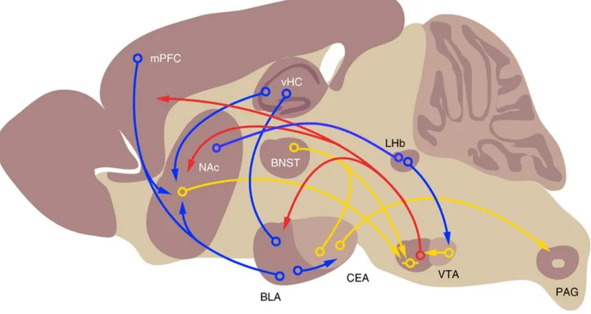
neurons from VTA project to nucleus accumbens (NAc) as well as to the mPFC (Fields et al, 2007). Synaptic connections between mPFC, amygdala, hippocampus, VTA and NAc reflect
a neural circuit that underlies pathological behavior responses in anxiety-related disorders
(Figure 1).
Figure 1. Brain circuits involved in fear perception. Simplified scheme depicting different
projections from distinct brain regions for regulation of fear response. Red, blue and yellow
arrows represent DA, glutamate, and GABA projections, respectively. Adapted from Lüthi
and Lüscher (Lüthi and Lüscher, 2014).
1.2.4.1. Prefrontal cortex
The prefrontal cortex integrates sensory information received from various brain regions
within the limbic system, regulates emotions and initiates goal-oriented behaviour (Gray et al, 2002; Miller et al, 2002a). Preclinical studies have shown that medial prefrontal cortex (mPFC) plays a crucial role in both the acquisition and extinction of fear response (Markham
1. INTRODUCTION
13 prelimbic (PrL) and infralimbic (IL) cortices. PrL is associated with increased fear expression
while IL has inhibitory functions for the fear response (Corcoran and Quirk, 2007; Fenton et al, 2014; Laurent and Westbrook, 2009). PTSD is associated with diminished responsivity in the adjacent ventral mPFC (vmPFC) (Shin and Liberzon, 2010). Functional neural imaging
studies of PTSD revealed a failure or decreased activation of ACC during the presentation of
trauma-related stimuli (Hou et al, 2007; Yang et al, 2004) and negative, non-traumatic stimuli (Kim et al, 2008; Williams et al, 2006). In addition, activation of ACC appears to be inversely related to PTSD symptom severity (Kim et al., 2008; Williams et al., 2006) and
positively associated with symptomatic improvement following treatment (Felmingham et al, 2007; Peres et al, 2007). Furthermore, decreases in vmPFC (Shin et al, 2010) and ACC (Corbo et al, 2005) volumes were found in PTSD patients and smaller ACC volumes have been linked with greater PTSD symptom severity (Woodward et al, 2006). Studies with rats exposed to chronic retrain stress demonstrated reductions in prefrontocortical (ACC and
prelimbic area) dendrite lengths (Radley et al, 2004) which might underlie the mechanism for PFC volume loss and cognitive impairments in stress-related disorders.
1.2.4.2. Hippocampus
The hippocampus is involved in formation and maintenance of contextual and declarative
memory, as well as in memory consolidation for generating long-term memory (Elzinga and
Bremner, 2002). Hypoactivation of hippocampus was found in PTSD patients and inversely
correlated with symptom severity (Shin and Liberzon, 2010). Other studies, however, have
1. INTRODUCTION
14
Some, but not all studies reported a reduction in hippocampal volume in PTSD patients
(Bossini et al, 2008; Golier et al, 2005; Karl et al, 2006; Pederson et al, 2004). Studies with twins demonstrated decreased hippocampal volumes may be a familial risk factor for
developing PTSD following traumatic events (Gilbertson et al, 2002). Moreover, N-acetylaspartate (NAA), a marker for neuronal integrity, has been associated with
dysfunctional hippocampal neuronal networks in PTSD patients (Schuff et al, 2006), a finding also observed in the PTSD mouse model applied in the current study (Siegmund et al, 2009). The mechanisms underlying stress-induced hippocampal shrinkage still remain elusive.
GC has been proposed to be involved in the development of hippocampal volume loss.
Chronic exposure to GC has neurotoxic effects (You et al, 2009), partially mediated by GC-induced increase in hippocampal glutamate levels and subsequent excitotoxic effects
(McEwen, 1997). Studies in PTSD rat models suggested that stress-stimulated alterations in
hippocampal expression levels of GC receptors (Kohda et al, 2007) and NMDA receptors (Yamamoto et al, 2010) might aggravate the neurotoxic effects of increased GC and glutamate levels. Furthermore, Golub et al. found a long-lasting reduced hippocampal volume
possibly due to shrinkage of axonal protrusions in an electric foot shock-induced PTSD
mouse model (Golub et al, 2011).
1.2.4.3. Amygdala
Hyperactivation of the amygdala has been repeatedly shown in PTSD patients (Bremner et al, 2005b; Morey et al, 2009; Vermetten et al, 2007). In addition, some studies have demonstrated that the activation of amygdala correlated positively with PTSD symptom
1. INTRODUCTION
15 enhancement of amygdala hyperresponsivity which in turns deteriorates emotional processing
upon stress (Rauch et al, 2006). Moreover, PTSD resilience was associated with reduced amygdala activation (Osuch et al, 2008) and amygdala lesions may decrease the occurrence of PTSD (Koenigs et al, 2008).
Several studies reported shrinkage of amygdala volume in PTSD patients (Karl et al, 2006; Rogers et al, 2009). Findings from animal studies suggested that apoptosis in the amygdala contributes extensively to the volume loss (Ding et al, 2010; Liu et al, 2011). Increases in dendritic arborisation and spine density were found in animal models of PTSD and were
proposed to be involved in hyperactivation of the amygdala (Adamec et al, 2012; Mitra et al, 2005). Furthermore, the abnormalities in the dendritic spine density are thought to affect the
long-term maintenance of fear memories, which is found disturbed in PTSD (Mitra et al., 2005).
1.2.4.4. Nucleus accumbens
Nucleus accumbens (NAc) is the brain structure involved in motivation and addiction. Upon
acute stressor (Wang et al, 2005) or arousing environmental stimuli (Merali et al, 2004), CRH is released from paraventricular nucleus of hypothalamus and acts on the NAc.
Subsequently, dopamine release is increased through co-activation of CRH R1 and R2 in the
NAc, and facilitates cue-elicited motivation (Peciña et al, 2006) and social bonding (Lim et al, 2007). In a mouse study, severe stress exposure abolished CRH’s capacity to regulate
dopamine release in the NAc and switch the positive-affective state of CRH-dopamine
interaction to an aversive state (Lemos et al, 2012). These studies demonstrated that severe stress produces a persistent dysregulation of CRH-dopamine interactions in the NAc and
1. INTRODUCTION
16
protein levels in NAc of social defeat susceptible mice. Furthermore, reducing BDNF release
from ventral tegmental area (VTA) to NAc enhances resistance to stress (Krishnan et al, 2007).
In both animal and human studies, increased alcohol consumption and drug addiction are
closely associated with traumatic event encounter (Bremner et al, 1996; Cross et al, 2015; Volpicelli and Ulm, 1990). Studies on women exposed to childhood rape report turning to
alcohol to reduce PTSD symptoms (Epstein et al, 1998). Furthermore, 40% of inpatients receiving treatment for substance abuse also met the criteria or PTSD (Dansky et al, 1997). It is known that drug addiction acts on the mesocorticolimbic system, which consists of the
VTA and the brain regions that are innervated by projections from VTA, such as NAc, PFC
and amygdala (Swanson, 1982). Studies have shown that addictive drug use induces synaptic
potentiation in the VTA and subsequently triggers synaptic alteration in downstream brain
structures (NAc and PFC), with further drug exposure (van Huijstee and Mansvelder, 2014).
These findings indicate that abnormal neuronal signaling in the NAc, which is part of the
mesocorticolimbic system, plays an important role in the comorbidity of alcohol and drug
addiction in PTSD patients.
1.3.
Risk factors and biomarkers for PTSD
1.3.1. Risk factors
The term risk factor refers to factors that preclude a causal involvement in disease. On the
other hand a biomarker serves as an indicator for disease development and therapy response.
As mentioned in chapter 1.1, only 10-20% of individual exposed to severe trauma will
develop PTSD. To date, several important risk factors both externally (e.g. lack of social
1. INTRODUCTION
17 development. A prior exposure to trauma or chronic stress increases dramatically the
occurrence of PTSD (Davidson et al, 1991). In addition, the type and exposure time of the trauma are potent factors, with prior assaults at a young age upon subsequent traumatization
raises the prevalence of PTSD significantly (Bremner et al, 1993; Breslau et al, 1999). Social and demographic factors, such as family instability, lower education and income, and being
widowed or divorced are also associated with PTSD development (King et al, 1996). A past history of psychological problems or psychiatric disorder increases the risk for the occurrence
of PTSD (McFarlane, 1989). Furthermore, epidemiologic studies showed that antisocial
(Schnurr and Vielhauer, 1999) or neurotic personalities (Breslau et al, 1998) prior to the traumatic event predicted the development of PTSD.
Abnormalities in cortisol secretion and HPA axis activity underlie the pathophysiological
mechanism for PTSD. Lower basal cortisol has been proposed to be a pre-existing risk factor
for PTSD (Zoladz and Diamond, 2013). Studies showed that individuals with lower cortisol
levels immediately after trauma have a greater risk for developing PTSD (Delahanty et al, 2005; Ehring et al, 2008; McFarlane et al, 2011). It is possible that lower cortisol leads to chronic enhancement of glucocorticoid receptor hypersensitivity which in turn results in the
development of later PTSD after trauma (van Zuiden et al, 2013; Wingenfeld and Wolf,
2011).
Additional risk factors including genetic and epigenetic ones are considered to contribute to
an individual’s predisposition to develop PTSD. Studies on trauma-exposed twin pairs
revealed that approximately 30% of the PTSD symptoms are heritable and comorbidity of
PTSD with other disorders may be partly due to shared environmental and genetic influences
(Afifi et al, 2010). Investigations on the candidate genes for association of Single Nucleotide Polymorphisms (SNP) with PTSD symptoms have aimed to identify specific genetic regions
1. INTRODUCTION
18
searching for genetic variations associated with the HPA axis. SNPs in the CRH receptor 1
(CRHR1) gene (Amstadter et al, 2011) and in the FK506 binding protein 5 (FKBP5) gene (Binder et al, 2008) were found to be associated with PTSD symptoms. FKBP5 regulates the cortisol-binding affinity of the glucocorticoid receptor and it is hypothesized that genetic
variations in the CRHR1 and FKBP5 genes lead to the dysregulation of the HPA axis upon stress exposure and result in PTSD development. Besides genetic variations, epigenetic
modifications caused by DNA methylation or histone modification also contribute to the risk
for PTSD occurrence. Environmental factors like early-life stress can modify gene activity
via epigenetic mechanisms. A study on holocaust survivors showed that maternal PTSD
increases the risk for offspring to develop PTSD. This finding illustrates the transgenerational
transmission of potential trauma-induced epigenetic modifications (Yehuda et al, 2008). In a
study of survivors of the Rwandan genocide, increased DNA methylation at the nerve growth
factor-induced protein A (NGFI-A) binding site of the GR gene was found to be related to less intrusive memory of the traumatic event and reduced PTSD risk in male but not female
survivors. These findings indicate that epigenetic modification of GRs is associated with
gender-specific PTSD risk (Vukojevic et al, 2014). A recent study showed that a SNP in the
PAC1 receptor, which is involved in stress response by stimulating CRH gene expression, is associated with PTSD in female patients. Furthermore, methylation of the PAC1 receptor gene has been correlated with PTSD symptom severity (Ressler et al, 2011). Taken together,
the findings on genetic and epigenetic variations of HPA axis associated genes underscore the
importance of the neuroendocrine stress response system for PTSD pathogenesis and
1. INTRODUCTION
19 1.3.2. Biomarkers
The term biomarker was defined by the Biomarker Definitions Working Group at National
Institutes of Health as “a characteristic that is objectively measured and evaluated as an
indicator of normal biological processes, pathogenic processes, or pharmacological responses
to a therapeutic intervention” (Biomarkers Definitions Working Group, 2001). In addition to
providing a diagnostic tool in the clinical setting, the search for new biomarkers aims to
further elucidate the mechanisms underlying pathogenic processes. A wide variety of
indicators can serve as biomarkers: genomic alterations such as single nucleotide
polymorphisms (SNPs), alterations in gene or protein expression, or changes in metabolite
profile. Genomic alterations or mutations are trait biomarkers that can predict the likelihood
of developing a disease and indicate disease susceptibility. On the other hand, proteins and
metabolites represent dynamic biomarkers, able to reflect the disease state. Besides molecular
markers, altered brain activity evaluated by fMRI or PET imaging and other physiological
parameters such as blood pressure or heart rate can also serve as biomarkers (Hampel et al, 2010; Zhang et al, 2009).
Biomarkers for PTSD comprise imaging, psychological, endocrine, and molecular measures.
Due to the complex etiology and polygenic character of psychiatric disorders including PTSD,
biosignatures rather than a single biomarker are expected to reflect more accurately PTSD
pathophysiology.
HPA-axis dysregulation has been frequently found to be associated with PTSD. Elevated
1. INTRODUCTION
20
function and reactivity in larger cohorts of PTSD patients to clarify the inconsistencies.
Hyperactivity of the SAM was found in PTSD patients (Pitman et al, 2012; Strawn and Geracioti, 2008), which is reflected by elevated urine and plasma norepinephrine levels
(Kosten et al, 1987; Yehuda et al, 1998). In addition, adrenoreceptor blockers were reported to improve PTSD symptoms, but not in all studies. Norepinephrinergic hyperactivity has been
associated with several symptoms of PTSD, including night mare and exaggerated startle
response (Dierks et al, 2007). Besides the association with SAM hyperactivity, startle response was found to be positively correlated with cortisol levels (Grillon et al, 2006) and cortisol suppression by dexamethasone reduces exaggerated startle responses in PTSD
patients (Jovanovic et al, 2011). Brain region specific structural and functional abnormalities were reported in PTSD patients and animal models of PTSD. Reductions in the volume of the
amygdala, the hippocampus and the anterior cingulate cortex have been described (Karl et al, 2006). Furthermore, lower levels of NAA, a marker for neuronal density, have been reported
in the hippocampus of PTSD patients (Karl and Werner, 2010a; Schuff et al, 2008). NAA levels not only serve as a risk factor for PTSD susceptibility as described in chapter 1.3.1, but
also as a diagnostic marker for PTSD.
Protein biomarkers for PTSD are mostly immunological factors. The cytokines IL-2, IL-6 and
IL-8 have been reported to be increased in PTSD patients (Baker et al, 2001; Maes et al, 1999; Song et al, 2007). On the other hand, proteins involved in the immune response, such as C-reactive protein, serum amyloid, and neopterin were found to have decreased levels in
PTSD patients (Atmaca et al, 2002; Söndergaard et al, 2004; von Känel et al, 2010).
1.4.
Pharmaco- and psychological therapy of PTSD
1. INTRODUCTION
21 to Weir Mitchell in 1885 (Mitchell, 1885). Following exposure to trauma, he
self-administrated and described ‘the mischievous tale of bromides, opium, chloral and brandy’.
Due to the complex etiology and symptomatology of PTSD, not all patients respond to
treatment and at present there is no drug that acts on all the PTSD symptoms mentioned in
chapter 1.1. The main goals of PTSD pharmacotherapy can be summarized as follows: (1) to
reduce core PTSD symptoms; (2) to improve resilience to stress; (3) to reduce comorbidity,
and (4) to prevent relapse over follow-up (Brunello et al, 2001). Serotonergic agents, such as selective serotonin reuptake inhibitors (SSRIs), are currently the most commonly used
pharmaco-treatment options. SSRIs increase serotonin functioning by inhibiting its uptake
from the synaptic cleft into pre-synapse. Treatment with SSRIs increased tolerance to
aversion and decreased stress-fear response. Currently, SSRIs including fluoxetine,
paroxetine, sertraline and fluvoxamine have been officially approved by the U.S. food and
Drug Administration (FDA) for treatment of PTSD (Ravindran and Stein, 2009). In addition
to SSRIs, noradrenergic agents, such as the α2-adrenoreceptor agonist clonidine and β-
adrenoreceptor antagonist propranolol, have been proven to be effective for the treatment of
PTSD associated hyperarousal symptoms by correcting dysregulated norepinephrine
signaling (Ravindran and Stein, 2009). However, other clinical trials have shown mixed
results (Pitman et al, 2002; Stein et al, 2007). Meanwhile, dysregulation of HPA axis activity was found in PTSD patients. It is known that GCs regulate memory formation, inhibit
unrelated information and enhance the consolidation of extinction memory (de Quervain et al, 2009; Joëls et al, 2006; Karst et al, 2005) which makes targeting glucocorticoids a possible option for treating PTSD. In a double-blind study, one-month of low-dose cortisol treatment
of chronic PTSD patients inhibited memory retrieval of traumatic events and reduced
symptoms even beyond the treatment period (de Quervain, 2008). Besides the above
1. INTRODUCTION
22
compounds are potential treatment options for PTSD (Aga-Mizrachi et al, 2014; Steckler and Risbrough, 2012).
Currently, a combination of trauma-psychotherapy and pharmacotherapy, mainly employing
SSRIs, is considered as the golden standard for PTSD treatment (Berger et al, 2009). Cognitive-behavioral therapy, the dominant psychological treatment for PTSD, applies the
approaches of exposure and cognitive therapies. Exposure therapy aims to reduce emotional
association with traumatic memories. It helps PTSD patients to learn effective regulation of
stress with relaxation training and stress coping techniques while they are being progressively
exposed to the fearful stimulus (Smith et al, 2015). The cognitive therapy aims to modify the inaccurate thoughts and emotions attributed to the traumatic memories of the patients. Studies
from Resick and Schnicke (1992) showed that cognitive therapy improved PTSD and
depression measures and the improvements were maintained for 6 months in rape victims.
1.5.
Animal models of PTSD
To study the underlying mechanisms of psychiatric disorders and potential pharmaceutical
treatments, animal models provide researchers a nice tool to investigate the affected pathways.
However, modeling psychiatric disorders in animals is challenging since diagnosis of the
symptoms are not objectively measureable and it is impossible to model the complex etiology
and expression of human psychiatric disorders. Therefore, animal research focusses more on
the basic emotional process which is shared by humans and other mammals (LeDoux, 2000;
Ohman and Mineka, 2001). The aim for animal research is to model selected endophenotypes
proposed to reflect features of the respective psychiatric disorder.
A valid animal model should fulfil the following three criteria (Chadman et al, 2009; Siegmund et al, 2006):
1. INTRODUCTION
23 the animal model. In the case of PTSD, the key to differentiate it from other anxiety disorders
is the encounter of a traumatic event (see chapter 1.5.1). The stressor should be intense but of
short duration and the severity of symptoms increase with the intensity of the stressors. The
core symptoms for PTSD need to include behavioral phenotypes of hyper-responding
(exaggerated fear responses to trauma-related cues), hyperarousal and hypo-responding
(emotional numbing). In addition, behavioral alterations are expected to last for a long time
from weeks to months.
Construct validity represents the underlying pathophysiological mechanisms for disorders
which are similar or correlate with humans with the animal model employed. In the animal
model of PTSD, studies have suggested hippocampal hypotrophy and hyperactivity of the
amygdala or the CRH system which correlates with the observations in PTSD patients
(Bremner, 2002; Thoeringer et al, 2012).
Predictive validity represents the treatment options human disorders are applicable to and
can be verified in animal research. Although there is no golden standard for treatment of
PTSD, SSRIs have shown to be effective in PTSD patients (Albucher and Liberzon, 2002).
Therefore, application of SSRIs can be verified for the predictive validity in animal models.
1.5.1. PTSD animal model
PTSD animal models can be generated by applying physical, psychosocial, psychogenic or
early life stressors. Restraint stress is induced by placing a mouse or rat in restraining tubes
for 2-6 hours which results in increased anxiety behaviour and changes in neuronal
morphology within brain regions involved in fear and anxiety (Miller and McEwen, 2006;
Vyas et al, 2002). Another physical stressor model, single prolonged stress (SPS), combines the administration of different stressors (restrain stress, forced swim and ether exposure). SPS
1. INTRODUCTION
24
mimics the situation for PTSD development in humans (Liberzon et al, 1997; Pitman et al, 2012; Takei et al, 2011). In another model animals are subjected to a variety of psychosocial stressors, such as social defeat and social isolation. When exposing to chronic social
instability, animals exhibit long-lasting changes in anxiety-like behaviors resembling human
PTSD symptoms (Zoladz et al, 2008). In another commonly used PTSD animal model a predator is used as a psychogenic stressor (Dielenberg and McGregor, 2001). In this model,
animals receive threat but no pain. The exposure to species-relevant predators or odor/scent
lead to long-lasting manifestations of anxiety and acoustic startle response (Hebb et al, 2003; Roseboom et al, 2007; Zoladz et al, 2012). In another model, animals were re-exposed to a trauma reminder which allows the study of underlying neural mechanisms for persistent
re-experiencing and intrusive memories, the core symptoms of PTSD (Ritov et al, 2014). In humans, trauma exposure during development leads to long-term HPA axis abnormality and
increases the risk of developing PTSD later in life (Delahanty and Nugent, 2006). Similarly,
in the early life stress induced PTSD mouse model, maternal separation results in enhanced
stress and anxiety responses upon exposure to severe stress later in life (Diehl et al, 2012; Tsoory et al, 2007).
The PTSD model used in the current study fulfils all the above mentioned criteria (face,
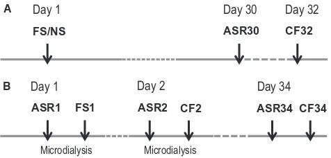
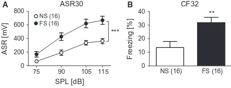
construct and predictive validities) (Siegmund and Wotjak, 2007). Two inescapable electric
foot shocks (FS) with 1.5mA amplitude and 2 seconds duration were given to the mice as
traumatic encounters. After 28 days of incubation, the shocked mice were assessed for
behavioral phenotypes of PTSD-like symptoms. In the shocked mice, both the conditioned
fear and generalized fear responses are increased. The conditioned fear response is evaluated
by the freezing behavior in the shock context and the generalized fear response is assessed by
measuring the freezing response in a completely neutral context or in a context resembling
1. INTRODUCTION
25 addition, shocked mice show a hyperarousal phenotype which can be evaluated by the
intensity of the acoustic startle response when presenting the mice different intensities of
neutral tone (Siegmund and Wotjak, 2007) or to startle-response provoking white noise
(Golub et al. 2009). For the treatment of PTSD, early intervention and chronic administration
of fluoxetine in shocked mice has been shown to ameliorate PTSD-like symptoms (Siegmund
and Wotjak, 2007). Until now, the pathophysiological mechanisms and affected molecular
pathways underlying PTSD development still remain elusive. Shrinkage of hippocampus
volume and dysregulations of CRH system were observed in the PTSD model we applied,
which resembles the clinical findings in PTSD patients (Golub et al, 2011; Thoeringer et al, 2012). Taken together, the face validity (traumatic foot shock, increased contextual fear and
hyperarousal phenotype), predictive validity (fluoxetine treatment) and constructive validity
(sharing similar biological alterations) are met in our PTSD mouse model.
1.6.
Hypothesis-free –omics: quantitative proteomics and metabolomics,
and RNA-seq transcriptomics
PTSD is a complex and polygenic disease. Therefore data integration from a variety of
approaches including -omics-based systems biology is critical for a better understanding of
disease pertinent molecular pathways (Barabási and Oltvai, 2004; Kitano, 2002). Functional
genomics, including transcriptomics, proteomics and metabolomics add an important
dimension to the study of psychiatric disorders for the analysis of spatial and temporal
alterations (Geschwind and Konopka, 2009; Turck et al, 2005). Both transcriptomics and proteomics approaches have been applied to study the underlying mechanisms of PTSD
(Henningsen et al, 2012; O'Donovan et al, 2011; Tylee et al, 2015). Transcriptomics analysis examines mRNA expression which reflects the genes that are actively expressed at any given
1. INTRODUCTION
26
addition to transcriptomics, the proteomics approach facilitates organelle specific analyses,
e.g. synapse specific alterations and is able to detect protein post-translational modifications
(PTM) which play an important role in protein function and activity. Since mRNA levels do
not always reflect protein expression levels complementary proteomics analyses have merit.
The metabolome represents the final product of interactions between gene expression, protein
expression, gene and protein interaction and the cellular environment (Kaddurah-Daouk and
Krishnan, 2009). With the advance of instrumentation and data analysis strategies,
metabolomics technology is able to complement transcriptomics and proteomics
investigations in the field of systems biology and pathway analysis. The integration of
transcriptomics, proteomics and metabolomics analyses can help delineate dysregulated
molecular pathways in disease. The following chapters describe proteomics (1.6.1)
metabolomics (1.6.2) and transcriptomics (1.6.3) technologies with a focus on the methods
that were used over the course of the thesis project.
1.6.1. Quantitative proteomics
Traditional proteomics strategies mainly exploited two-dimensional gel electrophoresis for
high resolution protein separation and relative protein quantification. Complex protein
mixtures are first separated based on their isoelectric point in the first dimension and by
molecular weight in the second dimension. Staining of the proteins allows the detection of
both quantitative (expression level) and qualitative (post-translational modification) protein
differences. Differentially expressed proteins between disease/treatment vs. control can then
be identified using mass spectrometry (O'Farrell, 1975). Today quantitative proteomics is
1. INTRODUCTION
27 utilizing sensitive MS in combination with multidimensional protein and peptide separation
techniques thousands of proteins can be identified and quantified in a sample.
1.6.1.1. Mass spectrometry based proteomics
The main components of a mass spectrometer are an ionization source, a mass analyzer and
an ion detector. A variety of ionization methods are available for introducing the ions into the
mass analyzer. For proteins and peptides the preferred methods are
matrix-assisted-laser-desorption ionization (MALDI) (Karas and Hillenkamp, 1988)and electrospray ionization
(ESI) (Whitehouse et al, 1985; Wilm and Mann, 1996a; Wilm et al, 1996b). Depending on the analytical question, different kinds of mass analyzers exhibiting distinct strengths can be
utilized: linear ion trap (LTQ), Orbitrap, time-of-flight (TOF), quadrupole (Q) mass filter,
magnetic sector and combinations thereof like LTQ-Orbitrap, Q-TOF or triple-quadrupole
(QqQ) instruments.
MS measures the mass-to-charge (m/z) ratio of ions. The mass spectrum is the final readout
of the mass spectrometer and represents the signal intensity of the ions and their m/z ratios
(Steen and Mann, 2004). Precursor ions can be fragmented to product ions via methods such
as collision-induced dissociation (CID), electron transfer dissociation (ETD) or high energy
collision dissociation (HCD). Parent and fragment masses are compared against a database
for protein identification (Eng et al, 1994; Perkins et al, 1999).
With its high mass accuracy (<5 ppm) and high sensitivity (sub-femtomol range for peptides)
the LTQ-Orbitrap hybrid mass spectrometer fulfils the requirements for sensitive and
comprehensive proteomics analysis. In addition, the LTQ-Orbitrap has a high dynamic range
(>103) and high mass resolution (up to 100.000 at m/z 400) (Yates et al, 2006).
1. INTRODUCTION
28
According to the isotope dilution theory (de Leenheer and Thienpont, 1992), peptides that
differ only in isotopic composition behave identical during an MS experiment. As a result
relative protein amounts are reflected by the unlabeled and labeled peptide signal ratios of the
extracted ion chromatograms (Steen and Mann, 2004). A great variety of methods are
available for relative protein quantification with stable isotopes (2H, 13C, 18O and 15N). These
include post-synthesis labeling such as isotope-coded protein label (ICPL) (Schmidt, 2005),
isotope-coded affinity tag (ICAT) (Gygi et al, 1999) and isobaric tag for relative and absolute quantitation (iTRAQ) (Ross et al, 2004). ICPL is based on adding a stable isotope tag to the free amino groups of intact proteins (N-terminus and lysine side chains) with the help of
specific reagents. Protein mixtures are first subjected to reduction and alkylation. For a
comparison of four samples the free amino groups are then derivatized with the ICPL_0,
ICPL_4 (4 Deuteriums), ICPL_6 (6 13C) and ICPL_10 (4 Deuteriums + 6 13C) reagents. The
samples are then combined for further separation and enzymatic digestion. After enzymatic
cleavage, the relative abundance of identical peptides, which due to their different tags differ
in their mass, can be quantified according to their signal intensities (Lottspeich and
Kellermann, 2011; Schmidt et al, 2005). The chemical probes used for the ICAT method are composed of three elements: a reactive group that is able to label a specific amino acid side
chain, an isotopically coded linker and a tag (e.g. biotin) for the affinity isolation of labeled
peptides/proteins. For quantitative analysis of two protein samples, one sample is labeled
with isotopically light (d0) tag while the other sample is labeled with the isotopically heavy
(d8) tag. Both samples are subsequently mixed and subjected to enzyme (mostly trypsin)
digestion, followed by an avidin affinity chromatography to isolate peptides labeled with
isotope-coded tagging reagents. Relative protein levels of the two samples are then calculated
1. INTRODUCTION
29 groups using tags of different masses. The derivatized peptides are isobaric and yield reporter
ions following CID which can then be used to identify and quantify relative peptide
abundance (Shadforth et al, 2005).
Alternatively, stable isotopes are introduced into proteins via metabolic labeling with either
‘stable isotope labeling using amino acids in cell culture’ (SILAC) (Ong et al, 2002) or ‘stable isotope labeling of mammals’ (SILAM) (Oda et al, 1999) methods.
For SILAC, one cell sample is cultured in media with standard essential amino acids, while
the other cell sample is grown in media supplemented with the heavy stable isotope form of
amino acids (e.g. 13C-lysine). Proteins are harvested from the two experimental cell samples
and combined. After extraction and fractionation proteins are digested and peptides subjected
to MS analysis. Unlabeled and labeled peptide pair peak intensity ratios reflect the relative
abundance of the two proteins in the cell samples (Ong et al, 2002). SILAM has been applied to investigate proteome changes in animals. Animal proteomes can be uniformly labeled with
15
N through feeding with a protein-based, 15N-labeled diet (Filiou et al, 2011). Labeled tissues can then be used as an internal standard when mixed with tissues of interest from an
animal model. For SILAC and SILAM unlabeled and labeled samples are combined at the
very beginning of sample preparation which results in high quantitation accuracy because
potential experimental biases affect both labeled and unlabeled proteins in the same manner
(Bantscheff et al, 2012). Therefore SILAC and SILAM represent the current gold standard for accurate relative protein quantification.


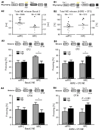
![Fig. 6. U-shape relationship between norepinephrine (NE) and behavior. (A)pus (HPC) upon stress presentations on day 2 [ASR2(CF)2 NE] after FS correlated with CF response on day 34 (CF34) in aninverted U-shape manner (adjustedexpressed as area under the cu](https://thumb-us.123doks.com/thumbv2/123dok_us/1841304.1238625/144.595.45.281.311.418/relationship-norepinephrine-behavior-presentations-correlated-response-aninverted-adjustedexpressed.webp)

