_____________________________________________________________________________________________________ *Corresponding author: E-mail: rhnmemon222@gmail.com;
28(7): 1-11, 2018; Article no.JAMMR.46700
ISSN: 2456-8899
(Past name: British Journal of Medicine and Medical Research, Past ISSN: 2231-0614, NLM ID: 101570965)
Outcomes of Proximal Femoral Nail in
Subtrochanteric Femur Fractures”- A Case Series
Rohan R. Memon
1*, Neel M. Bhavsar
1, Pankaj Patel
1and Nirav Patel
11
Department of Orthopaedics, NHL Medical College, VS General Hospital, Ahmedabad, India.
Authors’ contributions
This work was carried out in collaboration between all authors. All authors read and approved the final manuscript.
Article Information
DOI: 10.9734/JAMMR/2018/46700 Editor(s): (1) Dr. Ikem, Innocent Chiedu, Professor, Department of Orthopaedic Surgery and Traumatology, Obafemi Awolowo University, Ile-Ife, Nigeria. (2)Dr. Thomas I. Nathaniel, University of South Carolina, School of Medicine-Greenville, Greenville, USA. Reviewers: (1)Danúbia da Cunha de Sá Caputo, Universidade do Estado do Rio de Janeiro, Brazil.
(2)Vijaya Krishnan, MGM College of Physiotherapy, India. (3)Serghei Covantev, University of Medicine and Pharmacy “Nicolae Testemitanu”, Republic of Moldova. Complete Peer review History:http://www.sdiarticle3.com/review-history/46700
Received 26 October 2018 Accepted 13 January 2019 Published 14 January 2019
ABSTRACT
Background: Subtrochanteric fractures are defined as those occurring below the lesser trochanter and extend distally up to 5 cm in the shaft of the femur: Subtrochanteric fractures of the femur remain some of the most challenging fractures facing Orthopaedic surgeons. Internal fixation of these fractures has gained widespread acceptance but the problems i.e. malunion, nonunion, implant failure, refracture and infection encountered after surgical treatment of these fractures have prompted continued development of new devices and treatment programs. We study the outcome of these fractures treated with long proximal femoral nail.
Aims: Here we present a study evaluating the results of subtrochanteric femur fractures treated with proximal femur nail.
Study Design: This is a prospective observational type of study.
Place and Duration of Study: The present study consist of the patients admitted in orthopaedics unit of VS General hospital Ahmedabad from June 2013 till August 2017.
Methodology: All patients above 16 years of age who presented to our emergency department with subtrochanteric fracture of the femur were included in the study. Radiographs were taken and all the fractures were classified according to the Seinsheimers classification. All patients underwent
Memon et al.; JAMMR, 28(7): 1-11, 2018; Article no.JAMMR.46700
fixation with the proximal femoral nail. The functional outcomes of the patients were assessed using the Harris hip score.
Results: Most commonly seen fractures pattern in this study is Seinschemer‘s type III A. In our study 74.28% (26) patients did not require any support for walking and 5(14.28%) patients required canes for long walks and only one patient was mobilising with the help of crutch. Squatting was possible in 15(42.85%) patients with ease and with difficulty in 06 (17.14%) patients. 14 patients were unable to squat. In this study sitting cross legged with ease is possible in 18 (51.42%) patients. 10(28.57%) patients were able to sit cross legged but with difficulty.07 (17.14%) patients were unable to sit cross legged.
Conclusion: Proximal Femoral Nail is a good implant for the treatment of unstable subtrochanteric fractures of femur. In our study we had good results with the proximal femoral nail, it requires minimal exposure and achieves biological fixation. It allows early weight bearing which is beneficial and has fewer implant related complications. Proximal femoral nail is thus a choice of implant for fixation of subtrochanteric fractures.
Keywords: Proximal femur nail; subtrochnateric fractures; stresses; muscle forces.
1. INTRODUCTION
Fractures of the femur are commonly
encountered in Orthopaedic practice. They account for 10 to 15% of all hip fractures [1]. Subtrochanteric fractures are defined as those occurring below the lesser trochanter and extend distally up to 5 cm in the shaft of the femur. Fielding and Magliato have defined it as fractures occurring between a line extending from the superior border of the lesser trochanter to a line 7.5 cm distal to it [2]. In younger patients, the fracture is more commonly caused by high energy trauma. In older age groups, the fractures occur with low energy trauma as in a simple fall [3] Management of this fracture is difficult because this zone of femur is subjected to maximum amount of mechanical stresses. Open reduction and Internal fixation of these fractures has gained widespread acceptance but the problems i.e. malunion, nonunion, implant failure, refracture and infection encountered after surgical treatment of these fractures have
prompted continued development of new
devices. The theoretical and biomechanical advantages of cephalomedullary implants over plate fixation are attributed to a reduced distance between the hip joint and the implant (Long proximal femur nail) [4]. These further results in a reduced bending movement across the implant and fracture site and allow the load to be transferred directly to the femoral shaft, bypassing the calcar femorale.
2. MATERIALS AND METHODOLOGY
The present study consist of 35 patients admitted in orthopaedics unit of VS General hospital Ahmedabad from June 2013 till August 2017. All
patients who were above 18 years of age with fracture of subtrochanteric region of femur of traumatic origin and who were able to ambulate prior to the fracture were included in the study. Patients with pathological fractures, patients with
associated neurological problems and
polytrauma patients were excluded from the study. All patients who had a minimum follow up of at least one year, were included in the study.
Radiographs were taken and all the fractures were classified according to the Seinsheimer’s classification. Patients were worked up and pre anesthetic checkup was done. Preoperatively antibiotics were given according to the hospital protocol. All patients underwent fixation with the proximal femoral nail.
2.1 Operative Technique
Memon et al.; JAMMR, 28(7): 1-11, 2018; Article no.JAMMR.46700
the holes for the hip screws is checked in the C-arm for the depth of the nail. Guide wires for the screws are inserted via the jig and the drill sleeve. The ideal position of the guide wires is parallel and in the lower half of the neck in AP views, in a single line in the centre of the neck in the lateral views. The guide pins are inserted up to 5 mm from the articular surface of the femoral head and size of the lag screw determined, reaming and tapping for lag screw done .Insertion of the screw: First the 8 mm hip screw is inserted after reaming over the distal wire and then the 6mm cervical screw (Figs. 4,5). The hip screw should be 5 mm away from the sub-chondral bone. Distal screws: one or two static or dynamic 4.9 mm interlocking bolts are inserted in to the distal part of the nail (Fig. 6). Out of which one is a static and another is a dynamic hole. It should be done after removing the traction along with the tightening of the proximal screws. It is done free hand with the help of IITV and the jig is removed.
2.1.1 Post operative care
Operated limb was elevated for a day, Broad spectrum antibiotics were given for 5 days and than shifted to oral antibiotics. Iv fluids were given till the patient started orally. Static quadriceps exercises were begun on 2nd postoperative day. Active quadriceps exercises and hip flexion exercises were started on 4th postoperative day..Sutures were removed on 12th day (alternate) and complete suture removal
was done on 14th postoperative day. Partial weight bearing was started after reviewing
clinically and radiographically 6 weeks
postoperatively, Full weight bearing allowed after confirmation of clinical and radiological union.
Patients were discharged 5 days postoperatively.
2.1.1.1.1 Follow up
All the patients were followed up every month. On follow up following points were noted xray with both hip AP-view and lateral view of operated hip were looked for:
Signs of union
Neck shaft angle
Failure of fixation
Failure of implant
FUNCTIONAL RESULTS OF SURGERY
Assessed based following hip scoring system adopted Table 1.
Table 1. Grading of Harris Hip Score
Harris hip score Functional outcome
90-100 Excellent
80-90 Good
70-79 Fair
<70 Poor
Memon et al.; JAMMR, 28(7): 1-11, 2018; Article no.JAMMR.46700
Fig. 3. Nail passed Fig. 4. Proximal screws passed
Fig. 5. Proximal screws and nail Fig. 6. Distal locking done
3. OBSERVATIONS AND RESULTS
35 patients with subtrochanteric fractures were included in the study, The average age of the patients was 46 ±3.41. In the present series 69.23% (18) males sustained this
injury because of high velocity injury. Where as
in females they are most often caused by low velocity injury compared to their counter
parts. In this study 66.66%(06) females sustained injury because of low velocity injury (Graph 1). Most commonly seen fractures pattern
in this study is Seinschemer‘s type III A (Graph 2). Average time to union is 3.58± 0.54
months.
railing (Table 5). Squatting was 15(42.85%) patients with ease and in 06 (17.14%) patients. 14 patients to squat (Table 6). In this study legged with ease is possible in patients. 10(28.57%) patients were cross legged but with difficulty. patients were unable to sit cross (Table 7).
The complications which we saw in patients include superficial infection
Graph 1. showing distribution
Graph 2. Showing distribution
Memon et al.; JAMMR, 28(7): 1-11, 2018; Article
was possible in and with difficulty patients were unable sitting cross 18 (51.42%) were able to sit 07 (17.14%)
cross legged
in our series of infection in 2 patients,
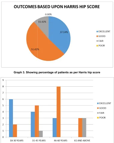
lateral migration or backout of the patients, 1 case of deep infection, fracture of the nail from the distal tip, proximal screw breakage (Table 8). no cases of implant failure or non treatment with proximal femur nail based on Harris hip score (Graph 3)
In this study all the patients in groups has excellent and good results age group patients has good and (Graph 4).
distribution of male and female patients as per mode of
distribution of the patients as per seinsheimers classification
no.JAMMR.46700
the screws in 2 one case with tip, one case of 8). There were non-union after nail overall results
3).
in younger age results and older and fair outcomes
of injury
Graph 3. Showing
Graph 4. Showing age
Table 2. Showing distribution of per pain charecteristics
Quality of pain No of patients Percentage
None or ignores 16 45.71
Slight ocassional 09 25.71
Mild 05 14.28
Moderate 04 11.42
Totally disabled 01 2.85
Memon et al.; JAMMR, 28(7): 1-11, 2018; Article
Showing percentage of patients as per Harris hip score
wise distribution of patients as per Harris hip score
of patients as charecteristics
Percentage
45.71 25.71 14.28 11.42 2.85
Table 3. Showing distribution of per limping
Limp No of patients Percentage
none 20 57.14
slight 11 31.42
modearte 04 11.42
severe 00 00
no.JAMMR.46700
score
of patients as
Percentage
Memon et al.; JAMMR, 28(7): 1-11, 2018; Article no.JAMMR.46700
Table 4. Showing distribution of patients as per walking ability
Walking ability No of patients
Percentage
None 26 74.28
Cane for long walks 05 14.28
Cane most of the time 03 8.57
Crutch 01 2.85
Not able to walk 00 00
Table 5. Showing distribution of patients as per stair climbing
Stair climbing No of patients
Percentage
Without using a railing 12 34.28
Using a railing 18 51.42
In any manner 03 8.57
unable 02 5.71
Table 6. Showing distribution of patients as per sqvatting
Percentage No of patients
With ease 15 42.85
With difficulty 06 17.14
Uable 14 40.00
Table 7. Showing distribution of patients as per sitting cross legged
Sitting cross legged No of patients
percentage
With ease 18 51.42
With difficulty 10 28.57
unable 07 20.00
Table 8. Showing complications after fixation with proximal femur nail
Complications No of patients
Superficial infection 02
Deep infection 01
Back out of screws 02
Breakage of proximal screws 01 Fracture from distal tip of nail 01
4. DISCUSSION
Subtrochanteric fractures of the femur are usually the result of high energy trauma, there is
a significant displacement of fracture
fragments,closed reduction is not possible in these type of fractures. Because of the high incidence of malunion, non-union and delayed
union, there is no role of conservative treatment as previously advocated by Lee etal [5]. Two main operative modalities used for the fixation of
subtrochanteric femur fractures are the
intramedullary implants and extramedullary
implnats. Extramedullary fixation of these fractures with implants like the dynamic hip screw or the dynamic condylar screw is complicated by extensive exposure, more blood loss which then leads on to problems in fracture union and also implant failure. Intramedullary fixation is a more biological fixation and has mechanical benefits over extramedullary fixation [6].
The proximal femoral nail being used for subtrochanteric femur fractures acts like an intramedullary splint and can bear a large axial load, this allows the patient early weight bearing. It is performed through a small surgical incision, so it is minimally invasive and reduces blood loss [7,8]. Proximal femur nail is also associated with cut out of implant and backout of proximal screws [9,10].
Fig. 7. Preoperative xray
Fig. 10. 1 year follow up
Clinical case 1. Pre-operative X-ray (Fig. 7) of left
lateral (Fig. 8) radiographs of left hip showing satisfactorily antero-posterior and lateral radiographs(figs.
Memon et al.; JAMMR
8
Fig. 8. Postoperative xray Fig. 9.
Fig. 11. Squatting Fig. 12.
left hip joint of a 34-year-old male with subtrochanteric fracture satisfactorily maintained fracture reduction and implant in situ. 9,10) of left hip showing fracture union with good alignment and
showing hip and knee range of motion
JAMMR, 28(7): 1-11, 2018; Article no.JAMMR.46700
9. 6 months follow up
12. cross legged sit
Fig. 13. Preoperative xray
Fig. 16. 1 Year follow up
Clinical case 2. Pre-operative X-ray (Figs. 13) of and lateral (Figs. 14) radiographs of left hip showing follow-up antero-posterior and lateral radiographs
Memon et al.; JAMMR
9
Fig. 14. Postoperativexray Fig. 15.
Fig. 17. Squatting Fig.
of left hip joint of a 44-year-old male with subtrochanteric fracture. showing satisfactorily maintained fracture reduction and implant
radiographs (Figs. 15,16) of left hip showing fracture union with good alignment (Figs. 17,18) showing hip and knee range of motion
JAMMR, 28(7): 1-11, 2018; Article no.JAMMR.46700
15. 6 months follow up
Fig. 18. Cross legg sit
Memon et al.; JAMMR, 28(7): 1-11, 2018; Article no.JAMMR.46700
nail [15]. A total of 41 patients were studied. There was a failure rate of 6 (29%) patients in the patients treated with the 95 degree blade plate whereas there was no failure in the patients treated with the PFN. They concluded that
internal fixation of subtrochanteric femur
fractures with a 95-degree angled blade plate is associated with increased implant failure and revision compared to closed intra-medullary nailing using a proximal femoral nail. Jiang LS et
al. did a study on 49 patients with
subtrochanteric fractures treated with the long proximal femoral nail [16]. They achieved union in all their cases but one case had delayed union. They had no complications like cut out or breakage of the implant. They concluded that long proximal femoral nail or long gamma nail is a reliable implant in treatment of subtrochanteric fractures Sahin EK et al. did a comparison of proximal femoral nail antirotation with dynamic condylar screw in the elderly in the treatment of pertrochanteric fracture of the femur [17]. They found that the mean salvati- wilson hip score was 31 in the PFNA group and 26 in the DCS group. They had good results in 73.9% of the patients in the PFNA group and 70% in the DCS group. They concluded that PFNA is a better choice as it has minimal exposure, reduce blood loss and achieves biological fixation.
Limitations of this study are as follows. First, this study is not a comparative study with that of other fixation methods especially dynamic condylar screw or proximal femur plate. Second, this study has a small number of cases and short term follow up period.
5. CONCLUSION
In our study majority of the patients had excellent and good functional outcomes as per harris hip scoring after fixation with proximal femur nails, it
requires minimal exposure and achieves
biological fixation. It allows early weight bearing which is beneficial and has fewer implant related complications. In our study there is not a single case of implant failure and fixation failure.
Proximal femoral nail is a good choice of implant for fixation of subtrochanteric
fractures.
CONSENT
As per international standard or university standard, patient’s written consent has been collected and preserved by the authors.
ETHICAL APPROVAL
As per international standard or university standard, written approval of Ethics committee has been collected and preserved by the authors.
COMPETING INTERESTS
Authors have declared that no competing interests exist.
REFERENCES
1. Kyle RF, Cabanela ME, Russell TA.
Fractures of the proximal part of the femur. Inst Course Lect. 1995;44:227-53.
2. Fielding JW, Magliato HJ. Subtrochanteric
fractures. Surg Gynecol Obstet. 1966; 122:555-60.
3. Bergman GD, Winquist RA, Mayo KA.
Subtrochanteric fracture of the femur: fixation using the zickel nail. J Bone Joint Surg. 1987;69(7):1032-9.
4. Lee JY, Lee SY. Treatment of the proximal
femoral extracapsular fracture with
proximal femoral nail antirotation (PFNA): Comparison with proximal femoral nail (PFN) J Korean Hip Soc. 2007;19:183– 189.
5. Lee JC, Clanton TO, Rockwood CA Jr.
Closed treatment of subtrochanteric
fractures of the femur in a modified cast-brace. J Bone Joint Surg Am. 1981; 63:773-9.
6. Brien WW, Wiss DA, Becker V Jr, Lehman
T. Subtrochanteric femur fractures: A comparison of the Zickel nail, 95-degree blade plate, and interlocking nail. J Orthop Trauma. 1991;5:458-64.
7. Jiang LS, Shen L, Dai LY. Intramedullary fixation of subtrochanteric fractures with long proximal femoral nail or long gamma nail: Technical notes and preliminary results. Ann Acad Med Singapore. 2007; 36:821–826.
8. Tencer AF, Johnson KD, Johnston DW,
Gill K. A biomechanical comparison of
various methods of stabilization of
subtrochanteric fractures of the femur. J Orthop Res. 1984;2:297–305.
9. Xu Y, Geng D, Yang H, Wang X, Zhu G.
Memon et al.; JAMMR, 28(7): 1-11, 2018; Article no.JAMMR.46700
10. Kristek D, Lovric I, Kristek J, Biljan M, Kristek G, Sakic K. The proximal femoral nail antirotation (PFNA) in the treatment of proximal femoral fractures. Coll Antropol. 2010;34(3):937-40.
11. Borens O, Wettstein M, Kombot C,
Chevalley F, Mouhsine E, Garofalo R. Long gamma nail in the treatment of subtrochanteric fractures. Arch Orthop Trauma Surg. 2004;124:443–447.
12. Kim JW, Chang JS, Lee H, Bae JY, Kim
JJ. Clinical results of femoral
subtrochanteric fractures. J Korean Hip Soc. 2010;22:222–226.
13. Kish B, Sapir O, Carmel A, Regev A,
Masrawa S, Stern A et al. Full weight
bearing after unstable per and
subtrochanteric fracture using proximal
femoral nail. J Bone Joint Surg.
2001;83:289.
14. Menezes, Daniel FA, Gamulin, Axel,
Noesberger, Bruno. Is the proximal femoral
nail a suitable implant for treatment of all trochanteric fractures? Clin Orthop Rel Res. 2005;439:221-7.
15. Harris I, Rahme D. A prospective
randomized controlled trial of
subtrochanteric fractures treated with the proximal femoral nail and the 95 degree blade plate. J Bone Joint Surg. 2005;87-B:310-1.
16. Jiang LS, Sheng L, Dai LY. Intramedullary
fixation of subtrochanteric fractures with long proximal femoral nail or long gamma nail: technical notes and preliminary
results. Ann Acad Med Singapore.
2007;36:821-6.
17. Sahin E, Imerci A, Kinik H, Karapinar L, Canbek U, Savran A. Comparison of proximal femoral nail antirotation (PFNA) with AO dynamic condylar screws (DCS) for the unstable peritrochanteric femoral fractures. Eur J Orthop Surg Traumatol. 2014;24(3):347-52.
_________________________________________________________________________________ © 2018 Memon et al.; This is an Open Access article distributed under the terms of the Creative Commons Attribution License (http://creativecommons.org/licenses/by/4.0), which permits unrestricted use, distribution, and reproduction in any medium, provided the original work is properly cited.
Peer-review history: