BY 1H AND 13C NUCLEAR MAGNETIC RESONANCE
Thesis by
Donald David Macmurchie
In Partial Fulfillment of the Requirements for the Degree of
Doctor of Philosophy
California Institute of Technology Pasadena, California
1973
ACKNOWLEDGMENT
I am grateful to Dr. M.A. Raftery for his advice ..
and direction and for the use of his laboratory, to my associates for many intriguing discussions, and to the California Institute of Technology for the education.
I've spent some time in study, Oh, I've taken my degrees; I've memorized my formulae,
My A's and B's and C's, But what I know came long ago
And not from such as these,
And I'm going to be a country girl again.
ABSTRACT
Introduction
1H nmr spectrum: assignment 1H nmr spectrum: interpretation Infrared spectrum
Structural implications
13C nmr spectrum: assignment 13C n.mr spectrum: interpretation Conformational processes
Discussion References
page 1 6 10
21
21
31
Introduction
Among the most interesting of the properties of natural
membranes are their selective permeability to various solutes, and, '
related to their selective cation permeability, their unusual electrical characteristics. These effects are manifested in the resting state of biological membrane systems, especially nervous systems, in which a concentration differential is maintained of approximately 15: 1
outside vs. inside with respect to Na+ and approximately 1: 50 outside vs. inside with respect to K+ (Katz,. 1966).
Propagation of nerve impulses involves disruption of this resting (equilibrium) potential by the flow of ionic species, notably Na + and K+, across the lipid barrier of the nerve axon membrane in a direction perpendicular to the direction of impulse propagation, but it remains obscure exactly how this ready flow of highly polar species through a distinctly hydrophobic medium is accomplished; the same question arises in contemplation of mechanisms for the "sodium pump" which maintains the appropriate membrane resting polariza-tion. There have been three basic hypotheses as to this means of transport, implicating, as structures resp.onsible for ion flow through the membrane , isolated carrier species (Haynes, 1969; Ohnishi and
Urry, 1969) organized pores of flux-mediating molecules or membrane structures modified locally by interaction with particular molecules to form areas of ready penetration (Duax, 1972). The discovery that
across natural and artificial membranes (Pressman, 1968 and
references therein; Mueller and Rudin, 1967) provided model systems
with properties in many ways similar to those exhibited by the nerve
cell membrane system.
Many of these ionophores have differential affinities for
various cations and of them the most selective is valinomycin (VM)
which has an affinity for~+ nearly 300 t~es as great as for Na+
(Mueller and Rudin,1967a). Produced by Streptomyces fulvissimus,
valinomycin is a cyclic trimer of the four-residue subunit
(1-lactate-1-valine-d-a-hydroxy-isovalerate-d-valine). The noteworthy features
of this sequence are the alternating ester and amide linkages in the
backbone, the exclusively hydrophobic nature of the side chains, and
the potential for three-fold symmetry offered by the trimeric
primary structure. The unique transport selectivity of the molecule
and the fact that no three-dimensional structure was known (although
conjectures had been made: Warner, 1966; Mueller and Rudin, 1967a)
made. it an interesting species for investigation by spectroscopic means, with ·the object of ascertaining the molecular conformation and from that seeking to learn about the ion-selective and transport
properties of the molecule. Nuclear magnetic resonance (nmr) spectroscopy was chosen as the chief means of investigation because of the great sensitivity of the method to changes in conformational
parameters, its possibility of revealing dynamic aspects of the
molecule's behavior and, not least, because of the convenient
In the interval since the start of this research, substantial progress toward understanding the behavior of valinomycin has been made by a number of groups and a brief review of past events is in order 0 The assignment of the
1H nmr spectrum described in the
,
following pages was soon found to be in agreement with that
reported by Haynes and co-workers (1969) in experiments performed at lower field (1401 Kguass). On the basis of measurement (1969) of the lactyl methyl line widths Haynes et al. concluded that the exchange process
was immeasurably slow in non-polar solvents, and from the lack of concentration effects on the spectrum, concluded that valinomycin did not ag·gregate substantially in solution. These conclusions were offered as arguments against a pore-forming mechanism for
valinomycin-mediated K+ transport, and in favor of an individual carrier complex; no tertiary structural information was reported.
Shortly thereafter, Ivanov et al. (1969) reported infrared (ir), optical rotatery dispersion (ORD) and circular dichroism (CD)
data and proposed tertiary structures both for valinomycin alone and for the VM o K+ complex which provided substantial refinement on the structure described here. The nmr data presented by Ivanov et al. were in agreement with those obtained previously (Haynes et al. ,
offered. Ivanov et al. interpreted their ORD data in media of low polarity as indicating an equilibrium between a number of conformers,
and from the observation of both H-bonded and non-H-bonded amide
N-H and .:=:: C=O stretching modes in the ir spectrum, concluded
that a major contributor tp the equilibrium was a conformer in which
each amide carbonyl was involved in H -bonding with the proton of the
neighboring amide group. A minor contributor to the equilibrium
structure in non-polar media, increasing in importance as solvent
polarity increased, was considered to be a structure in which some
number of amide carbonyls were involved in hydrogen bonds to solvent
molecules. In the complex with K+ Ivanov et al.. (1969) also concluded
from the ir spectrum that the ester carbonyls were involved in an
ion-dipole interaction and further that all amide protons were involved in
H-bonds; from ORD spectra they concluded that no conformational
equilibrium was present.
The spectroscopic conformational predictions were supported
by an analysis of the crystal structure of the valinomycin-K+ complex
(Pinkerton et ai. , 1969) which showed that the depsi-peptide backbone
of the molecule adopted the three-fold symmetric "bracelet"
con-..
figuration proposed for it (Ivanov, 1969), but which did not report
side-chain orientations or atomic coordinates. Subsequent publications
..
by other groups (Haynes et al., 1971; Ohnishi and Urry, 1969, 1970;
Mayers and Urry, 1972) confirmed the spectroscopic and
crystallo-graphic conclusions and indicated the popularity of the problem; in
determined independently by at least three groups (Ivanov, 1969;
Ohnishi, 1969;. Macmurchie, unpublished). Further
informa-tion was sought from the spectrum of the tsc resonances, and the
assignments and spectral changes upon K+ complex formation
described here are in agreement with the recent work of Ohnishi
et al. (1972) and Bystrov et al. (1972).
..
Observing spectroscopic indications of symmetry, most
ii).vestigators (Ivanov, 1969; Pinkerton, 1969; Ohnishi and Urry,
1969; Mayers and Urry, 1971; Macmurchie, unpublished)
had asswned for valinomycin alone, a conformation generally similar
to that which it had been shown to adopt in the presence of K+, and
so the crystallographic results of Duax et al. (1972), indicating an
..
entirely asymmetric structure for the unoccupied antibiotic, are
extremely interesting. If the conformation in solution is the same as in
the crystal one must ask why certain of its features are not revealed
in the nmr spectra; if the structures are not the same, he must
decide which is most appropriate to the environment in which the
molecule functions. It would be desirable to reconcile the results of
both techniques, and the possibility of doing this as well as its
1
H spectrum: assignment
The 1H nmr spectra of valinomycin (Fig.· 1) and its complex
with K+ were recorded using Varian HR-220, XL-100/15~· HA-100,"
..
A-60 and T-60 spectrometers, with the 220 MHz instrument, by
virtue of its greater resolution and sensitivity, being most frequently
employed. Valinomycin was obtained from Cal Biochem, lot 860009
and used without further purification; all solvents were spectro-quality
and tetramethylsilane (TMS) was used as an internal reference.
The 1H resonances were assigned as indicated in Table 1 on
the assumption that the spectrum could be interpreted in terms of a
single four-residue subunit, i.e., each line was produced by the
resonances of three magnetically equivalent nuclei; and according to
the following rationale.
After equilibration of the sample in a solution of 10% D20/ acetone
the two. doublets at lowest field (6
=
7. 84, 7. 89 ppm) were no longer..
present in the spectrum, and this fact, in addition to their appropriate
chemical shifts, makes them attributable to the amide protons of the
d- and 1-valyl residues, these being the only protons considered
likely to undergo exchange with the solvent. On elimination of the
amide resonances, the two quartets at 3. 99 ppm and 4.14 ppm
collapsed to doublets, and these resonances therefore are assigned
to the a-protons of the valyl residues. Inspection of the
proton-pro-ton couplings (Table 1) leads to the conclusion that the lower field
amide _resonance is coupled to the lower field a-proton resonance,
Figure 1: Valinomycin 1H nmr spectrum at 220 MHz
in CDC13; each trace is 500Hz, continuous
The otlfer a-proton resonances, at 6
=
5. 34 ppm and 6=
5. 02 ppm,are therefore those of the lactyl and hydroxy iso-valeryl residues
respectively, as indicated by their multiplicity. The {3-proton
resonances of the d- and 1-valyl and hydroxyiso-valeryl residues
..
are expected to be patterns of at least eight lines each, and these
three groups compose the poorly resolved envelope at approximately
2.4 ppm from TMS. The lactyl methyl resonance at 6 = 1.46 ppm
was assigned on the basis of its position separate from other
...
resonances in the methyl region and its intensity, the single aHiv
a-proton giving rise to the only other doublet. The remaining
methyl resonances, arising from the d- and 1-valyl and
hydroxyiso-valeryl residues, overlap one another extensively, as do the
{3-protons of the same residues, obscuring their assignments.
1
H spectrum: interpretation
The 1H resonance spectrum affords a considerable amount
of information and in a molecule such as valinomycin which lacks
functional side chains, the most detailed of this information is
frequently obtained from •the study of proton-proton couplings; in
valinomycin these couplings are all between protons bonded to
adjacent carbons or nitrogens, i.e., three-bond couplings. For
these it is possibie to relate the magnitude of the coupling constant.
3JHCCH to the dihedral angle between the bonds involving the two
protons (Karplus, 1959). The Karplus equation for protons on
adjacent carbons, as modified by Abraham and McLauchlan (1963)
3JaJ,1
=
10. 5 cos 28 - 0. 28Hz for 0°
<
8 ~goo 1a and3
Ja{3
=
13.7 cos 28 - 0. 28Hz for goo<8
~ 180° 1bIn addition, Bystrov and co-workers (1g69) have determined a similar relation for the 3JHNCH proton-proton coupli!lgs for amino acids:
. .. 2
=
8. 9 COS 2e
-
0. 9 COS 8 + 0. 9 Sin () 2or
3 2 . . .
JHNCH = 8. 0 cos () - 0. 9 cos () + 0. 9
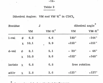
Using these relations one may estimate dihedral angles ¢ and
x
(Fig. 2) at various locations in both valinomycin and the complex with K+ as indicated in Table 2. It is to be noted, however~· that in
general, four distinct angles of rotation can give rise to the same
coupling constant, and that some other criteria are necessary if one is to choose among them. It has been shown by Gibbons et al.
(1970) that a combination of spin-coupling data and conformational
energy considerations (Scott and Scheraga.., 1966) can reduce the
number of possible configurations corresponding to a particular
coupling constant; even so, an exact knowledge of the Ca-N dihedral
angle (¢) is not sufficient to determine the backbone configuration of a peptide (Gibbons et al. , 1970) in which the amide moiety is
Figure 2: IUPAC-IUB peptide torsional angles;
* *
IR---1
~----H*
/
N---;
I
I
I
I
0
'====<
...I
'
I
' , ,
...
01
I
H
4>
=180°
..._
I
~~--
75);4>
'/y~
1
1
x1
"'=
180°
I
~
I
111
I
X=
180°
I
N
/I
1 I1
I
l/~11111111
I
I
l)
I
I
Table 2
Dihedral Angles: VM and VM·K+ in CDC13
Residue J dihedral angle
*
VM VM·K+ VM VM·K+
1-val ¢ 6.2 4.6 180° -144°
X 10.1 9.9 +153° +151°
d-val ¢ 8.1 5.1 - 39° - 46°
X 10.0 9.6 +152° +148°
lactate X 6.8 6.8 free rotation aHiv X 2.8 3.6 +123<::> +127°
[image:20.570.46.482.53.408.2]1953; Bystrov et al., 1970). Valinomycin, as a depsi-peptide, enjoys
the greater conformational freedom endowed by its ester linkages,
and thus, in order to specify its conformation exactly, even more information is required.
The proton chemical shifts of valinomycin undergo changes on variation of solvent and on formation of the cation complex. This is particularly evident in the shifts of the methyl resonances, which
in the absence of the cation, reflect the polarity of the medium as they progress from a poorly defined envelope in D20/acetone toward a relatively well-resolved system of doublets in CC14 (Fig. 3). As the factors giving rise to proton chemical shifts are of comparable
importance (Carrington and McLachlan, 1967; Pople et al., 1959)
it is tenuous to attribute these changes to a particular
source, but it is reasonable to say that in chemically similar nuclei,
similarity of chemical shift indicates similarity of chemical and magnetic environment, and that in this respect, the changes in methyl proton resonance positions with solvent polarity are indica-tive of a particularization of methyl-group environments as polarity decreases. This is to say that in the relatively polar D20/acetone
(10% v/v) the methyl groups of the valyl and a-hydroxy-iso-valeryl side chains find themselves in relatively equivalent positions with respect to moieties that can affect their chemical shifts, whether they are solvent molecules or other groups on the antibiotic, but in
..
the less polar solvents CC14 or CHC13:, eac}f methyl. group
d-ACETONE
d-ACETONE + 10% D20
-this implies a higher degree of intramolecular organization in the less polar media, which is in agreement with the ORD studies of Ivanov et al. (1969).
In the case of the lower-field resonances, more specific effects on change or solvent were observed. Especially; it was
observed that when the CDC13 solvent was saturated with D20, the amide proton resonance positions were altered to unequal degrees,
•
with the result that these resonances, which had originally appeared as a pair of doublets, now occurred closer together, appearing almost as a triplet.. This result was interpreted as indicating
..
different hydration properties of the am ides themselves, a conclusion that has been confirmed by Haynes et al. (1969) and by studies
involving the temperature dependence of these resonances (Ohnishi and Urry, 1969); this conclusion is also in accord with that of Ivanov et al. (1969) that, depending upon solvent, one set of valine amide
..
protons may form intramolecular H -bonds, while the other amide
protons form H-bonds to solvent.
Proton resonance positions were also observed to experience alterations on formation of the cation complex. The chemical shifts of the methyl resonances are nearer to those of valinomycin in non-polar than in non-polar solvent, and are less solvent-dependent than those of the free antibiotic; this again is taken as an indication of structural integrity and of uniqueness of environment of those protons. The lower field resonances undergo changes in chemical shift as
Table 3
1
H Resonance Shifts on Complex Formation
in CDC13
Resonance A ppm
lac Me -0.10
1-Val(a) +0.27
d-val(a) +0.31
aHiv(a) -0.43
lac (a) -0.39
1-val(NH) -0.50
[image:25.568.62.495.65.714.2]hydroxy acids occurred to lower field in the complex than in the
free species, while those of the amino acids were shifted to higher
field. Paramagnetic shifts in the presence of the electric field of the
cation might be predicted (Ems ley et al. , 1965; Carrington and
McLachlan, 1967), but the upfield shifts of the valine a-protons are
more interesting, in that these protons are alpha to the carbonyls
which are involved in the ion-dipole interaction (p. 21 ), an
argument based simply on electronic induction would predict shifts
--to lower field; thus it is apparent that additional fac--tors, presumably
arising from conformational changes, are contributing to chemical
shift changes on complex formation. These effects may be related
Ir spectrum of Valinomycin:
The infrared absorption spectrum of valinomycin (Fig. 4a) is well-resolved and the depsi-peptide nature of the molecule is
evidenced by the presence of both ester and amide carbonyl stretching modes, noted in Table 4; other salient features of the ir spectrum
includ~
the N-H stretching band at 3320 em -1 and the alkyl oxygen stretching absorptions at 1185 em -1 and 1132 cm-1•· On formation
of the VM · K+ complex the spectrum was altered noticeably (Fig. 4c) with the most striking changes occurring in the ester bands. The ester :::::: C=O stretching mode was sharpened and decreased in frequency. from 1755 em - l to 1738 em - l ," while the alkyl oxygen
stretching bands were present at decreased intensity; the amide carbonyl frequencies on the other hand were largely unaffected by
•
complex formation, with only a slight sharpening of the peaks taking
place. These observations are consistent with polarization of the
ester carbonyls by. a nearby positive charge (Nakanishi, 1962) and strongly implicated the valine residues in the interaction with the metal cation. This conclusion was also reached by Ivanov et al.
(1969), who in addition observed the disappearance of shoulders on the N-H stretching band (ca.3390 em -1) and the amide C=O band
(1678 em -1
) on complex formation and att~ibuted these effects to
constraint of all the amide protons in intramolecular H-bonds.
Implications for Valinomycin structure:
·
pro-Figure 4: Infrared absorption spectra of
a) valinomycin
b) valinomycin-KSCN, 10:4
Table 4
Infrared Spectral Assigmnents
~ (em -1) intensity assigmnent
native valinomycin
3310 m ~N -H stretch
1755 s
>c
=0 (ester) stretch1660 s
:::-c==o
(amide) stretch1540 ms >N-H deformation
\ /
1185 ms 0 (ester) asym. stretch
1151 m gem dimethyl skeletal
1132 m \ 0 / (ester) sym. stretch
1103 m gem dimethyl skeletal
bone angle ¢ were not by themselves sufficient to define a structure for
valinomycin. Knowledge that the ester carbonyls were apparently
uniquely involved in cation stabilization, however, substantially ..
reduced the number of conformational choices, and, bearing in
mind the apparent symmetry of the molecule as indicated by the nmr
spectra, the six participating carbonyls were most logically located
in an octahedral arrangement, which constrained the backbone of
the molecule to a serpentine configuration enclosing the ion. Using
,. space-filling models, it was possible to assemble a structural
representation of the VM: K+ complex which was consistent with the
spectral data and allowed coordination of the metal ion by the six
ester moieties (Fig. 5). This structure resembles in some of its
features that of the nonactin~K+ complex (Kilbourn et al. , 1967)
which was described as resembling the seam on a tennis balJ; in this
context, the valinomycin complex resembles one and one-half
tennis ball seams, with a coordinating carbonyl group at each bend.
This arrangement provides a hydrophilic enclosure for the metal
ion and a hydrophobic exterior suitable for situation in a lipid
environ-ment, as was also found to be the case for nonactin. Incorporation
of the intramolecular H-bonds proposed by Ivanov et al. (1969)
further constrains the structure and these constraints allowed Ivanov
. s
and co-workers, on the basis of an independent study of JHNCH ' ' .
coupling constants (Bystrov et al., 1969), to decide between two
Figure 5: Proposed non-hydrogen-bonded
[image:32.572.69.479.50.669.2]Figure 6: Proposed structures of:
a) valinomycin
b) valinomycin • K+
o-
/p--<(
\cr.
\0
Ib I
o---.q
\ 0... ...~
\ \ bb
(a)
.
(b)
I I
p
I---<{_
'
,A ....
' Cf
'o
?"'" __
o
'
I I0
\/):>... ...
cf
' 0in Fig. 5; their choice (Fig. 6) which was later confirmed by the
The use of 13C nmr offers several advantages in
comparison to 1H runr: a smaller number of resonances is usually
observed, the chemical shifts are larger (and thus more sensitive
to subtle effects), and information is obtained directly from the
framework of the molecule, tending to minimize intermolecular
effects on the spectrum. The method has the disadvantages of
the low sensitivity of the 13C nucleus (1. 6% of that of protons)
and the fact that the natural abundance of 13C is only 1.1 %, but
technical advances, especially the use of Fourier transform nmr,
and the benefit of the Overhauser effect on decoupling from
protons, can aid in overcoming these difficulties. In addition,
recent progress in the correlation of 13C che:r;nical shifts with
substituent effects has allowed the prediction of various resonance
positions (Grant and: Paul, 1964; Horsley et al. , 1969; Gibbons et al. ,
1970). In the following pages the 13C resonance positions are
compared with those calculated and with those of the 1H resonances
in order to derive additional information about the VM - VM · K+
system.
Valinomycin was obtained from Cal Biochem (Lot 860021
and used without further purification; solvents were Matheson,
Coleman and Bell spectroquality. Spectra were recorded using
with noise decoupling from protons and employing a 2H
field-frequency lock signal from solvent or, in the case of non-deuterated
solvent, from external 2H20. Tetramethylsilane was used as an
internal reference. Preliminary experiments were performed
using a 14.1 K gauss instrument made available through the
kind-ness of the Department of Chemistry, University of California,
Berkeley~'
As was the case with the 1H spectrwn, the number of
resonances observed in the 13C spectrum (Fig. 7) corresponded
to the nwnber of carbon atoms in one of valinomycin's three
subunits, reinforcing the inference that each chemically identical
part of the molecule senses an identical magnetic environment, and
the assignments (Table 5) were again made on that basis. The .
group of four resonances at lowest field was readily identified
as those of the carbonyls by virtue of their characteristic
chemical shifts and low intensities as a result of lack of nuclear
Overhauser enhancement (Kuhlman and Grant, 1968).
Assign-ment of the individual peaks was more difficult. Following
pre-liminary experiments in acetone-d6, it was observed that the two
carbonyl resonances at highest field were somewhat broader
and lower in peak height than the two at lowest field, an effect
that could be attributed to relaxation by a nearby quadrupolar
Figure 7: 13C FT runr spectrum of valinomycin in
at 25. 2 KHz. 1H noise de coupled, spectrum
.+. i
l
1
<:>.
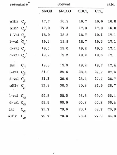
~Table 5
Solvent Dependence of Valinomycin 13
C Chemical Shifts (ppm from 'IMS)
*
resonance Solvent calc.
MeOH Me2CO CDC13 CC14
aHiv
c,
17.7 16.9 16.7 16.6 16.0a
Hiec
Iy 17.9 17.3 17.0 17.0 16.0
1-Val
cY
18.9 18.8 18.7 19.1 17.11-val
c
Iy 19.3 18.8 18.7 19.3 17.1
d-val
c
y 19.5 19.0 19.2 19.5 17.1d-val
c
Iy 19.7 19.2 19.2 19.6 17.1
lac
cf3
19.8 19.3 19.2 19.7 17.4l-val
cf3
31.0 29.6 28.4 27.7 27.3d-val
cf3
31.3 29.6 28.4 27.7 28.7aHiv
cf3
31.8 30.3 30.2 27.9 28.7I
1-val
ca
59.8 58.5 58.8 59.0 66.4d-val
ca
59.8 60.0 60.2 60.2 66.4lac
ca
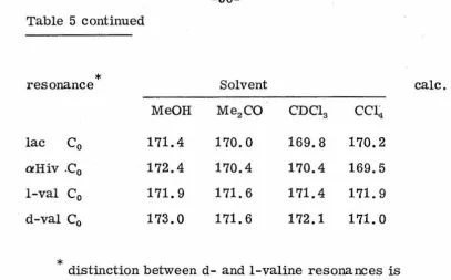
71.7 70.6 70.1 69.7 76.9 [image:41.574.49.451.149.680.2]Table 5 continued
*
resonancelac C0
aHiv .C0
1-val C0
d-val C0
MeOH
171.4
172.4
171.9
173.0
SolventMe2CO
170.0
170.4
171.6
171.6
CDC13 CCI 4
169.8
170.2
170.4
169.5
171.4
171.9
172.1
171.0
*
distinction between d- and 1-valine resonances istentative.
[image:42.574.59.467.49.302.2]two lower field resonances were tentatively assigned to the valine
residues and the remaining two to the hydroxy acids; a similar
argument was later used by Ohnishi and co-workers (1972) in
their assignment of these carbonyl resonances. Assignment of
the C , Cr-2 and C resonances was based on comparison with the
a tJ Y
predicted chemical shifts of these resonances in the various
residues, obtained from calculations using the substituent
para-meters employed by Horsley and co-workers (1969, 1970) and
by comparison with the 1H resonance spectrum, bearing in mind
the general correlation between the chemical shifts of 13 C nuclei
and those of protons bonded to them (Emsley et al. , 1965; Del Re
et al., 1963; Horsley et al., 1970). In the cases of resonances
of nuclei at corresponding positions in the d- and 1-valine
residues, and of chemically equivalent positions in the same
residue, the lines fall closely together and a distinction was not
readily made; tentatively, the higher-field resonances have
been assigned to the 1-valines in observance of the situation
in the 1H spectrum. These assignments are in agreement with ·
those of Bystrov and co-workers (1972) and Ohnishi and co-workers
(1972).
Interpretation:
The sensitivity of the molecular conformation to solvent
changes, noted in the discussion of the 1H spectrum (p. 15)
is manifested again in the spectrum of 13 C resonances. The range of methyl resonance positions, for example, is increased by nearly 50% as the dielectric constant of the solvent changes from 31.7 in MeOH to 2.2 in CC14 ; furthermore the observed range of these shifts (3.1 ppm) substantially exceeds both that calculated for the nuclei concerned (1. 4 ppm) and that observed for sequence analogues of the valinomycin residues (1. 8 ppm, Ohnishi et al. , 1972). Thus on incorporation into the tertiary structure of the molecule, the various residues experience effects
·-which confe~ on each of them particular chemical shifts, and
these must be related to the conformation of the species. Decrease of solvent polarity, therefore, leads to conformation changes
in the molecule which increase the uniqueness of each methyl group's environment.
Alteration of solvent polarity also affects the positions of the Ca and
c
13
resonances, of which the former are the moreamide carbonyls resulting from loss of interaction with polar solvent molecules, an interpretation requiring a structu;re where-in the ester carbonyls are excluded from such an interaction with solvent. Direct solvent effects on methynyl 13C resonances are
expected to be negligible (Batchelor et al. , 1972) and this,
taken in conjunction with consideration of the carbonyl 13C shifts, indicates that other effects, probably arising from conformation changes, are also important.
Applying the peak-height argument offered earlier (p. 37 ) and by Ohnishi et al. (1972), for the identification of the ester
and amide carbonyl resonances, it is apparent that at least in the two solvents CD30D and CC14 , the order of the resonances in
the spectrum is substantially altered. Ohnishi et al. (1. 972) found
that in CD30D the order was, from lowest field: ester, amide,
ester, amide, and apparently this order is maintained in CDCl3
(Fig. 8a); in CC14, however, the order is clearly reversed
(Fig. 8b). ·In considering the magnitudes of these chemical
shift changes (Table 5), it was observed that the carbonyl
resonances of the valines were shifted to lower field by at least
two ppm on change of solvent from CC14 to MeOH, while the
corresponding resonances of the hydroxy acids were shifted
to lower field by only half that amount. In the case of the Ca
Figure 8: Carbonyl 13C resonances in (left) CDC1 8
[image:46.572.53.493.35.705.2]one ppm to lower field and the other in fact moved 0. 4 ppm to
higher field, while the C resonances of the hydroxy acids were
. a
shifted to lower field by an average of 1. 9 ppm each. Assuming the shifts of the carbonyl resonances to be governed largely by
7T-bond polarity (Stothers and Lautebur, 1964; Maciel and Natterstad •.
1965; Maciel, 1965), if the inductive effect of the carbonyl group
on the C a electron density were the chief . factor in determining
the Ca chemical shift, one would expect the Ca. shifts to follow
those of the carbonyl carbons of the same residue. Since they do not, it is probable that other effects are significant contributors
to the observed chemical shifts, and these are likely to arise
from conformational differences such as reorientation of the
atom in question with respect to a nearby anisitropic group, for
example another carbonyl as has been suggested in the 1H
spectrum by Ohnishi and Urry (1969). Evidence for structural
alteration on alteration of the medium, then, is found throughout
the 13C spectrum as well as the 1
H spectrum.
The chemical shifts of the 13C resonances in the VM· K+
complex are given for various solvents in Table 6, and were
assigned by comparison to the spectrum of free valinomycin.
The resonances of the side-chain carbons experience small
Table 6
Solvent Dependence of VM·K+ 13C Chemical Shifts (ppm from
TMS)
*
resonance Solvent
MeOH Me2CO CDC13 CC14
aHiv
ca.
17.3 17.1 17.0 16.9a.Hiv
c
Ia
17.9 17.8 17.6 17.41-val
Ca
19.4 19.1 19.0 19.0d-val
ca
19.4 19.3 19.3 19.3d-val
c
IQ! 20.6 20.4 20.3 20.5
1-val
ca
29.8 29.1 28.3 28.1d-val
cfJ
29.8 29.1 28.4 28.2aHiv
cfJ
31.9 29.1 30.3 30.01-val
ca
63.0 62.5 61.6 61.4d-val
Ca
63.2 62.7 61.8 61.6lac
ca
72.6 72.3 71.1 70.9 [image:49.574.82.461.147.701.2]Table 6 continued
*
resonancelac C0
aHiv C0
1-va1 C0
d-val C0
MeOH
172.7
174.1
176.5
176.9
Solvent
Me2CO
172.0
173.1
176.0
176.6
CDC13 CC14
170.6
171.4
172.0
172.8
174.4
172.9
175.0
173.6
*
distinction between d- and 1-va1ine resonances is [image:50.570.69.472.75.711.2]one ppm to lower field, while the shifts of the backbone carbon
resonances are more significant. The participation of the ester
carbonyls in the interaction with the ion is clearly evidenced
by their substantial shift to lower field on complexation, in
accordance with increased polarization of those moieties by the
ion's electric field. In addition, the 13C resonances of the amide
carbonyls also experience shifts to lower field, which have
been attributed (Bystrov et aL , 1972) to participation of those
groups in an ion-dipole interaction with K+. However, inspection
of models produced from spectroscopic (Ivanov et al., 1969;
Macmurchie, unpublished) and X-ray (Pinkerton et al., 1969)
studies shows that the amide carbonyls are to be found in an
orientation perpendicular to the metal ion, one in which the ion
might not be expected to exert a great polarizing influence
(.Millett and Raftery, 1972). Accordingly, it is reasonable to
suggest that enhanced intramolecular hydrogen bonding (Ivanov
'
et al., 1969; Ohnishi and Urry, 1969) may be important in
producing these shifts. Such an interpretation would be consistent
with the observation (Maciel and Natterstad, 1965) of the significance
of H-bonding in determining 13C chemical shifts in carbonyl grrups.
It is seen, therefore, that the .ts C nmr spectrum is
of the groups involved in complexation, the valine carbonyls,
is obtained, and the greater range and simplicity of the 13C
spectrum allows the dependence of the molecular conformation
Conformational Processes in Valinomycin:
In the preceeding pages, it has been emphasized. that
conformation changes take place in valinomycin on alteration of its medium and on formation of a complex with K+; in the case of the K+ complex, X-ray analysis of the crystaline material (Pinkerton et al., 1969) has confirmed the structure first proposed (Ivanov et al., 1969) on the basis of spectroscopic studies, and has revealed the three-fold symmetry that seemed apparent from the sequence and the simplicity of the nmr spectra. The structure of the antibiotic when not complexing a cation, although obviously not identical to that of the complex, was generally assumed (Ivanov et al., 1969,
Pinkerton et al., 1969) to have a similar configuration and H-bonding network. The recent publication (Duax et al. , 1972) of an X-ray analysis of the crystal structure of the free species is therefore of
great interest, particularly in that this structure (Fig. 9) is entirely asymmetric and involves a hydrogen-bonding arrangement not pre-viously suspected for valinomycin. Inspection of the structure pro-posed by Duax and co-workers indicates that various chemically identical parts of the molecule are in situations which could lead to non-equivalence of their magnetic environments. Remembering the effect of H-bonding on the 13C resonances of carbonyl groups (Maciel
and Savitsky, 1964; Maciel and Natterstad, 1965), one might expect to distinguish on this basis among, for example, the resonances of the lactate carbonyls, of which two are hydrogen-bonded in the
among the other chemically identical carbonyl groups, and in all one could predict the occurrence of eight resonances in the carbonyl region, in contr:;tst to the four that were observed. Other chemically identical nuclei, such as the lactate Ca 's are also found in environ-ments that have great potential for magnetic non-equivalence, by virtue of their differing orientation with respect to nearby anisotropic groups, specifically the carbonyls of the d-valines. Considering the proton spectrum, the efforts of Bystrov and co-workers (1969) have demonstrated the dependence of the coupling constant 3JHNCH on the
- . ..
conformational angle¢ (Fig. 2, eqn. 2). Estimation of these angles in a molecular model of the Duax structure (Fig. 9) leads to the pre-diction of three doublets arising from the 1-valine amides, with
coupling ,constants of approximately 1Hz, 7Hz, and 10Hz, and three from the d-valine amide protons, with couplings of 3 Hz, 7Hz and 10Hz. Similarly, using equation (1), the aHiv a-protons would be expected to produce two doublets with large couplings, 3J a{3 ~ 9Hz, and one with 3J
a
/3
!:!:! 3 Hz. Since, even at the highest fields employed,
only one doublet is seen for each of these chemically identical
atoms, there must be additional features to the solution conformation of free valinomycin that are not revealed in the crystal structure.
It is possible, of course, that the configuration of the molecule in solution is not at all related to that in the crystal form, but it
seems more attractive to reconcile both sets of data in a model requiring the antibiotic to undergo a continual exchange of conforma-tions, as illustrated in Fig. 10. Bearing in mind that the "ends"
J
~
i
o(
0/
·
roY-
·
--._fh
_
_
'(
-t
o
~o---N
N
'>=o
01 --
~
·
-·~ro-.~
·
~·-
·
~
-
~
3-1.'
-
.
1.
-~
·
-N
-<
ol,
'-.-(~o
o-·-' 0 I
r
H
"'--~~
v
· I 0 " '
y
A
..&0-·- ·-N 0.·--
.
~
. 0 /.
r
o ·-
o
___ .-N
0
_),=o-~
-
·
-·N
>=o-
·
N
.
-~o
-(_~o-·--r-
·
~N
o~;!(
0 I ' y
"'-- I
that they are identical in overall conformation, but that any
individual atom, or group of atoms, finds itself in a different
environment in each. If the molecule were to equilibrate rapidly
.. among three structures such as these, an averaged nmr spectrum,
similar to that observeq., could result.
·In order to determine kinetic parameters from nmr spectra,
.
it is necessary to know the resonance frequency of a nucleus in
each site in which it may be found, and this in turn requires obtaining
a spectrum in the slow exchange limit, i.e., when r » (27T Av) - t
where T is the time constant for exchange and AV is the difference in
resonance positions, in Hz, of the sites involved. This limit would
be most readily attained in the 13C spectrum, considering its large
range of chemical shifts, but that spectrum when recorded at the
highest field and lowest temperature available (52 Kgauss and
-50° (', respectively, Fig. 11) is not one characteristic ofthat
..
type of exchange. Nevertheless, examination of the line widths
in the 1H spectrum indicates that an exchange process may indeed
be taking place; particular attention is drawn to the doublets
pro-duced by the J3-protons of the aHiv and lactate residues. From
the spin-spin coupling constants of these resonances (2. 8 Hz
and 6. 8Hz, respectively) it is apparent that the aHiv has restricted
rotation about the Ca-C{3 bond (¢ = . + 123\ Table 2) whereas
the lactate methyl group •is likely to be rotating freely. This being
the case, one would expect the resonance of the lactate protons to
..
Figure 11: 13C nmr spectrum of valinomycin in CDC~
at -50° C, 55 MHz. 1H noise decoupled, spectrum width 5KHz; inverted signals are
it is broader by more than 20% at ambient temperature and 100 MHz (Av
=
1.
72Hz and 1. 41Hz, respectively; Fig. 12'); it is alsoobserved that the width of the lactate methyl resonance is less when the complex with K+ is formed than in the absence of the ion (Fig. 13). These results would be produced by a situation in which the molecule experiences rapid exchange among conformations having greater chemical shift differences for the lactate than the aHiv residues, and in which the exchange is eliminated by formation of a
symmetric complex structure. In addition the lactate methyl line-width exhibits a field and temperature dependence (Table 7) that is compatible with such a scheme.
Conformation changes associated with complex formation also lead to exchange phenomena, as has been observed in the 1H
. .. 13 .
spectrum by Haynes et al. (1969). In the C spectrum, less-than-saturating amounts of K+ produce different exchange conditions depending on the solvent employed: Fig. 14 compares these spectra in methanol, acetone and CC14• Taking the line widths of the
hydroxy acid C
a
resonances and employing the relation for reasonably fast exchange (Pople et al., 1959)= 3
Figure 12: 1H resonances in CDC13 of (top) lactate CH3 and (bottom) a-hydroxy-iso-valerate
1
N I (\.) (\.)1
N
:r:
0
1
NI
Table 7
Temperature and Field Dependence of Lactate C1H3 Line Width
Field (Kgauss) ~v(lac) ~~~ ('IMS) corrected All (lac) 14.1 1. 58±. 06Hz 0. 59±. 06Hz 0. 99± .13Hz 23.5 1. 71 ±. 02 0. 62 ±. 04 1. 09±. 06 51.7 2.27±.10 0. 74±. 05
1..
53± .15Temperature
32°
c
1. 71 ±. 02 Hz 0. 62 ±. 04 1. 09±. 06Hzoo
2. 83 ± .10 0.39±.02 2. 44± .12 -20° 3.55±.10o.
58±. 05 2.97±.15 [image:67.574.66.468.146.348.2], , 'I' , o •I o o •• I • o o o 1' o o o 1 • • • 11• o 1 o 1 ' o •t•• o 'I o o o o t • •I + I o o • o I o o t l I o t o • I' o • I' • • Itt o • o I I • ' I I' • t o I I • • • I • o •
>-
·•
"'
1---40ppm~
Table 8
VM·K+ VM + K+ Exchange Rates:
C
a
Resonances1 PA
+ PB + PA2PB2 (wA
-un)
2 (r A +TB)=
T I
2 T2A T2B
Ar (Hz) Aw/ 21T (Hz)
K+:VM lactate aHiv lactate aHiv MeOH
0:1 3.5±0.5 3.0±0.5
2:5 8.0 8.0 26± 1 28± 1 1:1 4.0 4.0
r A + r B
=
(2. 7± 0. 3) x 10-3 sec Me2CO0:1 6.0±0.5 7.0±0.5
[image:70.577.46.492.42.661.2]the solubilities of KSCN in those solvents and, as the rate of
ion uptake is likely to be extremely rapid (cf: kON = 3 x 10⁸ M-1
sec-1 for monactin-Na+ complex, Eigen and Winkler, 1971), it is
probable that the exchange rates are determined by the propensity
Discussion
The exact means whereby valinomycin, and other
iono-phores, mediates the passage of an ion through a non-polar, lipid
barrier has been the subject of some discussion: some authors
(Ohnishi and Urry, 1969) have suggested a "pore" mechanism,
in which the molecules stack on top of one another so as to present
a hydrophilic channel through a membrane, while others
(Haynes et al. , 1969; Pinkerton et al. , 1969; Tosteson, 1971)
have argued for an individual carrier, shuttling back and forth,
picking an ion up at one side and releasing it at the other. It has also been suggested (Haynes et al. , 1969; Duax et al., 1972)
that aspects of both processes are involved, and that while acting
independently of other ionophores, a single one may remain
relatively fixed in the membrane and allow ions to pass through it
to the opposite side. It is quite possible, of course, that different
ionophores act in. different ways, and since none of these compounds
have been detected in nervous tissue (indeed, the "crown ethers"
(Pederson, 1967) are not natural products at all) that the re lation-ship that they bear to the mechanisms of nervous conduction
may seem obscure. Nevertheless, they serve as useful models and it is to be hoped that their study may lead to at least some
understanding of more complex systems.
In the case of valinomycin, the structure proposed here,
(1969) has characteristics appropriate to a single-carrier species.
The iso-propyl and methyl side chains effectively envelop the
ion and its surrounding carbonyls, providing the complex with an
exterior that is compatible with the hydrophobic environment
through which the ion must pass. The six ester carbonyls,
octahedrally arranged at a distance of about 3
A.;
provide the K+ion with a local environment similar to that which it experiences
in aqueous solution, in which the hydration number if four or
five (Bell, 1958); thus the complex is a 11water-drop" in
non-aqueous media, analogous to the "oil-drop" model in which many
proteins have been cast.
The probability of the molecules acting independently of
one another is heightened by the facts that the complex structure
does not allow intermolecular H-bonding in the axial direction,
.,
that the molecule has no ionizable groups, and that hydrophobic
interactions are not likely to be important in the lipid environment.
These observations are strengthened by the lack of concentration
effects in the nmr spectra reported by Haynes and co-workers
~·\ \ \ \.. 1.~ ' : ...
(1969) as well as in this work (p. ) and also by the observation
(Pinkerton et al., 1969) that the rate of valinomycin-mediated
equilibration of ion concentrations in aqueous solutions separated
by chloroform was highest at less than saturating ion
concentra-tions. This latter observation was interpreted as meaning that
side was necessary, and was prevented by high K+
concentra-tions on the receiving side; thus a mechanisms of independent
carrier molecules was implicated.
The outstanding difference between the VM • K+ structure
proposed on the basis of early work in this thesis (Fig. 5) and
that proposed by Ivanov and co-workers (1969, Fig. 6) lies in
the disposition of the amide >C=O and .:::=N-H moieties. In
·
-Figure 5, these are seen to be of two distinct types, the lactyl
amides having their carbonyl groups relatively exposed and their
amide protons more or less buried, while the iso-valeryl
amides are to be found with their carbonyls secluded and their
amide protons more exposed to solvent. This arrangement is
in accordance with the observation of different solvation properties
for the two types of amide protons (p. 18 ) and observes the
convention of amide group planarity. Completion of the
intra-molecular H-bo1_1ding network suggested by Ivanov and co-workers
(1969) slightly compacts the VM· K+ structure, makes the
distinction between the amide groups more subtle, and judging from
carefully constructed space-filling models, causes the am ides
to be substantially deformed from a planar configuration. This
possibility has not been mentioned by Ivanov (1969) or Pinkerton
(1969), but has been taken as a necessity in conformational
energy calculations (Mayers and Urry, 1972). Prior to
co-workers (1972) most others had assumed the free species
to resemble closely the complex structure (cf. Fig. 6a and b).
In this arrangement, the ester carbonyl oxygens would be free
to withdraw slightly from the center of the cavity in the absence
of K+ and it appears possible for the amide groups to enjoy a
planar configuration under those circumstances. A situation
of this type could provide a mechanism for maintaining the carrier
in a more open, or receptive, conformation before the arrival
of the ion and allowing it to close about the ion as it transports
it through the hydrophobic region.
While not denying the possibility of a carrier-diffusion
mechanism for valinomycin action, Duax and co-workers (1972)
have suggested that the asymmetric crystal structure confers
upon valinomycin properties such that it ''could be lodged in
a membrane pore and act as a pump,. In this context it is of
particular importance to consider the possibility of different
structures in solution and in the crystal state, or ways in which
a single structure could give rise to these disparate observations.
In interpreting their ORD data, Ivanov and co-workers (1969)
favored a situation in which equilibrium existed between two
conformers of greater and lesser symmetry, with the equilibrium
constant changing according to solvent polarity; it is possible
that the same. results could arise from a single conformation,
which itself altered with the polarity of the medium. Such a
reorganiza-tion of the type previously hypothesized (p. 55 ) and produce an averaged nmr spectrum of the type observed, but which varied with solvent polarity (pp. 15, .38).
As has already been mentioned, one cannot determine exchange rates from nmr spectra without knowledge of resonance positions in each state; however, an approximation may be useful. Variations in H-bonding for chemically identical moieties in the asymmetric structure have been pointed out (p. 50 ); for
carbonyl groups, Maciel and Savitsky (1964) have found that H-bond formation results in shifts of the 1-sC resonances to lower
13 ..
field by 3-7 ppm. At 52 Kgauss, 3 ppm :::: 166 Hz for C, so taking a typical line width of (211'T/ )-1 ::::10Hz and asswning a line-width in the absence of exchange of (27TT2)-1
=
1 Hz," onemay calculate an exchange rate for the simplified case of two sites from Eqn. 3: this turns out to be 1. 5 x 10⁴ sec-\ and line broadening contributions from other sources would increase
this figure. It is unlikely that treatment of an exchange as among three sites would lead to a significantly different exchange rate
molecule undergoes a conformational equilibrium with a time constant in the microsecond range, although the nature of the
conformation change was unspecified; in view of this, an extremely rapid equilibration of asymmetric structures for valinomycin
may not be totally unthinkable. Failing such a condition, one might suggest that in the crystal state packing forces may produce a slightly abnormal configuration, a situation that has been found in the crystal structures of smaller molecules
(Benedetti et al., 1968; Ganis et al., 1970; Pedone et al., 1970). However the apparent dilemma presented by the
..
crystallographic and spectroscopic data is resolved, one must conclude that the molecule exhibits remarkable flexibility and this is almost certainly a significant factor in its mechanism of action. The data presented and reviewed here tend to favor a single carrier in a lipid membrane; indeed, if as rapid a
References
. ..
Abraham, R.J. andK.A. McLauchlan, Mol. Phys. 5, 513 (1963). "'
Batchelor, J.G., J.H. Prestegard, R.J. Cushley and S.R. Lipsky,
Biochem. Biophys. Res. Commun. ~' 70 (1972).
Bell, R. P., Endeavour,31 (1958).
·
-Benedetti, E., P. Corradini, M. Goodman and C. Pedone, Proc.
Natl. Acad. Sci. 62, 650 (1968).
"""'
Bystrov, V.J., S.L. Portnova, V.I. Tsetlin, V.T. Ivanovand
Yu. A. Ovchinnikov, Tetrahedron~' 493 (1969).
Bystrov, V.J., V.T. Ivanov, S.A. Koz'min, I.I, Mikhaleva,
K. Kh. Khalilulina, Yu.A. Ovchinnikov, E. I. Fedin and
P. V. Petrovskii, _F_E_B_S_L_e_t_te_r_s
!!,
34 (1972).Carrington, A. and A. D. McLachlan , Introduction to Magnetic
Resonance, New York, Harper and Row (1967).
Corey, R. B. and L. Pauling, Proc. Roy. Soc. B .!i!_, 10 (1953).
DelRe, G., B. Pullman andY. Yonezawa, Biochem. Biophys.
Acta 75, 153 (1963).
- -
"""'Duax, W. L., H. Hauptman, C.M. Weeks and D. A. Norton, Science
176, 911 (1972). ~
Eigen, M. and R. ·Winkler, Neurosciences Res. Prog. Bull. ~' 330 (1971).
Emsley, J. W., J. Feeney and L.H. Sutcliffe, High Resolution
Nuclear Magnetic Resonance Spectroscopy,. Oxford, Per gammon
Press (1965).
Faulstich, H., W. Burgermeister and .. Th. Wieland, Biochem.
Biophys. Res~ Commun. 47, 975 (1972).
Ganis, P., G. Avitabile, E. Benedetti, C. Pedone and M. Goodman, Proc. Natl. Acad. Sci. ~' 426 (1970).
Gibbons, W.A., G. Nemethy, A. Stern and L.C. Craig, Proc. Natl. Acad. Sci. 67, 239 (1970a).
"""-Gibbons, W.A., J.A. Sogn, A. Stern, L.C. CraigandL.F. Johnson, Nature
W,
840 (1970b).Grant; D.M. and E.G. Paul, J. Amer. Chern. Soc. 86, 2984 (1964).
"""'
Haynes, D. H., A. Kowalsky and B. C. Pressman, J. Biol. Chern.
~' 502 (1969).
Haynes, D. H., B. C. Pressman and A. Kowalsky, Biochem. 10.,.,_,... ,
853 (1971).
Horsley, W. and H. Sternlicht, Biochem. Biophys. Res. Commun. 37, 47 (1969).
Horsley, W. H. Sternlicht and J.S. Cohen, J. Amer. Chern. Soc.
~' 680 (1970).
Ivanov., V.T., I.A. Laine, N.D. Abdulaev, L.B. Senyavina,
E. M. Popov, Yu. A. Ovchin.nikov and M. M. Shemyakin,
..
Biochem. Biophys. Res. Comrnun. 34, 803 .,.,_,... (1969) .
Jensen, F.R., D.S. Noyce, C.H. Sederholm and A.J. Berlin, J. Amer. Chern. Soc. 82, 1256 (1960).
~
Karplus, M. , J. Chern. Phys. 30~ , 11 (1959).
Katz, B., Nerve Muscle and Synapse, New York, McGraw-Hill (1966). Kilbourn, B.T., J.D. Dunitz, L.A.R. Pioda and W. Simon, J.
Mol. Biol. 30, 559 (1967). ~
Maciel, G.E., J. Chern. Phys.
g,
2746 (1965).Maciel, G. E. and J.J. Natterstad, J. Chern. Phys. 42, 2752 (1965) . ...,...
. .
Maciel, G. E. and G. B. Savitsky, J. Phys. Chern. 68, 437 (1964) . ...,... Mayers, D.F. andD.W. Urry, J. Arner. Chern. Soc. 94, 77 (1972) • ...,... Millett, F. andM.A. Raftery, Biochern. Biophys. Res. Cornrnun.
47' 625 (1972). ~
Mueller, P. and D. 0. Rudin, Biochern. Biophys. Res. Cornmun. 26, 398 (1967b) .
...,...
Nakanishi, K., Infrared Absorption Spectroscopy--Practical, San Francisco, Holden-Day (1962).
Ohnishi, M. and D. W. Urry, Biochern. Biophys. Res. Cornrnun. 36, 194 (1969) .
.,...,...
Ohnishi, M. and D.W. Urry, Science 168, 1091 (1970).
~
Ohnishi, M., M. -C. Fedarko, J.D. Baldeschwieler and L. F. John-son, Biochern. Biophys. Res. Cornrnun. ~' 312 (1972).
Pederson, C.J., J. Arner. Chern. Soc. -~' 7017 (1969).
Pedone, C., E. Benedetti, A. IrnmirziandC. Allegra, J. Arner. Chern. Soc. 92, 3549 (1970). ,..,...
Pinkerton, M., L.K. Steinrauf and P. Dawkins, Biochem. Biophys. Res. Cornrnun. 35, 512 (1969) . .,...,...
Pople, J. A. , W. G. Schneider and J. H. Bernstein, High Resolution Nuclear Magnetic Resonance, New York, McGraw-Hill (1959). Pressman, B. C., Federation Proc. 27, 1233 (1968).
~
..
Stothers, J.B. and P.C. Lautebur, Can. J. Chern. 42, 1563 (1964).
""""
Tosteson, D.C., Neurosciences Res. Prog. Bull. 9, 339 (1971).
"'
Warner, D.R., J. Am. Oil Chemists Soc. 44, 593 (1966).
PROPOSITION I
It is proposed to investigate the specificity of Ile-tRNAile synthetase in its acylation of tRNA 's and its deacylation of
mischarged aminoacyl-tRNA 's in methanol-water mixtures.
The precision of translation of the genetic message during the process of protein synthesis is widely remarked upon, and the efficiency with which aminoacyl-transfer ribonucleic acid
(aa + tRNA) synthetases recognize their substrates in the catalysis of reactions 1a and 1b is very great. Despite the interest
a a + ATR ~
aa- AMP+ PP. 1a
~
1
aa
-
AMP+ tRNA ~ -..:::-- aa - tRNA + AMP lba a - tRNA ~ aa + tRNA 2
-.;:;:---in them, aa-tRNA synthetases remain poorly understood enzymes: only one has been crystallized (Chirikjian et al.,
1972) and the molecular structure is not yet known. Some of the enzymes appear to be complexes of either identical (Chirikjian et al., 1972) or non-identical (Lapointe and Soll, 1972) subunits while
others are single chains (Baldwin and Berg, 1956). Among the best known of these enzymes is E. coli Ue-tRNAile synthetase (IRS, Baldwin and Berg, 1956) which has the interesting property,
'
deacylation as well as an aminoacylation site (Eldred and
Schimmel, 1972; Yarus, 1972a); it has been shown that this
deacylation, reaction 2, which does not depend on inorganic
phosphate {PP.) or adenylic acid (AMP), is not the reverse process
1
of aminoacylation, and it has been suggested as a mechanism for ..
correction of mischarging errors {Schreier and Schimmel, 1972)
which, in spite of the enzyme's renowned specificity, do occur.
In addition to deacylating m.ischarged
Val
-
tRNAile~
IRS will, ata slower rate, deacylate Ile-tRNAile {Schreier and Schimmel, 1972).
Mischarging apparently occurs by the binding of incorrect tRNA 's
to the aa-tRNA synthetase, rather than by transfer of an incorrect
amino acid to the appropriate tRNA (Roe and Dudock, 1972; Yarus,
1972 a, b) and the exact means whereby the synthetase recognizes
the appropriate tRNA is a subject of considerable interest. If the
deacyration site has indeed evolved as an error-correcting
mechanism, the deacylation of correctly charged Ile-tRNAile
could be regarded as being itself a mistake in eliminating a desired
product; in that respect it would seem that the major specificities
of the enzyme are complementary: the aminoacylation site does
not transfer other than, in this case, isoleucine, and the deacylation
site does not remove amino acids from other than tRNAile.
It has been observed {Yarus, 1972b) that incubation of E. coli
a vastly increased rate of mischarging of tRNA Phe; this phenomenon
has been attributed to loosening of the tRNAPhe structure so that
it resembles, at crucial points, the structure of tRNAile and is
recognized by the IRS acylation site. It is proposed to investigate
the deacylation
a~tivity
of the enzyme toward Val-tRNA Phe underthose conditions and compare it to the deacylation of Val-tRNAile.
If the tRNA Phe structure in 20% MeOH does resemble that of
tRNAile, increased deacylation should result; conversely," if the
addition of methanol so alters the tRNAPhe structure, it might
well cause the tRNAile structure to be unrecognizable and so allow
mischarged Val-tRNAile to escape.
If these effects were observed, one would be encouraged
to undertake a survey of aa-tRNA synthetases and their substrates, attempting to ascertain which could be made to resemble which others and from this to learn about the recognition process of
the synthetase; if they were not observed one might seek alternative interpretations for the behavior of the enzyme in alcoholic solution.
References
Baldwin, A. N. arid P. Berg, J. Biol. Chern. ~' 831 (1956).
Chrikjian, J.G., H. T. Wright and J.R. Fresco, Proc. Natl. Acad.
Sci. ~' 1638 (1972).
..
Lapointe, J. and D. Soll, J. Bioi. Chern. 247,4966 (1972). ,.._,._,.._ Roe, B. and B. Dudock, Biochem. Biophys. Res. Commun. 49, .,.._,.._
399 (1972).
Schreier, A. A. and P.R. Schimmel, Biochemistry 11, 1582 {1972). -"'-"'
PROPOSITION II
It is proposed to prepare and study the properties of cis- and trans-2-aminomethylcyclohexaneacetic acid lactams.
It appears likely that a number of interesting peptide compounds have at least some of their amide groups in conforma-tions other than the planar arrangement that has been assumed for them for many years (Winkler and Dunitz, 1971). Among these are valinomy-cin (Mayers and Urry, 1972), enniatin B
(Shemyakin et al. , 1969) and antamanide (Faulstich et al. , 1972); these observations have been made on the basis of model building and theoretical bases, in the first case, and from optical
rotatory dispersion measurements in the latter two. It would be desirable to have other means of observing such situations, especially in solution, since the presence of deformed amide groups in several crystal structures has been attributed to inter-molecular interactions in that state (Benedetti et al., 1968; Ganis et al., 1970; Pedone et al., 1970).
Conjugation of the type shown (I) is responsible for the 0
II
c
•
..
/ ' /
N I
preference of amides for a planar configuration (Corey and
Pauling, 1953); disruption of that resonance must accompany
deformations from planarity. In the cases of conjugated ketones,
it has been observed {Dhami and Stothers, 1964) that sterically
induced deformations from planarity result in characteristic
de-shielding of the carbonyl 13C nmr signal, and that a good
correlation may be made between chemical shift and deformation
angle. Earlier, Lantebur (1962 a, b) had observed similar effects
in the spectra of analines and nitrobenzenes. It would be
reason-able to expect the 13C=O resonances of amides to be effected in
a like manner if those entities