Function and regulation of Platelet-Derived Growth Factor
Receptor Alpha during development
Tao SUN
MRC Laboratory for Molecular Cell Biology
and Department of Biology,
University College London,
Gower Street
London, WCIE 6BT
September 1999
A thesis submitted to the University of London
ProQuest Number: U642389
All rights reserved
INFORMATION TO ALL USERS
The quality of this reproduction is dependent upon the quality of the copy submitted.
In the unlikely event that the author did not send a complete manuscript and there are missing pages, these will be noted. Also, if material had to be removed,
a note will indicate the deletion.
uest.
ProQuest U642389
Published by ProQuest LLC(2015). Copyright of the Dissertation is held by the Author.
All rights reserved.
This work is protected against unauthorized copying under Title 17, United States Code. Microform Edition © ProQuest LLC.
ProQuest LLC
789 East Eisenhower Parkway P.O. Box 1346
To my parents
ABSTRACT
Platelet-Derived GroAvth Factor Receptor Alpha (PDGFRa) plays a vital role in the development o f vertebrate embryos, since mice lacking this protein die at mid-gestation. The PDGFRa gene displays a complex time- and tissue-specific expression pattern during development, and participates in the development of many diverse tissues and organs. Among its many functions, PD G FRa is essential for the development of oligodendrocyte progenitors (OLPs), which originate from the ventral spinal cord in the central nervous system (CNS).
To gain more insight into the transcriptional regulation of the PDGFRa gene, I analyzed
the relative promoter activities o f a 6 kb upstream fragment of the murine PDGFRa
promoter and a 2.2 kb human PDGFRa promoter by transient transfection assay in
CG4 cells, an OLP cell line. I also mapped the transcription start site of PDGFRa in OLPs by primer extension and nuclease-Sl protection assay. These results suggest that distant cis-acting regulatory elements are required for PDGFRa expression in OLPs. To
study PDGFi? a regulation, particularly in OLPs, I generated a transgenic mouse model by pronuclear injection of a 380 kb yeast artificial chromosome (YAC) containing the entire human PDGFRa gene and flanking sequences. The human PDGFRa transgene was faithfully expressed in OLPs in the spinal cord, which is not observed with conventional transgenes containing up to 6 kb of 5’ flanking sequence, and also in many tissues outside of the CNS. There was also ectopic expression at sites that normally express c-kit and flk-1, which map close downstream of PDGFRa. Despite this, the YAC transgene rescued the profound craniofacial abnormalities and spina bifida in the PDGFRa null mutant mouse and prolonged survival until birth. In addition, PDGFRa null mice rescued with the YAC transgene died after birth from respiratory failure.
in the ventral-most part o f the Pax^-positive ventricular zone, which at earlier times generated somatic j-positive) MNs. In Pax6 mutant mice, the site of origin of OLPs was shifted dorsally and production of both OLPs and MNs was delayed by about a day. I suggest that OLPs and somatic MNs are derived from the same pool of precursors whose positional specification depends on Pax6. Neuron-glia fate switching might be a preprogrammed property of these precursors or a response to feedback from newly generated neurons. Oligodendrocytes developed normally in explants of
ACKNOWLEDGMENTS
I would like to thank my supervisor Prof. Bill Richardson, for his inspirational discussion, consistent encouragement and kind help throughout the four years, and also for reading this thesis.
Many thanks to Dr. Hazel Smith, who is my second supervisor, for her help in Pax6 work and YAC technique and helpful discussion.
I would like to thank all colleagues in this lab: Nigel for help and kind contribution of Isll(-/~) data in this thesis, Paris for help in skeletal preparation and interesting
discussion, Marcus for accompanying me in the transgenic room to set up my YAC transgenics, Damith for help of boring genotyping work, Rachel and Paul for friendly help and discussion, also for help from Anita, Ellen, Andy and Caroline who have left this lab. Many thanks to Adrian and Richard in Hazel’s lab, for their help in Pax6 work, and Richard in Animal House, for his looking after my transgenic mice.
I also would like to thank members o f the Raff lab, Robert and Anna, people in the Marsh lab, especially Wolfgang, Marie, P.J. and Nathalie. Whenever I come to work during the weekend, they are always around and do not make me feel alone.
TABLE OF CONTENTS
Title page 1
Dedication 2
Abstract 3
Acknowledgments 5
Table of contents 6
List of figures 12
List of tables 14
Abbreviations 15
Chapter 1. Introduction 17
1.1. Initial formation of the neural structure: from the neural plate
to the neural tube 18
1.2. Induction of the neural plate: studies in BMP signaling in Xenopus 20 1.3. Pattern formation of the neural plate: two independent signaling systems 22 1.3.1. Pattern formation along the anteroposterior axis of the neural tube 22
1.3.1.1. FGF family and posterior neural induction 23
1.3.1.2. Retinoid acid and anteroposterior neural patterning 24 1.3.1.3. Wnt signaling and posterior neural induction 25 1.3.2. Pattern formation along the dorsoventral axis of the neural tube 25
1.3.2.1. Diversity and pattern in the ventral neural tube:
Shh signaling pathway 25
1.3.2.2. Diversity and pattern in the dorsal neural tube 27 1.3.2.3. Common features in patterning dorsal and ventral neural tube 29 1.3.2.4. Transcription factors and pattern formation o f the neural tube 30
A. HNF-Sp and floor plate development 31
B. Pax gene family and the dorsoventral patterning of the neural
C. Gli gene family and Shh signalling pathway 34
1 A. Cell fates determination in neurogenesis 37
1.4.1. Specification of motor neurons and intemeurons in the neural tube 37
1.4.2. Glial cell fates determination 39
1.5. Aims of this thesis 42
Chapter 2. Materials and methods 44
2.1. Bacteriology 46
2.1.1. Bacterial strains, growth and storage 46
2.1.2. Preparation and transformation o f competent bacteria 46
2.2. Molecular biology 47
2.2.1. Extraction o f DNA/RNA with phenol/chloroform/iso-amyl alcohol 47 2.2.2. Precipitation of DNA/RNA with ethanol or isopropanol 48
2.2.3. Agarose gel electrophoresis of DNA 48
2.2.4. Gel purification of DNA 49
2.2.5. Small scale preparation of plasmid DNA by alkaline lysis(miniprep) 49 2.2.6. Large-scale preparation of plasmid DNA by caesium chloride
equilibrium centrifugation (maxiprep) 50
2.2.7. Isolation of genomic DNA 51
2.2.8. Total RNA extraction 52
2.2.9. Random-primer labelling o f DNA probes with [a-^^P]dCTP 52
2.2.10. 5’ termini labelling o f oligonucleotides probes with [y-^^P]ATP 53
2.2.11. Southern blot analysis of genomic DNA 53
2.2.12. Nouthem blot analysis of RNA 54
2.2.13. Avoidance of RNase contamination 54
2.2.14. Primer extension 55
2.2.15. Nuclease-Sl protection assay 56
2.2.16. Polymerase chain reaction (PCR) 57
2.2.17. Preparation o f digoxygenin (DIG) -labelled antisense RNA probes
2.3. Cell Biology 59
2.3.1. Cell culture of B 104 neuroblastoma cells 59
2.3.2. Cell culture of CG4 cells 59
2.3.3. Transfection of CG4 cells by electroporation 60
2.3.4. Spinal cord cell cultures 60
2.3.5. Immunolabelling of primary spinal cord cultures 61
2.3.6. Spinal cord explant cultures 61
2.3.7. Immunolabelling of spinal cord explant cultures 62
2.3.8. Preparation of APES-coated slides 62
2.3.9. Preparation o f tissue sections 63
2.3.10. In situ hybridisation using digoxygenin (DIG) -labelled
RNA probes 63
2.3.11. Combined BrdU immunolabeling and in situ hybridization 64 2.3.12. Mapping PDGFR c t oligodendrocyte progenitors in the
dorsal-ventral axis 65
2.3.13. Whole mount in situ hybridization of mouse embryos 65
2.3.14. Skeletal preparations 66
2.4. Yeast Artificial Chromosome (YAC) and production of YAC transgenic
mice 67
2.4.1. Yeast strains, media and storage 67
2.4.2. Transfer of YACs into Window Strains 67
2.4.3. Rapid assessment of yeast mating type by PCR 68
2.4.4. Preparation of plugs containing YAC DNA from 50ml yeast
cultures 69
2.4.5. Analysis o f YAC DNA by Southern Blot 69
2.4.6. Checking the integrity of YAC DNA by PCR 70
2.4.7. Preparation of high density yeast plugs containing YAC DNA for
microinjection 71
2.4.8. Isolation of intact YAC DNA for microinjection 72
2.4.10. Production of YAC transgenic mice by microinjection 74
2.4.11. Genotyping o f transgenic mice 75
2.4.12. Copy number analysis of YAC transgenic mice 76
2.4.13. Staging of embryonic mice 76
2.4.14. Rescue analysis of PDGFRa null mutant mice 76
Chapter 3. Investigating the relative promoter activities of PD G FR a
gene fragments and 5’ end structure of PD GFRa mRNA 80
Introduction 81
Results 84
1. Functional analysis of a promoters in CG4 cells 84
2. Northern blot analysis o f PDGFRa mRNA 85
3. Transcription initiation site o f rat PDGFRa mRNA 86
Discussion 87
1. Up to 6 kb o f 5’ sequence is not enough to drive expression of
PDGFRa in OLPs 87
2. Transcription of PDGFRa may be initiated from the same site in the
CNS and outside the CNS 88
Chapter 4: YAC complementation of craniofacial and neural tube defects
in PD G F R a knockout mice 96
Introduction 97
Results 98
1. Characterization and modification o f human PDGFRa YACs 98
2. Generation o f human PDGFRa YAC transgenic mice 100
3. Determination of transgene copy number 101
4. Expression o f the human PDGFRa transgene in non-neural tissues 101
oligodendrocyte progenitors in the CNS 103 6. Ectopic expression o f the PDGFJ?a transgene partly mimics
c-kit and flk-1 104
7. The human PDGFRa YAC rescues craniofacial and skeletal defects in
PDGFRa null mutant mice 105
8. PDGFRa null mice rescued with the transgene die after birth from
respiratory failure 107
9. The human transgene rescues oligodendrocyte development in
PDGFRa null mice 109
Discussion 110
1. The 380 kb YAC contains regulatory elements specific for PDGFRa
expression in oligodendrocyte progenitors 111
2. Regulatory elements that normally regulate c-kit or flk-1 promoters are
within this 380 kb YAC 112
3. PDGFRa signalling is essential for mouse early development 114
Chapter 5: Pax6 influences the time and site of origin of oligodendrocyte
progenitors in the ventral spinal cord 134
Introduction 135
Results 138
1. Oligodendrocyte progenitors originate in the ventral-most part o f the
Paxd-positive domain 138
2. Appearance o f oligodendrocyte progenitors is delayed in Sey/Sey mice 139 3. Oligodendrocyte progenitor cell cycle dynamics in Sey/Sey embryos 140 4. The site of origin of oligodendrocyte progenitors in the ventricular zone
is shifted dorsally in Sey/Sey embryos 141
5. Appearance o f specific neuronal populations is delayed in Sey/Sey
embryos 141
neurons 142
D iscussion 143
1. Motor neurons and oligodendrocyte progenitors may decend from the same neuroepithelial precursors in a cell-intrinsic program 143 2. The lack of Pax6 causes the dorsal shift of ventral cell fates in Sey/Sey
spinal cord 145
C h ap ter 6: F u tu re directions 156
Specific regulatory elements for PDGFRa transcript in oligodendrocyte
progenitors: summary and future work 157
LIST OF FIGURES
Fig. 2.1. Schematic drawing of the two-step gel isolation of YAC DNA for
microinjection. 78
Fig. 3.1. Relative promoter activities of the PDGFRa in CG4 cells. 91
Fig. 3.2. Northern blot analysis of PDGFRa RNA from PO rat brain, El 4 rat
embryo and CG4 cells. 92
Fig. 3.3. Primer extension analysis of the RDGF7?a transcription initiation site. 93
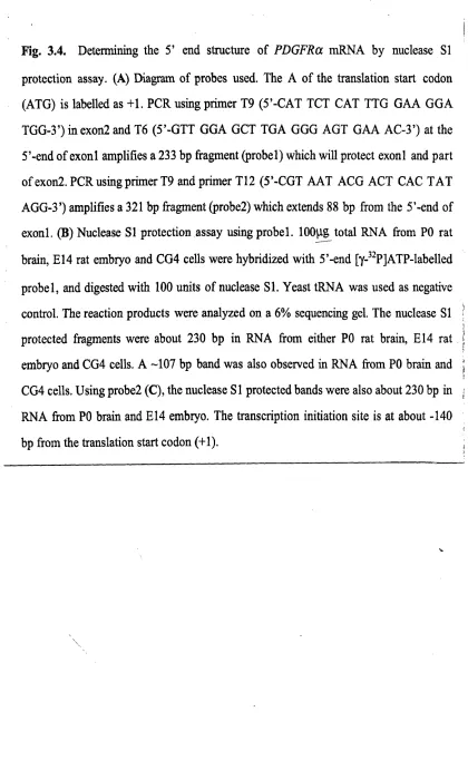
Fig. 3.4. Determining the 5’ end structure of PD G F R am R N A by nuclease SI
protection assay. 94
Fig. 3.5. Two possibilities that initiate specific expression of PDGFRa in OLPs. 95 Fig. 4.1. Analysis of CEPH 449C2 and CEPH 29E11 YACs carrying human
PDGFRa. 116
Fig. 4.2. Determination of mating type in window yeast strains carrying 449C2
and 29E11 YACs by PCR. 117
Fig. 4.3. Analysis o f 449C2 and 29E11 YACs transferred into window strains
byPFG E. 118
Fig. 4.4. Two-step gel purification of intact YAC DNA for microinjection. 119 Fig. 4.5. Genotyping of the human PDGFRa YAC transgenic mice by PCR. 120
Fig. 4.6. Genotyping o f the human PDGFRa YAC transgenic mice by
Southern blots. 121
Fig. 4.7. Determining the numbers of integrated YAC transgenes in transgenic
lines. 122
Fig. 4.8. Expression of endogenous mouse PDGFRa and human transgene-derived
PDGFR a outside the CN S. 123
Fig. 4.9. Expression of endogenous and transgene-derived PDGFRa transcripts
in embryonic spinal cord. 124
Fig. 4.11. Craniofacial and skeletal defects in homozygous PDGFRa nuW (KO)
mice were rescued by the human PDGFRa YAC. 126
Fig. 4.12. Comparison of (+/-), wt-YAC and KO-YAC lungs at PO. 127 Fig. 4.13. Abnormal lung development in KG-YAC mice. 128 Fig. 4.14. PDGFRa and MBP expression in spinal cords of wild type, PDGFRa
(+/-), YAC transgenic (wt-YAC), PDGFRa mA\ (KO)and KG-YAC mice. 129
Fig. 4.15. Spinal cord cell cultures of E14.5 wild type, PDGFRa wmW (KG) and
KG-YAC embryos. 130
Fig. 5.1. Site of origin of PD GFRct GLPs relative to the Pax6- and 2-positive
domains o f the wild type spinal cord. 147
Fig. 5.2. Development o f PDGFRot GLPs in wild type and Sey/Sey spinal cords. 148
Fig. 5.3. Cell cycle dynamics of PDGFRot GLPs analyzed by BrdU incorporation
in vivo. 149
Fig. 5.4. Site o f origin of PDGFRot GLPs in the ventral VZ o f Sey/Sey spinal cord
shifts dorsally relative to wild-type. 150
Fig. 5.5. Expression o f LIM-domain transcription factors in E9.5 wild-type and
Sey/Sey spinal cords. 151
Fig. 5.6. Expression of LIM domain transcription factors in E l 0.5 wild type and
Sey/Sey spinal cords. 152
Fig. 5.7. Expression of LIM domain transcription factors in E l 1.5 wild type and
Sey/Sey spinal cords. 153
Fig. 5.8. Expression o f LIM domain transcription factors in E l3.5 wild type and
Sey/Sey spinal cords. 154
Fig. 5.9. Gligodendrocytes develop normally in explant cultures of Isll(-/~) spinal
LIST OF TABLES
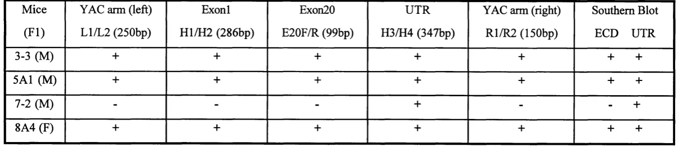
Table 2.1. Preparation of antisense RNA probes for in situ hybridization 79 Table 4.1. Summary of genotyping of human PDGFRa YAC transgenic mice 131
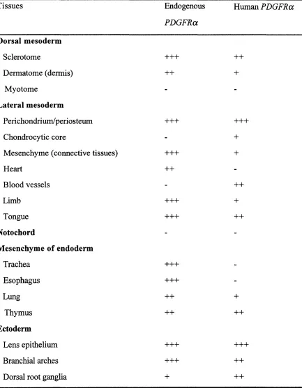
Table 4.2. Expression of endogenous mouse PDGFRa and the human PDGFRa
ABBREVIATIONS
BCIP 5-bromo-4-chloro-3-indoIy 1-phosphate
bp base pair
BrdU bromodeoxyuridine
CNS Central nervous system
DEPC diethylpyrocarbonate
DIG Digoxygenin
DM EM Dulbecco’s modified Eagle’s medium
DTT dithiothreitol
EBSS Earle’s balanced salt solution
ECO Extracellular domain
PCS Fetal calf serum
FGF Fibroblast growth factor
GC Galactocerebroside
IN Intemeuron
kb kilobase
KG Knock out
LMP Low melting point
M Molar
MBP Myelin basic protein
M N Motor neuron
mRNA Messenger RNA
NBT Nitroblue tétrazolium salt
OL Oligodendrocyte
OLP Oligodendrocyte progenitor
PBS Phosphate-buffered saline
PCR polymerase chain reaction
PDGFRa platelet-derived growth factor receptor alpha
PNS Peripheral nervous system
Sey Small eye
Shh Sonic hedgehog
TEMED T etraethy Imethy lenediamine
TGF Transforming growth factor
UTR Untranslated region
VZ Ventricular zone
Chapter 1
1.1. Initial formation of the neural structure: from the neural plate
to the neural tube
Neurulation is one of the most widely studied morphogenetic processes during embryonic development. The entire nervous tissue develops from the ectoderm. Signals from the notochord are thought to induce the formation of neural plate from ectoderm. In mouse embryos, the neural plate is first distinguishable soon after Embryonic day 7.5 (E7.5). Subsequently, the neural plate changes shape dramatically. The neural plate deepens to form the neural groove and its lateral edges rise to form the neural folds. A t this stage, in cross section, the neuroectoderm comes to resemble a “V” with the “hinge point” being the midline overlying the notochord. The cells of the medial hinge point will afterwards give rise to the floor plate. At about E8 to E8.5, the neural folds fuse along the dorsal midline at the level of the fourth and fifth somites. Then the neural tube is formed. Subsequently, fusion proceeds both rostrally and caudally in a zipper-like fashion from the cervical region of the neural tube. The anterior open end of the neural tube (anterior neuropore), is closed completely at about E9 (15-20-somite stage), whereas the posterior open end of the neural tube (posterior neuropore), remains open until ElO to E l 0.5 (32-somite stage).
After the neural tube has formed, the neuroepithelial cells continue to divide and the wall of the neural tube becomes even thicker. In the anterior region of the neural tube, the neuroepithelial cells rapidly divide and afterwards differentiate into primitive nerve cells or neuroblasts which form an easily recognized layer (subventricular zone). In the posterior region o f the neural tube — the spinal cord, the neuroepithelial cells give rise directly to post-mitotic neuronal progenitors. In both cases, the immature neuronal progenitors migrate outwards to their final places along the processes of radial glia. They then differentiate and extend their axons to their target cells.
along the anteroposterior neural tube — the forebrain, midbrain, hindbrain and spinal cord. It is thought that the dorsoventral polarity of the neural plate is also established early in development before closure of the neural tube. At the cellular level, the differentiation o f specific neurons in the neural tube follows a stereotyped dorsoventral pattern. Motor neurons are located ventrally in the spinal cord, whereas commisural neurons and the neural crest cells arise in dorsal positions.
Once the neural folds are formed, the cells of the neural crest temporarily appear at the two lateral folds. After the neural folds have fused, the neural crest cells separate fi-om the folds. This population of cells originates in the dorsal part of the neural tube. Afterwards, they form a continuous plate between the neuroepithelium and the surface ectoderm. In mouse embryos, neural crest cells first appear on the dorsal surface o f the neural tube at the level corresponding to the third or fourth somite. Thereafter, they both emerge and migrate in a rostral-to-caudal sequence. Neural crest cells migrate along two pathways: a ventral pathway through the rostral half o f the somites and a dorsolateral pathway between the dermamyotome and the epidermis. Once they arrive their locations, neural crest cells differentiate into a wide variety of cell types, such as, bone and cartilage, melonocytes, glial cells including Schwann cells, and sensory neurons.
1.2. Induction of the neural plate: studies in BMP signaling in
Xenopus
The development of the vertebrate nervous system requires induction of neural plate from undifferentiated ectodermal cells. Spemann and Mangold (1924) demonstrated that a small region of the Xenopus embryo, the organizer or dorsal lip, could direct neighbouring tissues to form dorsal mesodermal fates, such as notochord and muscle. The organizer grafted to the ventral side of another embryo can induce a complete secondary axis containing the central nervous system (Spemann and Mangold, 1924). This then raises the question of how the nervous system is induced.
Dissociation of blastula-stage ectoderm into single cells, which presumably prevents intercellular signaling, is sufficient to elicit the formation of neural tissue (Godsave and Slack, 1989; Sato and Sargent, 1989). This observation suggests the possibility that neural induction might result from the inactivation of a signaling pathway that represses neural fate. Members of a branch of the transforming growth factor-13 (TGF-p) -like protein family, the bone morphogenic proteins (BMPs) were suggested to mediate this repressive signal. In Xenopus embryonic development, BMP4 is a strong ventralizing molecule that is able to overcome the dorsalizing effects of activin and o f the Spemann Organizer itself (Dale et al., 1992; Jones et al., 1992). BMP7, which heterodimerizes with BMP4, is expressed widely in the gastrula and is also considered an important component o f the ventralizing signal (Aono et al., 1995; Hawley et al., 1995). Besides the ventralizing activity o f BMPs, an intact BMP signaling pathway is also required for the ectodermal patterning. In situ hybridization shows that BMP4 is present in the entire animal cap at the start of gastrulation. At later stages, BMP4 transcription disappears from the portion of the ectoderm that becomes the neural plate (Fainsod et al., 1994; Hemmati-Brivanlou and Thomsen, 1995).
Xenopus animal cap explants from the early gastrula develops as epidermis (ventral
neural tissue (dorsal ectoderm). Interestingly, soluble BMP4 can induce epidermis in dispersed cells and suppress neural tissue formation (Wilson and Hemmati-Brivanlou, 1995). Microinjection of dominant-negative BMP receptor (Xu et al., 1995a), or dominant-negative ligands (Hawley et al., 1995) or BMP4 antisense RNA (Sasai et al., 1995) causes animal cap explants to develop as neural tissue. Thus, endogenous BMP4 present in the animal cap appears to repress neural development. Similar evidence has also been found in Drosophila. Drosophila embryonic dorsal-ventral patterning is accomplished partly through the action of Decapentaplegic (Dpp), which is a TGF-p member most closely related to vertebrate BMP2 and BMP4. The loss-of-function phenotype o f dpp mutation involves expansion of the neurogenic ectoderm at the expense of epidermis (Irish and Gelbart, 1987), whereas, ectopic expression o f dpp leads to the expansion o f epidermis and reduction of the neurogenic ectoderm (Ferguson and Anderson, 1992a; Ferguson and Anderson, 1992b; Wharton et al., 1993).
Therefore, an attractive hypothesis is that the diffusible organizer signals might neuralize ectoderm and dorsalize mesoderm by antagonising the ventral BMP signaling pathway. Support for this idea has come from the demonstration that three secreted factors produced by Spemann’s organizer, noggin, follistatin and chordin, can act in this manner. Noggin is the first molecule shown to have the properties expected of an organizer signal (Smith and Harland, 1992). Noggin can bind to BMP4 and block its binding to cognate cell-surface receptors thus interrupting BMP4 signaling (Zimmerman et al., 1996). Chordin is a soluble protein secreted by organizer cells during gastrulation (Sasai et al., 1994). Chordin also induces neural tissue (Sasai et al., 1995) by specifically binding to mature BMP4 proteins and antagonising BMP4 signaling pathway (Piccolo et al., 1996; Sasai et al., 1995). Likewise, Follistatin is expressed by organizer cells, and binds to and inhibits BMPs (Hemmati-Brivanlou et al., 1994).
epidermal inducer. Understanding of this pathway requires identification of more downstream components and investigation of combined function analysis.
1.3. Pattern formation of the neural tube: two independent signaling
system s
Development o f the vertebrate CNS is characterised by the formation o f the neural tube with distinct anteroposterior (A-P) and dorsoventral (D-V) polarity. The signaling system which controls the A-P pattern establishes the formation o f the forebrain, midbrain, hindbrain and spinal cord. The second signaling system patterns the neural plate along its mediolateral axis, which later becomes the dorsoventral axis of the neural tube.
1,3A, Pattern formation along the anteroposterior axis o f the neural tube
Recent studies have shown that noggin (Lamb et al., 1993), follistatin (Hemmati- Brivanlou et al., 1994) and chordin (Sasai et al., 1995), secreted proteins expressed in the organizer, are potential candidates for anterior neural inducers, since all of them induce neural tissue that is anterior in character. No direct neural inducer isolated so far has the ability to induce posterior neural tissue such as spinal cord. These observations are consistent with the concept of the primary neural inducing signal (activation) in the two- step model. Recently, three kinds of secreted protein factors have been suggested as candidate molecules for the posterior transformation signal (the second step).
1.3.1.1. FGF family and posterior neural induction
Fibroblast growth factor (FGF), previously studied as a mesoderm inducer, has also been proposed to be a neural inducer. Basic FGF (bFGF) is capable o f specifically inducing dissociated ectoderm cells at the gastrula stage of Xenopus embryo to produce CNS neurons (Kengaku and Okamoto, 1993). In chick embryos, FGF-soaked beads can induce expression o f the early neural markers in extraembryonic epiblast cells (Alvarez et al., 1998; Storey et al., 1998). Inhibiting FGF signaling by the dominant negative FGF receptor also prevents neural induction in animal cap explants (Launay et al., 1996).
Xenopus (Pownall et al., 1996). In chick embryos, FGF-soaked beads can specifically
induce expression oïcash4 and Sax-1, which are markers of the early posterior nervous system, but not that of anterior neural markers (Henrique et al., 1997; Storey et al., 1998). Thus, it is possible to derive posterior neural fates from prospective anterior regions o f neural tissue. These findings have provided strong evidence for the function of the second signal (transformation).
However, the two-step model is not fitted to the following findings. bFGF can mimic the organizer action by inducing Xenopus ectoderm cells in culture to express position- specific neural markers in a dosage-dependent manner, i.e., with lower doses eliciting more anterior markers and higher doses more posterior markers (Kengaku and Okamoto,
1995). Therefore, it seems that bFGF alone can drive both steps (induction and transformation) by establishing a concentration gradient. Moreover, in chick embryos, FGF signaling can induce expression of early posterior neural genes in the absence of anterior neural tissue (Storey et ah, 1998).
1.3.1.2. Retinoid acid and anteroposterior neural patterning
Retinoid acid (RA) was first proposed to be involved in neural development when it was demonstrated that exogenously applied RA produces a concentration-dependent truncation of anterior and enhancement of posterior structures in Xenopus embryos (Durston et ah, 1989; Sive et ah, 1990). In Xenopus embryo at the neurula stage, RA concentration in the posterior end is 10 times higher than in the anterior end (Chen et al., 1994). This concentration gradient implys the positional induction o f RA in anteroposterior axis formation. RA can mimic the action o f endogenous signals involved in inducing posterior gene expression in the Xenopus nervous system (Sharpe, 1991). If animal caps expressing noggin are treated with RA, expression of anterior neural markers is lost while expression of posterior markers is maintained (Papalopulu and Kintner,
al., 1997). These findings suggest that retinoid signaling is required for the expression of posterior neural markers and the correct spatial restriction of anterior markers.
1.3.1.3. Wnt signaling and posterior neural induction
The Wnt gene family encodes a group of cell signaling molecules that play important roles in vertebrate development (Parr et al., 1993). In Xenopus, Xwnt-3a is expressed in the neural ectoderm of the early neurula (Wolda et al., 1993). This expression indicates that Xwnt-3 A may participate in patterning the neural axis. Coinjection o f Xwnt-3a and noggin or follistatin mRNAs induces posterior neural markers in animal caps while
Xwnt-3a alone can not induce neural tissue (McGrew et al., 1995). These findings
demonstrate that Wnts may synergize with noggin and follistatin to provide posteriorizing signals during development of the nervous system. Thus, Wnt-3A is another good candidate for a posterior transformation signal.
In summary, noggin, follistatin and chordin which are expressed in the organizer during early development are reasonable candidates for neural inducers working at the activation step of the two-step model. FGFs, RA and Wnt-3A seem to be the transforming signals to posteriorize neural tissues.
1.3.2. Pattern formation along the dorsoventral axis o f the neural tube
1.3.2.1. Diversity and pattern in the ventral neural tube: Shh signaling pathway
neuron differentiation and an increase in the total number of motor neurons. Floor plate grafts can mimic the effect of the notochord. These results suggest that the notochord is the source of two inductive signals: a local signal that induces floor plate differentiation and a longer-range signal that induces other neural cells (Yamada et al., 1993).
An excellent candidate for this inducing molecule emanating from the notochord is the secreted protein Sonic hedgehog (Shh) (Echelard et al., 1993; Krauss et al., 1993; Roelink et al., 1995). Shh is a vertebrate homolog of the Drosophila segment polarity gene hedgehog (hh) (Lee et al., 1992). In situ hybridization and immunostaining have shown
that Shh is expressed in the notochord and the floor plate in early embryonic development (Echelard et al., 1993; Marti et al., 1995b). Ectopic Shh expression under Wnt-1 enhancer activates floor plate gene HNF-3p expression in the brain (Echelard et
al., 1993). In addition, antibodies that inhibit Shh signaling in vitro block the ability of the notochord and floor plate to induce ventral cell types (Ericson et al., 1996; Marti et al., 1995a).
The direct role o f Shh in normal embryonic patterning was investigated in Shh gene mutant mouse (Chiang et al., 1996). Shh mutant mice were generated by targeted disruption of Shh to produce a truncated protein. Homozygous Shh mutant embryos show the absence of a morphologically distinct floor plate and rostral loss o f notochord tissue. These findings demonstrate that Shh is required for the induction of floor plate and maintenance of notochord. In the Shh mutant neural tube, expression o f Pax3, Pax6 and Paxl expands ventrally to the ventral midline. Thus, Shh plays a direct role in dorsoventral patterning o f the neural tube. Furthermore, Isll expressing motor neurons are also absent from the Shh mutant neural tube. All these studies show that Shh is necessary and sufficient for the induction of ventral cell types.
low concentration threshold (0.4nM in vitro) to convert naive neural plate cells into ventralized progenitor cells. The loss of Pax3 and Pax7 expression in the medial region of the neural plate reflects this conversion process (Ericson et al., 1996; Tremblay et al.,
1996). Ventralized progenitors at the midline of the neural plate respond to a high concentration of Shh with the generation of floor plate cells (Roelink et al., 1995). Moreover, Shh signaling induces ventralized progenitors to give rise to motor neurons at an intermediate Shh concentration threshold (>1.2nM in vitro) (Ericson et al., 1996). A t the onset of motor neuron differentiation in vertebrates, the notochord is displaced ventrally and is no longer close to the neural tube. Since Shh is also expressed in the floor plate cells once they have been induced (Echelard et al., 1993; Marti et al., 1995b), the Shh that is required to convert ventralized progenitors into motor neurons is most likely to derive from the floor plate (Ericson et al., 1996). However, ventralized progenitors generate Liml/Lim2 intemeurons rather than motor neurons when the late period of Shh signaling is blocked. These studies suggest that the identity and pattern of ventral cell types in the neural tube is controlled largely by Shh signaling, through actions at multiple concentration thresholds. It will be very interesting to investigate how small differences in extracellular Shh concentration generate distinct neural cell types.
1.3.2.2. Diversity and pattern in the dorsal neural tube
Pax3, Pax7 and Msx are expressed in the dorsal spinal cord. Dorsalin-1 (dsl-1), a novel
BMP-like member of the TGF-p superfamily, is also expressed in the dorsal region of the spinal cord (Easier et al., 1993). Cultured ventral neural plate explants from stage 10 chick embryos do not show any Msx and dsl-1 expression and very low Pax3 expression. Thus, cells in the ventral neural plate explants that have been exposed to notochord-derived signals do not express definitive dorsal cell markers (Liem et al.,
1995). Whereas, culture of intermediate and dorsal neural plate explants that have not been exposed to notochord-derived signals shows high Msx, dsl-1 and Pax3 expression. Certain genes characteristic o f dorsal neural tube cells are acquired in the absence of ventralizing signals (Liem et al., 1995).
However, neural crest cell differentiation suggests the existence of dorsalizing signals from epidermal ectoderm. Neural crest cells have been shown to arise from the neural folds and subsequently migrate from the dorsal neural tube towards their target sites (Bronner-Fraser and Fraser, 1989; Bronner-Fraser and Fraser, 1988; Serbedzija et al., 1989). Grafting o f the prospective neural plate in chick embryos into tissue culture does not show expression of HNK-1, a marker of neural crest cells. Thus, early neural plate alone is unable to form neural crest derivatives (Selleck and Bronner-Fraser, 1995). However, coculturing either early (stage 4-5 chick embryos) or later (stage 6-10 chick embryos) neural plate plus epidermis can induce the formation of the neural crest derivatives (Liem et al., 1995; Selleck and Bronner-Fraser, 1995).
et al., 1993), Liem et al. (1995) have tested functions of other members o f TGF-P family, especially BMP4 and BMP7, in dorsal induction. BMP4 and BMP? are expressed in the lateral neural plate. Afterwards, BMP4 and BMP? expression is lost from the majority of the epidermal ectoderm, except BMP4 expression in the region directly above the dorsal neural tube. BMP4 and BMP? are also expressed within the caudal dorsal neural tube. Importantly, BMP4 or BMP7 induce the expression of dorsal markers, such as Pax3, Dsl-1, Msx, slug and HNK-1, in ventral neural plate explants. Thus, BMP4 and BMP7 can mimic the ability of epidermal ectoderm to induce the expression of markers of dorsal neural tube and to promote the differentiation of neural crest cells (Liem et al., 1995).
1.3.2.3. Common features in patterning dorsal and ventral neural tube
There are common features in pattern formation in dorsal and ventral neural tube. Both dorsal and ventral cell types are induced by signals secreted from non-neural tissues: the epidermal ectoderm and the notochord, respectively. Both dorsal and ventral signaling factors (BMPs and Shh) are initially expressed by non-neural tissues, then by cells at the midline of the neural tube ( the roof plate and floor plate ).
A morphogen is a diffusible molecule that establishes a gradient away from its source and triggers distinct cellular responses at different distances (Nellen et al., 1996; Reilly and Melton, 1996). Shh acts as a morphogen to induce the differentiation of ventral cell types. Are BMPs also morphogens? In Drosophila, the BMP homolog Decapentaplegic (Dpp) has been shown to act as a morphogen. Ectopic expression of dpp in wing disc cells induces the transcription of its target genes omb and spalt at the different distances from the source (Nellen et al., 1996). These results suggest that omb and spalt genes respond to differrent threshold concentration of Dpp.
fates are induced by noggin in a dose-dependent manner, with low doses inducing most strongly, whereas higher doses repressing dorsal fates (Knecht and Harland, 1997). Since BMP4 antagonizes the action of noggin, BMP4 may also act in a concentration- dependent manner. In fact, the highest concentrations of BMP4 induce epidermis (Wilson and Hemmati-Brivanlou, 1995), slightly lower concentrations induce neural crest cells (LaBonne and Bronner-Fraser, 1998) and dorsal neural fates (Knecht and Harland, 1997). In chicken, BMP4 induces a graded response in mediolateral patterning of somitic and lateral mesoderm (Tonegawa et al., 1997). All this evidence suggests that BMPs also act as morphogens in pattern formation. Shh and BMPs may induce ventral and dorsal cell types in a similar concentration-dependent manner.
Shh and BMPs act alone to induce ventral and dorsal cell fates in the neural tube. In addition, Shh and BMPs have antagonistic actions that specify intermediate cell fates (Basler et al., 1993; Liem et al., 1995). However, Shh and BMPs also cooperate to determine neural cell fates in the rostral diencephalon. In explants of rat intermediate neural plate, prechordal mesoderm can induce the expression of rostral diencephalic markers, BMP7 and Nkx2.1 (Dale et al., 1997). Like notochord, prechordal mesoderm expresses Shh but also BMP7. Notochord and caudal neural plate explants cultured with BMP7 generate rostral diencephalic ventral midline cells, whereas when BMP7 activity is blocked with an anti-BMP7 antibody, only floor plate cells are induced by notochord (Dale et al., 1997). Thus, the induction o f rostral diencephalic ventral midline cells by prechordal mesoderm requires the actions of both Shh and BMP7.
1.3.2.4. Transcription factors and pattern formation o f the neural tube
A. HNF-3p and floor plate development
Hepatocyte nuclear factor (HNF) was originally identified as a transcription factor for liver-specific genes (Lai et al., 1991). HNF-3p contains the Drosophila forkhead (fkh) domain which encodes a well-conserved 101 amino acid DNA-binding domain.
In mouse embryonic development, HNF-Sp is expressed in the notochord first and then
in the floor plate (Monaghan et al., 1993). Since HNF-3P expression precedes that of Shh in both the midline mesoderm and the ventral neural tube, the initial activation of
Shh expression may be regulated by HNF-3p (Echelard et al., 1993). Ectopic expression
of HNF-3p under the control of the midbrain/hindbrain specific En-2 promoter causes
spatial expression of floor plate-specific genes {BMP-1 and HNF-3a) in transgenic
brains (Sasaki and Hogan, 1994). These findings suggest that HNF-3p fimctions as a regulator of floor plate development. In HNF-3P null mutant embryos, no notochord or floor plate is ever formed (Ang and Rossant, 1994). Although neural tissue is formed, Shh expression is not detected in the neural tube. The D-V patterning o f the neural tube is severely affected. The expression regions of Pax3 and Pax6 have also been extended ventrally. These results demonstrate that HNF-3p is a critical component in initiating the formation o f the notochord.
In the Shh mutant embryos, the early expression of HNF-3p is initiated at a low level in
the notochord. However, HNF-3p expression in the axial mesoderm is completely absent and its expression in the ventral neural tube is never initiated (Chiang et al., 1996). These findings show that Shh is not necessary for the initiation o f HNF-3P
expression. But once HNF-3P expression is initiated, Shh and HNF-3p expression become interdependent.
Through the homology to Drosophila genes which control early development, a large number of vertebrate genes have been identified, such as homoebox genes and paired box genes. The paired box was first identified in Drosophila segmentation genes (Bopp et al., 1986; Frigerio et al., 1986). After that, paired box genes were also isolated in the mouse (Deutsch et al., 1988). Until now, nine Pax genes have been identified, referred to as Paxl to Pax9 (Mansouri et al., 1994).
The paired box is a 384 bp DNA sequence which encodes 128 amino acid DNA binding domain (Bopp et al., 1986). Other conserved sequence motifs are the octapeptide and the paired-type homeodomain which is 61 amino acids in length. Secondary structure predictions indicate that the paired domain contains three a-helices (Epstein et al., 1994). The structure of paired domain and homeodomain indicates that Pax proteins act as transcription factors that regulate gene expression (Chalepakis et al., 1991; Czerny et al., 1993; Epstein et al., 1994; Xu et al., 1995b; Zannini et al., 1992). Pax genes may be involved in the control of morphogenesis and pattern formation, since these genes are expressed in a spatially and temporally restricted pattern during embryonic development.
Pax genes show restricted expression regions along the dorsoventral neural tube during
markers E nl and Evxl are coexpressed with Pax2 in a combinatorial manner to regulate intemeuron identity.
Signals secreted from notochord regulate the dorsoventral expression pattern of Pax genes in the neural tube. In the chick embryos, after removing the notochord, the expression regions of PaxS and Pax6 extend further ventrally (Goulding et al., 1993). On the other hand, a dorsal shift in expression domains of PaxS and Pax6 occurs in regions of the spinal cord adjacent to the site where a second notochord has been placed. One of the candidate signals from the notochord is Shh (Echelard et al., 1993; Ericson et al.,
1997; Krauss et al., 1993; Roelink et al., 1995). Graded Shh signaling is sufficient to establish graded Pax6 expression in the neural tube (Ericson et al., 1997). However, Pax6 expression is not initiated by Shh signaling. Using in vitro explant culture and
grafting experiments, Pituell and colleagues (1999) showed that Pax6 expression can be induced independantly of the presence of Shh-expressing cells when neural plates are maintained in culture in the presence of paraxial mesoderm. Pax6 expression disappears from the neural tube when a somite is replaced by presomitic mesoderm (Pituello et al., 1999). These results suggest that somites initiate and activate Pax6 expression, but Shh plays a major role in regulating the level of Pax6 expression.
The functions of Pax genes have been investigated in Pax gene mutant mice. The mutation of PaxS in Splotch mice leads to defects in neural crest derivatives and in limb muscle (Franz, 1990; Moase and Trasler, 1989). PaxS may play an important role in neural crest development. Similarly, homozygous Pax7 mutant mice die shortly after weaning (Mansouri et al., 1996). They exhibit malformations in facial structures involving the maxilla and nose. Pax7 also plays a vital role in neural crest development. On the other hand, Tremblay and colleagues (1996) produced transgenic mice expressing PaxS in the entire neural tube under the Hoxb4 promoter region A enhancer. In this
transgenic mouse, floor plate is absent in the neural tube and expression of Shh and HNF-Sp is also missing in the floor plate area (Tremblay et al., 1996). These results
Pax6 mutant mice and rats display an eyeless phenotype in the homozygous condition
(Hill et ah, 1991; Matsuo et al., 1993). Homozygous Small eye {Sey) mice lack eyes and nasal carvities and die soon after birth. These findings show that Pax6 plays a vital role
in eye development. Pax6 acts in eye development in a dosage dependent manner.
Overexpressing Pax6 in yeast artificial chromosome transgenic mice carrying the human Pax6 locus causes abnormalities of the eye (Schedl et al., 1996). In the neural tube of
Pax6 mutant mice, Nkx2.2 expression region which is at most ventral part o f the neural
tube expands dorsally (Ericson et al., 1997). In the hindbrain, the loss o f Pax6 causes an apparent transformation of hypoglossal motor neurons (expressing Isll and Isl2, and transiently Lim3 and Gsh4) into vagal motor neurons (expressing Nkx2.2, Isll and Gsh4) (Ericson et al., 1997; Osumi et al., 1997). In the cervical spinal cord, the total number o f motor neurons is decreased and somatic motor neurons are transformed into Sim l positive cell fate, which normally develop from the Pax6-negative domain adjacent
to the floor plate (Ericson et al., 1997). These results demonstrate that the loss of Pax6
results in a dorsal-to-ventral transformation in the identity of progenitor cells. For the intemeurons, there is a loss of a ventral population o f Pax2 expressing intemeurons in the Sey/Sey spinal cord. E nl expressing intemeurons are also not detected (Burrill et al.,
1997). In addition to neurons, the site of origin of oligodendrocyte progenitors is also affected. In Sey/Sey neural tube, the origin of oligodendrocyte progenitors is shifted dorsally (Sun et al., 1998 and this thesis). The time o f their appearance is also delayed up to a day.
C. Gli gene family and Shh signaling pathway
In Drosophila, the actions of Hh have been shown to be transduced by the activity of four genes: fuse ifu), specifying a putative protein-serine/threonine kinase; costal 2 {cos2'), identified as encoding a distant relative of the kinesin motor proteins; pka,
encoding protein kinase A; cubitus interruptus {Ci), a zinc finger transcription factor (Ruiz i Altaba, 1997).
Two candidate receptors for Hh have also been identified, encoded by genes known as patched (ptc), and smoothened (smo) (Alcedo et al., 1996; Hooper and Scott, 1989;
Nakano et al., 1989; van den Heuvel and Ingham, 1996). The work o f many labs has established the early events in the Hh signaling pathway. Normally, Ptc binds to and inhibits Smo function. Following Hh binding, Ptc is released from Smo, allowing the activity of Smo to be expressed. Acting through C0S2, PKA and FU, this triggers Ci- mediated activation of Hh target genes (Aza-Blanc et al., 1997; Robbins et al., 1997; Sisson et al., 1997).
In vertebrates, three homologs of Ci have been identified as GUI (Kinzler et al., 1988; Kinzler and Vogelstein, 1990), GH2 and GU3 (Hui et al., 1994; Ruppert et al., 1990; Walterhouse et al., 1993). In fi*og and mouse neural plate, GUI is expressed in midline cells at the time that they are induced to become floor plate, and in cells lateral to the midline (Hui et al., 1994; Lee et al., 1997; Sasaki et al., 1997). GH2 is expressed throughout the neural plate with the exceptions of the midline and GU3 is absent from the midline and shows a graded expression with highest levels laterally (Lee et al., 1997; Sasaki et al., 1997). Subsequently, the expression of GUI, GH2 and GU3 becomes restricted to overlapping but distinct demains along the dorsoventral axis in the neural tube. GUI is expressed in the ventral most domain adjacent to the floor plate, GU2 is restricted to ventral and intermediate regions, while GU3 is only expressed dorsally (Hui et al., 1994; Lee et al., 1997; Sasaki et al., 1997). The similar roles of vertebrate Shh to Drosophila Hh, the homology of vertebrate Gli genes to Drosophila Ci gene and the
GUI proteins, but not 0113, injected into frog embryos can induce ectopic expression of HNF-3p expression in floor plate. Glil binding site is required for the activity of the
minimal floor plate enhancer of HNF-3p in vivo (Sasaki et al., 1997). These results demonstrate that Glil is involved in induction of floor plate (Lee et al., 1997). Ectopic expression of Glil in transgenic mice under the En-2 promoter-enhancer in the dorsal midbrain and hindbrain suppresses dorsal cell markers and induces ventral cell markers (Hynes et al., 1997). Moreover, Glil injected into frog embryos induces ectopic ventral neuronal differentiation (Lee et al., 1997). In addition, ectopic Shh induces Glil expression in both Xenopus and mice (Hynes et al., 1997; Lee et al., 1997). Shh also can directly up-regulate Glil expression in a cell line (Sasaki et al., 1997). Thus, Glil may act as a target and mediator of Shh signaling in floor plate and ventral neuronal differentiation.
To the contrary, in frog embryos, only Glil can induce floor plate differentiation. GU2 and GU3 antagonise the effects of GUI. The same regulation is also observed in the induction of ventral cell differentiation in forebrain (Ruiz i Altaba, 1998). Glil and GU2 induce motor neuron differentiation, whereas Gli3 represses this induction. These results suggest that Glil and/or GU2 mediate the Shh induction of motor neurons and GU3 acts as a general repressor of Shh induction (Ruiz i Altaba, 1998). Thus it appears
that the functions of Glil and Gli2 are not conserved among species.
1.4. Cell fates determination in neurogenesis
1.4.1. Specification o f motor neurons and intemeurons in the neural tube
There are three general classes of motor neurons at hindbrain and spinal cord levels: the branchiomotor neurons that innervate muscles from the branchial arches; somatic motor neurons that innervate skeletal muscles, and visceral motor neurons that innervate parasympathetic and sympathetic neurons. The somatic motor neurons are subdivided into two groups according to the position of their cell bodies and the location of the muscles they innervate. Motor neurons in the medial part of the medial motor column (MMCm) project their axons to axial muscles that lie close to the vertebral column. Motor neurons in the lateral subdivision of the MMC (M M Cl) project their axons to body wall muscles. Motor neurons in the lateral motor column (LMC) innervate muscles in the limb. LMC can be subdivided into lateral LMC (LMCl) and medial LMC
(LMCm) according to motor neurons in them project to dorsal limb muscles and ventral
limb muscles, respectively.
Isll, Isl2 and Lim3, while motor neurons in M M C l coexpress Isll and Isl2. M otor neurons in LMCm express Isll and Isl2, while motor neurons in LM Cl express Isl2 and Lim l, and transiently express Isll (Tsuchida et al., 1994).
(Tanabe et al., 1998). Taken together, homeodomain transcription factor MNR2 is sufficient to direct somatic motor neuron cell fates in the vertebrate spinal cord, whereas Lim3 appears sufficient to specify V2 intemeuron fates. It will be interesting to identify other factors that specify neuronal differentiation in the future.
1,4,2, Glial cell fa te determination
Neurons and glia show great diversity with respect to position, morphology and physiological and biochemical properties. However, they can arise from common lineages in both the central nervous system and the peripheral nervous system (PNS). The relative simplicity of the Drosophila embryonic CNS makes it a suitable model system for the study of neuroglial lineages and the molecules that specify them.
In Drosophila, the origin of glia in the CNS is known from 3 types of progenitors. The longitudinal glia arise from the longitudinal glioblast (Jacobs et al., 1989). Londitudinal glia express repo which encodes a paired-like homeobox protein (Halter et al., 1995; Xiong et al., 1994). Repo is specifically expressed in most o f the glia in embryonic CNS and PNS from an early stage. Longitudinal glia also express the homeodomain protein encoded by the prospero (pros) gene (Doe et al., 1991) and the PI form of the Ets family o f transcription factor encoded by the pointed (put) gene (Klaes et al., 1994). Midline glia arise from midline mesectodermal progenitors (Klambt et al., 1991). Midline glia neither express repo nor pros, but they express P2 form of the Pnt protein (Klaes et al., 1994). Other glia arise from neuroblasts (NBs) which act as common progenitors for glia and neurons (Udolph et al., 1993). For example, the NBl-1 divides asymmetricly into a ganghon mother cell (CMC) and another NB. After several divisions, NBs generate a series of GMCs. Each CMC divides to produce either neuron or glia.
glide gene (Hosoya et al., 1995; Jones et al., 1995; Vincent et al., 1996). Drosophila
GCM is a transcription factor with novel DNA binding domain that recognises the motif AT(G/A)CGGGT (Akiyama et al., 1996; Schreiber et al., 1997). In the CNS o f loss-of- function gem mutants, nearly all glia (except midline gha) are missing. Strikingly, the presumptive ghal cells differentiate into neurons. To the contrary, after ectopic expression o f gem in presumptive neurons, many presumptive neurons are transformed into glia. In the PNS of loss-of-ftmction gem mutants, the presumptive glial cells are also transformed into neurons. Ectopic expression of gem in presumptive neurons also causes transformation of presumptive neurons into gha (Hosoya et al., 1995; Jones et al., 1995).
The genetic network underlying glial cell development in Drosophila is also studied. A t least three genes are known to be involved in ghal ceh differentiation. In repo putative null alleles mutant, gha progenitors originate normally and show normal migration behaviour. However, the differentiation of the presumptive gha seems to be severely affected (Halter et al., 1995). Pnt is also required for ghal cell differentiation (Klaes et al., 1994). Ectopic expression of Pnt in the lateral CNS causes additional glial-like cells. In addition, tramtraek (ttk) encodes two different zinc-finger-type transcription factors, ttkp69 and ttkp88. Ttkp69 is expressed in all CNS gha. Ttkp69 may act to block the
competence of cells to differentiate as neurons within the CNS (Giesen et al., 1997). Since the pnt-ttk double mutant phenotype (a fused commissure phenotype) is different from the gem mutant phenotype and ectopic gem expression is still capable to induce ectopic repo expression in pnt-ttk double mutants, the expression of repo, pnt and ttk is downstream o f gem (Giesen et al., 1997). Thus, GCM acts as a master regulator for ghal development. GCM may control ghal cell differentiation by concomitant activation of ghal differentiation (function of pnt and repo) and suppression o f neuronal differentiation (function of ttk).
repression o f mesoderm specific genes and in the loss of muscle morphology (Bemardoni et a l, 1998). These findings suggest that gliogenesis does not require a ground neural state, and that gem transcription must be tightly regulated in order to ensure correct embryonic development.
Mammalian homologs of gem have been isolated in mouse (Akiyama et al., 1996; Altshuller et al., 1996) and rat (Kim et al., 1998). The conserved DNA-binding GCM motif, has been identified. However, the mammalian gem is either expressed at very low levels during neuro- and ghogenesis, or not expressed in embryonic neural tissues (Altshuller et al., 1996; Kim et al., 1998). The fimction of gem, at least those isoforms discoverd to date, is not conserved from Drosophila to mammals. However, loss-of- function mutants of mammalian gem have yet to be described and these will be important for our understanding of gem function.
olfactory bulb to generate neurons (Doetsch et al., 1999). Whether these lineage studies identify dedicated progenitors or multipotent precursors presumably depends on how far back in the lineage tree are looked.
In mouse spinal cord, oligodendrocyte progenitor cells which express Platelet-Derived Growth Factor Receptor Alpha (PDGFRa) derive from the ventral ventricular zone as bilateral foci of cells (Noll and Miller, 1993; Ono et al., 1995; Pringle and Richardson, 1993; Yu et al., 1994). After that, they proliferate and migrate from the ventral spinal cord and finally spread through the whole spinal cord (Pringle and Richardson, 1993; Yu et al., 1994). The first oligodendrocyte progenitors appear in the ventricular zone on E12.5 in the mouse (E14.5 in the rat), shortly after the end of motor neuron production. Moreover, oligodendrocytes are induced in explant cultures of dorsal or intermediate neural tube at Shh concentration that overlap those required for motor neuron induction (Poncet et al., 1996; Pringle et al., 1996). These findings suggest that the same population of progenitor cells might give rise first to motor neurons and then to oligodendrocyte progenitors (Richardson et al., 1997). Support for this idea comes from the analysis of origins of oligodendrocyte progenitors and somatic motor neurons in the Pax6 mutant spinal cord (Sun et al., 1998 and this thesis). In Sey/Sey spinal cord, the
appearance of oligodendrocyte progenitors is delayed by about a day. Interestingly, the appearance of Isl2 and Lim3 expressing motor neurons which are also generated in the ventral ventricular zone is also delayed by up to a day. These results indicate a close relationship between Isl2/Lim3 motor neurons and oligodendrocyte progenitors, and also suggest that both cell types may derived from common neuron-oligodendrocyte progenitor cells.
1.5. Aims of this thesis
progenitor (OLP) development. In this thesis, I mapped the transcriptional start site of PDGFRa in the CNS and found that PDGFRa transcribes from the same site in the
CNS as in other tissues. To analyze the remote cis-acting regulatory elements that are required for PDGFi?a expression in the CNS, I generated transgenic mice byjpronuclear microinjection of a 380 kb yeast artificial chromosome (YAC) containing entire human PDGFRa gQViQ. This YAC transgene is faithfully expressed in OLFs in the spinal cord
and correctly expressed in most non-neural tissues. This YAC transgene can rescue the defect of oligodendrocyte development in PDGFRa null mutant mice. I also analyzed
functions of PDGFRa in craniofacial and lung development. In addition, to investigate factors that affect OLP development, I mapped origins o f OLPs in both wild-type and Pax6 mutant Small eye {Sey) mice spinal cords. A dorsal shift o f OLP origin was
Chapter 2
General chemicals and reagents were all purchased from Sigma-Aldrich Co Ltd or BDH Chemicals Ltd unless otherwise stated, and were Molecular Biology grade where available.
Restriction and DNA/RNA modifying enzymes were all purchased from Promega Ltd or New England Biolabs (UK) Ltd unless otherwise stated.
Bacterial media and yeast media components were obtained from Difco Laboratories Ltd, or from the in-house facility.
All radio-chemicals were purchased from Amersham International.
All tissue culture media components were purchased from GIB CO BRL Life Technologies Company.
2.1. Bacteriology
2.1.1. Bacterial strains^ growth and storage
All general cloning and sub-cloning was carried out using Escherichia coli (E. Cali) strain XL 1-Blue (supE44 h s d R ll recAl endAl gyrA46 thi relAl lac F ’ \proAB^ lacP LacZAM \5 In lO (Jef)]). Bacteria were grown at 37°C in Luria Broth (LB, lOg bacto-
tryptone, 5g bacto-yeast extract and lOg NaCl per liter) or on LB-agar plates containing LB with 15g/L bacto-agar. Ampicillin was added to the LB or molten LB-agar (after cooling to 55°C) at a final concentration of 25-lOOjLig/ml (lOOmg/ml stock in H2O,
0.22|xm filter-sterilised and stored in aliquots at -20°C). Liquid cultures were continually agitated in a rotating environmental shaker at 300 rpm.
For short term storage of bacterial strains and clones, they were stored on LB agar plates at 4°C for two or three weeks. For long term storage, 75% glycerol was added to overnight cultures to a final concentration of 15%. This mixture was stored in 1ml aliquots at -20°C or -80°C if necessary, for a few years.
2.1.2. Preparation and transformation o f electrocompetent bacteria
1ml of an overnight liquid culture of E. Coli strain XL 1-Blue (grown from a single colony) was transferred into IL of LB. This culture was grown until it was in mid-log phase (an absorbance of -0.6 at OD600nm), then chilled on ice and pelleted by centrifugation (4,000rpm, 4°C, 10 minutes). The bacterial pellet was gently resuspended in IL of ice-cold H2O, pelleted as before, resuspended in 500ml ice-cold H2O and
pelleted again. This pellet was then resuspended in 20ml ice-cold 10% (v/v) sterile glycerol, centrifuged as before, and finally resuspended in 2 .5ml ice-cold 1 0% (v/v)
bacteria can be transformed at frequencies >1 0^ transformed colonies per microgram of
supercoiled plasmid DNA.
For transformation of recombined plasmids into the electrocompetent cells, the bacteria aliquots were transferred on ice directly from liquid nitrogen , and thawed slowly before use. 2 0til of competent cells was transferred to a chilled disposable electroporation
cuvette (Bio-Rad Laboratories Ltd) which 2|il of the plamid DNA (~100pg) had been preloaded. Competent cells were mixed with the DNA with a gel-loading tip. The mixture was then pulsed using a Gene Puiser electroporator equiped with a Pulse Controller (Bio-Rad Laboratories Ltd) set to 2,500V, 2000, 25}iF. This resulted in a time constant of about 5.0 to 5.3 ms. The pulsed bacteria were transferred to 100|il prewarmed LB without ampicillin and incubated at 37°C for 30 minutes before plating on LB-agar-ampicillin plates.
When cloning into vectors where the multiple cloning site formed part of the (3- galactosidase gene, a blue/white colour screen could be used to pick colonies containing vectors with inserts. In this case each plate was spread with lOOjxl o f 1.25% (w/v) X-gal (5-bromo-4-chloro-3-indolyl-p-D-galactopyranoside) and lOmM IPTG (Isopropyl p- D-thiogalactopyranoside) and incubated at 37°C for 30 minutes before use. The X-gal
stock was 2.5% (w/v) in dimethyl formamide and was kept at -20°C in dark. The IPTG
stock was O.IM in H2O and was stored at 4°C. Colonies containing religated vector are
blue under these conditions, whereas colonies containing vectors with inserts remain white.
2.2. Molecular biology
The phenol stock was extracted with Tris-HCl pH 7.5 for DNA and pH 4.0 for RNA. The extracted phenol was stored in aliquots at -20°C and kept at 4°C for routine use. The chloroform stock was made by mixing chloroform with iso-amyl alcohol (24:1, v/v) and kept at 4°C in dark. The DNA/RNA samples dissolved in TE (lOmM Tris-HCl pH
8, ImM EDTA pH 8) or Diethyl Pyrocarbonate (DEPC)-treated water were extracted
with an equal volume of phenol/chloroform. The sample was then vortexed and centrifuged at 13,000rpm for 5 minutes. The upper aqueous phase was collected, re extracted with an equal volume of chloroform, and then vortexed, spun and collected as before.
2.2.2. Precipitation o f DNA/RNA with ethanol or isopropanol
0.1 volume o f 3M NaOAc pH 5.2 was added to the DNA/RNA samples and then 2.5 volumes of cold ethanol (or 0.6 volumes of isopropanol) was added. The samples were vortexed, kept on ice or at -20°C for 10 to 20 minutes and then centrifuged at 4°C, 13,000rpm for 10 minutes to precipitate the DNA/RNA. If necessary (i.e. less than 1-2 fig or a very low concentration of DNA), lOjig of glycogen (from muscles molecular biology grade, Boehringer Mannheim) was added to the DNA as a carrier before the precipitation. The DNA/RNA pellet was washed with 500|il 70% (v/v) ethanol, centrifuged briefly at 13,000rpm, air dried and resuspended in an appropriate volume of TE or DEPC-treated water.
2.2.3, Agarose gel electrophoresis o f DNA
lOx DNA loading buffer containing 0.25% (w/v) bromophenol blue, 0.25% (w/v) xylene cyanol FF and 15% (w/v) Ficoll (Type 400, Pharmacia) was added to the DNA samples. The gel was run at 5V/cm. The DNA was visualised on an ultraviolet (UV) transilluminator at 302nm and recorded using a CCD camera and thermal printer.
2.2.4, Gel purification o f DNA
The Geneclean II kit (Bio 101) was used to purify DNA bands from agarose gel. The DNA bands were cut out of the gel and collected in an Eppendorf tube. Three volumes of 6M sodium iodide was added, assuming a gel density of Ig/ml. The gel containing
DNA bands was incubated at 55°C for more than 5 minutes until the gel had melted, then chilled on ice. 5|i,l of the supplied silica matrix was then added. The suspension was vortexed and incubated on ice for 5 minutes. After pelleting in a microcentrifuge, the matrix with bound DNA was washed 3 times in the supplied wash buffer and resuspended in an appropriate volume of TE. The DNA was eluted from the matrix at 55°C for 5 minutes. Then the matrix was spun again and the DNA-containing supernatant was recovered.
2.2.5. Small scale preparation o f plasmid DNA by alkaline lysis (miniprep)