Regulation of caspase activation during
programmed cell death
Victoria Haigh Cowling
A thesis submitted to University College London,
University of London for the degree of Doctor of
Philosophy
Signal Transduction Lab
Cancer Research UK
ProQuest Number: U642472
All rights reserved
INFORMATION TO ALL USERS
The quality of this reproduction is dependent upon the quality of the copy submitted.
In the unlikely event that the author did not send a complete manuscript and there are missing pages, these will be noted. Also, if material had to be removed,
a note will indicate the deletion.
uest.
ProQuest U642472
Published by ProQuest LLC(2015). Copyright of the Dissertation is held by the Author.
All rights reserved.
This work is protected against unauthorized copying under Title 17, United States Code. Microform Edition © ProQuest LLC.
ProQuest LLC
789 East Eisenhower Parkway P.O. Box 1346
Acknowledgements
I have been able to carry out this PhD only with the assistance and support o f many people. I would like to thank my supervisor Julian Downward and the past and present members of the Signal Transduction Lab.
I would like to thank my former supervisor Gerard Evan and the BCN Lab. The support of my family and friends has been invaluable.
Abstract
Most triggers of programmed cell death or apoptosis promote cytochrome c release from the mitochondria into the cytosol which initiates the activation of a panel of pro-apoptotic caspases. Activation of caspase-8 in this pathway is not well defined but may be important because it activates Bid which promotes further cytochrome c release. A range of
experimental approaches have suggested different mechanisms for caspase-8 activation in this pathway. In order to identify the proteases directly responsible for caspase-8
activation, caspase-8 cleaving activity was purified from cytochrome c activated cytosolic extracts. Caspase-6 was found to be the only soluble protease in cytochrome c activated cytosolic extracts that has significant caspase-8 cleaving activity. Furthermore, caspase-6 was sufficient to induce Bid-dependent cytochrome c releasing activity in cell extracts. Inhibition of caspase-6 activity in cells significantly inhibited caspase-8 cleavage and apoptosis, therefore establishing caspase-6 as a major activator of caspase-8 in vivo and confirming that this pathway can have a critical role in promotion of apoptosis. The intrinsic activity of caspase-6 was found to be dependent on removal of the short
Table of contents
Chapter 1 Introduction ____________________________________________________ 14
1.1. Programmed cell death_________________________________________ 14
1.1.1. Programmed cell death is a physiological process_____________________________________ 14 1.1.2. Discovery of ap o p to sis___________________________________________________________ 15 1.1.3. Clinical relevance of ap o p to sis_____________________________________________________ 16 1.1.4. Initial discoveries of the protein families involved in apoptosis _________________________ 17
1.2. Caspases have a major role in apoptosis_____________________________20
1.2.1. Caspase fa m ily __________________________________________________________________ 20 1.2.2. Caspase activation of cytokines_____________________________________________________20 1.2.3. Caspase activation during ap o p to sis________________________________________________ 22 1.2.4. Caspase-independent apoptotic pathw ays____________________________________________23
1.3. Caspase activation_____________________________________________ 24
1.3.1. Procaspase structure._____________________________________________________________ 24 1.3.2. Active caspase stru c tu re__________________________________________________________ 25 1.3.3. Auto cleavage of caspases_________________________________________________________ 27 1.3.4. Caspase mediated trans cleavage of c a sp a se s________________________________________ 27 1.3.5. Methods used to define caspase cleavage of caspases_________________________________ 28 1.3.6. Other proteases mediate trans cleavage of caspases.___________________________________30 1.3.7. Caspase post-translational m o d ificatio n _____________________________________________ 30 1.3.8. Caspase localisation _____________________________________________________________ 32
1.4. Caspase inhibition_____________________________________________ 33
1.4.1. Procaspase activation and mature caspase activity can be inhibited______________________ 33 1.4.2. Inhibition of procaspase cleavage by caspase h om ologues_____________________________ 33 1.4.3. Regulation of F L I P s _____________________________________________________________ 35 1.4.4. Caspase inhibition by lA P s ________________________________________________________ 36 1.4.5. Caspase inhibition by CrmA and p 3 5 _______________________________________________ 37 1.4.6. Down regulation of caspase expression in tumour c e lls ._______________________________ 37
1.5. The role of non-caspase proteases in apoptosis_______________________ 38
1.6. The Bcl-2 family integrates pro- and anti-apoptotic signals____________ 41
1.6.1. Bcl-2 fa m ily ____________________________________________________________________ 41 1.6.2. Bcl-2 family function ____________________________________________________________ 42 1.6.3. The Bcl-2 family regulates cytochrome c re le a s e _____________________________________ 43 1.6.4. A wide range of signals modulate the BH3-only proteins______________________________ 44 1.6.5. Role of Bcl-2 family members at the endoplasmic reticulum and nuclear m em brane_______ 46
1.7. Inhibitor of apoptosis proteins (TAP)______________________________ 46
1.7.1. TAP family______________________________________________________________________ 46 1.7.2. XIAP inhibits caspase activity_____________________________________________________ 47 1.7.3. c-IA Pl and C -IA P2______________________________________________________________ 49
1.8. Cytochrome c induced apoptosis pathway__________________________ 49
1.8.1. Initiators of apoptosis promote cytochrome c re le a s e __________________________________49 1.8.2. Initial discovery__________________________________________________________________ 52 1.8.3. Release of Holocytochrome c initiates apoptosis______________________________________ 52 1.8.4. Cytochrome c triggers formation of Apaf-l-caspase-9 h oloenzym e_____________________ 53 1.8.5. The apoptosome recruits and activates effector caspases_______________________________ 54 1.8.6. Positive feedback loop of caspase activation and cytochrome c release___________________ 55
1.9. Other mitochondrial proteins promote apoptosis____________________ 58
1.9.1. SM A C__________________________________________________________________________58 1.9.2. H trA 2 _________________________________________________________________________ 59 1.9.3. A I F ____________________________________________________________________________ 59 1.9.4. Endonuclease G__________________________________________________________________60
1.10. Mechanisms of Caspase-8 activation_______________________________ 60
1.10.1. Caspase-8 is activated by several m echanism s_______________________________________ 60 1.10.2. Caspase-8 activation in the ligated Death Receptor com plexes__________________________61 1.10.3. Caspase-8 activation in the cytochrome c initiated apoptosis pathw ay.___________________ 63 1.10.4. Caspase-8 activation in Huntington's D isease________________________________________ 65 1.10.5. Caspase-8 activation at the endoplasmic reticulum ____________________________________66
Chapter 2 Materials and M ethods__________________________________________ 67
2.1. Materials_____________________________________________________ 67
2.1.1. R eag en ts______________________________________________________________________ 67 2.1.2. Antibodies_____________________________________________________________________ 67 2.1.3. C onstructs_____________________________________________________________________ 68
2.2.1. Cell maintenance________________________________________________________________ 69 2.2.2. Transient tran sfectio n s___________________________________________________________ 69 2.2.3. Apoptosis induction______________________________________________________________ 69
2.3. Bacterial culture 69
2.3.1. Preparation of competent E .c o li___________________________________________________ 69 2.3.2. Transformation of E.coli by heat shock______________________________________________70
2.4. DNA techniques_______________________________________________70
2.4.1. Purification of plasmid DNA______________________________________________________ 70 2.4.2. Quantitation of D N A _____________________________________________________________ 71 2.4.3. DNA agarose gel electrophoresis __________________________________________________ 71 2.4.4. Restriction digestion and fragment ligation__________________________________________ 71 2.4.5. Nucleotide sequencing ___________________________________________________________ 71
2.4.6. In vitro transcription and translation________________________________________________72
2.5. Generation of caspase-6 and -8 constructs__________________________72
2.5.1. Generation of caspase-6 D23A and C173A__________________________________________ 72 2.5.2. Generation of caspase-8 511-1440 _________________________________________________ 73
2.6. Protein techniques_____________________________________________73
2.6.1. Protein quantitation_______________ 73 2.6.2. Lysis in SDS-sample buffer_______________________________________________________ 73 2.6.3. SDS-polyacrylamide gel electrophoresis____________________________________________ 74 2.6.4. 2-Dimensional gel electrophoresis._________________________________________________ 74 2.6.5. Western B lotting________________________________________________________________ 75 2.6.6. Phosphor Imaging. ______________________________________________________________ 75 2.6.7. Calculation of isoelectric p o in t____________________________________________________ 76
2.7. Cell fractionation______________________________________________76
2.7.1. Isolation o f mouse liver mitochondria_______________________________________________76 2.7.2. Cytosolic preparation by Dounce homogenisation_____________________________________76 2.7.3. Rapid cytosol preparation using digitonin___________________________________________ 77
2.8. Biochemical techniques_________________________________________77
2.8.1. EKD-Biotin labelling of activated caspases__________________________________________ 77 2.8.2. Assay for cytochrome c release from m itochondria.__________________________________ 77 2.8.3. Immunoprécipitation. ____________________________________________________________ 78 2.8.4. Im m unodepletion._______________________________________________________________ 78
2.9. Column Chromatography_____________________________________ 79
2.9.1. General procedures______________________________________________________________ 79 2.9.2. Gel Filtration___________________________________________________________ 79 2.9.3. Ion-exchange chromatography ____________________________________________________ 80 2.9.4. Hydrophobic Interaction Chromatography __________________________________________ 80 2.9.5. Affinity Chrom atography______________________________________________________ 80
Chapter 3 Results: Mechanism o f caspase-8 activation in the cytochrome c initiated
apoptosis p a th w a y__________________________________________________________81
3.1. Introduction__________________________________________________81
3.2. Results_______________________________________________________81
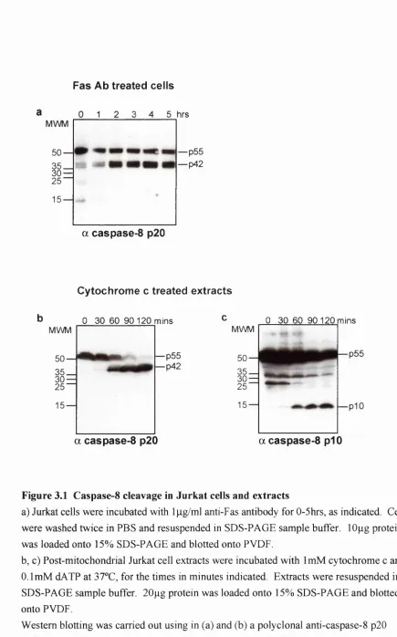
3.2.1. Caspase-8 cleavage in Jurkat cells and extracts_______________________________________ 81 3.2.2. Caspase-8 remains in the same sized complex during activation._______________________ 84 3.2.3. Cytochrome c induces trans activation of caspase-8: the prodomain is not required.________86 3.2.4. Caspase-8 is cleaved by transient interaction with a smaller p ro tease ____________________ 87 3.2.5. Gel filtration analysis of the cytochrome c initiated apoptosis pathw ay.__________________91
3.3. Discussion____________________________________________________95
3.3.1. Cytochrome c initiated caspase-8 activation_________________________________________ 95 3.3.2. Caspase-8 is cleaved by trans activation; by transient interaction with a smaller
protease or proteases. __ 96
3.3.3. Limitations of the in vitro sy ste m ______________________________________________ 97
Chapter 4 Results: Purification, identification and characterisation o f the caspase-8
cleaving activity____________________________________________________________98
4.1. Introduction__________________________________________________ 98
4.2. Results_______________________________________________________ 98
4.2.1. Purification of caspase-8 cleaving activity __________________________________________ 98 4.2.2. Identification of the 17kDa band as the catalytic subunit of the caspase-8 cleaving activity __
101
4.2.3. The 13kDa band is also a subunit of the caspase-8 cleaving activity____________________103 4.2.4. Confirmation that caspase-6 is the caspase-8 cleaving activity.________________________ 105 4.2.5. The caspase-6 prodomain must be removed for caspase-6 to be active._________________ 107 4.2.6. Caspase-6 remains in the same size complex during activation_______________________ 110 4.2.7. Caspase-6 is the only factor purified with significant caspase-8 cleaving activity_________111
4.3. Discussion_____________________________________________ 113
4.3.2. C asp ase-6 _____________________________________________________________________ 115 4.3.3. Specificity of caspase-6 cleavage of caspase-8______________________________________ 115 4.3.4. Caspase-6 activation requires removal o f the prodom ain._____________________________ 117
Chapter 5 Results: Action o f caspase-6______________________________________119
5.1. Introduction_________________________________________________ 119
5.2. Results______________________________________________________ 119
5.2.1. Caspase-6 has specificity for cleaving caspase-8 and c a sp ase-6 ________________________ 119 5.2.2. Caspase-6 promotes Bid dependent cytochrome c releasing a c tiv ity .___________________ 122 5.2.3. Inhibition of caspase-6 inhibits caspase-8 activation and apoptosis in cells_______________124
5.3. Discussion___________________________________________________ 130
5.3.1. Caspase-6 has specificity for caspase-8 and caspase-6 prodom ain._____________________ 130 5.3.2. Caspase-6 indirectly activates cytochrome c releasing activ ity _________________________ 131 5.3.3. Inhibition of caspase-6 in cells inhibits caspase-8 activation and apoptosis_______________132
Chapter 6 Results: Purification and identification o f caspase-6 cleaving factors 134
6.1. Introduction_________________________________________________ 134
6.2. Results______________________________________________________ 137
6.2.1. Caspase-6 is cleaved by transient interaction with >lGGOkDa and IGGkDa protein
complexes. ___________________________________________________________________ 137 6.2.2. Caspase-6 cleaving factors are inhibited by E K D -B iotin._____________________________ 139 6.2.3. Purification of caspase-6 p2G/plG cleaving activity.__________________________________14G 6.2.4. Purification of caspase-6 prodomain/p2G cleaving activity.____________________________ 147 6.2.5. Immunodepletion of caspase-3 but not caspase-7 inhibits caspase-6 cleavage at both
sites _________________________________________________________________________ 148
6.3. Discussion___________________________________________________ 150
6.3.1. Caspase-3 efficinetly cleaves both sites of caspase-6 ________________________________ 15G 6.3.2. Limitations of sy ste m ___________________________________________________________ 152 6.3.3. Preferred peptide substrates_______________________________________________________152
Chapter 7 General Discussion_____________________________________________154
7.1.1. Cytochrome c induced caspase-8 activ atio n ________________________________________ 154 7.1.2. Cytochrome c induced caspase-6 activation ________________________________________ 155 7.1.3. Caspase-3 and caspase-7 cleave caspase-6 differentially______________________________ 156
7.2. Further experiments___________________________________________ 157
7.2.1. Role of caspase-6 in apoptosis____________________________________________________ 157 7.2.2. Caspase-6 localisation___________________________________________________________ 158 7.2.3. Compensatory activation of caspase-6______________________________________________ 158 7.2.4. Mechanism of action of caspase-6 prodomain_______________________________________ 159
7.3. Role of effector caspases _______________________________________ 160
Table of figures, diagrams and tables
Chapter 1 Introduction____________________________________________________ 14
Table 1.1 Members of the conserved caspase activation c o m p le x ___________________________ 19 Figure 1.1 C.ele^ans caspase CED-3 and the mammalian caspase fa m ily ____________________ 21 Figure 1.2 Diagram of the structure of the three Bcl-2 sub-fam ilies_________________________ 41 Figure 1.3 Smac and HtrA2 inhibit XIAP inhibition of caspase-9____________________________ 48 Figure 1.4 Feedback loop of caspase activation and cytochrome c release ____________________ 55 Figure 1.5 Caspase-8 auto activation in the Fas induced D ISC ______________________________ 61
Chapter 3 Results: Mechanism o f caspase-8 activation in the cytochrome c initiated
apoptosis p a th w a y__________________________________________________________81
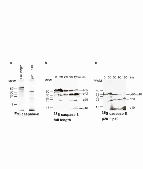
Figure 3.1 Caspase-8 cleavage in Jurkat cells and extracts __________________________________83 Figure 3.2 Caspase-8 is in a -lOOkDa complex before and during activ atio n __________________ 85 Figure 3.3 IVT caspase-8 is activated in Jurkat cell e x tra c ts _____________________________ 88 Figure 3.4 Caspase-8 is cleaved by transient interaction with a smaller protease_______________ 90 Figure 3.5 Gel Filtration analysis of known components of the cytochrome c induced apoptotic
pathway in Jurkat c e lls______________________________________________________ 92 Figure 3.6 Gel Filtration analysis of known components of the cytochrome c induced apoptotic
pathway in Jurkat c e lls______________________________________________________ 93
Chapter 4 Results: Purification, identification and characterisation o f the caspase-8
cleaving activity____________________________________________________________98
Figure 4.1 Purification of caspase-8 cleaving activity____________________________________ 100 Figure 4.2 EKD-Biotin inhibits purified caspase-8 cleaving activity and binds to a single
17kDa band ______________________________________________________________ 102 Figure 4.3 Anti-caspase-6 antibody binds to the p i 3 of the caspase-8 cleaving a c tiv ity 104 Figure 4.4 Caspase-8 cleaving activity is caspase-6_______________________________________ 106 Figure 4.5 The pro-domain of caspase-6 must be removed for caspase-8 cleavage.____________ 108 Diagram 4.1 SP Sepharose column allows separation of inactive caspase-6 p20/pl0 from
active caspase-6 p i7 /p 1 0 ___________________________________________________ 110 Figure 4.6 Caspase-6 remains in the same size complex before and during activation._________ 112 Figure 4.7 Caspase-3 does not have significant caspase-8 cleaving activity _________________ 114
Chapter 5 Results: Action o f caspase-6_____________________________________ 119
Figure 5.2 Purified caspase-6 activates cytochrome c releasing factor in cell extracts_________ 123 Figure 5.3 Cos-7 cells expressing caspase-6 mutants were produced._______________________126 Figure 5.4 Serum deprivation induced caspase-8 activation is delayed by inhibiting
production of mature active caspase-6 _______________________________________ 128 Figure 5.5 Serum deprivation induced apoptosis is delayed by inhibiting production of
mature active casp ase-6___________________________________________________ 129
Chapter 6 Results: Purification and identification o f caspase-6 cleaving factors 134
Diagram 6.1 Procaspase-6 cleavage s ite s ________________________________________________ 135 Figure 6.1 Caspase-6 is cleaved by transient interaction with >1000kDa and ~100kDa
protease complexes_______________________________________________________ 138 Figure 6.2 Caspase-6 activation in cytochrome c activated cell extracts.____________________ 141 Figure 6.3 Purification of caspase-6 cleaving activity ____________________________________ 143 Figure 6.4 Caspase-3 and caspase-7 cleave caspase-6 with preference for alternate s i t e s 144 Figure 6.5 Caspase-3 and caspase-7 are the purified proteases which cleave caspase-6________ 145 Figure 6.6 Immunodepletion of caspase-3 and caspase-7__________________________________149 Figure 6.7 Immunodepletion of caspase-3 but not caspase-7 inhibits cytochrome c induced
caspase-6 activation______________________________________________________ 151
Chapter 7 General Discussion____________________________________________ 154
Abbreviations
2D gel ( N H 4 ) 2 S 0 4 ,
ANT APS BIR BSA CaCl2 CARD CV dATP DD DED DISC DMSO DNA DTT ECL EDTA EGTA EKD-Biotin FPLC PCS HD Hepes HIV-1 lAP lEF IP IVT
2-dimensional gel (electrophoresis) Ammonium Sulphate
Adenine Nucleotide Translocase Ammonium Persulphate
Baculovirus lAP repeat Bovine Serum Albumin Calcium Chloride
Caspase activation and recruitment domain Column volume
Deoxy adenosine tri-phosphate Death domain
Death effector domain
Death Inducing Signalling Complex Dimethyl Sulphoxide
Deoxyribonucleic Acid Dithiotheritol
Enhanced chemiluminescence
1 -(4-Isothiocyanatobenzyl) ethylenediamine-N,N,N',N'- tetraacetic acid
Ethylene glycol-bis(2-aminoethylether)-N,N,N0,Nji-tetraacetic acid
z-Glu-Lys (Biotinyl)-Asp-CH2-DMB Fast Protein Liquid Chromatography Foetal Calf Serum
Huntington’s Disease
4-(2-Hydroxyethyl)piperazine-1 -ethanesulfonic acid Human Immunodeficiency Virus-1
Inhibitor of Apoptosis Protein Iso-electric focussing
KCl Potassium Chloride
KOH Potassium Hydroxide
LB Luria-Bertani Medium
MEF Mouse Embryonic Fibroblasts
MgClz Magnesium Chloride.
MnCL Manganese Chloride
NaCl Sodium Chloride
PAGE Polyacrylamide gel electrophoresis
PBS Phosphate buffered saline
PCR Polymerase Chain Reaction
PI Iso-electric point
Pipes 1,4-piperazinebis(ethanesulfonic acid)
PMSF Phenylmethysulphonyl fluoride
FTP Permeability Transition Pore Complex
PVDF Polyv/nylfluoridd .
SDS Sodium Doecyl Sulphate
TBS Tris buffered saline
TEMED N,N,N0,N0-T etramethylethylenediamine
TNFa Tumour Necrosis Factor alpha
Tween-20 Polyethylene glycol sorbitan monolaurate
UPR Unfolded protein response
UV Ultra-violet radiation
VDAC Voltage Dependent Anion Channel
Chapter 1 Introduction
1.1.
Programmed cell death
1.1.1. Programmed cell death is a physiological process
Programmed cell death or apoptosis is a physiological process by which cells are killed (Kerr et a l, 1972). Apoptosis has a critical role in development, tissue homeostasis and immune defence. During development, apoptosis is used to sculpt organs, remove auto reactive immune cells and remove neurons which have not made proper connections (Meier et al., 2000a). In the adult, a balance of cell division and apoptosis is maintained in tissue homeostasis (King and Cidlowski, 1998). Cells undergo apoptosis if there are too many, if they are growing in the wrong place or if their DNA has become damaged beyond repair. Immune cells trigger apoptosis in an infected cell (Krammer, 2000).
When viewed under the light and electron microscope, apoptosis is observed to be a dynamic process. Classical morphological features of an apoptotic cell are plasma
membrane blebbing, cytoplasmic condensation and chromatin condensation (Wyllie et al., 1980). Adherent cells detach from the dish and become small and spherical. Latterly in the process, a cell may become broken up into several vesicles or apoptotic bodies which are engulfed by neighbouring healthy cells. A significant feature of this process is that the contents of the cell are not released and therefore, the cell is killed without an inflammatory response.
of phosphatidylserine on the external surface of the plasma membrane (Strasser et al., 2000). Although cell type specific and trigger specific differences do occur in the initial stages o f apoptosis, once committed to apoptosis cells undergo similar morphological and biochemical changes.
1.1.2. Discovery of apoptosis
Over the last 150 years the process of programmed cell death had been investigated independently by three major approaches (Wyllie et al., 1980). Firstly, cell death had been found to occur in response to disease. Treatment of cells with toxic agents was carried out to investigate this process in vitro. Secondly, cell death had been observed during
development. Hormones and genetic background were found to regulate this form of cell death. Thirdly, kinetic studies had shown that the growth of some tissues was less than had been expected from the rate of proliferation of cells in that tissue. Cell death or migration had been suggested as reasons for this disparity. The review by Wyllie, Kerr and Currie in
1980, began to show the relationship between these forms of cell death and the common features that they share (Wyllie et al., 1980). This review also began to classify features of apoptosis. The result was that subsequent studies all used the same terminology which allowed a rapid understanding of the uniform nature of the execution phase of apoptosis, regardless of the stimulus, cell type or organism.
Apoptosis research gained momentum when genes necessary for promotion and inhibition of apoptosis were found in C.elegans (Ellis and Horvitz, 1986), and homologues were found in mammals (Hengartner, 2000).
1997). In particular, inhibition of apoptosis has a major role in cancer progression (Hanahan and Weinberg, 2000).
1.1.3. Clinical relevance of apoptosis
Apoptosis is essential for development, tissue homeostasis and immune defence. Therefore, deregulation of this process is associated with a range of developmental
problems as well as genetic and acquired diseases (Rudin and Thompson, 1997). Examples o f the medical problems associated with disregulation of apoptosis are described below.
Disregulation of programmed cell death in the central nervous system is observed in a range o f neuropsychiatrie diseases (Drouet et a l, 2000; Ho et al., 2001). Neuronal
apoptosis is fundamental to normal development with over half o f neurons produced being removed by apoptosis. Neurodevelopmental diseases are associated with both excess and insufficient apoptosis and neurodegenerative disease is associated with apoptosis in adult neurons. The mechanism of apoptosis and specifically caspase-8 activation in
Huntingt:)n’s disease and other polyglutamine repeat diseases is discussed in section 1.10.4.
different tissues, the resistance mechanisms found in cancer cells are often cell type specific. Tumour cells are found to have several anti-apoptotic lesions to inhibit the
redundant pathways operating in apoptosis signalling. Resistance to apoptosis is thought to be a component of resistance to chemotherapy (Kaufmann and Eamshaw, 2000).
1.1.4. Initial discoveries of the protein families involved in apoptosis
The major protein families that drive apoptosis are well conserved across the species. The initial discoveries of these proteins and their mode of action were made when Horvitz and colleagues were studying the development of the nematode Caenorhabditis elegans (Sulston and Horvitz, 1977). They made the intriguing discovery that during the development of C. elegans. out of the 1090 cells that are made, 131 cells undergo
programmed cell death. This invariant developmental apoptosis in a model organism allowed screening for genes that function in this pathway (Ellis and Horvitz, 1986). Two genes found by these screens, ced~3 and ced-4, were found to have an essential role in the cell death programme. Recessive mutations in either of these genes prevented most of the cell deaths (Yuan and Horvitz, 1990).
CED-3 protein was found to have homology to two mammalian cysteine proteases, human ICE (interleukin-1 ^-converting enzyme), and murine NEDD-2 (Yuan et al., 1993), now known as caspase-1 and caspase-2, respectively (Alnemri et a l, 1996).
Overexpression of caspase-1 or CED-3 induced apoptosis in mammalian cells which was dependent on the cysteine protease activity (Miura et a l, 1993). This was the first evidence that the caspase family of cysteine proteases may function in programmed cell death. Fourteen mammalian caspases have now been isolated and activation of a subset of these is essential for rapid and morphologically complete apoptosis. The caspase family are
Genetic screens in C.elegans also found a gene which negatively regulates
programmed cell death, ced-9 (Hengartner et a l, 1992). A ced-9 gain-of function mutation prevented the death of cells that normally die during development and mutations that inactivated ced-9 caused cells to die that normally live. On this occasion the mammalian field was ahead and CED-9 protein was found to be a homologue, in sequence and function, of the mammalian proto-oncogene Bcl-2 (Hengartner et al., 1992). Bcl-2 was cloned as a protein over expressed in B cells in follicular lymphomas due to a common translocation, t(14:18) (Tsujimoto et al., 1984). In mammalian systems, Bcl-2 had been shown to inhibit apoptosis initiated by a range of treatments and a number of the other mammalian homologues had already been cloned (Hawkins and Vaux, 1994). Some of these were found to behave in a yi way to Bcl-2, but others were found to actually promote apoptosis. This family is discussed in section 1.6.
When ced-4 was cloned, it had no known homologues and CED-4 protein did not appear to be a protease (Yuan and Horvitz, 1992). Genetically, it could be placed between ced-9 and ced-3 (Shaham and Horvitz, 1996), and was found to be able to bind
simultaneously to both CED-9 and CED-3 proteins (Chinnaiyan et al., 1997b; Wu et al., 1997). CED-4 could promote activation of CED-3 but not the enzymatic activity
(Chinnaiyan et al., 1997a). This led to the model that CED-4 may be an adaptor protein which in the absence of CED-9, promotes CED-3 auto activation.
al., 2000). It has been proposed that other mammalian CED-4 homologues may exist which can be directly inhibited by binding to Bcl-2 family members.
In D.melano2aster, the homologues of CED-3 and CED-4 are DRONC and DARK,
respectively (Dorstyn et al., 1999; Rodriguez et al., 1999). Although expression of Bcl-2 in the developing drosophila eye or in drosphila cells inhibits apoptosis, no anti-apoptotic Bcl- 2 family member has yet been found (Vemooy et al., 2000). Two pro-apoptotic Bcl-2 homologues have been found known as drob-1 /dBorg- 1/debcl/dbok and buffy/dBorg-2. These proteins may have anti-apoptotic function under certain conditions (Brachmann et a l, 2000).
C.eleeans D.melanoeaster H.saniens
Inhibitor CED-9 Bcl-2 like? Bcl-2
Adaptor CED-4 DARK Apaf-1
Initiator caspase CED-3 DRONC Caspase-9
Table 1.1 Members of the conserved caspase activation complex
1.2.
Caspases have a major role in apoptosis
1.2.1. Caspase family
Activation of caspases is essential for efficient apoptosis in almost every case of apoptosis investigated. Caspases are defined as cysteine proteases which cleave directly C- terminal to aspartic acid residues (Alnemri et al., 1996).
Figure 1.1 is a diagram of the 14 mammalian caspases that have been identified, of which 13 are found in humans. Analysis of the sequenced human genome revealed no other close human homologues (Aravind et al., 2001). The caspases activated during apoptosis cleave a diverse set of proteins, altering their properties in such a way to promote the dynamic biochemical and morphological changes that kill the cell and label it for engulfment. Another subset of caspases do not have a major role in apoptosis (or no apparent role in the case of some caspases), but instead are involved in activating pro- inflammatory cytokines.
1.2.2. Caspase activation of cvtokines
The first caspase to be cloned was initially found to have a role in the inflammatory response not in apoptosis. Caspase-1 (or ICE), was identified by its ability to cleave the interleuken-ip precursor, producing the mature form (Cerretti et al., 1992; Thomberry et al., 1992). In addition to caspase-1, several other caspases appear to have a role in the inflammatory response, based on functional studies or sequence homology (Zeuner et al., 1999).
Prodomain large domain small domain (large subunit) (small subunit)
(p20) (p10)
CED-3 GARD caspase-10 caspase-9 caspase-2 caspase-12 caspase-3 caspase-6 caspase-7 caspase-1 caspase-4 caspase-5 caspase-11 caspase-13 caspase-14
caspase-8 DED DED
DED DED
CARDE
>
■0 O -0 O (/) C/) 01
5z
m>
o
H Initiator Caspases Effector CaspasesFigure 1.1 C.elesans caspase CED-3 and the mammalian caspase family.
The caspases are divided into those that promote apoptosis and those that generally promote cytokine activation (although they may also be activated during apoptosis). DED = death effector domain. CARD = caspase activation and recruitment domain.
role in this process, since overexpression of many proteases would induce apoptosis (Yuan et a l, 1993; Zhu et al., 1995). Other experiments have shown that inhibitors of caspase-1 can inhibit apoptosis, however these inhibitors are not very specific and may be inhibiting true “pro-apoptotic” caspases (Garcia-Calvo et al., 1998; Hirata et al., 1998). Although there is probably not a broad role for “pro-inflammatory” caspases in apoptosis, there are some specific cases of involvement. For example, in the caspase-1 deficient mouse, there is an inhibition of Fas induced apoptosis in thymocytes (Kuida et al., 1995; Li et al., 1995).
In contrast, some “pro-apoptotic” caspases have been proposed to have a role in cytokine production. Inhibition of the “pro-apoptotic” caspase-3 has been shown to inhibit cytokine IL-16 production (Ludwiczek et al., 2001; Sciaky et al., 2000). However, the caspase-3 inhibitor peptides used in these studies may be cross-reacting with other “pro- inflammatory” caspases to inhibit cytokine production.
1.2.3. Caspase activation during apoptosis
Caspases were first proposed to have a role in apoptosis when CED-3, a protein necessary for nearly all cell deaths in C.elegans, was found to have homology to human caspase-1 (Yuan et al., 1993). Confirmation that caspases were required to be active during apoptosis came from use of the cowpox virus protein CrmA, which was known to inhibit caspase-1. Fas and TN Fa induced apoptosis and NGF withdrawal induced apoptosis could be inhibited by CrmA (Gagliardini et al., 1994). With a more comprehensive knowledge of the caspase family, it is now knovm that CrmA inhibits apoptosis not by inhibiting just caspase-1 but by inhibiting the pro-apoptotic caspases such as caspase-3, -8, and -9 (Garcia-Calvo et al., 1998).
activated subsequently are known as “effector caspases”. They are activated by different mechanisms which are discussed in section 1.4.
The contribution of individual caspases to apoptosis is measured in a number of ways. Cell lines cultured from caspase-deficient mice are a clean system in which to look at the effect of deficiency of a particular caspase (Zheng et al., 1999). In these studies, the initiator caspases are found to be critical for apoptosis induced by the relevant apoptotic trigger. For example, a deficiency in caspase-9 inhibits the cytochrome c pathway and a deficiency in caspase-8 inhibits the Fas pathway, (Hakem et al., 1998; Kuida et al., 1998; Varfolomeev et al., 1998).
Biochemical studies have been more suitable to establish the role of the effector caspases, because although substrates specific for individual effector caspases have been found, these caspases have a degree of redundancy for substrate cleavage and for promoting the morphological features of apoptosis (Strasser et al., 2000). The effector caspases are found to be abundant and cleave a wide range of substrates during apoptosis (Eamshaw et al., 1999). Inhibition o f cleavage of these substrates generally results in morphologically incomplete or delayed apoptosis.
1.2.4. Caspase-independent apoptotic pathwavs
Mitochondrial proteins have been implicated in mediating caspase-independent cell death. Expression of the Bcl-2 homologues, Bak or Bax, which promote release o f pro- apoptotic proteins from mitochondria, induce cell death with some morphological features of apoptosis in the absence of caspase activation (McCarthy et al., 1997; Xiang et al., 1996). Caspase independent features of apoptosis may be mediated by the mitochondrial proteins apoptosis inducing factor (AIF), endonuclease G and HtrA2 (Li et al., 2001; Susin et al., 1999; Suzuki et al., 2001a). All of these proteins, when released into the cytosol, induce some of the morphological changes observed during apoptosis. Their role in apoptosis is discussed in greater detail in section 1.9.
Non-caspase proteases also promote apoptotic events. The different protease families appear to have roles in apoptosis initiated by specific triggers. For example, calpains promote neuronal apoptosis and granzymes promote apoptosis induced by cytotoxic T lymphocytes. However, no one protease family appears to have such an extensive role as the caspase family which is activated in response to most triggers of apoptosis in most tissues. Non-caspase proteases activated during apoptosis are discussed in section 1.5
1.3.
Caspase activation
1.3.1. Procaspase structure.
All caspases are synthesised as inactive pro-enzymes, facilitating rapid cleavage and activation on the appropriate trigger. A diagram of the mammalian procaspases and
The large domain contains the conserved active site motif, QACXG (X is R, Q, orG).
Some caspases contain a short linker between the large and small domains. The N-terminal prodomain varies between the members in size and sequence. The prodomain of initiator caspases may contain either a CARD (Caspase activation and recruitment domain), or a DED (Death effector domain) which are involved in homotypic interactions with adaptor proteins during activation. The prodomain of effector caspases are only about 30 amino acids long. The caspase-2 and -7 prodomains have also been found to have a role in caspase localisation (Colussi et al., 1998; Yaoita, 2002).
1.3.2. Active caspase structure
In general, caspases are activated by cleavage between the large and small domains and sometimes between the prodomain and large subunit (Cerretti et al., 1992). The mature active enzyme is a hetero-tetramer consisting of two large subunits and two small subunits. A notable exception in many ways is caspase-9. In the mature enzyme, the caspase-9 prodomain needs to be retained for caspase activity because cleaved caspase-9 has very little activity without binding to Apaf-1 via a CARD-CARD interaction. (Rodriguez and Lazebnik, 1999). Caspase-9 is actually active when bound to Apaf-1 independently of cleavage between the large domain and small domain (Bratton et al., 2001; Stennicke et al., 1999). This is the exception rather than the rule.
Removal of the linker region between the large and small domains influences the activity or activation of some caspases. In the case of caspase-9, cleavage after the large domain at Asp-315 but not Asp-330 retains the linker on the small subunit. As described later, the N-terminal sequence o f the linker but not the N-terminal sequence of the minimal small subunit has the requisite sequence for binding to XIAP, a caspase inhibitor
sequence composed of three sequential aspartate residues. Disruption of the ionic
interactions of this sequence by acidification or other factors was found to be necessary for increased trans cleavage of caspase-3 and increased caspase-3 activity (Roy et al., 2001). Caspase-7 is the only other caspase that contains a linker sequence which may function in the same way. However, these residues did not show multiple ionic interactions in the crystal structure of caspase-7 (Chai et al., 2001b).
Initial three-dimensional structure studies showed that the arrangement of mature caspases in hetero-tetramers consisted of two adjacent small subunits surrounded by two large subunits (Shi, 2002). The tetramer has two active sites which are formed with amino acids from the small and large subunits. These active sites are cavities on either side of the tetramer which may function independently.
An exception is the apoptosome which is the high moLjcul ar weight complex of active caspases and Apaf-1. A recent study used cryoelectron microscopy to determine the structure of a recombinant apoptosome consisting of Apaf-1 and caspase-9 (Acehan et al., 2002). The structure is complex but the basic form is described as a “seven-spoked wheel”. Caspases-3 and -7 are also components of the apoptosome purified from cells but their structure within the apoptosome has not yet been well defined.
The indications are that other caspases may be found to be active in other forms than as a dimer. For example, caspase-3 and possibly caspase-6 in mouse fibroblasts were found to be cleaved and active in high molecular weight complexes distinct from the apoptosome, i.e. Apaf-1 and caspase-9 were not detectable (Kilic et al., 2002). There are other examples of cleaved caspases being present in other high molecular weight
Inducing Signalling Complex) has never been isolated as an active complex and therefore it is not known whether caspase-8 is active when part of the DISC or only when released.
1.3.3. Auto cleavage of caspases
Caspases are cleaved by either auto cleavage or trans cleavage.
Auto cleavage has been observed only for caspases with long prodomains. The CARD or DED present in the prodomain interacts with a homologous domain in an adaptor protein. For example the DED in procaspase-8 interacts with the DED in F ADD and the CARD in procaspase-9 interacts with the CARD in Apaf-1 (Li et al., 1997c; Medema et al., 1997). These procaspases have been shown to have enough residual activity to allow auto cleavage when two or more caspases are brought into close association by binding to oligomerised adaptors (Muzio et al., 1998; Srinivasula et a l, 1998).
1.3.4. Caspase mediated trans cleavage of caspases
Procaspases can be cleaved by active caspases (Eamshaw et al., 1999). The first evidence for this was that during apoptosis different caspase activities were found to be produced sequentially in a caspase activation cascade (Greidinger et al., 1996). Three distinct cleavage phases were distinguished using a range of peptide-based inhibitors with different selectivities.
which has been previously known as an integrin recognition motif (Buckley et al., 1999). Although this activation mechanism has not been confirmed to occur in vivo, it may have therapeutic value.
1.3.5. Methods used to define caspase cleavage of caspases
As more caspases have been isolated, the activation relationships between them have been investigated by a number of different approaches.
Peptide Inhibitors. To investigate caspase substrate specificity in general, a systematic approach using combinatory peptide fluorogenic substrates was employed (Thomberry et al., 1997). The results of this study could be used to infer the procaspase substrates of active caspases. This information has also been used to create caspase inhibitor peptides which have been used in vitro and in cells to determine the sequence of caspase activation following specific triggers of apoptosis (Hirata et al., 1998; Sun et al.,
1999). From this work, caspase activation cascades are now known to be branched rather than linear and distinct cascades are employed depending on the initial trigger.
Recombinant caspases. To look at the specificity o f caspase induced caspase activation directly, recombinant caspases have been incubated with procaspases and combinations of active caspases and procaspase have been expressed in yeast (Kang et al., 1999; Muzio et al., 1997). The benefit of these experiments, over those carried out in cells, is that they are clean systems because bacteria and yeast do not contain caspases and any caspase activity has to be supplied.
necessity of initiator caspases-8 or —9, depending on the stimulus, for effector caspase activation (Hakem et al., 1998; Kuida et al., 1998; Varfolomeev et al., 1998).
Observations made using caspase deficient mice highlight the gaps in our current knowledge about tissue specific differences in apoptotic pathways . In response to the same apoptotic stimulus, a deficiency in one caspase has often been found to differentially protect cells from different tissues. For example, in the caspase-9 knock-out mouse, MEFs and ES cells were resistant to UV irradiation whereas in thymocytes and splenocytes UV irradiation still triggered apoptosis (Hakem et al., 1998). In the caspase-3 knock-out
mouse, MEFs were resistant to TN Fa induced apoptosis but thymocytes remained sensitive (Woo et al., 1998). As yet, differences between the cell types which could account for these observations are not known.
Another intriguing observation is that although caspase deficient mice have some major developmental problems, most of the organs form normally, despite the significant role played by apoptosis in development. Redundancy and compensation may maintain the apoptotic response in the absence of one caspase enough to prevent developmental
problems in most tissues. Loss of certain caspases was shown to be compensated for by earlier activation of other caspases (Troy et al., 2001 ; Zheng et al., 2000). In certain cases, the caspase which is activated in compensation for deficiency in another, was been found to be upregulated and caspase inhibitors downregulated.
being used to deplete caspases in cells and this combined with expression of caspase mutants should be a powerful technique in the future (Paddison et al., 2002).
Biochemical studies. Investigation of caspase activation in cell extracts allows more manipulation of the system than is possible in cells. Caspases can been depleted from cell extracts revealing the dependency of caspases on others for activation (Slee et al., 1999). Caspases have been purified from cell extracts on the basis o f their ability to cleave other caspases. This powerful technique defined the early events in the cytochrome c mediated pathway (Li et al., 1997c; Liu et a l, 1996b) and has been used to isolate a novel caspase (Liu et al., 1996a). Both of these techniques have the limitation of only being able to study the activation of caspases by other caspases which are soluble at the time of cell extract preparation.
As may be expected from using such a range of different approaches, contradictory results have been gained.
1.3.6. Other proteases mediate trans cleavage of caspases.
Several other types of protease are activated during apoptosis that can cleave
caspases including cathepsins, calpains, granzymes. They are discussed at greater length in section 1.5.
1.3.7. Caspase post-translational modification
Caspases have been described to be post-translationally modified in a number of ways. Some of these modifications may be constitu tive and/or inconsequential. Other modifications may be regulated and have an effect on caspase activity.
was found to promote caspase-9 phosphorylation (Cardone et al., 1998; Marte and Downward, 1997). This Akt driven modification was suggested to be part of the
mechanism of Akt mediated protection because phosphorylated caspase-9 was not active; it could not undergo auto cleavage or activate downstream caspases. The general
significance of this finding was questioned when it was found that the Akt phosphorylation site in caspase-9 was not found in other species which could also be protected by Akt (Fujitae/fl/., 1999).
A number of caspases have been found to be modified by nitrosylation (Dimmeler et al., 1997; Li et al., 1997b). Nitric oxide is toxic to cells at high concentrations but at lower concentrations has a protective effect (Kim et al., 2001). A component of this protective effect is mediated by nitrosylation of caspases which inhibits their activity. This caspase modification has been found to be modulated by other signalling pathways. Fas and TN Fa were found to induce caspase-3 activity not only by cleaving the caspase but also by reversing caspase-3 nitrosylation (Hoffmann et al., 2001 ; Mannick et al., 1999).
1.3.8. Caspase localisation
Caspase substrates are found in various subcellular compartments and therefore active caspases have to be present in these compartments at least transiently. All caspases are present in the cytosol and some are present in other compartments too.
In some cases, a specific caspase in a specific location is required to initiate apoptosis. Procaspase-8 is recruited from the cytosol to the plasma membrane when it is activated in complex with the Fas Receptor (Medema et al., 1997). Caspase-12 is localised to the ER and is activated to mediate a response to ER stress. (Nakagawa et al., 2000).
Some caspases are translocated during apoptosis. Procaspases-3 and -7 have a high homology and similar peptide substrate specificity. Differences between them in function were implied when it was found that during Fas induced apoptosis in hepatocytes, caspase- 7 but not caspase-3 becomes localised to the mitochondrial and microsomal fractions (Chandler et al., 1998).
The signals which determine where caspases are localised are not well described. Constitutive localisation o f caspase-2 in the nucleus is mediated by the prodomain (Colussi et al., 1998). At least in response to Fas, the nuclear caspase-2 is activated before the cytosolic caspase-2 (Zhivotovsky et al., 1999). Other caspases are present in the cytosol until induction of apoptosis when they are translocated to the nucleus in a mechanism mediated by caspase-9. During apoptosis, active caspase-9 inactivates nuclear transport and increases the diffusion limit of the nuclear pores (Faleiro and Lazebnik, 2000). Caspase-3 (and other proteins) can now enter or leave the nucleus by diffusion. The
1.4.
Caspase inhibition
1.4.1. Procaspase activation and mature caspase activity can be inhibited
Inappropriate apoptosis causes damage to an organism. In healthy cells, apoptosis pathways are inhibited at many levels, including direct inhibition o f caspase activation and activity. In virally infected cells, apoptosis must be inhibited to allow time for viral
replication. Expression of viral gene products inhibits a range o f pro-apoptotic proteins including caspases. Caspase activity is inhibited in cancer cells by down regulation of caspase transcription.
Procaspase activation and mature caspase activity is regulated in the cell in a variety of ways, by a number of inhibitor families. Procaspase activation is inhibited by
competition with cellular and viral homologues of caspases. Mature caspase activity is inhibited by binding to the viral inhibitors, CrmA, p35 and inhibitor of apoptosis proteins (lAP). The cellular lAP, X-linked inhibitor of apoptosis also binds directly to caspases. The effect o f post-translational modification on caspase activation and activity is reviewed in section 1.3.7.
1.4.2. Inhibition of procaspase cleavage bv caspase homologues
Catalytically inactive homologues of nearly all pro-apoptotic caspases have been isolated. These can be cellular; either splice variants of caspases (Boldin et al., 1996; Fernandes-Alnemri et a l, 1995a; Fernandes-Alnemri et al., 1995b; Himeji et a l, 2002; Seol and Billiar, 1999; Srinivasula et al., 1999; Wang et al., 1994), or expressed from distinct genes (Goltsev et al., 1997; Hu et al., 1997b; Inohara et al., 1997; Irmler et al.,
Thome et a l, 1997). Those that have been investigated are mostly found to inhibit caspase activation.
The inactive homologues of both caspase-8 and caspase-9, appears to inhibit caspase activation by competing with the procaspase for binding to adaptor proteins. The inactive splice variant Caspase-9S (or-9b) which lacks the large domain competes with procaspase-9 for interaction with the adaptor protein Apaf-1 (Seol and Billiar, 1999; Srinivasula et a l, 1999). Although overexpression of caspase-9S protects against a range of apoptotic triggers, expression of this splice variant has not yet been found to be
modulated.
The inactive caspase-8 homologues, known as FLICE inhibitory proteins (FLIP), are viral, v-FLIP and cellular, c-FLIP. v-FLIP is homologous to the prodomain of caspase- 8, consisting of two DEDs and was found initially in a range o f y herpes viruses as a protein which inhibits apoptosis in the host cell (Thome et a l, 1997). Fas and TN Fa induced apoptosis are inhibited by v-FLIP. c-FLIP exist in two major forms; c-FLIPs (c-FLIP short) is homologous to v-FLIP and c- F L I Pl(c-FLIP-long) is homologous to full length caspase-8 with an inactive catalytic site (reviewed in (Thome and Tschopp, 2001).
Expression of FLIPs in cells can promote apoptosis under certain conditions. This result may be misleading because high FLIP expression may act in the same way as
expression of catalytically inactive caspase-8, and promote wild type caspase-8 aggregation and activation (Perez and White, 1998; Siegel et a l, 1998). In support of a role for c-FLIP in inhibition of caspase-8 activation, c-FLIP -/- mouse embryonic fibroblasts were W to TN Fa and Fas induced caspase-8 activation and apoptosis (Yeh et a l, 2000).
which has neither caspase-2 or caspase-2S, neuronal apoptosis is accelerated (Bergeron et a l, 1998). This may suggest that the CASP-2 gene encodes a suppressor of apoptosis, presumably caspase-2S, which works independently of caspase-2. However, it cannot be discounted that this neuronal apoptosis phenotype is due to an indirect effect of the loss of full length caspase-2 expression. For example, caspase-9 expression is upregulated in caspase-2 deficient neurons and in tissue culture, caspase-9 can be activated in
compensation for caspase-2 in neuronal apoptosis (Troy et al., 2001).
The effector caspases also have inactive splice variants (Fernandes-Alnemri et al., 1995a; Fernandes-Alnemri et al., 1995b). Their mode of action is less well described; presumably they can compete with effector caspases for interaction with activators and possibly effectors.
1.4.3. Regulation of FLIPs
The inhibitors of caspase activation only have real significance in apoptosis regulation if their expression level can be modulated. The role of v-FLIPs, together with viral Bcl-2 and lAP homologues, is to inhibit apoptosis following viral infection, allowing viral replication (Bertin et al., 1997; Hu et al., 1997a; Wang et al., 1997). The mechanism of v-FLIP protection may utilise an additional process, in addition to competing with caspase-8. Mutants of MCI 59 v-FLIP which can still bind to F ADD and caspase-8 could not inhibit apoptosis, (Garvey et al., 2002).
recruits intermediate adaptor proteins that promote upregulation of NF-kB (Kataoka et al.,
2000).
The significance of c-FLIP in protecting against apoptosis is seen in a number of clinical situations. For example, c-FLIP has been implicated in protecting cardiac myocytes from apoptosis. Following ischemia/reperfusion injury, cardiac infarcts associated with caspase-3 activation were found where c-FLIP levels were low; whereas high c-FLIP expression was found in surrounding unaffected tissue (Rasper et al., 1998). There appears to be an additional role for c-FLIP along with F ADD and caspase-8 in the cardiac myocytes. Although in the FLIP knock-out, apoptosis is promoted and in the F ADD and caspase-8 knock-out apoptosis is inhibited, all three knock-out mice have a similar defect of impaired heart development (Varfolomeev et al., 1998; Yeh et al., 2000; Yeh et al., 1998).
One of the several ways that tumour cells acquire resistance to apoptosis is by inhibiting caspase activation. High levels of c-FLIP expression have been observed in tumour cell lines, e.g. in melanoma cell lines, malignant melanoma tumours and non- Hodgkins lymphoma cell lines (Bullani et al., 2001 ; Irisarri et al., 2000; Irmler et al., 1997; Thomas et al., 2002).
1.4.4. Caspase inhibition bv lAPs
1.4.5. Caspase inhibition bv CrmA and p35
The cowpox virus protein CrniA and the baculovirus protein p35 inhibit caspases by acting as suicide substrates (Bump et al., 1995; Ray et al., 1992). Both inhibit a broad, although not complete, spectrum of caspases. They are cleaved by caspases and remain bound to the active site, inhibiting interaction with other substrates. Inhibition o f apoptosis by these inhibitors provided some of the first evidence that caspases were necessary for apoptosis (Gagliardini et al., 1994).
1.4.6. Down regulation of caspase expression in tumour cells.
The expression of caspases has been found to be regulated . As described previously, caspases can be ubiquitinated by lAPs and subsequently degraded. Caspase expression can also be regulated at the level of transcription.
Silencing of caspases and caspase adaptor genes, by DNA méthylation or gene deletion, is one mechanism by which tumour cells avoid apoptosis. In neuroblastoma, loss of caspase-8 and caspase-10 by gene méthylation or gene deletion was observed which correlated with a lack of sensitivity to a range of apoptotic triggers (Eggert et al., 2001; Teitz et ah, 2000). In melanomas, the silencing of the caspase-9 adaptor protein, Apaf-1, by DNA méthylation or gene deletion correlated with lack of chemosensitivity (Soengas et ah, 2001). Silencing of caspase-1 is associated with renal tumours and silencing of
caspase-5^endometrial and gastrointestinal cancers (Schwartz et ah, 1999; Ueki et ah, 2001) The initiator caspases appear to be preferentially silenced over the effector caspases which may be because effector caspases have a high degree of redundancy. The silencing of pro-inflammatory caspases may provide an environment that promotes tumour
1.5.
The role of non-caspase proteases in apoptosis
1.5.1. Non-caspase proteases in apoptosis signalling
Besides the caspases, several other protease families are activated or translocated during apoptosis. Unlike caspases, these other protease families appear to be only utilised under specific conditions. For proteases such as Granzyme B, initiation of apoptosis appears to be the primary function. Other proteases such as calpains and cathepsins, have important functions in healthy cells as well as during apoptosis. As with the caspases, only a subset of these protease families appears to be involved in apoptosis. Determining the role of proteases in apoptosis which have a function in healthy cells as well is difficult because inhibition of these proteases impedes the function o f the healthy cell and can lead indirectly to apoptosis.
1.5.2. Granzvme B
Granzyme B is a serine protease which can initiate caspase activation and apoptosis. Cytotoxic T lymphocytes and Natural killer cells induce apoptosis in target cells by
stimulating the Fas pathway and by granule exocytosis which is the transfer of the contents of cytoplasmic granules into the target cell (Shresta et a/., 1995). Two components of these granules which together induce apoptosis are perforin and the serine protease granzyme B.
Granzyme B cannot induce apoptosis without perforin which facilitates the entry and/or activity of granzyme B in the cell. Perforin has been reported to form a pore in the plasma membrane to allow entry of granzyme B, release granzyme B from vesicles in the cell or facilitate granzyme B transport to the nucleus (Johnson, 2000).
peptide library, the optimal peptide substrate was found to be the same as for caspase-8 (Thomberry et al., 1997).
Granzyme A is also released from cytotoxic T lymphocytes and although it is not as cytotoxic as granzyme A, it can promote some morphological features*apoptosis (Masson et al., 1986). Granzyme A does not appear to activate caspases but promotes nuclear
condensation and DNA cleavage in a caspase-independent manner (Johnson, 2000).
1.5.3. Calpains
Calpains are cytosolic cysteine endopeptidases which are calcium activated (Saido et al., 1994). They have been implicated in cell proliferation, differentiation and apoptosis; inhibition of calpains reduces cell migration and invasion (Perrin and Huttenlocher, 2002). Their role in mediating apoptosis has been mainly associated with neuronal cell death and calpain inhibitors have neuroprotective effect in a range of models of neurological damage (Wang, 2000). The calpain family has a broad tissue distribution with some members being ubiquitously expressed and therefore there is the potential for calpains to have roles in apoptosis in other tissues.
1.5.4. Cathepsins
The cathepsirtare found in lysosomes where they mediate the terminal degradation of proteins and they are secreted to degrade the extracellular matrix. More recently, cathepsins have been found to be released in a controlled manner from the lysosomes into the cytosol during apoptosis (Leist and Jaattela, 2001). The role o f cysteine cathepsins in apoptosis may have been underestimated previously because they are inhibited by zVAD- fmk which was thought to be specific for caspases (Schotte et al., 1999).
Specific cathepsins were found to be released in relatively small amounts from the lysosomes following different apoptotic triggers. For example, cathepsin B was released during bile salt induced apoptosis in hepatocytes (Roberts et al., 1999), and cathepsin D was released following oxidative stress (Roberg and Ollinger, 1998). The role of cathepsins has most clearly been described in TN Fa induced apoptosis (Guicciardi et al., 2000). Following TN Fa treatment, a deficiency of cathepsin B inhibited apoptosis and in vitro caspase-8 could induce cathepsin B release from the lysosomes. Another study showed that cathepsin B could cleave Bid and promote cytochrome c release from isolated mitochondria (Stoka et al., 2001).
The mechanism and timing of cathepsin release from the lysosomes during
apoptosis is not known and this information may indicate the significance of this pathway in apoptosis.
1.5.5. Other proteases.
HtrA2 which is a serine protease which is released from the mitochondria and neutralises XIAP by binding to it during apoptosis. A role of the protease domain of HtrA2 has not been found. The role of HtrA2 is discussed in section 1.9.
1.6.
The Bcl-2 family integrates pro- and anti-apoptotic signals
1.6.1. B cl-2 fam ily
Each cell continuously receives several pro- and anti-apoptotic signals and the Bcl-2 family members act as integrators of these signals (Adams and Cory, 2001). The state of the Bcl-2 complexes which result from these signals, can dictate whether cytochrome c and other pro-apoptotic factors are released from the mitochondria.
There are three groups of Bcl-2 family members defined by structure and function (Gross et ciL, 1999). The first group are homologues of Bcl-2 which inhibit cytochrome c release and apoptosis initiated a large range of triggers. The second group are homologues
Anti-Apoptotic - Bcl-2 like
Pro-Apoptotlc - Bax like
BH3-onlv - variable structure ()BH 3) . . . 4 W ' )
Figure 1.2 Diagram of the structure of the three Bcl-2 sub-families.
BH domains and transmembrane domains (TM) are indicated. The BH3 only members have a the largest variation, indicated by the “dashed” protein backbone.
of Bax which promote cytochrome c release and apoptosis. These two groups both have three domains in comm on, the Bcl-2 homology motifs (BH) and have a similar structure. The third group, the “BH3 only” members also promote apoptosis. They all have one BH domain, BH3, but apart from this are not homologous. Many Bcl-2 family members (Bid and Bim being two notable exceptions) have a hydrophobic C terminus which allows interaction with the mitochondrial, ER and nuclear membranes. In healthy cells, anti- apoptotic members and some pro-apoptotic members are found on these membranes; other pro-apoptotic Bcl-2 family members are observed to migrate to the mitochondrial
membrane following an apoptotie trigger.
1.6.2. Bcl-2 familv function
The Bcl-2 family members are sensors of a wide range o f signals whieh regulate their pro- and anti-apoptotic functions. In a cell, where often several members of each group are expressed, pro- and anti-apoptotic signals can be integrated because the different Bcl-2 members can bind to each other (Adams and Cory, 2001). Through this mechanism, the balance of pro- and anti-apoptotic signals is read to determine whether the eell survives.
The Bcl-2 homologue in C.elegans, CED-9, binds to and inhibits CED-4 directly (Chinnaiyan et al., 1997b; Wu et al., 1997). Although interactions between the mammalian homologues, B c1 -X l and Apaf-1 have been observed in overexpression systems (Hu et al.,
1998a; Pan et al., 1998), these interactions have not been observed with endogenous protein (Moriishi et al., 1999; Newmeyer et al., 2000). The assumption that Bcl-2 may be acting in this way in mammals persists because Bcl-2 can rescue cells in ce<7-P-deficient nematodes (Hengartner and Horvitz, 1994). This certainly does support the argument that the Bcl-2 family members from different species may have a common function. It also shows that Bcl-2 can have a function besides blocking cytochrome c release which is not required for CED-3 activation and apoptosis in nematodes (Hengartner, 2000). In mammals, the Bcl-2 family members may be binding to other mammalian CED-4 homologues besides Apaf-1 to inhibit apoptosis.
1.6.3. The Bcl-2 familv regulates cvtochrome c release
Several overlapping theories exist to explain how the Bcl-2 proteins regulate cytochrome c release. Bcl-2 proteins may form a pore in the mitochondrial membrane. The 3 -D structure of B c1 -X l resembles membrane-penetrating bacteria toxins, and other members can form channels in liposomes (Muchmore et al., 1996). However, it is not clear whether these structures form in the cell. Alternatively, Bcl-2 proteins may influence transport through the permeability transition pore (FTP) (Crompton, 1999). Components of this pore have been found to bind to Bcl-2 proteins.
MEFs, all the Bcl-2-like and the BH3-only proteins must eventually signal to the Bax-like proteins to regulate cytochrome c release. Bak and Bax have been described as a “requisite gateway to mitochondrial dysfunction” (Wei et a l, 2001). In other cell types this degree of resistance may require a deficiency in a different a combination of Bax-like proteins.
Bax and Bak are activated by a conformational change which is detected using site specific antibodies (Desagher and Martinou, 2000; Goping et al., 1998; Nechushtan et ah, 1999). Integration of apoptotic signals appears to centre on modulation of the
conformational change. Bcl-2 or B c1 -X l directly inhibit the conformational change,
whereas pro-apoptotic, BH3-only proteins promote the conformational change (Desagher et al., 1999; Marani et al., 2002). Bcl-2 and B c1 -X l can also be inactivated by caspase
cleavage (Cheng et al., 1997; Woo et a l, 1999).
1.6.4. A wide range of signals modulate the BH3-onlv proteins
The BH3 only proteins are a diverse group in sequence, structure and localisation. They are activated in a number of ways, by a range of signalling pathways to promote the conformational change in the Bax-like proteins. Individual members are often found to have their activity potentiated by several mechanisms. The regulation of three BH3-only proteins is discussed below.