ISSN 2286-4822 www.euacademic.org
Impact Factor: 3.4546 (UIF) DRJI Value: 5.9 (B+)
Assessment of Alternative Methods for Diagnosing
of Ascariasis in Piglets
YLLKA (MIJA) ÇANI
Doctoral School, Faculty of Veterinary Medicine Agricultural University of Tirana, Albania
BEJO BIZHGA1 Faculty of Veterinary Medicine Agricultural University of Tirana, Albania
Abstract:
The incidence of ascariasis is high in Albania having adverse affects across the entire levels of the breeding system with far-reaching damage to all pig categories. Ascariasis is rightly assumed to be causing significant damage to pigs until they are bound for slaughter after just 7 months of age with symptoms correlating with high prevalence rate and a heavy parasite load. The losses incurred from Ascaris suum in piglets is compelling us to gear for a series of diagnostic and prophylactic alternatives to be applied in slaughterhouses with the view to boosting the much sought-after profitability. The study was conducted in the country’s slaughterhouses involving as many as 162 pigs in which was detected the presence of ascariasis through several alternative diagnostic methods. The coproscopical examination of the samples collected from the slaughter-houses revealed that as many as 124 pigs (76.54% of the total) were infested. Further to this, an examination of the liver clearly showed the presence of the livery milk spots in 49 piglets or 30.24 % of the samples collected and examined. Under examination of the small intestines indicated that the adult parasites were detected in the small intestines of some 78 pigs or an equivalent of 48.14% of the examined samples. During the examination of the lungs as many as 34 piglets or a proportion of 20.98 % of the total samples examined tested positive
with parasitic pneumonia or with well-established signs of parasitic migration. A closer inspection of nose-leak swabs demonstrated that a total of 26 piglets or roughly 16.04% of the total tested positive with larvae infestation/presence. Coproscopic examination in turn was identified as the most effective and easy-to-use method while liver examination proved to be quite as practical and very useful in terms of the observations occurring in the slaughterhouse. In addition, the examination of intestines was deemed a highly relevant tool in the diagnosis of ascariasis in the slaughterhouse. Whereas the examination of the lungs can serve as a very practical diagnostic tool in the observations done in slaughterhouses, while the examination of the nose swabs turned out to be an alternative method, both in the case of the living piglet and the slaughtered ones in the slaughterhouse.
Key words: Ascariasis, coproscopy, milk spots, intestine, lung, nose swabs piglets.
MATERIALS AND METHODS
method of study. During the post-mortem control of biological properties of A.suum were detected variations in the liver and lungs along with lesions easily noticeable in the migration stage. These observations were done in the organs of the piglets from which a series of samples were collected for the intents of macroscopic, histological and microscopic examinations. As for the biological properties of A. suum it should pointed out that livers carrying white spots were identified. In the slaughterhouse the piglets’ livers were subjected to the macroscopic examination focusing primarily on the liver milk spots. The livers were carefully examined for hot spots due to the migration of the ascarids larvae. The observations were administered upon the same piglets from which sampling was done for purposes of coproscopical examinations. To distinguish between parasitic spots and spots caused by mold the liver samples were stained with Wright Gimsae and Ziehl Neelsen.
already examined. In the case of the adult parasites the paper concentrated on the identification of the species type and their corresponding final counting.
The lungs were also observed in the same piglets. Upon the observation of parasitic pneumonia or in case of suspicions triggered from migrations of larvae to lungs haemorragical lesions and intensive filtrations of eozionphiles around alveoli were detected (from larvae migrating to the bronchial tree). Repeated infections produce haemorrhage, oedema and emphysema. The lungs which proved to be suspicious of affected were sampled partially or in their entirety to be examined in the laboratories. In the laboratories the full lungs were examined through the perfusion method. While the sections/ components were examined in the microscopic swabs (strish) which were acquired from the liquids in the bronchial tree. While in the slaughterhouse swabs were prepared from nose leaks of the same piglets as used in the experiment. The swabs were observed to detect migratory larvae. When larvae were detected they were quantified effectively. A Chi-square test was conducted to investigate the association between the exposure and the outcome (Agresti, 2012). The odds ratio and its 95% confidence limits were calculated to measure the magnitude of the association. A 5% level of significance was used to evaluate significance of association, i.e the p-value was considered significant if it was less than 0.05 . The analyses were conducted using Statulator, an online statistical program (Dhand and Khatkar, 2014).
RESULTS AND DISCUSSION
the prevalence of infestation as well as the parasitic load. It can also be applied both in living pigs and slaughtered piglets (carcasses) in slaughterhouses.
Tabele no. 1. The examination results of piglets in slaughterhouses.
Method No. of
examined piglets
Positive no
Positive %
Coprologic examination of piglets in slaughterhouses
162 124 76.54
Presence of hot spots in liver 162 49 30.24
Ascarids in intestines 162 78 48.14
Examination of lungs 162 34 20.98
Examination of nose swabs 162 26 16.04
It was established that as many as 49 piglets or 30.24% of the observed ones were found to be suffering from damage due to migration to the liver. As far as the relationship between macroscopic and histological models was concerned, white and compact milk spots have generally been produced by interstitial eosinophilic hepatitis.
Figure no 1. Milk spots in piglets liver
commonly referred to as "milk spots". In the absence of re-infections these lesions will begin to regress (retreat) after the larvae migrate out of the liver and the organ will recover completely after 4 to 6 weeks. It could be argued that their presence in necrosis is an indication of the final infestation. In pigs which experience multiple re-infestations during their life cycle, the spots become noticeably fibrotic.
It should be highlighted that in the experimental pigs intestinal examinations were carried out to track and count up the adult ascarids. During the microscopic examinations of the small intestines it was revealed that out of a total of 162 piglets were found to have adult parasites residing in them some 78 piglets or an equivalent of 48.14% of piglets examined. There were also variations in the number of adult parasites quantified in the intestines. The piglets which resulted to have been infested with adult parasites in the intestines was found through post-mortem examination, and consequently the paper concentrated on their categorization.
The nose swabs were collected from the piglets examined and the microscopic ones were examined in the stereomicroscope as moist and dry solutions. The A. suum migrating larvae were found in these swabs. The technique proved to be quite useful under these conditions since it is well-known that larvae appear in the infested pig's noses on the 7 and 9 days upon infestation. The larvae in the nose and mouth are ingested to go down to the intestines or they are coughed up (sneezed up) leaked out/discharged through the nose to the outer environment. The diagnostic technique proved to be very easy-to-use, extremely efficient and very effective. The reason has to do with the fragility method and the biological properties of Ascaris suum's which for a relatively short period of time can be found in the nose leaks. This coincides with the period when it migrates into the bronchioles and through the mucocilial apparatus it comes straight into the mouth to be swallowed and then to be discharged through the leaks out into the open. The time is rather limited and values will be much smaller. Using the swabs in the nose leaks it was established that 26 piglets or a proportion of 16.04% of the total samples tested positive with the presence of larvae. The advantages of this method are that swab examinations can yield invaluable data in the living piglets (not necessarily slaughtered ones) and positivity along with the parasitic loads appear to be of significant value.
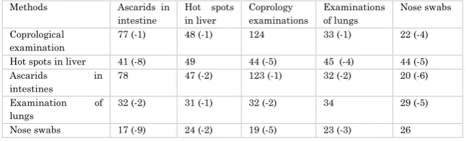
Table no 2. Comparable data for the positivity in piglets dependent upon the methods being used.
Methods Ascarids in intestine
Hot spots in liver
Coprology examinations
Examinations of lungs
Nose swabs
Coprological examination
77 (-1) 48 (-1) 124 33 (-1) 22 (-4)
Hot spots in liver 41 (-8) 49 44 (-5) 45 (-4) 44 (-5) Ascarids in
intestines
78 47 (-2) 123 (-1) 32 (-2) 20 (-6)
Examination of lungs
32 (-2) 31 (-1) 32 (-2) 34 29 (-5)
It was only 1 out of a total of 124 piglets which tested negative with adult ascarids in intestines during the coproscopical examination. In this case in point this can be fully accounted for by the fecundity of the parasites, the peculiarities of the parasitic populations while, if probability were to be weighing in, it might happen that, regardless of the large number of eggs laid by a female parasite, it might well be the case that 1 out of 124 samples might have escaped the microscopic observation. Meantime only 1 piglet tested negative for the presence of hot spots in the liver. By estimating the data from the wide-ranging available literature and the research experience to this day this is easily understandable, and it relates closely to the physiological traits of the animal and more specifically to the immunity of piglets.
Method Post-mortem (white spot) Total
Positive Negative
Coprological
Positive 41 83 124
Negative 8 30 38
Total 49 113 162
The odds ratio indicates that the positive animals in coprological examinations has 1.85 times the odds of the presence of white spots in liver than the negative animals. Also, we are 95% confident that the odds ratio in the population would be between 0.78 and 4.40.
Association between the positive results and the outcome was not significant [P-value: 0.158; odds ratio and 95% CI: 1.85 (0.78, 4.40)] (Dhand and Khatkar, 2014).
Only 1 piglet which tested positive during the coproscopic examination tested negative for the presence of larvae during the lung examination. While this number increased to 4 piglets when the examination of the nose swabs was undertaken. The differences are accounted for from the stages of the biological cycle of parasites and the specific traits of the immunity of piglets.
not display any positive result for ascarids in the intestines, with 5 of them testing negative through the coproscopic examination and 4 others testing positive through the lung examination as well as 5 others testing negative during the examination of nose swabs. The differences proved to be significant in this case especially in the case of ascarids breeding in the intestines and larvae in the noses. We assume that this could be accounted for only by the physiological traits of the piglets, the specific immunity of the piglets which has a direct effect even upon the biological cycle of the ascarids. In the case of individuals possessing strong immunity the intestinal and hepatobronchial migration stages is significantly halted.
Method Post-mortem (presence of ascarides) Total Positive Negative
Coprological
Positive 122 2 124
Negative 1 37 38
Total 123 39 162
The odds ratio indicates that the positive animals in coprological examinations has 2257.00. times the odds of the presence of ascarides in intestine than the negative animals. There was a significant association between positive coprological results and (P-value: <0.001) with presence of ascarides in intestine than the animals with negative results (odds ratio 95% CI: 198.99, 25599.11).
reason to assume that a piglet which tests positive through the coproscopy does not display any obvious signs for the presence of the adult parasites in the intestines. In this case there is no justification whatever for the presence of eggs in faeces. By looking at this as a case in isolation this could be easily attributable to the fact of piglets being adult ascarids-free as much as to the presence of prepatente ascarids which are not yet fully matured and fertilized to be laying eggs. Meanwhile 2 piglets which tested positive during the examination of the intestines tested negative for the presence of larvae during the examination of the lungs and that of nose swabs with the number jumping to 6 piglets. The differences could be accounted for through the traits of the parasitic biological cycle and the specific characteristics of immunity in piglets which directly affect the migration of larvae. This indicates that the body reacts forcefully by getting rid of a large number of them. Only a limited number, say, out of a thousand (1-40) could succeed in establishing themselves and becoming mature in the piglets’ intestines.
numbers diminishing along the way with only a handful of them managing to mature sexually.
Method Post-mortem (lungs lessions) Total Positive Negative
Coprological
Positive 32 92 124
Negative 2 36 38
Total 34 128 162
There is a significant association between the animals with positive coprological results (P-value: 0.007) and lung lesions, and they have 6.26 times the odds than animals with negative coprological results (odds ratio 95% CI: 1.43, 27.49).
is much more unstable for the presence of adult parasites in the intestines.
Method Post-mortem (results of nasal swabs) Total Positive Negative
Coprological
Positive 22 122 124
Negative 4 14 28
Total 26 136 162
There was an association between coprological results and results of nasal swabs, however it was not significant [P-value: 0.449; odds ratio and 95% CI: 0.63 (0.19, 2.10)]. Logically this correlation should have proven otherwise stable, but the immunity mechanisms in piglets allows for the body to fight off the penetration of the larvae to the mucosa, hence reducing the number of those (1/2) which stand a chance to mature and test positive in the coprological analysis (confidence interval at 95 %).
CONCLUSIONS
lungs when signs of migrations and pneumonia can be established for the presence of invasive larvae along with intestinal control to evaluate adult ascarids can be applied as quite successful diagnostic techniques in the post-mortem diagnosis of ascariasis in pigs. While in the examination of piglets an analysis of the nose leaks can be administered for the presence of larvae as a highly efficient and cost-effective alternative method in diagnosing and monitoring ascaridiosis.
REFERENCES
1. Agresti, A. (2012). Categorical Data Analysis (3rd ed.). Hoboken, New Jersey: John Wiley and Sons.
2. Anderson TJ (2001) The dangers of using single locus markers in parasite epidemiology: Ascaris as a case study. Trends in parasitology 17: 183–188.
3. Arizono N, Yoshimura Y, Tohzaka N, Yamada M, Tegoshi T, et al. (2010) Ascariasis in Japan: is pig-derived Ascaris infecting humans? Japanese journal of infectious diseases 63: 447–448.
4. Coates, S. (2000). Modelling the population dynamics of Ascaris suum in pigs. Ph.D. thesis, Danish Centre for Experimental Parasitology, Copenhagen, Denmark and the University of Warwick, Coventry, UK.
5. Dhand, N.K., Khatkar, M.S. 2014. Statulator: an online statistical calculator. Sample size calculator for
comparing two independent proportions.
http://statulator.com/SampleSize/ss2P.html
6. Dold C, Holland CV (2011) Investigating the underlying mechanism of resistance to Ascaris infection. Microbes and infection/Institut Pasteur 13: 624–631.
potential manipulation for future vaccine control strategies. Parasitology research 1–13.
8. Helwigh, A. B. and Nansen, P. (1999). Establishment of Ascaris suum in the pig : development of immunity following a single primary infection. Acta Veterinaria Scandinavica 40, 121–132.
9. Holland CV (2009) Predisposition to ascariasis: patterns, mechanisms and implications. Parasitology 136: 1537– 1547.
10.Leles D, Gardner SL, Reinhard K, Iniguez A, Araujo A (2012) Are Ascaris lumbricoides and Ascaris suum a single species? Parasites & vectors 5: 42.
11.Masure D, Vlaminck J, Wang T, Chiers K, Van den Broeck W, et al. (2013) A role for eosinophils in the intestinal immunity against infective Ascaris suum larvae. PLoS Negl Trop Dis 7: e2138.
12.Mejer, H., Wendt, S., Thomsen, L. E., Roepstorff, A. and
Hindsbo, O. (2000). Nose-rings and transmission of helminth parasites in outdoor pigs. Acta Veterinaria Scandinavica 41, 153–165.
13.Miquel N, Roepstorff A, Bailey M, Eriksen L (2005) Host immune reactions and worm kinetics during the expulsion of Ascaris suum in pigs. Parasite Immunol 27: 79–88.
14.Miquel, N., Roepstorff, A., Bailey, M. and Eriksen, L.
(2005). Host immune reactions and worm kinetics during the expulsion of Ascaris suum in pigs. Parasite Immunology 27, 79–88.
15.Morris RG, Jordan HE, Luce WG, Coburn TC, Maxwell CV, 1984. Prevalence of gastrointestinal parasitism in Oklahoma swine. Am Vet Res, 45: 2421-2423.
17.Nejsum P, Parker ED Jr, Frydenberg J, Roepstorff A, Boes J, et al. (2005) Ascariasis is a zoonosis in denmark. Journal of clinical microbiology 43: 1142–1148.
18.Nejsum P, Parker ED Jr., Frydenberg J, Roepstorff A, Boes J, Haque R, Astrup I, Prag J, Sørensen UBS, 2005. Ascariasis is a zoonosis in Denmark. J Clin Microbiol, 43(3): 1142-1148.
19.Ngowi HA, Kassuku AA, Maeda GE, Boa ME, Willingham AL, 2004. A slaughter slab survey for extra-intestinal porchine helminth infections in northern Tanzania. Trop Anim Health Prod, 36:335-340.
20.Nsoso SJ, Mosala KP, Ndebele RT, Ramabu SS, 2000. The prevalence of internal and external parasites in pigs of different ages and sexes in Southeast District, Bostwana. Onderstepoort J Vet Res, 67: 217-220.
21.Roepstorff A, Eriksen L, Slotved HC, Nansen P (1997) Experimental Ascaris suum infection in the pig: worm population kinetics following single inoculations with three doses of infective eggs. Parasitology 115 (Pt 4) 443–452.
22.Roepstorff, A. (2003). Ascaris suum in pigs : population
biology and epidemiology. Doctorate thesis, the Royal Veterinary and Agricultural University, Copenhagen, Denmark.
23.Roepstorff, A. and Murrell, K. D. (1997). Transmission
dynamics of helminth parasites of pigs on continuous pasture: Ascaris suum and Trichuris suis. International Journal for Parasitology 27, 563–572.
24.Roepstorff, A. and Nansen, P. (1994). Epidemiology and
control of helminth infections in pigs under intensive and non-intensive production systems. Veterinary Parasitology 54, 69–85.
suum which presented with eosinophilic pneumonia and multiple intra-hepatic lesions with severe eosinophil infiltration-outbreak in a Japanese area other than Kyushu. Intern Med, 1: 574-579.
26.Shamri R, Xenakis JJ, Spencer LA (2011) Eosinophils in innate immunity: an evolving story. Cell and tissue research 343: 57–83.
27.Tokojima M, Ashitani J, Nakazato M, 2004. A case of eosinophilic pneumonia caused by visceral larva migrans due to Ascaris suum. Kansenshogaku Zasshi, 78: 1036-1040.
28.Xhelil Koleci, Chris L. S. Coryn Kristin A. Hobson, Rruzhdi Keci (2011). Probability Sampling Designs for Veterinary Epidemiology. Albanian j. agric. pg 1-16. 29.Yllka Mija Çani, Bejo Bizhga. Ascaris suum Infection
Estimate. Anglisticum, Journal of the Association for Anglo-American Studies. Vol 5, No 9, Pg. 8-13. (2016). 30.Yllka Mija Çani, Bejo Bizhga. About diagnose of swine
ascariasis and their risk. Annals of the University of Craiova - Agriculture, Montanology, Cadastre Series, Vol. XLVI, pp. 90-96, 2016.
31.Yllka Mija Çani, Bejo Bizhga. Ascariasis in pigs, diagnose and alternative. Albanian Journal of Agricultural Sciences (AJAS). Volume 16, Pg. 375 – 380. 2017.