Cancer Management and Research
Dove
press
O R i g i n a l R e s e a R C h
open access to scientific and medical research
Open Access Full Text Article
Orchestration of immune checkpoints in tumor
immune contexture and their prognostic
significance in esophageal squamous cell
carcinoma
Jing-Jing Zhao1,2,*
Zi-Qi Zhou1,2,*
Peng Wang3,4,*
Chang-long Chen1,2
Yuan liu1,2
Qiu-Zhong Pan1,2
Qian Zhu1,2
Yan Tang1,2
De-sheng Weng1,2
Jian-Chuan Xia1,2
1Department of Experimental
Research, sun Yat-sen University Cancer Center, state Key laboratory of Oncology in South China, Collaborative Innovation Center for Cancer Medicine, guangzhou, China;
2Department of Biotherapy, sun
Yat-sen University Cancer Center, guangzhou, China; 3Department
of emergency Medicine, sun Sen Memorial Hospital, Sun Yat-sen University, guangzhou, China;
4Department of Medical Research,
Guangdong Provincial Key Laboratory of Malignant Tumor Epigenetics and Gene Regulation, Sun Yat-Sen Memorial Hospital, Sun Yat-Sen University, guangzhou, China
*These authors contributed equally to this work
Introduction: Esophageal squamous cell carcinoma (ESCC) develops in a background of chronic inflammation; therefore, it is a promising candidate for treatment by immunotherapy. Although tumor immunity is critically involved in tumor growth and metastasis in ESCC, important gaps exist in our understanding of its immune microenvironment. This study aimed to investigate the expression and prognostic significance of immune checkpoint proteins in ESCC and the associated T-cell densities.
Materials and methods: We investigated the infiltration of CD8+ T cells and the expressions
of immune checkpoint proteins (PD-1, TIGIT, PD-L1, and PD-L2) in 154 primary ESCC patients by immunohistochemistry. The correlation of immune checkpoint proteins’ expression and clini-cal outcomes was determined by Kaplan–Meier test and multivariate Cox regression analysis. Results:PD-L1 and PD-L2 expression were detected in 45.5 and 59.7% of the ESCC samples, respectively. The high densities of PD-1+ and TIGIT+ tumor-infiltrating lymphocytes (TILs)
were expressed in 47.4 and 49.4% of the ESCC patients, respectively. The number of PD-1+
TILs was significantly positively correlated with CD8+ TILs (P<0.001). Cases displaying high
PD-L1 expression exhibited consistently high CD8+ T-cell infiltration (P=0.0157). Increased
numbers of PD-1+ and TIGIT+ TILs alone or both, as well as PD-L1 and PD-L2 expression
alone or both, were significantly and associated with a shorter overall survival among these patients. The combined analysis of the expression of PD-1, TIGIT, PD-L1, and PD-L2 found that a group of patients with PD-1+/TIGIT+ TILs and PD-L1- and/or PD-L2-positive tumor cells
had the worst prognosis in primary ESCC.
Conclusion: These immune profiles of checkpoint proteins expression should guide the selec-tion of ESCC patients to receive suitable immunotherapies.
Keywords:biomarker, PD-1, prognostic significance, immune microenvironment,
tumor-infiltrating lymphocytes
Introduction
Esophageal cancer is one of the most aggressive and lethal malignancies among gas-trointestinal cancer and is the sixth leading cause of cancer death.1 It has the
follow-ing two main subtypes: squamous-cell carcinoma and adenocarcinoma. Esophageal squamous cell carcinoma (ESCC) accounts for more than 90% of esophageal cancer cases worldwide. Although advances have been made in the therapy with neoadjuvant chemotherapy or radiochemotherapy, it still remains a major cause of morbidity and mortality in Asia, Africa, and South America.1 Therefore, new clinical parameters for Correspondence: Jian-Chuan Xia;
De-sheng Weng
Department of Biotherapy, sun Yat-sen University Cancer Center, 651 Dongfeng Road east, guangzhou 510060, China Tel +86 20 8734 5699
Fax +86 20 8734 3392 Email xiajch@mail.sysu.edu.cn; wengdsh@sysucc.org.cn
Journal name: Cancer Management and Research Article Designation: Original Research Year: 2018
Volume: 10
Running head verso: Zhao et al
Running head recto: Prognostic significance of immune checkpoints DOI: http://dx.doi.org/10.2147/CMAR.S181949
Cancer Management and Research downloaded from https://www.dovepress.com/ by 118.70.13.36 on 20-Aug-2020
For personal use only.
Dovepress Zhao et al
prognosis and new treatment approaches for adjuvant treat-ment are needed.
A solid tumor is an intricate and dynamic ecosystem comprising tumor cells, immune cells, fibroblasts, blood and lymphoid vessels, nerves, extracellular matrix proteins, endothelial cells, and pericytes.2 The density and
composi-tion of immune cell populacomposi-tions is heterogeneous and is the major factor that determines the fate of cancer, such as the prevention or encouragement of cancer initiation, metastasis and invasion, and angiogenesis.3 In various
human solid cancers, tumor-infiltrating lymphocytes (TILs) are considered to play important roles in orchestrating the immune response to cancer. Among TILs, most CD8+
T cells are cytotoxic T lymphocytes that were generally considered as the main force against cancer.4 In the vast
majority of cancers, the presence, type, and location of CD8+ T cell infiltrates in the tumor mass are associated
with longer patient survival.5
The expression of immunosuppressive proteins (immune checkpoints) on tumor-infiltrating T cells sug-gests that they help the tumor to evade host immune surveil-lance. PD-1 and its ligands PD-L1 and PD-L2 are important immune checkpoints.6–8 PD-1 is an inhibitory co-signal
on activated lymphocytes, and PD-1⁄PD-L pathway plays a critical role in inactivation of the endogenous antitumor immune defense.9 In ESCC, several studies have shown
that the expression of the PD-1 on the immune cells,10 or
its ligand PD-L1 and PD-L2 on tumor cells,11,12 is
associ-ated with a poor clinical outcome. Interestingly, in a recent Phase III trial, favorable responses and survival outcomes were obtained using nivolumab (an anti-PD-1 monoclonal antibody) in advanced squamous cell non-small-cell lung cancer (NSCLC), which is genetically similar to ESCC.13,14
Furthermore, a favorable response and durable efficacy of anti-PD-1 monoclonal antibodies for ESCC were estab-lished in early clinical trials.15 Another inhibitory molecule
that has received attention recently is T-cell Ig and ITIM domain (TIGIT), which is co-expressed with PD-1 on CD8+ TILs in melanoma, which regulates T-cell function
synergistically with PD-1.16,17
Immune checkpoint blockade has changed the treat-ment landscape for a variety of cancers, most prominently melanoma, NSCLC, renal cell carcinoma, and cancers.13,18,19
These marked successes have led to an increased interest in evaluating these agents in several other malignancies including ESCC. It is important to define the combination of immune-based biomarkers that will predict a patients’ prognosis and further guide immunotherapeutic approaches.
No published study has systematically examined the co-expression of PD-1, TIGIT, PD-L1, and PD-L2 in ESCC patients before. Thus, this study aimed to investigate the expression and prognostic significance of immune check-point receptors and their paired ligands on ESCC in relation to CD8+ T-cell densities.
Materials and methods
Patient population and tissue samples
Tissue specimens from 154 patients who underwent surgi-cal resection for ESCC between 2002 and 2005 at the Sun Yat-Sen University Cancer Center (Guangzhou, China) were studied. All specimens were fixed in 10% formalin and embedded in paraffin wax. Patients received no immu-notherapy or chemotherapy prior surgery. The demographic characteristics of the cohort are shown in Table S1. The origi-nal histological diagnosis was classified according to WHO criteria. All tumors were staged pathologically according to the American Joint Committee on Cancer (AJCC, 2002) TNM staging system. Postoperative follow-up was carried out in our outpatient department and included regular clinical and laboratory examinations as follows: every 3 months for the first 2 years, every 6 months for the following 2 years, and annually for an additional 5 years or until patient death, whichever occurred first. This study was approved by the Ethics Committee of the Sun Yat-Sen University Cancer Center and was performed according to the Declaration of Helsinki. Written informed consents had been obtained from all patients.Immunohistochemical (IHC) staining
Serial 4 µm formalin-fixed paraffin-embedded tissue sec-tions from ESCC were stained for IHC analysis. Deparaf-finization was carried out with xylene, and the sections were subsequently hydrated with an ethanol gradient. For antigen retrieval, the tissue sections were immersed in EDTA (1 mmol/L, pH 9.0) and maintained at 100°C for 15 minutes, before cooling at room temperature for 2 hours. The sections were then washed with PBS (pH 7.4) and immersed in 3% H2O2 for 15 minutes to eliminate endogenous peroxidase activity. After incubation in 10% normal goat serum (Thermo Fisher Scientific, Waltham, MA, USA) for 30 minutes at room temperature to block non-specific antigens, sections were then incubated overnight at 4°C with the primary detection antibody against CD8 (Abcam, Cambridge, MA, USA), PD-1 (Abcam), PD-L1 (Abcam), PD-L2 (Cell Signaling Technology, Danvers, MA, USA), and TIGIT (Thermo Fisher Scientific). The sections were then washed with PBS threeCancer Management and Research downloaded from https://www.dovepress.com/ by 118.70.13.36 on 20-Aug-2020
Dovepress Prognostic significance of immune checkpoints
times. Subsequently, the sections were incubated with horse-radish peroxidase-conjugated secondary antibody (EnVision Detection Kit; Dako Denmark A/S, Glostrup, Denmark) at room temperature for 30 minutes. After washing three times with PBS, the sections were stained with 3,3′ -diaminoben-zidine for 1 minute and nuclei were counterstained with hematoxylin. Slides were dehydrated in an ethanol gradient, mounted with neutral gum, and stored at room temperature for later observation.
Evaluation of immunostaining
IHC of PD-L1 and PD-L2 was scored as 0 (no staining), 1+ (weak membranous staining in <10% of the tumor cells), 2+ (weak-to-moderate membranous staining in ≥10% of the tumor cells), and 3+ (strong membranous staining in ≥10% of the tumor cells). Cases that were scored as 2+ or 3+ were considered to be positive for PD-L1 or PD-L2 expression, respectively. Cases that were scored as 0 or 1+ were considered to be negative for PD-L1 or PD-L2 expres-sion, respectively.
The density of CD8+, PD-1+, and TIGIT+ TILs was
determined as lymphocytes that infiltrated into cancer nests. The median TILs’ count was used as a cutoff to categorize each case into either a high (+) TILs group or a low (–) TILs group. Immune cells were identified by their specific mark-ers (CD8, PD-1, and TIGIT). For each section, a minimum of five areas of a representative field of tumor were assessed with a microscopic field of ×200 (0.933 mm2). The average
number of immune cells was calculated as the final density of each section (cells/mm2). All scoring and counting were
performed independently by two investigators without knowl-edge of clinical information.
Statistical analyses
Comparisons among the demographic and pathological features, immune marker densities, and PD-L1 and PD-L2 expressions were evaluated using a Chi-squared test or Fisher’s exact test. The difference of TILs density between PD-L1/L2 expression positive and negative was analyzed by the paired t-test. Associations of TILs density between CD8+, PD-1+, and TIGIT+ were examined by calculating
Pearson’s correlation coefficient. OS was evaluated using the Kaplan–Meier method, and the differences between sur-vival curves were tested for statistical significance using the log-rank test. The Cox proportional hazards model was used to estimate the independent prognostic factors for OS. All statistical analyses were carried out using SPSS 19.0 (IBM
Corporation, Armonk, NY, USA), and a two-sided P-value of <0.05 was considered statistically significant.
Results
Clinical characteristics of patients
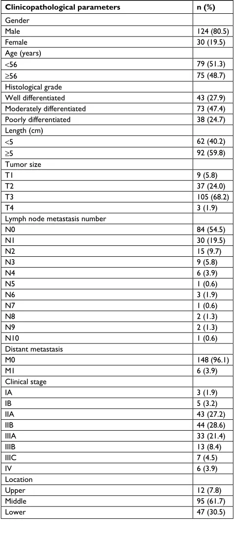
A total of 154 (124 males and 30 females) patients were included in the study. The median age was 55 years (range, 37–48 years). The median length of the tumor was 5 cm (range, 2–10 cm). Tumor locations were upper thoracic in 12 patients, middle thoracic in 95 patients, and lower thoracic in 47 patients. The histopathological differentiations were poor in 38 cases, moderate in 73 cases, and well in 43 cases. A total of 108 patients (70.1%) had T3/T4 tumors, and 70 patients (45.5%) had positive lymph nodes. The pathological stages were stage I in eight patients, stage II in 87 patients, stage III in 53 patients, and stage IV in six patients. The estimated 1-, 3-, and 5-year overall survival (OS) rates were 85.1, 61.0, and 51.9%, respectively. The median OS was 41.5 months (range, 1–82 months). Patient characteristics are listed in Table S1.
The expression pattern of PD-l1 and
PD-L2 and the infiltration of PD-1
+,
TigiT
+, and CD8
+Tils in esCC
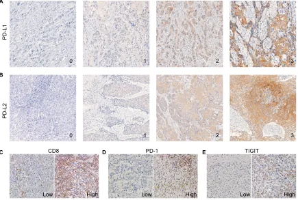
The PD-1+, TIGIT+, and CD8+ TILs in cancer nests and
the expression of PD-L1 and PD-L2 in tumor tissues were observed by IHC staining. Representative IHC images of PD-L1, PD-L2, PD-1, TIGIT, and CD8 are presented in Figure 1A–E. PD-L1 and PD-L2 were highly expressed in 45.5 and 59.7% of the ESCC samples, respectively (Table 1 and Figure 1A and B). Among 154 ESCC specimens, the mean number of infiltrating PD-1+, TIGIT+, and CD8+ T cells
was 59.21±61.31 TILs/mm2 (median 43.8, range 0–250.0),
41.20±29.90 TILs/mm2 (median 41.5, range 0–133.0), and
323.31±140.47 TILs/mm2 (median 290.0, range 0–790.0),
respectively. The high densities of CD8+, PD-1+, and TIGIT+
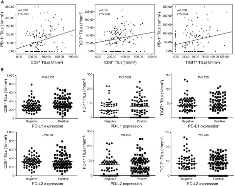
cells were expressed in 41.6, 47.4, and 49.4% of the ESCC patients, respectively (Table 2 and Figure 1C, D, and E). In addition, a significant positive correlation was found between the densities of PD-1+ and CD8+ T cells (r=0.278, P<0.001),
TIGIT+ and CD8+ T cells (r=0.18, P=0.025), and PD-1+ and
TIGIT+ cells (r=0.236, P=0.003) in the tumor tissues of the
154 primary ESCC cases (Figure 2A).
To investigate the relationship between PD-Ls’ expres-sion by tumor cells and immune cell-related parameters, we performed a comparative analysis of PD-L1 and PD-L2 expressions by tumor cells and the number of PD-1+, CD8+,
and TIGIT+ TILs. High expression of PD-L1 in ESCC was
Cancer Management and Research downloaded from https://www.dovepress.com/ by 118.70.13.36 on 20-Aug-2020
Dovepress Zhao et al
associated significantly with the number of CD8+ T cells
but not with PD-1+ and TIGIT+ TILs (P=0.0157, Figure
2B). However, no significant relationship between PD-L2 expression and the number of PD-1+, TIGIT+, or CD8+ TILs
was detected (Figure 2B).
Simultaneous expression of immune
checkpoints in primary ESCC identifies
patients with poor clinical outcome
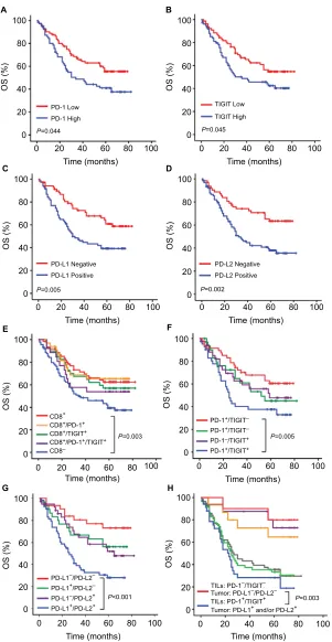
Kaplan–Meier survival analysis revealed that the patients carrying a high number of PD-1+ TILs (n=73/154, 47.4%) or
TIGIT+ TILs (n=76/154, 49.4%) tended to exhibit a shorter
OS (P=0.044, Figure 3A, and P=0.045, Figure 3B). Patients with PD-L1-positive tumors (n=83/154, 53.9%) had a shorter OS than those with PD-L1-negative tumors (n=71/154, 46.1%) (P=0.005, Figure 3C). Patients with PD-L2-positive tumors (n=92/154, 59.7%) displayed a shorter OS compared with those with PD-L2-negative tumor cells (n=62/154, 40.3%) (P=0.002, Figure 3D).
Multivariate analysis of the patients as grouped was per-formed with other clinicopathological predictors for survival
time using the Cox regression model. The results indicated that PD-1+ TILs, TIGIT+ TILs, PD-L1, and PD-L2-positive
expressions in cancer were the independent unfavorable prognostic factors in ESCC (Table 3).
To investigate the impact of immune checkpoint mol-ecules on the prognostic impact of the CD8+ TILs group,
we analyzed the protein expression of PD-1 and TIGIT and co-expression in this group of tumors. Kaplan–Meier sur-vival analysis showed that patients carrying a high number of CD8+ TILs (n=64/154, 41.6%) displayed a longer OS than
those carrying a low number of CD8+ TILs (CD8-, n=90/154,
58.4%) (P=0.003, Figure 3E). However, no significant dif-ference in OS was observed between those with high levels of CD8+/PD-1+ TILs (n=42/154, 27.3%), CD8+/TIGIT+ TILs
(n=35/154, 22.7%), or CD8+/PD-1+/TIGIT+ (n=27/154,
17.5%) and those with high levels of CD8+ TILs (Figure 3E).
To evaluate the possibility that a high level of both PD-1+
and TIGIT+ T-cell infiltrations in cancer might correlate with
unfavorable patient prognosis, the patients were classified into the following four groups: PD-1+/TIGIT+ TILs (n=42/154,
27.3%), PD-1+/TIGIT- TILs (n=31/154, 20.1%), PD-1-/
PD-L
1
PD-L
2
0 1 2 3
0
CD8
Low High Low High Low High
PD-1 TIGIT
1 2 3
A
B
C D E
Figure 1 Expression patterns of PD-L1, PD-L2, CD8, PD-1, and TIGIT in ESCC samples.
Notes: (A and B) Representative immunohistochemical images of PD-L1 and PD-L2 expressions, which were scored from 0 to 3+. Cases displaying scores of 0 or 1+
were considered negative for PD-L1 and PD-L2 expressions, whereas those displaying scores of 2+ or 3+ were considered positive (original magnification, 200×). (C–E) Representative immunohistochemical images from cases with low vs high numbers of CD8+, PD-1+, and TigiT+ TILs (original magnification, 200×).
Abbreviations: ESCC, esophageal squamous cell carcinoma; TILs, tumor-infiltrating lymphocytes.
Cancer Management and Research downloaded from https://www.dovepress.com/ by 118.70.13.36 on 20-Aug-2020
Dovepress Prognostic significance of immune checkpoints
TIGIT+ TILs (n=34/154, 22.1%), and PD-1-/TIGIT- TILs
(n=47/154, 30.5%). The Kaplan–Meier survival results showed that PD-1+/TIGIT+ TILs in cancer demonstrated
significantly lower survival rates than patients with PD-1-/
TIGIT- TILs (P=0.005, Figure 3F). However, no significant
difference in OS was observed between PD-1+/TIGIT- and
PD-1-/TIGIT+ TILs in patients with cancer and patients
with PD-1-/TIGIT- TILs (P=0.205 and 0.250, respectively, Figure 3F). The median survival time in four groups is 24.5, 42, 39, and 59 months. Similarly, to evaluate the possibility that a high level of both PD-L1 and PD-L2 expressions in cancer might correlate with unfavorable patient prognosis, the patients were classified into the following four groups: PD-L1 positive/PD-L2 positive (n=54/154, 35.1%), PD-L1 positive/PD-L2 negative (n=30/154, 19.5%), PD-L1 negative/ PD-L2 positive (n=39/154, 25.3%), and PD-L1 negative/ PD-L2 negative (n=31/154, 20.1%). The Kaplan–Meier survival results showed that PD-L1-positive/PD-L2-positive patients demonstrated significantly lower survival rates than
PD-L1-negative/PD-L2-negative patients (P<0.001, Figure 3G). However, no significant difference in OS was observed between PD-L1-positive/PD-L2-negative or PD-L1-negative/ PD-L2-positive patients and PD-L1-negative/PD-L2-negative patients (P=0.195 and 0.065, Figure 3G). The median survival time in four groups is 25, 59.5, 42, and 66 months.
To identify the good and poor prognosis of the patients, the combined expression of PD-1, TIGIT in TILs, and PD-L1 and PD-L2 in tumor was analyzed. According to the above prognosis results, we found that patients with PD-1+/
TIGIT+ TILs and PD-L1- and/or PD-L2-positive tumor cells
(n=34/154, 22.1%) had a markedly shorter OS than patients with PD-1-/TIGIT- TILs and PD-L1- and PD-L2-negative
tumor cells (n=10/154, 6.5%) (P=0.003, Figure 3H). The median survival time in two groups is 20 and 72.5 months, respectively. In addition, no significant differences were found between PD-1+ or TIGIT+ alone and both PD-1+/TIGIT+
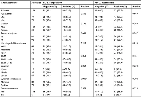
TILs in the patients with PD-L1- and/or PD-L2-positive tumor cells (Figure 3H). Similarly, no significant differences Table 1 Clinicopathological characteristics of ESCC patients according to PD-L1 and PD-L2 expressions
Characteristics All cases PD-L1 expression PD-L2 expression
Negative (%) Positive (%) P-value Negative (%) Positive (%) P-value
All cases 154 71 (46.1) 83 (53.9) 62 (40.3) 92 (59.7)
Age (years) 0.646 0.949
<56 79 35 (44.3) 44 (55.7) 32 (40.5) 47 (59.5)
≥56 75 36 (48.0) 39 (52.0) 30 (40.0) 45 (60.0)
gender 0.196 0.389
Male 124 54 (43.5) 70 (56.5) 52 41.9) 72 (58.1)
Female 30 17 (56.7) 13 (43.3) 10 (33.3) 20 (66.7)
Tumor size (cm) 0.641 0.747
<5 62 30 (48.4) 32 (51.6) 24 (38.7) 38 (61.3)
≥5 92 41 (44.6) 51 (55.4) 38 (41.3) 54 (58.7)
Histological differentiation 0.913 0.015a
Well 43 21 (48.8) 22 (51.2) 25 (58.1) 18 (41.9)
Moderate 73 33 (45.2) 40 (54.8) 26 (35.6) 47 (64.4)
Poor 38 17 (44.7) 21 (55.3) 11 (28.9) 27 (71.1)
TnM 0.051 0.121
TNM (1+2) 98 51 (52.0) 47 (48.0) 44 (44.9) 54 (55.1)
TNM (3+4) 56 20 (35.7) 36 (64.3) 18 (32.1) 38 (67.9)
location 0.064 0.370
Upper 12 6 (50.0) 6 (50.0) 5 (41.7) 7 (58.3)
Middle 95 50 (52.0) 45 (48.0) 42 (44.2) 53 (55.8)
lower 47 15 (31.3) 32 (68.7) 15 (31.9) 32 (68.1)
lymphatic metastasis 0.042a 0.294
negative 84 45 (53.6) 39 (46.4) 37 (44.0) 47 (56.0)
Positive 70 26 (37.1) 44 (62.9) 25 (35.7) 45 (64.3)
Distant metastasis 0.572 0.229
no 148 68 (45.9) 80 (54.1) 61 (41.2) 87 (58.8)
Yes 6 3 (50.0) 3 (50.0) 1 (16.7) 5 (83.3)
Note:aP<0.05.
Abbreviation: ESCC, esophageal squamous cell carcinoma.
Cancer Management and Research downloaded from https://www.dovepress.com/ by 118.70.13.36 on 20-Aug-2020
Dovepress Zhao et al
were found between PD-1- or TIGIT- alone and both PD-1-/ TIGIT- TILs in the patients with PD-L1- and PD-L2-negative tumor cells (Figure 3H).
Multivariate analysis of the patients as grouped was per-formed with other clinicopathological predictors for survival time using the Cox regression model. The results indicated that PD-1+/TIGIT+ TILs, PD-L1-positive/PD-L2-positive and
combined PD-1+/TIGIT+ TILs, and PD-L1 positive and/or
PD-L2 positive in cancer were the independent unfavorable prognostic factors in ESCC (Table 3).
Discussion
The exploration of immune-based biomarkers involved in tumor microenvironment is becoming a useful strategy to predict patient’s outcomes and guide immunotherapy. How-ever, the intricate and dynamic immune microenvironment is heterogeneous in different tumor types. Emerging studies have suggested that tumor may evade host immune response
through the expression of immune checkpoints such as PD-1, TIGIT, PD-L1, and PD-L2. A comprehensive detection and assessment of immune checkpoint factors influencing prog-nosis are important for improving patient management of ESCC. However, there is no previous study on the expression of PD-1, TIGIT, PD-L1, and PD-L2 simultaneously in ESCC tissues. Our results showed that PD-1, TIGIT, PD-L1, and PD-L2 were aberrantly overexpressed in ESCC tissues and these proteins expression alone or together had a significantly poorer prognosis than the negative patients. We also com-pared and identified the patients’ cohort with worst prognosis according the combined expression of PD-1, TIGIT, PD-L1, and PD-L2 in ESCC.
ESCC has been regarded as a proinflammatory neoplasm, where tumor cells produce several cytokines (such as TGF-β, IL-6, IL-8, CCL5, and VEGF) that could lead to the recruit-ment and activation of polyclonal CD8+ T cells.20–26 CD8+ T
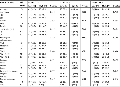
cell is a crucial component of cell-mediated immunity as it Table 2 Clinicopathological characteristics of ESCC patients according to the numbers of PD-1+, CD8+, and TigiT+ Tils
Characteristics All
cases
PD-1+ TILs CD8+ TILs TIGIT+ TILs
Low (%) High (%) P-value Low (%) High (%) P-value Low (%) High (%) P-value
All cases 154 81 (52.6) 73 (47.4) 90 (58.4) 64 (41.6) 78 (50.6) 76 (49.4)
Age (years) 0.640 0.300 0.750
<56 79 43 (54.4) 36 (45.6) 43 (54.4) 36 (45.6) 41 (51.9) 38 (48.1)
≥56 75 38 (50.7) 37 (49.3) 47 (62.7) 28 (37.3) 37 (49.3) 38 (50.7)
gender 0.928 0.308 0.627
Male 124 65 (52.4) 59 (47.6) 70 (56.5) 54 (43.5) 64 (51.6) 60 (48.4) Female 30 16 (53.3) 14 (46.7) 20 (66.7) 10 (33.3) 14 (46.7) 16 (53.3)
Tumor size (cm) 0.647 0.938 0.645
<5 62 34 (54.8) 28 (45.2) 36 (58.1) 26 (41.9) 30 (48.4) 32 (51.6)
≥5 92 47 (51.1) 45 (48.9) 54 (58.7) 38 (41.3) 48 (52.2) 44 (47.8)
Histological differentiation
0.174 0.862 0.145
Well 43 27 (62.8) 16 (37.2) 26 (60.5) 17 (39.5) 27 (62.8) 16 (37.2)
Moderate 73 33 (45.2) 40 (54.8) 41 (56.2) 32 (48.3) 35 (47.9) 38 (52.1)
Poor 38 21 (55.3) 17 (44.7) 23 (60.5) 15 (39.5) 16 (42.1) 22 (57.9)
TnM 0.005a 0.926 0.648
TNM (1+2) 98 60 (61.2) 38 (38.8) 57 (58.2) 41 (41.8) 51 (52.0) 47 (48.0) TNM (3+4) 56 21 (37.5) 35 (62.5) 33 (58.9) 23 (41.1) 27 (48.2) 29 (51.8)
location 0.794 0.448 0.674
Upper 12 7 (58.3) 5 (41.7) 5 (41.7) 7 (58.3) 5 (41.7) 7 (58.3)
Middle 95 48 (50.5) 47 (49.5) 56 (58.9) 39 (41.1) 51 (53.7) 44 (46.3)
lower 47 26 (55.3) 21 (44.7) 29 (61.7) 18 (38.3) 22 (46.8) 25 (53.2)
lymphatic metastasis 0.004a 0.720 0.263
negative 84 53 (63.1) 31 (36.9) 48 (57.1) 36 (42.9) 46 (54.8) 38 (45.2)
Positive 70 28 (40.0) 42 (60.0) 42 (60.0) 28 (40.0) 32 (45.7) 38 (54.3)
Distant metastasis 0.897 0.677 0.423
no 148 78 (52.7) 70 (47.3) 86 (58.1) 62 (41.9) 74 (50.0) 74 (50.0)
Yes 6 3 (50) 3 (50) 4 (66.7) 2 (33.3) 4 (66.7) 2 (33.3)
Note:aP<0.05.
Abbreviations: ESCC, esophageal squamous cell carcinoma; TILs, tumor-infiltrating lymphocytes.
Cancer Management and Research downloaded from https://www.dovepress.com/ by 118.70.13.36 on 20-Aug-2020
Dovepress Prognostic significance of immune checkpoints
produces interferon-γ (IFN-γ) upon interaction with tumor targets, and CD8+ TILs have been correlated with a favorable
outcome in several tumor types.27–29 However, recent findings
demonstrated that high infiltration of CD8+ T cells correlates
with poor survival in renal cell cancer,30 diffuse large B-cell
lymphoma,31 and Hodgkin lymphoma.32 It has been reported
that IFN-γ can induce PD-L1 expression on tumor cells.33,34 In
our study, we found that PD-L1-positive tumor had more CD8+
T cells than PD-L1-negative tumor. In addition, a significant positive correlation was found between the densities of PD-1+
or TIGIT+ and CD8+ T cells. Survival analysis showed that
patients with high CD8 expression had a significantly better clinical outcome than low CD8 expression, but co-expression
of PD-1 or TIGIT with CD8 did not significantly decrease the OS in ESCC patients. Therefore, CD8+ T cells may play an
important role in suppressing tumor progression in ESCC. To the best of our knowledge, this is the first report of the poor prognosis associated with high densities of TIGIT+
cells in ESCC. In addition, we presented evidence that the expression of PD-1 and TIGIT is highly correlated in ESCC. Patients with high expression of PD-1 or TIGIT have signifi-cantly shorter OS than low expression of PD-1 and TIGIT patients, and they have similar survival rate, suggesting that TIGIT may have similar effect with PD-1 in immune inhibition in ESCC. Some studies on mouse models have shown a synergistic effect of the inhibition of both pathways
CD8+ TILs(1/mm2)
PD-L1 expression Negative 0 200 400 600 800 1,000 Positive PD-L2 expression Negative Positive PD-L2 expression Negative Positive PD-L2 expression Negative Positive PD-L1 expression Negative Positive PD-L1 expression Negative Positive CD8
+ TILs (1/m
m 2) 0 200 0 100 200 0 50 100 150 300 400 600 800 1,000 CD8
+ TILs (1/m
m
2)
PD-1
+ TILs (1/m
m
2)
TIGIT
+ TILs (1/m
m 2) 0 100 200 300 PD-1
+ TILs (1/m
m 2) 0 50 100 150 TIGIT
+ TILs (1/m
m 2) 0.00 50.00 100.00 150.00 200.00 250.00 A B PD-1
+ TILs (1/m
m
2)
0.00 200.00 400.00 600.00 800.00
r=0.278
P<0.001
P=0.0157 P=0.0582 P=0.709
P=0.994 P=0.450 P=0.646
0.00 50.00 100.00 150.00 200.00 250.00 PD-1
+ TILs (1/m
m
2)
0.00 25.00 50.00 75.00 100.00 125.00
r=0.236
P=0.003
CD8+ TILs(1/mm2) TIGIT+ TILs(1/mm2) 0.00 0.00 25.00 50.00 75.00 100.00 125.00 TIGIT
+ TILs (1/m
m
2)
200.00 400.00 600.00 800.00
r=0.18
P=0.025
Figure 2 The correlation between the densities of PD-1+, CD8+, and TigiT+ cells and association with PD-L1 and PD-L2 expression status in ESCC samples.
Notes: (A) The number of PD-1+ TILs was significantly positively correlated with the number of CD8+ Tils and TigiT+ TILs per unit area (mm2). (B) Comparing the number
of CD8+, PD-1+, and TigiT+ Tils according to the PD-l1 and PD-l2 expression status in esCC patients. The number of CD8+ TILs was much higher in ESCC cases displaying
PD-L1 expression compared with those lacking PD-L1 expression (P=0.0157). However, the number of PD-1+ Tils and TigiT+ Tils did not differ according to the PD-l1
expression status. The number of CD8+ Tils, PD-1+ Tils, and TigiT+ Tils did not differ according to the PD-l2 expression status. Abbreviations: ESCC, esophageal squamous cell carcinoma; TILs, tumor-infiltrating lymphocytes.
Cancer Management and Research downloaded from https://www.dovepress.com/ by 118.70.13.36 on 20-Aug-2020
Dovepress Zhao et al
0 0 20 40 OS (% ) 60 80 100 A 20 40 Time (months) PD-1 Low PD-1 High P=0.044
60 80 100 0
0 20 40 OS (% ) 60 80 100 B 20 40 Time (months) TIGIT Low TIGIT High P=0.045
60 80 100
0 0 20 40 OS (% ) 60 80 100 C 20 40 Time (months) PD-L1 Negative PD-L1 Positive P=0.005
60 80 100 0
0 20 40 OS (% ) 60 80 100 D 20 40 Time (months) PD-L2 Negative PD-L2 Positive P=0.002
60 80 100
0 0 20 40 OS (% ) 60 80 100 E 20 40 Time (months) CD8+
CD8+/PD-1+ CD8+/TIGIT+ CD8+/PD-1+/TIGIT+ CD8–
P=0.003
PD-L1–/PD-L2– PD-L1+/PD-L2– PD-L1–/PD-L2+ PD-L1+/PD-L2+
TILs: PD-1–/TIGIT– Tumor: PD-L1–/PD-L2– TILs: PD-1+/TIGIT+ Tumor: PD-L1+ and/or PD-L2+
P<0.001 P=0.003
PD-1+/TIGIT– PD-1+/TIGIT– PD-1–/TIGIT+ PD-1+/TIGIT+
P=0.005
60 80 100 0 0 20 40 OS (% ) 60 80 100 F 20 40 Time (months)
60 80 100
0 0 20 40 OS (% ) 60 80 100 G 20 40 Time (months)
60 80 100 0 0 20 40 OS (% ) 60 80 100 H 20 40 Time (months)
60 80 100
Figure 3 Kaplan–Meier analyses of OS of 154 ESCC patients according to CD8, PD-1, TIGIT, PD-L1, and PD-L2 expressions alone or combined.
Notes: (A and B) Patients with a high number of PD-1+ or TigiT+ TILs tended to exhibit a shorter OS (P=0.044 and 0.045). (C and D) Patients with a positive expression
of PD-L1 or PD-L2 tended to exhibit a shorter OS (P=0.005 and 0.002). (E) Comparing the OS between CD8+/PD-1+ (yellow line), CD8+/TigiT+ (green line), CD8+/PD-1+/
TigiT+ (purple line), CD8- (blue line) TILs, and CD8+ TILs (red line) in ESCC. (F) Comparing the OS between PD-1+/TigiT- (green line), PD-1-/TigiT+ (purple line), PD-1+/
TigiT+ (blue line), and PD-1-/TigiT- (red line) TILs in ESCC. (G) Comparing the OS between tumor with PD-L1-positive/PD-L2-negative (green line),
PD-L1-negative/PD-L2-positive (purple line), PD-L1-positive/PD-PD-L1-negative/PD-L2-positive (blue line) expressions and those with PD-L1-negative/PD-L2-negative (red line) expression in ESCC. (H) Comparing
the Os between patients with PD-1+/TigiT+ TILs and PD-L1 and/or PD-L2-positive tumor cells (blue line) and patients with PD-1-/TigiT- Tils and PD-l1 and PD-l2-negative
tumor cells (red line) in ESCC. Comparing the OS between patients with PD-1+/TigiT- (purple line), PD-1-/TigiT+ (yellow line), and PD-1-/TigiT- (red line) TILs in PD-L1-
and PD-L2-negative tumor cells of ESCC. Comparing the OS between patients with PD-1+/TigiT- (gray line), PD-1-/TigiT+ (green line), and PD-1+/TigiT+ (blue line) TILs in
PD-L1- and/or PD-L2-positive tumor cells of ESCC.
Abbreviations: ESCC, esophageal squamous cell carcinoma; OS, overall survival; TILs, tumor-infiltrating lymphocytes.
Cancer Management and Research downloaded from https://www.dovepress.com/ by 118.70.13.36 on 20-Aug-2020
Dovepress Prognostic significance of immune checkpoints
in boosting the antitumor immune response.17 Thus, our
results suggest that cooperation between infiltrating PD-1+
and TIGIT+ lymphocytes in tumors might be important in
the progression of ESCC and support the rationale of dual blockade of these molecules in ESCC.
Our results found that the density of PD-1+ cells and
the tumor expression of PD-L1 in primary ESCC were associated with a poor clinical outcome. Furthermore, we described the prognostic significance of PD-L2. PD-L2 seems to be expressed in a higher proportion of tumors (59.7%) than PD-L1 (53.9%), and PD-L2 expression is associated with a poor clinical outcome. We also found that patients with PD-L1-positive/PD-L2-positive tumor had significantly shorter OS than PD-L1-negative/PD-L2-neg-ative patients, and PD-L1 positive/PD-L2 positive was an independent unfavorable prognostic factor in ESCC. This finding might have clinical relevance because anti-PD-L1 treatment alone seems to have a lower response rate than anti-PD-1.35,36 In addition, there are several PD-L1-negative
tumors that respond to anti-PD-1 treatment,35 implying that
there are other molecules beside PD-L1 that are involved in the PD-1 inhibition axis of ESCC. A few publications have reported PD-L2 expression in other tumors, including NSCLC,37 ovarian cancer, and primary mediastinal large
B-cell lymphoma,38,39 where it has shown a restricted impact
on patients’ prognosis.
The immunological status in the tumor microenvironment is now well recognized to be a critical determining factor in tumor prevention, development, and progression. Better understanding of the roles of immune cells and molecules in the tumor microenvironment will therefore be essential
in the development of histology-specific tumors. Combined analysis of PD-1, TIGIT expression in TILs, and tumorous PD-L1 and PD-L2 expressions in ESCC, we found that only a small proportion of patients (n=10/154, 6.5%) with PD-1-/ TIGIT- TILs and PD-L1- and PD-L2-negative tumor cells had best prognosis (median survival time, 72.5 months) and a higher proportion of patients (n=34/154, 22.1%) with PD-1+/
TIGIT+ TILs and PD-L1 and/or PD-L2-positive tumor cells
had worst prognosis (median survival time, 20 months). Mul-tivariate analysis showed that combined PD-1+/TIGIT+ TILs
and PD-L1 positive and/or PD-L2 positive in cancer were the independent unfavorable prognostic factors in ESCC. Our data suggest that patients with PD-1+/TIGIT+ TILs and
PD-L1- and/or PD-L2-positive tumor cells may benefit from immune checkpoints blockade therapy but need multiple blockade of these immune molecules or optimize immune checkpoints blockade in ESCC.
Conclusion
Our results evaluated novel prognostic factors in ESCC based on the concomitant quantification of densities of CD8+,
PD-1+, and TIGIT+ TILs in addition to PD-L1/PD-L2
expres-sion by tumor cells. These immune profiles should guide the selection of patients to receive suitable immunotherapies and need to be further validated in larger and independent cohorts.
Acknowledgment
This work was supported by grants from the National Natural Science Foundation of China (no 81402560) and the Guang-dong Province Science and Technology Plan Project (no 2017A020215029).
Table 3 Univariate and multivariate analyses of overall survival in ESCC
Variables Univariate analysis Multivariate analysis
HR 95% CI P-value HR 95% CI P-value
CD8 0.486 0.295–0.799 0.004a 0.308 0.177–0.534 <0.001a
PD1 1.659 1.052–2.616 0.029a 1.773 1.054–2.981 0.031a
TigiT 1.585 1.003–2.502 0.048a 1.808 1.119–2.919 0.016a
PD-l1 1.997 1.239–3.221 0.005a 2.138 1.300–3.517 0.003a
PD-l2 2.200 1.335–3.624 0.002a 1.495 0.884–2.528 0.134
PD1 and TigiT 1.518 1.124–2.048 0.006a 1.443 1.067–1.952 0.017a
PD-l1 and PD-l2 2.063 1.466–2.902 0.001a 1.910 1.342–2.717 <0.001a
PD1 and TigiT + PD-l1 and/or PD-l2 1.729 1.312–2.279 <0.001a 1.548 1.161–2.065 0.003a
Histological grade 1.701 1.229–2.356 0.001a 1.475 1.048–2.075 0.026a
Age (years) 1.101 0.700–1.734 0.677
gender 0.954 0.541–1.680 0.869
length 1.179 0.741–1.877 0.488
location 1.153 0.774–1.718 0.483
TnM 3.368 2.130–5.324 <0.001a 2.927 1.767–4.848 <0.001a
Note:aP<0.05.
Abbreviation: ESCC, esophageal squamous cell carcinoma.
Cancer Management and Research downloaded from https://www.dovepress.com/ by 118.70.13.36 on 20-Aug-2020
Dovepress Zhao et al
Author contributions
JJZ and ZQZ were the major contributors in writing the manuscript. PW analyzed the experimental data. ZQZ col-lected the tissues and clinical data of patients. CLC and YL drew the figures. QZP did the data interpretation. QZ and YT searched the literature. DSW and JCX designed this study. All authors contributed to data analysis, drafting and revising the article, gave final approval of the version to be published, and agree to be accountable for all aspects of the work.
Disclosure
The authors report no conflicts of interest in this work.
References
1. Rustgi AK, El-Serag HB. Esophageal carcinoma. N Engl J Med. 2014;371(26):2499–2509.
2. Hanahan D, Weinberg RA. The hallmarks of cancer. Cell. 2000;100(1):57–70.
3. Giraldo NA, Becht E, Remark R, Damotte D, Sautès-Fridman C, Frid-man WH. The immune contexture of primary and metastatic huFrid-man tumours. Curr Opin Immunol. 2014;27:8–15.
4. Zitvogel L, Galluzzi L, Kepp O, Smyth MJ, Kroemer G. Type I inter-ferons in anticancer immunity. Nat Rev Immunol. 2015;15(7):405–414. 5. Fridman WH, Pagès F, Sautès-Fridman C, Galon J. The immune contex-ture in human tumours: impact on clinical outcome. Nat Rev Cancer. 2012;12(4):298–306.
6. Dong H, Chen L. B7-H1 pathway and its role in the evasion of tumor immunity. J Mol Med (Berl). 2003;81(5):281–287.
7. Dong H, Zhu G, Tamada K, Chen L. B7-H1, a third member of the B7 family, co-stimulates T-cell proliferation and interleukin-10 secretion. Nat Med. 1999;5(12):1365–1369.
8. Latchman Y, Wood CR, Chernova T, et al. PD-L2 is a second ligand for PD-1 and inhibits T cell activation. Nat Immunol. 2001;2(3): 261–268.
9. Keir ME, Butte MJ, Freeman GJ, Sharpe AH. PD-1 and its ligands in tolerance and immunity. Annu Rev Immunol. 2008;26:677–704. 10. Feng Z, Xiang-Lei L, Hai-Tao W, et al. Programmed cell death 1
expres-sion in esophageal squamous cell carcinoma and association with clinical characteristics. Indian J Cancer. 2015;52 Suppl 3:E176–178. 11. Chen L, Deng H, Lu M, et al. B7-H1 expression associates with tumor
invasion and predicts patient’s survival in human esophageal cancer. Int J Clin Exp Pathol. 2014;7(9):6015–6023.
12. Ohigashi Y, Sho M, Yamada Y, et al. Clinical significance of programmed death-1 ligand-1 and programmed death-1 ligand-2 expression in human esophageal cancer. Clin Cancer Res. 2005;11(8):2947–2953. 13. Brahmer J, Reckamp KL, Baas P, et al. Nivolumab versus Docetaxel
in Advanced Squamous-Cell Non-Small-Cell Lung Cancer. N Engl J Med. 2015;373(2):123–135.
14. Song Y, Li L, Ou Y, et al. Identification of genomic alterations in oesophageal squamous cell cancer. Nature. 2014;509(7498):91–95. 15. Kojima T, Doi T. Immunotherapy for Esophageal Squamous Cell
Car-cinoma. Curr Oncol Rep. 2017;19(5):33.
16. Chauvin JM, Pagliano O, Fourcade J, et al. TIGIT and PD-1 impair tumor antigen-specific CD8+ T cells in melanoma patients. J Clin Invest. 2015;125(5):2046–2058.
17. Johnston RJ, Comps-Agrar L, Hackney J, et al. The immunoreceptor TIGIT regulates antitumor and antiviral CD8(+) T cell effector function. Cancer Cell. 2014;26(6):923–937.
18. Hodi FS, O’Day SJ, McDermott DF, et al. Improved survival with ipilimumab in patients with metastatic melanoma. N Engl J Med. 2010;363(8):711–723.
19. Motzer RJ, Escudier B, McDermott DF, et al. Nivolumab versus Everolimus in Advanced Renal-Cell Carcinoma. N Engl J Med. 2015;373(19):1803–1813.
20. Abdel-Latif MM, Duggan S, Reynolds JV, Kelleher D. Inflammation and esophageal carcinogenesis. Curr Opin Pharmacol. 2009;9(4):396–404. 21. Deans DA, Wigmore SJ, Gilmour H, Paterson-Brown S, Ross JA, Fearon KC. Elevated tumour interleukin-1beta is associated with systemic inflammation: A marker of reduced survival in gastro-oesophageal cancer. Br J Cancer. 2006;95(11):1568–1575.
22. Doyle SL, Donohoe CL, Finn SP, et al. IGF-1 and its receptor in esopha-geal cancer: association with adenocarcinoma and visceral obesity. Am J Gastroenterol. 2012;107(2):196–204.
23. Hong S, Lee HJ, Kim SJ, Hahm KB. Connection between inflammation and carcinogenesis in gastrointestinal tract: focus on TGF-beta signal-ing. World J Gastroenterol. 2010;16(17):2080–2093.
24. Jenkins GJ, Mikhail J, Alhamdani A, et al. Immunohistochemical study of nuclear factor-kappaB activity and interleukin-8 abundance in oesophageal adenocarcinoma; a useful strategy for monitoring these biomarkers. J Clin Pathol. 2007;60(11):1232–1237.
25. Liu J, Li F, Ping Y, et al. Local production of the chemokines CCL5 and CXCL10 attracts CD8+ T lymphocytes into esophageal squamous cell carcinoma. Oncotarget. 2015;6(28):24978–24989.
26. O’Sullivan KE, Phelan JJ, O’Hanlon C, Lysaght J, O’Sullivan JN, Reynolds JV. The role of inflammation in cancer of the esophagus. Expert Rev Gastroenterol Hepatol. 2014;8(7):749–760.
27. Shankaran V, Ikeda H, Bruce AT, et al. IFNgamma and lymphocytes prevent primary tumour development and shape tumour immunogenic-ity. Nature. 2001;410(6832):1107–1111.
28. Pagès F, Kirilovsky A, Mlecnik B, et al. In situ cytotoxic and memory T cells predict outcome in patients with early-stage colorectal cancer. J Clin Oncol. 2009;27(35):5944–5951.
29. Zhang L, Conejo-Garcia JR, Katsaros D, et al. Intratumoral T cells, recurrence, and survival in epithelial ovarian cancer. N Engl J Med. 2003;348(3):203–213.
30. Giraldo NA, Becht E, Pagès F, et al. Orchestration and Prognostic Signifi-cance of Immune Checkpoints in the Microenvironment of Primary and Metastatic Renal Cell Cancer. Clin Cancer Res. 2015;21(13):3031–3040. 31. Muris JJ, Meijer CJ, Cillessen SA, et al. Prognostic significance of
activated cytotoxic T-lymphocytes in primary nodal diffuse large B-cell lymphomas. Leukemia. 2004;18(3):589–596.
32. Scott DW, Chan FC, Hong F, et al. Gene expression-based model using formalin-fixed paraffin-embedded biopsies predicts overall sur-vival in advanced-stage classical Hodgkin lymphoma. J Clin Oncol. 2013;31(6):692–700.
33. Abiko K, Matsumura N, Hamanishi J, et al. IFN-γ from lymphocytes induces PD-L1 expression and promotes progression of ovarian cancer. Br J Cancer. 2015;112(9):1501–1509.
34. Bellucci R, Martin A, Bommarito D, et al. Interferon-γ-induced acti-vation of JAK1 and JAK2 suppresses tumor cell susceptibility to NK cells through upregulation of PD-L1 expression. Oncoimmunology. 2015;4(6):e1008824.
35. Brahmer JR, Tykodi SS, Chow LQ, et al. Safety and activity of anti-PD-L1 antibody in patients with advanced cancer. N Engl J Med. 2012;366(26):2455–2465.
36. Topalian SL, Hodi FS, Brahmer JR, et al. Safety, activity, and immune correlates of anti-PD-1 antibody in cancer. N Engl J Med. 2012;366(26):2443–2454.
37. Zhang Y, Wang L, Li Y, et al. Protein expression of programmed death 1 ligand 1 and ligand 2 independently predict poor prognosis in surgically resected lung adenocarcinoma. Onco Targets Ther. 2014;7:567–573. 38. Hamanishi J, Mandai M, Iwasaki M, et al. Programmed cell death 1 ligand
1 and tumor-infiltrating CD8+ T lymphocytes are prognostic factors of human ovarian cancer. Proc Natl Acad Sci U S A. 2007;104(9):3360–3365. 39. Shi M, Roemer MG, Chapuy B, et al. Expression of programmed cell
death 1 ligand 2 (PD-L2) is a distinguishing feature of primary medias-tinal (thymic) large B-cell lymphoma and associated with PDCD1LG2 copy gain. Am J Surg Pathol. 2014;38(12):1715–1723.
Cancer Management and Research downloaded from https://www.dovepress.com/ by 118.70.13.36 on 20-Aug-2020
Dovepress Prognostic significance of immune checkpoints
Supplementary material
Table S1 Clinicopathological features of the 154 esophageal cancer patients (at the time of initial diagnosis)
Clinicopathological parameters n (%)
gender
Male 124 (80.5)
Female 30 (19.5)
Age (years)
<56 79 (51.3)
≥56 75 (48.7)
Histological grade
Well differentiated 43 (27.9)
Moderately differentiated 73 (47.4)
Poorly differentiated 38 (24.7)
Length (cm)
<5 62 (40.2)
≥5 92 (59.8)
Tumor size
T1 9 (5.8)
T2 37 (24.0)
T3 105 (68.2)
T4 3 (1.9)
lymph node metastasis number
n0 84 (54.5)
n1 30 (19.5)
n2 15 (9.7)
n3 9 (5.8)
n4 6 (3.9)
n5 1 (0.6)
n6 3 (1.9)
n7 1 (0.6)
n8 2 (1.3)
n9 2 (1.3)
n10 1 (0.6)
Distant metastasis
M0 148 (96.1)
M1 6 (3.9)
Clinical stage
ia 3 (1.9)
iB 5 (3.2)
iia 43 (27.2)
iiB 44 (28.6)
iiia 33 (21.4)
iiiB 13 (8.4)
iiiC 7 (4.5)
iV 6 (3.9)
location
Upper 12 (7.8)
Middle 95 (61.7)
lower 47 (30.5)
Cancer Management and Research downloaded from https://www.dovepress.com/ by 118.70.13.36 on 20-Aug-2020
Dovepress
Cancer Management and Research
Publish your work in this journal
Submit your manuscript here: https://www.dovepress.com/cancer-management-and-research-journal
Cancer Management and Research is an international, peer-reviewed open access journal focusing on cancer research and the optimal use of preventative and integrated treatment interventions to achieve improved outcomes, enhanced survival and quality of life for the cancer patient. The manuscript management system is completely online and includes
a very quick and fair peer-review system, which is all easy to use. Visit http://www.dovepress.com/testimonials.php to read real quotes from published authors.
Dove
press
Zhao et al
Cancer Management and Research downloaded from https://www.dovepress.com/ by 118.70.13.36 on 20-Aug-2020