doi:10.1136/thorax.57.10.847
2002;57;847-852
Thorax
G C Donaldson, T A R Seemungal, A Bhowmik and J A Wedzicha
pulmonary disease
lung function decline in chronic obstructive
Relationship between exacerbation frequency and
http://thorax.bmj.com/cgi/content/full/57/10/847
Updated information and services can be found at:
These include:
References
http://thorax.bmj.com/cgi/content/full/57/10/847#otherarticles
98 online articles that cite this article can be accessed at:
http://thorax.bmj.com/cgi/content/full/57/10/847#BIBL
This article cites 25 articles, 18 of which can be accessed free at:
service
Email alerting
top right corner of the article
Receive free email alerts when new articles cite this article - sign up in the box at the
Correction
http://thorax.bmj.com/cgi/content/full/63/8/753
online at:
been appended to the original article in this reprint. The correction is also available
have
correctionA correction has been published for this article. The contents of the
Notes
http://journals.bmj.com/cgi/reprintform
To order reprints of this article go to:
go to:
Thorax
ORIGINAL ARTICLE
Relationship between exacerbation frequency and lung
function decline in chronic obstructive pulmonary disease
G C Donaldson, T A R Seemungal, A Bhowmik, J A Wedzicha
. . . .
Thorax 2002;57:847–852
Background:Chronic obstructive pulmonary disease (COPD) is characterised by both an accelerated decline in lung function and periods of acute deterioration in symptoms termed exacerbations. The aim of this study was to investigate whether these are related.
Methods:Over 4 years, peak expiratory flow (PEF) and symptoms were measured at home daily by 109 patients with COPD (81 men; median (IQR) age 68.1 (63–74) years; arterial oxygen tension (PaO2) 9.00 (8.3–9.5) kPa, forced expiratory volume in 1 second (FEV1) 1.00 (0.7–1.3) l, forced vital capacity (FVC) 2.51 (1.9–3.0) l); of these, 32 (29 men) recorded daily FEV1. Exacerbations were iden-tified from symptoms and the effect of frequent or infrequent exacerbations (> or < 2.92 per year) on lung function decline was examined using cross sectional, random effects models.
Results:The 109 patients experienced 757 exacerbations. Patients with frequent exacerbations had a significantly faster decline in FEV1 and peak expiratory flow (PEF) of –40.1 ml/year (n=16) and –2.9 l/min/year (n=46) than infrequent exacerbators in whom FEV1 changed by –32.1 ml/year (n=16) and PEF by –0.7 l/min/year (n=63). Frequent exacerbators also had a greater decline in FEV1 if allowance was made for smoking status. Patients with frequent exacerbations were more often admit-ted to hospital with longer length of stay. Frequent exacerbations were a consistent feature within a patient, with their number positively correlated (between years 1 and 2, 2 and 3, 3 and 4).
Conclusions:These results suggest that the frequency of exacerbations contributes to long term decline in lung function of patients with moderate to severe COPD.
C
hronic obstructive pulmonary disease (COPD) is in the top five leading causes of deaths in the world, and is associated with a significant health and economic bur-den through hospital admission and absenteeism from work.1 2The accelerated decline in forced expiratory volume in1 second (FEV1) with age has been related to the severity of
airway inflammation.3
Exacerbations of COPD are an important cause of unsched-uled physician visits and hospitalisation, and patients prone to frequent exacerbations have a reduced quality of life.4
We have recently shown that COPD exacerbations may increase airway inflammation5
and that a significant proportion may not show complete symptomatic or physiological resolution.6
Thus, it is possible that exacerbations by their incomplete recovery play a part in the decline in FEV1.
Previous studies of the relation of exacerbations and decline in lung function have been inconclusive. In 1976 Fletcher and colleagues7
concluded from their 10 year study that there was no relation between lung function decline and chest infections. However, their cohort had mild or no airflow obstruction, the diagnosis of exacerbation was retrospectively based on patient recollections, and the study possibly lacked the statistical power to identify any effect after adjustment for smoking, age, and body size. Kanner and colleagues8
reported in 1979 an association between decline in lung function and episodes of lower respiratory tract illnesses, although again the patients were relatively young and with mild airflow obstruction. More recently the Lung Health Study9
has reported that one lower respiratory tract illness per year, for which a physician was consulted, increased FEV1decline by
7 ml/year in continuing smokers and intermittent smokers. A similar effect was not observed in sustained quitters and the average number of illnesses was only 0.24 per year. However, whether the decline in FEV1 is related to exacerbation
frequency in patients with moderate to severe COPD is not
known. Such information would be of importance as currently only smoking cessation is known to affect FEV1 decline in
COPD.
In this study we have prospectively investigated a cohort of 109 patients with moderate to severe COPD over a 4 year period. Daily lung function and symptomatic data were collected to identify exacerbations and the relationship between decline in lung function and exacerbation frequency was then evaluated.
METHODS
Patients
Between November 1995 and November 1998, 161 patients with a diagnosis of COPD who attended outpatient clinics were recruited. Inclusion criteria were: FEV1<70% predicted
from age and height,β2 agonist reversibility <15% and/or
200 ml, and absence of asthma, bronchiectasis, bronchial car-cinoma, or other significant respiratory disease.10 11
To investi-gate changes over time, the analysis was confined to 109 of these 161 patients who had recorded data on at least 365 days. The reasons for failing to record 365 days of data were withdrawal from the study, death, or inadequate completion of diary cards combined with late enrolment. These 109 patients did not differ significantly from the 52 excluded patients in any of the characteristics reported in table 1 except in peak expiratory flow (PEF).
Baseline measurements were made of spirometric para-meters and PEF by rolling seal spirometer (Sensor Medic Cor-poration, Yorba Lindo, CA, USA) when the patients were stable—that is, with no history of an exacerbation requiring treatment during the previous 4 weeks. Measurements were also taken of reversibility to 400µg inhaled salbutamol and arterialised ear lobe blood gas tensions (model 278 Blood Gas Analyzer; Ciba-Corning, Medfield, MA, USA),12and a history
was taken of smoking, chronic symptoms, and long term
See end of article for authors’ affiliations
. . . .
Correspondence to: Professor J A Wedzicha, Academic Unit of Respiratory Medicine, Dominion House, St Bartholomew’s Hospital, London EC1A 7BE, UK; [email protected]
Revised version received 30 May 2002 Accepted for publication 5 June 2002
inhaled and oral steroid use. The patients recorded on diary cards daily PEF (Mini-Wright, Clement Clark International Ltd, Harlow, UK) measured after medication around 10.00 hours, and increase over normal in symptoms of dyspnoea, amount and purulence of sputum, wheeze, sore throat, cough, or nasal congestion/discharge. Due to funding restrictions only 32 patients (29 men) in addition measured daily FEV1at
home using a spirometer (Micro Medical Ltd, Chatham, Kent, UK). They were similar to the other 77 patients in all charac-teristics listed in table 1, except they had a higher PEF (245 (IQR 180–300) v 174 (IQR 150–240) l/min, p=0.003 (Wil-coxon test)).
The study was approved by the ethics committee of the East London and City Health Authority and the patients provided written informed consent.
Exacerbation identification and treatment
Every 3 months patients were reviewed in the outpatients clinic and their diary cards collected. Exacerbations were diagnosed according to previously accepted criteria4–6 13
if the following symptom patterns were experienced for at least two consecutive days: either two or more of three major symptoms (increase in dyspnoea, sputum purulence or sputum volume)
or any one major symptom together with any one of the flow-ing minor symptoms: increase in nasal discharge, wheeze, sore throat, cough or fever. Symptoms recorded continuously in the 5 day period before exacerbation onset were discounted when deciding whether an exacerbation had occurred or not. Some exacerbations where no diary card symptoms were recorded were identified by questioning the patient about their symptoms when at the clinic visits or on admission to hospi-tal. Patients experiencing increased symptoms were encour-aged to contact the clinical team and were generally seen before treatment within 48 hours. These exacerbations were classified as “reported exacerbations” with those unseen termed “unreported exacerbations”. During the study the number of hospital admissions and length of hospital stay for acute exacerbations of COPD were noted. Records of drug treatment, dosage, and course duration at these visits were kept from November 1996 and any treatment by primary care physicians was obtained where possible.
Exacerbation frequency
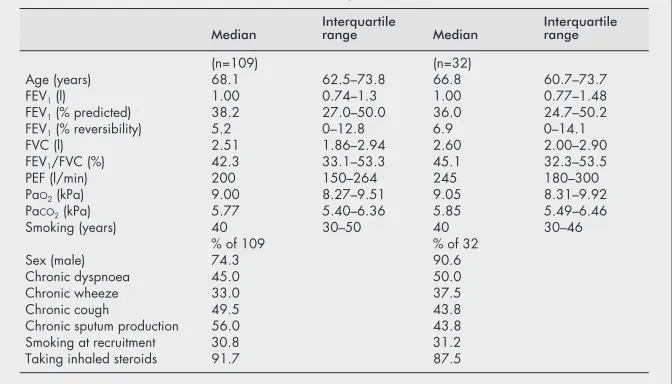
The annual exacerbation rate of each patient was calculated by dividing the number of exacerbations by the number of days they participated in the study, and multiplying by 365. As the Table 1 Characteristics of the 109 study patients with COPD measured at
recruitment and of the 32 who recorded FEV1
Median Interquartilerange Median Interquartilerange
(n=109) (n=32)
Age (years) 68.1 62.5–73.8 66.8 60.7–73.7 FEV1(l) 1.00 0.74–1.3 1.00 0.77–1.48
FEV1(% predicted) 38.2 27.0–50.0 36.0 24.7–50.2
FEV1(% reversibility) 5.2 0–12.8 6.9 0–14.1
FVC (l) 2.51 1.86–2.94 2.60 2.00–2.90 FEV1/FVC (%) 42.3 33.1–53.3 45.1 32.3–53.5
PEF (l/min) 200 150–264 245 180–300 PaO2(kPa) 9.00 8.27–9.51 9.05 8.31–9.92
PaCO2(kPa) 5.77 5.40–6.36 5.85 5.49–6.46
Smoking (years) 40 30–50 40 30–46 % of 109 % of 32
Sex (male) 74.3 90.6
Chronic dyspnoea 45.0 50.0
Chronic wheeze 33.0 37.5
Chronic cough 49.5 43.8
Chronic sputum production 56.0 43.8 Smoking at recruitment 30.8 31.2 Taking inhaled steroids 91.7 87.5
FEV1=forced expiratory volume in 1 second; FVC=forced vital capacity; PEF=peak expiratory flow; PaO2;
PaCO2=arterial oxygen and carbon dioxide tensions.
Table 2 Annual changes in treatment of exacerbations with oral steroids and antibiotics
Exponentiated regression
coefficient* 95% CI p value No ofexacerbations No ofsubjects†
Odds ratio
Oral steroids (infrequent
exacerbators) 0.89 0.59 to 1.35 0.589 165 49 Oral steroids (frequent
exacerbators)
1.20 0.90 to 1.60 0.208 396 45
Antibiotics (infrequent exacerbators)
0.87 0.61 to 1.26 0.477 165 49
Antibiotics (frequent
exacerbators) 1.106 0.87 to 1.41 0.418 396 45
*The regression coefficient is of the form eband is the odds ratio (OR) giving the relative amount by which
the odds of the outcome increase (OR >1) or decrease (OR <1) when the independent variable time is increased by 1 year.
†Data were not available on all 109 patients as records of medication were only kept from November 1996 and not all patients had exacerbations.
study focused on decline in FEV1, we calculated the median
exacerbation rate in the 32 patients recording FEV1 and
divided all the patients into two groups—those experiencing less than the median rate of 2.92 exacerbations per year (“infrequent exacerbators”) and those with more than 2.92 per year (“frequent exacerbators”). Exacerbation rates for each 12 month period for all the patients who took part in the study were also calculated from the date of their enrolment and used to assess consistency in exacerbation frequency from year to year.
Statistical analysis
Data are presented as mean (SD) or median (interquartile range; IQR) values and comparisons were performed by unpairedttest, Wilcoxon matched paired sign rank test, orχ2
tests as appropriate.
Cross sectional random effects models were used to determine the effect of exacerbation frequency on decline in lung function,14
with lung function as the dependent variable and time (in years) as the independent variable. These models allow examination of the time variations independently of the cross sectional variations in panel data. The lung function data were logarithmically transformed as the work of Fletcher7
suggests a non-linear decline in lung function over time; the transformation also gave higherr2
values for the regression analysis, indicating that it provided a better model. We calcu-lated the regression coefficients for infrequent and frequent exacerbators groups separately. The coefficients were then compared using their standard errors.15We also tried the more
conventional approach of testing the interaction between exacerbation frequency, as a binary variable, and time after allowance for their main effects. This showed frequent exacer-bations as having a highly significant faster decline in FEV1
(p<0.001). We believe the simpler comparison of standard errors, although not optimal, is statistically more secure as the sample size in this study is relatively small. We also repeated the analysis but stratified the patients into four groups by exacerbation frequency and whether or not the patient currently smoked. The analysis was also repeated after strati-fying the patients into two groups according to whether they had more or less than 1.5 reported exacerbations per year. This was the median rate for those exacerbations seen at clinic by the clinical team. The rationale for this was that the symptoms for these exacerbations were validated. Cross sectional, random effects models were also used to examine changes in binomial data on treatment with oral steroids and antibiotics over time assuming a logit relationship. The analysis was per-formed using Stata 5.0 (Stata Corporation) and the cross sec-tional models fitted using the Xtreg and Xtgee commands.
RESULTS
Patients
The 109 patients studied had moderate to severe COPD (table 1). Of these, 100 patients took inhaled steroids daily and 12 were on prednisolone taking a mean (SD) dose of 5.91 (3.0) mg /day; 10 patients used both oral and inhaled steroids. Data on exacerbation treatment collected over the last 3 years of the study showed that 124 of 561 exacerbations (22.1%) were treated with oral steroids and 341 of 561 (60.8%) with antibiotics. Table 2 shows how the probability of treatment changed per year in the frequent and infrequent exacerbator groups, and indicates that there was no significant change over time in the likelihood of treatment with either oral ster-oids or antibiotics. No significant change was seen if the groups were combined (data not shown).
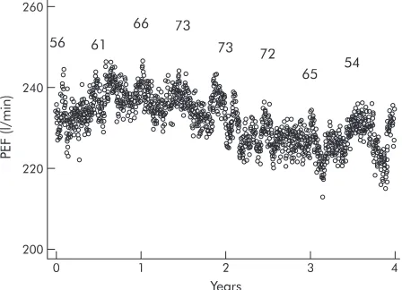
The patients participated in the study for a median of 958 days (range 405–1489) but diary card data were recorded on only a median of 801 days (range 366–1460) or 83% of this time (a total of 95 776 patient days). Figure 1 shows daily mean PEF over the 4 years of the study, although comparisons between days are not possible because patients withdrew and entered the study as it progressed. Non-compliance in record-ing data was due to diary card loss, non-completion while on holiday or in hospital, or when the patient felt too unwell. Eleven patients died and 20 withdrew before the end of the study.
Seven of the 63 infrequent exacerbators were admitted to hospital on a median of one occasion (IQR 1–1) for 10 days (IQR 4–12) while 20 of the 46 frequent exacerbators were admitted on a median of 1.5 occasions per year (IQR 1–3) for 10 days (IQR 7–14). Frequent exacerbators were therefore admitted more frequently (p=0.0001, Wilcoxon) and for longer (p=0.0032, Wilcoxon).
Exacerbations
One hundred of the 109 patients had one or more exacerbations, with a total of 757 of which 649 (85.7%) were
Figure 1 Mean morning PEF after treatment over 4 years. Numbers reflect the number of patients recording data on 1 November and 1 May each year.
PEF
(l/min)
200 220 240 260
0 1 2 3 4
Years 56 61
66
73 72
65 54 73
Table 3 Initial and annual change in lung function in patients with infrequent and frequent exacerbations
Starting value Annual change
Infrequent Frequent Infrequent Frequent
Exacerbations (reported
and unreported) <50% percentile,<2.92 per year > 50% percentile>2.92 per year (n=63) (n=46) PEF (l/min) 214 232 –0.72 –2.94***
(n=16) (n=16) FEV1(ml) 893 950 –32.1 –40.1*
PEF=peak expiratory flow; FEV1=forced expiratory volume in 1 second.
identified from the symptom data recorded by the patients on their diary cards. A total of 380 (50.1%) exacerbations were reported and seen by the clinical team. There were no differences in daily symptoms recorded by the patient at exac-erbation onset between those unreported and those reported to the clinic team (dyspnoea 61% and 62% respectively; sputum purulence 26% and 24%; sputum amount 44% and 39%; wheeze 33% and 31%; sore throat 13% and 11%; cough 23% and 28%) except in nasal congestion/discharge (22% and 34%; p<0.001, χ2
test). Including those patients who experienced no exacerbations over the study period, the median exacerbation rate was 2.53 (IQR 1.33–3.80) exacerba-tions per year, and for those patients recording FEV1 the
median rate was 2.92 (IQR 1.94–4.23) per year; these rates were not significantly different (p=0.210, Wilcoxon). Of the patients who recorded FEV1, those classified as frequent
exac-erbators had a median rate of 4.2 (IQR 3.5–6.2) per year and infrequent exacerbators had a median rate of 1.9 (IQR 1.0–2.4) per year.
The number of exacerbations a patient experienced over the first year was highly and positively correlated with the number suffered during the following year (Spearman’s rho=0.53 , p<0.001, n=109). For those patients with data for more than 2 years the correlation between the annual rates for years 2 and 3 was rho=0.56, p<0.001, n=67 and, similarly, between years 3 and 4 was rho=0.35, p=0.044, n=33.
Lung function decline and its relationship to exacerbation frequency
Table 3 shows that the decline in FEV1of 16 frequent
exacer-bators was at a significantly faster rate than in the 16 infrequent exacerbators (40.1 ml/year (95% CI 38 to 42) v 32.1 ml/year (95% CI 31 to 33), p<0.05). The difference between the two groups was therefore 8.0 ml/year (95% CI 2.6 to 13.5). Similarly, PEF showed a faster decline in 46 frequent exacerbators than in 63 patients with infrequent exacerba-tions (2.94 l/min/year (95% CI 2.8 to 3.1) v 0.72 l/min/year (95% CI 0.52 to 0.92), p<0.001). Figure 2 shows that the per-centage decline in FEV1of frequent exacerbators (4.22% per
year) was greater than for those with infrequent exacerba-tions (3.59% per year). Similar results were found if the patients were stratified by their annual rate of reported exac-erbations into two groups of <1.5 and >1.5 exacexac-erbations per year. In the group with infrequent exacerbations FEV1
declined by 25.3 ml/year compared with 46.1 ml/year in those with frequent exacerbations (p<0.001).
Table 4 shows there were no major differences in age, physi-cal size, sex, lung function, blood gas tensions, and smoking habits between the infrequent and frequent exacerbators. There were also no differences in long term inhaled steroid use. However, chronic symptoms of dyspnoea and wheeze were significantly more likely in the frequent exacerbators. Similar comparisons limited to the 32 patients who recorded FEV1showed, in addition to the chronic symptoms above,
dif-ferences in age (frequent exacerbators were slightly younger than those with infrequent exacerbations (median 61.6 years (IQR 56.9–70.5) v 71.5 years (IQR 63.7–74.0); p=0.033, Wilcoxon)) and in the chronic symptom of cough (75% v 12.5% (p=0.001,χ2
test).
In these 32 patients there were more current smokers among the frequent exacerbators (9 of 16) than among those
FEV
1
(l)
0.85
0.80
0.75 0.90 0.95
0 1 2 3 4
Years
Figure 2 Percentage change in FEV1with standard errors over 4 years. Open circles represent infrequent exacerbators; closed circles represent frequent exacerbators.
Table 4 Comparison between patients who experience infrequent exacerbations (<2.92 per year) and those who experience frequent exacerbations (>2.92 per year)
Infrequent Frequent
p value Wilcoxon test Median (n=63) IQR Median (n=46) IQR
Age (years) 69.8 64.4–73.9 65.6 58.8–73.5 0.088 Body mass index (kg/m2) 25.2 23.1–28.0 25.2 21.5–29.1 0.813
FEV1(l) 1.05 0.74–1.30 0.96 0.72–1.35 0.988
FEV1(% predicted) 38.5 26.1–49.8 37.7 28.0–52.8 0.766
FEV1(% reversibility) 5.4 0–13.6 3.6 0–12.6 0.497
FVC (l) 2.68 1.82–3.00 2.38 1.86–2.90 0.461
FEV1/FVC (%) 41.9 33.1–48.9 46.6 31.8–53.8 0.540
PEF (l/min) 188 147–250 210 160–300 0.129
PaO2(kPa) 8.95 8.41–9.44 9.09 7.98–9.57 0.578
PaCO2(kPa) 5.75 5.43–6.21 5.95 5.40–6.44 0.306
Smoking (years) 40 30–50 41.5 30–50.5 0.673
% % χ2test
Male 79.4 67.4 0.158
Chronic dyspnoea 30.2 65.2 <0.001
Chronic wheeze 22.2 47.8 0.005
Chronic cough 42.9 58.7 0.102
Chronic sputum production 52.4 60.1 0.378
History of smoking 93.7 95.6 0.651
Current smoking 22.2 39.3 0.056
Daily inhaled steroid use 88.7 95.7 0.186
FEV1=forced expiratory volume in 1 second; FVC=forced vital capacity; PEF=peak expiratory flow; PaO2; PaCO2=arterial oxygen and carbon dioxide
tensions.
with infrequent exacerbations (1 of 16). Analysis of the effect of smoking status at recruitment and exacerbation frequency on decline in FEV1 showed that frequent exacerbations
contributed 21.5 ml/year (95% CI 20.2 to 22.7) to the decline in FEV1 independently of smoking which added a further
6.8 ml/year (95% CI 4.1 to 9.7); both effects were significant (p<0.001).
DISCUSSION
This study has shown for the first time that patients with moderate to severe COPD who suffered frequent exacerbations (>2.92 per year) experienced a significantly greater decline in FEV1of 40 ml/year and in PEF of 2.9 l/min/year than patients
who had infrequent exacerbations (<2.92 per year) in whom FEV1declined by only 32 ml/year and PEF by 0.7 l/min/year.
Similar differences in the decline in FEV1were found whether
or not the patient visited a physician at exacerbation. Frequent exacerbations were also associated with a faster decline in FEV1 if allowance was made for smoking status, although
there was only a relatively small effect of smoking, possibly because there was only one smoker in the infrequent exacer-bator group. A faster decline in the group with frequent exac-erbations could be explained by less treatment of their exacer-bations, but during the study we found no change over time in the probability of treatment of exacerbations with antibiotics or oral steroids in the frequent and infrequent exacerbation groups. Patients with frequent exacerbations had significantly more hospital admissions and spent a longer time in hospital. Other studies have recently shown that exacerbation fre-quency is an important independent risk factor for treatment failure and hospitalisation.16 17
The mean decline in FEV1of 36 ml/year for all the patients
is consistent with other recent studies of patients treated with long term inhaled steroids; 92% of our patients were treated with inhaled steroids. Renkemaet al18
found a median decline of 30 ml/year with budesonide, but the ISOLDE study reported a 50 ml/year decline in FEV1with fluticasone.
19
Vestboet al20
reported values of 30.0 ml/year and 45.5 ml/year respectively for male non-smokers and heavy smokers without mucus hypersecretion, which suggests a figure of around 35 ml/year in a patient group comprising 30% current smokers, although their subjects did not have a diagnosis of COPD and were about 10–15 years younger than the patients in our study.
The difference in the decline in FEV1 between the
infrequent and frequent exacerbators was 8 ml/year, and their median exacerbation rates were 1.9 and 4.2 per year, respectively. In the Lung Health Study9
FEV1 in continuing
smokers and intermittent quitters declined by 7 ml/year with each lower respiratory tract illness, with 0.15–0.25 illnesses reported per year. This suggests that exacerbations in moder-ate to severe COPD contribute a gremoder-ater proportion to the decline in FEV1. In this study the difference of 8 ml/year
between the two groups represents a 25% increase in decline due to the effect of frequent exacerbations.
Patients in this study were monitored on a daily basis over a 4 year period with exacerbations identified using previously accepted criteria of respiratory symptom increase.4–6
About half the exacerbations identified from the patients’ self-reported symptoms were confirmed by the study team at the clinic. We are therefore confident that few exacerbations were missed or misdiagnosed. The median exacerbation rate for the whole cohort of 2.53 per year was higher than that previously reported for patients with COPD of similar severity of lung function (1–2 per year18
and 0.99 per year19
) This can be explained mainly by inclusion of about 50% of the exacerba-tions which were not seen in the clinic by the study team but which were diagnosed from diary cards. These unreported exacerbations would not have been included in the previous studies where definitions required a worsening of respiratory symptoms that involved physician intervention. However, we
found no symptomatic differences between reported and unreported exacerbations except that the incidence of nasal congestion was higher with reported exacerbations.
There are a number of mechanisms that might explain the association between frequent exacerbations and decline in lung function, but it is also possible that a common mechanism leads to both. The decline may be mediated by airway inflammation which would be further increased by COPD exacerbations,5
especially if triggered by viral or bacte-rial infections.21
The presence of bacteria in the airways has been associated with increased airway neutrophilia and inflammatory markers,22 23
and persistent bacterial colonisa-tion of airways is related to the severity of the underlying air-flow obstruction.24–26
We have recently found that patients with frequent exacerbation are more likely to have increased bacte-rial colonisation.27We have also shown that patients with
fre-quent exacerbations have higher levels of the cytokines inter-leukin (IL)-6 and IL-8 in induced sputum, which suggests that frequent COPD exacerbations are associated with increased airway inflammation.5The increased airway inflammation in
patients with frequent exacerbations may therefore lead to the accelerated decline in FEV1seen in this study.
We have shown that the annual exacerbation rate remains fairly consistent within a patient from one year to the next. The consistency is important in contributing to the acceler-ated decline in FEV1associated with frequent exacerbations.
We have previously shown that exacerbation frequency is strongly related to health status4
and to airway inflammatory markers. This finding confirms the basis for categorising patients into frequent and infrequent exacerbation groups, and emphasises the importance of targeting treatment at those with frequent exacerbations.
This study has shown that exacerbation frequency is an important determinant of decline in lung function in COPD. Strategies for prevention or ameliorating COPD exacerbations may have an important impact on the health burden of this common disease and thus improve the morbidity and mortality.
ACKNOWLEDGEMENTS
We would like to thank Dr Mark Roland for assistance with data col-lection and to express our gratitude to the British Lung Foundation for support for this study and to GlaxoSmithKline for financial assistance with the analysis of part of the data.
. . . . Authors’ affiliations
G C Donaldson, T A R Seemungal, A Bhowmik, J A Wedzicha, Academic Unit of Respiratory Medicine, St Bartholomew’s and Royal London School of Medicine and Dentistry, London EC1A 7BE, UK
Funding: The British Lung Foundation
REFERENCES
1Wise RA. Changing smoking patterns and mortality from chronic obstructive pulmonary disease.Prev Med 1997;26:418–21. 2Pauwels, RA, Buist AS, Calverley PM,et al. Global strategy for the
diagnosis, management, and prevention of chronic obstructive pulmonary disease.Am J Respir Crit Care Med 2001,163:1256–76.
3Di Stefano A, Capelli A, Lusuardi M,et al. Severity of airflow limitation is associated with severity of airway inflammation in smokers.Am J Respir Crit Care Med 1998;158:1277–85.
4Seemungal TAR, Donaldson GC, Paul EA,et al. Effect of exacerbation on quality of life in patients with chronic obstructive pulmonary disease. Am J Resp Crit Care Med 1998;157:1418–22.
5Bhowmik A, Seemungal TAR, Sapsford RJ,et al. Relation of sputum inflammatory markers to symptoms and lung function changes in COPD exacerbations.Thorax 2000;55:114–20.
6Seemungal TAR, Donaldson GC, Bhowmik A,et al. Time course and recovery of exacerbations in patients with chronic obstructive pulmonary disease.Am J Respir Crit Care Med 2000;161:1608–13.
7Fletcher CM, Peto R, Tinker CM,et al. The natural history of chronic bronchitis and emphysema. Oxford: Oxford University Press, 1976. 8Kanner RE, Renzetti AD, Klauber MR,et al. Variables associated with
9Kanner RE, Anthonisen, NR, Connett JE. Lower respiratory illnesses promote FEV1decline in current smokers but not ex-smokers with mild
chronic obstructive pulmonary disease.Am J Respir Crit Care Med 2001;164:358–64.
10American Thoracic Society. Standards for the diagnosis and care of patients with chronic obstructive pulmonary disease.Am J Respir Crit Care Med 1995;152: S78–83.
11British Thoracic Society. British Thoracic Society guidelines for the management of chronic obstructive pulmonary disease.Thorax 1997;52(Suppl 5):S1–12.
12Pitkin AD, Roberts CM, Wedzicha JA. Arterialised earlobe blood gas analysis: an underused technique.Thorax 1994;49:364–6.
13Anthonisen NR, Manfreda J, Warren CPW,et al. Antibiotic therapy in exacerbations of chronic obstructive pulmonary disease.Ann Intern Med 1987;106:196–200.
14Liang K-Y, Zeger SL. Longitudinal data analysis using generalized linear models.Biometrika 1986;73:13–22.
15Bland M.An introduction to medical statistics. 2nd ed. Oxford: Oxford Medical Publications, 1995.
16Dewan NA, Rafique S, Kamwar B,et al. Acute exacerbation of COPD. Factors associated with poor treatment outcomes.Chest
2000;117:662–71.
17Garcia-Aymerich J, Monso E, Marrades RM,et al. Risk factors for hospitalization for a chronic obstructive pulmonary disease exacerbation. EFRAM study.Am J Respir Crit Care Med 2001;164:1002–7. 18Renkema TE, Schouten JP, Koeter GH. Effects of long term treatment
with corticosteroids in COPD.Chest 1996;109:1156–62.
19Burge PS, Calverley PMA, Jones PW,et al. Randomised, double blind, placebo controlled study of fluticasone propionate in patients with moderate to severe chronic obstructive pulmonary disease: the ISOLDE trial.BMJ 2000;320:1297–303.
20Vestbo J, Prescott E, Lange P. Association of chronic mucus hypersecretion with FEV1decline and chronic obstructive pulmonary
disease morbidity.Am J Respir Crit Care Med 1996;153:1530–5. 21Seemungal T, Harper-Owen R, Bhowmik A,et al. Respiratory viruses,
symptoms, and inflammatory markers in acute exacerbations and stable chronic obstructive pulmonary disease.Am J Respir Crit Care Med 2001;164:1618–23.
22Soler N, Ewig S, Torres A,et al. Airway inflammation and bronchial microbial patterns in patients with stable chronic obstructive pulmonary disease.Eur Respir J 1999;14:1015–22.
23Hill AT, Campbell EJ, Hill SL,et al. Association between airway bacterial load and markers of airway inflammation in patients with chronic bronchitis.Am J Med 2000;109:288–95.
24Zalacain R, Sobradillo V, Amilibia J,et al. Predisposing factors to bacterial colonization in chronic obstructive pulmonary disease.Eur Respir J 1999;13:343–8.
25Monso E, Rosell A, Bonet G,et al. Risk factors for lower airway colonization in chronic bronchitis.Eur Respir J 1999;13:338–42 26Miravitlles M, Espinosa C, Fernandez-Laso E,et al. Relationship
between bacterial flora in sputum and functional impairment in patients with acute exacerbations of COPD.Chest 1999;116:40–6.
27Patel IS, Seemungal TAR, Wilks M,et al. Relationship between bacterial colonisation and the frequency, character and severity of COPD exacerbations.Thorax 2002;57:759–64.
rates are clearly significant, favouring var-enicline over nicotine replacement therapy (NRT) (p = 0.04).
In addition, while Hillmanet alquestion the justification of the additional cost of varenicline over NRT, the NICE technology appraisal guidance concluded that, over a lifetime, varenicline is more cost-effective than both bupropion SR and NRT.5
In conclusion, we feel that we honestly reported our results, not claiming any super-iority of varenicline over NRT in the long term, either in the abstract or in the conclusion of the paper. Rather, we hoped to convey the message that any intervention shown to be at least as clinically effective as NRT is an important additional option for smokers attempting to quit.
H-J Aubin,1A Bobak,2J R Britton,3C Oncken,4 C B Billing Jr,5J Gong,5K E Williams,5K R Reeves5 1Hoˆpital Emile Roux, Assistance Publique-Hopitaux de Paris,
Limeil-Bre´vannes, France; Centre d’Enseignement, de Recherche et de Traitement des Addictions, Hoˆpital Paul Brousse, Paris, France; Assistance Publique-Hoˆpitaux de Paris, Villejuif, France; INSERM, Paris, France;2Wandsworth
Medical Centre, London, UK;3University of Nottingham,
Nottingham, UK;4University of Connecticut Health Center,
Farmington, Connecticut, USA;5Pfizer Global Research and
Development, Groton, Connecticut, USA
Correspondence to:Dr H-J Aubin, Centre de Traitement des Addictions, Hoˆpital Emile Roux, F-94456 Limeil-Bre´vannes Cedex, France; [email protected]
Competing interests:H-JA has received sponsorship to attend scientific meetings, speaker honoraria and consultancy fees from GlaxoSmithKline, Pierre-Fabre Sante, Sanofi-Aventis, Merck-Lipha and Pfizer. AB has received sponsorship to attend scientific meetings, speaker honoraria and consultancy fees from Boehringer Ingelheim, GlaxoSmithKline, Novartis and Pfizer. In the past 5 years JRB has received consultancy fees from Xenova and Novartis and his employing institution has received consultancy fees and honoraria on his behalf from Pfizer. CO has received honoraria and consulting fees from Pfizer; nicotine and placebo products for research studies at no cost from GlaxoSmithKline; and honoraria from Pri-Med and CME outfitters. CBB, JG, KEW and KRR are employees of Pfizer.
REFERENCES
1. Hajek P,West R, Foulds J,et al. Randomized comparative trial of nicotine polacrilex, a transdermal patch, nasal spray, and an inhaler.Arch Intern Med 1999;159:2033–8.
2. Hurt RD,Sachs DPL, Glover ED,et al. A comparison of sustained-release bupropion and placebo for smoking cessation.N Engl J Med 1997;23:1195–202.
3. Jorenby DE,Scott PD, Leischow SJ,et al. A controlled trial of sustained-release bupropion, a nicotine patch, or both for smoking cessation. N Engl J Med1999;340:685–91.
4. West R,Hajek P, Stead L,et al. Outcome criteria in smoking cessation trials: proposal for a common standard.Addiction2005;100: 299–303.
5. National Institute for Health
and Clinical Excellence (NICE).Varenicline for smoking cessation. NICE Technology Appraisal Guidance 123. London: NICE, 2007. http://www.nice.org.uk/nicemedia/pdf/ TA123Guidance.pdf (accessed 28 March 2008).
CORRECTION
doi:10.1136/thorax.57.10.847corr1
G C Donaldson, T A R Seemungal, A Bhowmik, and J A Wedzicha. Relationship between exacerbation frequency and lung function decline in chronic obstructive pulmonary disease.Thorax2002;57:847–52.
The legend for fig 2 should read: ‘‘Change in FEV1with standard errors over 4 years.
Open circles represent frequent exacerba-tors; closed circles represent infrequent exacerbators.’’
ANSWER
From the question on page 746
The differential diagnosis for pulmonary infiltrates in the immunocompromised host includes opportunistic infections, drug toxicity, alveolar haemorrhage and progression of the primary disease. The diagnostic yield of bronchoscopy in immunocompromised patients with lung infiltrates is variable with a higher yield (,81%) for infectious aetiologies. On the basis of the larvae in the bronchoalveolar lavage fluid, our case was diagnosed asStrongyloideshyperinfection. Strongyloidiasis is caused by an infection with Strongyloides stercoralis, a helminth that can complete its life cycle entirely within the human host.1This autoinfection permits the organism
to persist for decades and allows hyperinfection to occur in states of impaired cell-mediated immunity.2Detection of a large number of larvae in the stool and/or bronchoalveolar lavage fluid
or sputum is a hallmark of hyperinfection. The diagnosis requires a high index of suspicion and patients who have peripheral eosinophilia, serpiginous rash or history of soil exposure in tropical and subtropical areas should be screened by stool studies before any form of immunosuppression. In disseminated disease or steroid exposure eosinophilia may be absent.
The respiratory symptoms in strongyloidosis are caused by the migrating larvae producing alveolar haemorrhage, oedema or inflammatory changes. Adult worms are known to cause chronic bronchitis or asthma-like symptoms. The chest radiograph or a CT scan in those with clinical signs and symptoms will usually show abnormal findings including fine miliary nodules or diffuse reticular infiltrates. As the infection progresses there may be bronchopneumonia with scattered ill-defined alveolar, segmental or even lobar opacities similar to those seen in Lo¨ffler’s syndrome or eosinophilic pneumonitis.
The current treatment of choice for strongyloidiasis is ivermectin with albendazole as an alternative. Our patient was treated with ivermectin for 5 days. He was extubated and his nodular infiltrates cleared slowly over 6 weeks.
Thorax2008;63:753. doi:10.1136/thx.2007.90100a
REFERENCES
1. Vadlamudi RS,Chi DS, Krishnaswamy G. Intestinal strongyloidiasis and hyperinfection syndrome.Clin Mol Allergy2006;4:8. 2. Keiser PB,Nutman TB. Strongyloides stercoralis in the immunocompromised population.Clin Microbiol Rev2004;17:208–17.