BACTERIAL
BIODEGRADATION
OFPOLYCYCLIC
AROMATLC
HYDROCARBONS
(PAH) AND
POTENTIAL
EFFECTS
OFSURFACTANTS
ON PAH BIOAVAILABILITYby
Michael D. Aitken, Stefan J. Grimberg, Janet Nagel, Robert D. Nagel and William T. Stringfellow Department of Environmental Sciences
and
EngineeringSchool of Public Health
The University of North Carolina at Chapel Hill Chapel Hill, North Carolina 27599-7400
The research on which this report is based was financed in part by the United States Department of the Interior, Geological Survey, through the UNC Water Resources Research Institute.
Contents of the publication do not necessarily reflect the views and policies of the United States Department of the Interior, nor does mention of trade names or commercial products constitute their endorsement by the United States Government.
We thank the U.S. Geological Survey (award # 14-08-0001-G2103) and the University of North Carolina Water Resources Research Institute (project # 20162) for financial support of this work. S.J. Grimberg also was supported in part by a Graduate Assistance in Areas of National Need Fellowship from the U.S. Department of Education. J. Nagel volunteered her services as a visiting scholar during the Fall of 1993. Some of the data presented in this report (in Chapters 3 and 4) were obtained in work supported by the National Institute of Environmental Health Sciences (grant # P42ES05948), but are included because they complemented and strengthened the research carried out under this project.
A number of individuals contributed materially and intellectually to this project. Soil samples were provided by Paul Flathman (OHM Remediation Services, Inc.), Hans Stroo (Remediation
Polycyclic aromatic hydrocarbons (PAH) represent one of the major classes of hydrophobic organic chemicals found in contaminated soils in the U.S. PAH are known to be biodegradable, and bioremediation is often considered as an option in treating PAH-contaminated soils. In many of the reported studies on biodegradation of PAH in soil, however, removal of PAH has been incomplete, particularly for the high molecular weight compounds. One of the primary factors believed to limit the extent of PAH degradation is the low water solubility of these compounds, which therefore limits their availability to microorganisms that otherwise are able to degrade them. The purposes of this project were to evaluate whether indigenous microorganisms from PAH-contaminated soils produce surfactants (biosurfactants) as a means of enhancing the bioavailability of PAH; to improve our understanding of the general physiology of a diverse group of PAH-degrading bacteria; and to study in general how surfactants influence the biodegradation of hydrophobic chemicals.
PAH-degrading bacteria were isolated from seven different PAH-contaminated soils by enrichment on phenanthrene as sole carbon source. All but one of these bacteria' were able to degrade a wide range of two-, three- and four-ring PAH (and in some cases the five-ring compound benzo[a]pyrene), but only one organism appeared to produce a biosurfactant during growth on PAH substrates. It therefore appears that biosurfactant production may not be a common trait among PAH-degrading soil bacteria. We learned, however, that growth on phenanthrene induces the ability t o degrade other PAH
substrates, and that multiple PAH are metabolized competitively in two different bacteria. Such results suggest that the same enzymes are involved in the degradation of multiple PAH within a given
organism. This finding has implications for quantifying the degradation kinetics of PAH mixtures, as well as for developing strategies to extend the biodegradation of the more recalcitrant PAH.
We also studied the influence of synthetic, nonionic surfactants on the kinetics of phenanthrene
Page
...
1
.
INTRODUCTION 1...
Scope and Objectives of Research 3
2
.
COMPARATIVE PHYSIOLOGY OF PHENANTHRENE DEGRADATION BY TWO DISSIMILAR...
PSEUDOMONADS ISOLATED FROM A CREOSOTE-CONTAMINATED SOIL 5
...
Introduction 5
...
Materials and Methods 5
...
Results
8
...
Discussion 16
3
.
COMPETITIVE METABOLISM OF NAPHTHALENE. METHYLNAPHTHALENES AND...
F'LUORENE BY PHENANTHRENE-DEGRADING PSEUDOMONADS 21
...
Introduction 21
...
Materials and Methods - 2 1
...
Results - 2 4
...
Discussion 30
4
.
CHARACTERISTICS OF AEROBIC BACTERIA ISOLATED FROM VARIOUS P A . -...
CONTAMINATED SOILS BY ENRICHMENT ON PHENANTHRENE 33
...
Introduction - 3 3
...
...
Materials and Methods i - 3 3
...
Results - 3 5
Discussion
...
44...
Conclusions 49
5
.
mNETICS OF PHENANTHRENE DISSOLUTION INTO WATER IN THE PRESENCEOF NONIONIC SURFACTANTS
...
$ 5 1 Introduction...
51 Background...
51 Materials and Methods...
55Results
...
- 5 7 Discussion...
60 Notation...
65...
6
.
BIODEGRADATION OF PHENANTHRENE SOLUBILIZED IN SURFACTANT MICELLES 67 Introduction...
67 Biodegradation Model...
- 6 7...
Materials and Methods
68
Results and Discussion
...
- 6 9LIST OF FIGURES
Page
2.1. Oxygen uptake as a function of phenanthrene concentration for resting cells of P. stutzeri
P-16 grown on peptone in the presence (induced condition) and absence (uninduced) of
...
phenanthrene.. 1 1
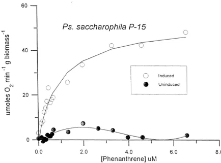
2.2. Oxygen uptake as a function of phenanthrene concentration for resting cells of P. saccharophila P-15 grown on peptone in the presence (induced condition) and absence
(uninduced) of phenanthrene..
...
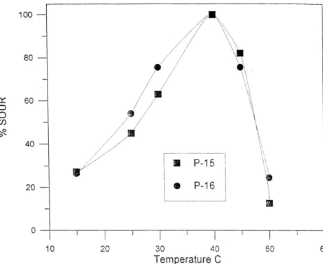
12 2.3. Relative specific oxygen uptake rate (% SOUR) as a function of temperature for P-15 andP-16 after growth on PEPIPAT at 25
+
0.1 O C...
132.4. Specific phenanthrene uptake rate in mg ruin-' (g biomass)-' (SPUR) as a function of
phenanthrene concentration for P-15 and P-16 at 25
+
0.1 O C...
14 2.5. Specific oxygen uptake rate in mg min" (g biomass)-' (SOUR) as a function of 1-hydroxy-2-naphthoic acid concentration for P-15 and P-16 at 25
+
0.1 O C...
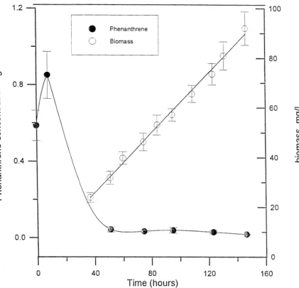
17 2.6. Biomass and aqueous phase phenanthrene concentrations during batch growth of P....
stutzeri P-16 in the presence of solid phenanthrene. 183.1. Lineweaver-Burk plot of initial phenanthrene degradation rate by P. stutzeri P-16 as a
...
function of phenanthrene concentration alone and in the presence of 30 pM naphthalene .273.2. Initial phenanthrene degradation rate by P. stutzeri P-16 as a function of increasing
...
naphthalene concentration in binary mixture with 5
pM
phenanthrene 283.3. Initial phenanthrene degradation rate by P. stutzeri P-16 as a function of increasing phenanthrene concentration in binary mixture with 30 p M naphthalene, expressed as a fraction of the initial specific phenanthrene degradation rate at the appropriate
concentration of phenanthrene alone
...
-29 4.1. Surface tension of distilled water containing C2 biosurfactant as a function of its...
concentration (relative to its aqueous solubility). . 4 4
5.1. Total dissolved phenanthrene concentration
(CT)
as a function of time in a batch shakeflask system containing T-Maz 20 at a concentration of 500 mg/L or no surfactant
...
58 5.2. Apparent saturation concentration of phenanthrene as a function of T-Maz 20concentration..
...
- 5 9 5.3. Observed mass transfer coefficient (kl,obs) for phenanthrene dissolution as a function ofsurfactant concentration for (a) T-Maz 20 and (b) Tergitol NP-10
...
.61 5.4. Maximum phenanthrene dissolution rate as a function of surfactant concentration for T-Maz 20.
...
62 5.5. Maximum rates of phenanthrene dissolution as a function of surfactant concentration forall six surfactants
...
63 6.1. Batch growth of P-16 in peptone medium in the absence of surfactant and in the presencePage
...
2.1. Biochemical characterization of phenanthrene degrading bacteria 9...
2.2. Phenanthrene oxidation kinetics 10
...
2.3. Phenanthrene uptake kinetics 15
...
2.4. Oxidation of pathway intermediates 15
...
3.1. Oxidation of low molecular weight PAH 24
...
3.2. Naphthalene oxidation kinetics 25
...
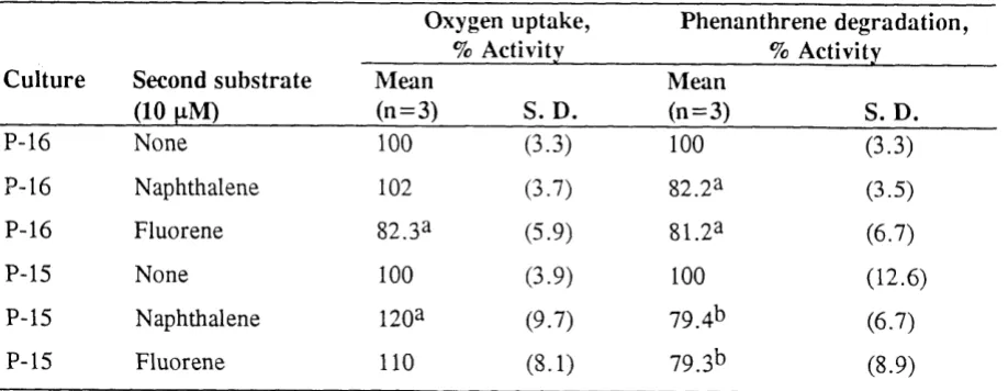
3.3. Effect of naphthalene and fluorene on phenanthrene degradation and oxidation 25...
3.4. Effect of methylnaphthalenes and indane on phenanthrene degradation 26...
3.5. Estimates of inhibition coefficients 29
4.1. Isolates from mixed phenanthrene-degrading cultures and their relative abundance in the
mixed culture
...
36...
4.2. Identification and characteristics of bacterial isolates 374.3. Michaelis-Menten kinetic coefficients for oxidation of phenanthrene by mixed cultures
...
growing on phenanthrene - 3 7
4.4. Michaelis-Menten kinetic coefficients for oxidation of phenanthrene by pure cultures
...
growing on peptone plus phenanthrene 38
4.5. Oxygen uptake rate at 0.8 mg/L phenanthrene for isolates grown on peptone alone or
...
peptone plus phenanthrene - 3 8
...
4.6. Oxidation of phenanthrene degradation intermediates 3 9 4.7. Growth substrate range for bacterial isolates ... 404.8. Oxygen uptake rate on various low molecular weight PAH substrates by phenanthrene-
grown pure cultures
...
- 4 1...
4.9. Removal of selected PAH by resting cells grown on peptonelphenanthrene medium 42...
4.10. Removal of selected PAH in killed controls 43
4.11. Surface tension of culture fluid from isolates grown on either phenanthrene or
naphthalene as sole carbon source
...
43...
4.12. Results of MATH assay for bacterial isolates 45
...
4.13. Combined substrate range of bacterial isolates 48
S U ~ ~ U R Y AND CONCLUSIONS
Both the microbiology of PAH degradation and the effects of surfactants on PAH dissolution and biodegradation were studied in this project. Bacteria able to degrade PAH compounds were readily isolated from a variety of PAH contaminated soils by standard enrichment culture techniques,
employing phenanthrene as a sole carbon source. The ease with which PAH degraders were isolated I from different sources is consistent with the fact that PAH compounds are both naturally occurring and widespread in the environment.
The PAH-degrading bacteria isolated during this project were useful models in studying various characteristics of PAH degradation. All but one of the isolates had a diverse range of PAH substrates that they could use as a sole carbon source for growth or otherwise degrade under non-growth
conditions, spanning
a
range of two-, three-, four- and five-ring compounds. These results suggest that biodegradation of the more recalcitrant PAH in contaminated environments, particularly the PAH with five and six rings, probably can be achieved by bacteria that use lower-molecular weight PAH as growth substrates. Furthermore, several of the bacteria were able to use chrysene, a four-ringcompound with an aqueous solubility in the low pg/L range, as a growth substrate, indicating that limited aqueous solubility alone is not responsible for an inability of microorganisms to grow on the higher-molecular weight PAH.
Only one of the eleven bacterial isolates studied
(Pseudomonas
strain C2) appeared to produce a biosurfactant during growth on PAH, indicating that biosurfactant production is probably not a common mechanism used by bacteria to enhance the bioavailability of these essentially insoluble compounds. At least one of the organisms(Pseudomonas
saccharophila P-15) grew as a biofilm directly on phenanthrene crystals, which is a likely mechanism by which the bioavailability of aninsoluble, crystalline substrate could be improved. Further experiments to test the effect of biofilm growth on phenanthrene degradation kinetics are planned. The only other organism tested for its ability to grow as a biofilm on phenanthrene crystals was
Pseudomonas stutzeri
P-16, and it was observed not to do so. Consequently, P-16 was selected as a model organism with which to study the effects of phenanthrene dissolution into the aqueous phase on biodegradation kinetics.The kinetics of phenanthrene degradation in aqueous solution followed the Michaelis-Menten model (Monod model in growth situations) for all of the isolates. This finding represents a basis on which more complex biodegradation models can be developed. For example, the known kinetics of isolate P-
16 have been used to demonstrate that biodegradation of solid phenanthrene is controlled by its rate of dissolution, and to study and model the degradation of phenanthrene solubilized in surfactant micelles. We have also demonstrated that low-molecular weight PAH are metabolized competitively by bacteria when present as binary mixtures. Since PAH are always present in contaminated environments as complex mixtures, this finding has implications for predicting the biodegradation of individual
compounds within such mixtures. Further work to develop quantitative models of competition among more than two substrates, and to extend the competitive metabolism model to mixtures of low- and high-molecular weight PAH, remains to be done.
Several lines of evidence derived from this work point to the likelihood that multiple PAH are degraded by a common set of enzymes within a given bacterium. This concept is the basis for the competitive metabolism of PAH in mixtures, but also has implications for controlling the degradation of PAH that are not growth substrates. If several PAH substrates are degradable by the same enzymes, then any inducer for the synthesis of those enzymes should induce the degradation of all of the
substrates. Growth of PAH-degrading bacteria on inducing substrates should enable those organisms
to sustain their ability to degrade non-growth substrates after the growth substrates have been depleted. Such a strategy might be particularly useful in enhancing the degradation of those PAH which are not removed completely during bioremediation of contaminated soils; most important among these are the high-molecular weight, carcinogenic PAH. Work on induction of high-molecular weight PAH
degradation is in progress.
Although PAHdegrading bacteria generally do not appear to produce biosurfactants during the biodegradation of PAH, the concept of enhancing PAH bioavailability through the use of synthetic surfactants, exogenously added biosurfactants or in situ production of biosurfactants by other organisms still warrants investigation. Early in this project we recognized that there was little fkndarnental information in the literature on how surfactants influence the biodegradation of
hydrophobic compounds. Knowing that the rate of biodegradation of a substance that is poorly soluble in water is controlled by its rate of dissolution, we studied how surfactants influence the rate of
dissolution of solid phenanthrene. This work led to the development of a mathematical model of dissolution kinetics in the presence of nonionic surfactants, which incorporates key concepts that can be extrapolated to a variety of physical systems. We are in the process of using this model to evaluate the effects of nonionic surfactants on biodegradation of solid phenanthrene in well-defined experimental systems.
We have also studied the effect of phenanthrene solubilization in surfactant micelles on its rate of biodegradation. In a system in which all of the phenanthrene is present in the liquid phase, either in micelles or in the aqueous phase itself, we have observed that phenanthrene
biodegradation can be described by assuming that only the amount present in the aqueous phase is available for biodegradation. A modified form of the Michaelis-Menten equation was
therefore developed to account for this observation, based on a simple partitioning equilibrium between the aqueous and micellar phases. Under these conditions (no excess hydrocarbon present or extremely under-saturated micelles) surfactants can actually inhibit biodegradation rates, suggesting that surfactant overdosing should be considered and avoided when possible. It should be noted, however, that the system used in this work was intentionally selected to avoid more complex interactions between the surfactant and the microorganism. For example, the surfactant selected for these studies does not influence the growth or metabolism of the organism
(P-16),
and is not biodegraded by the organism. Biodegradation of the surfactant, or inhibitory or toxic effects on microbial activity, would represent negative influences onbiodegradation of the target hydrophobic pollutant(s) but would be important to consider in practice. Positive interactions between surfactants and microorganisms can be envisioned as well. For example, an afinity between the surfactant micelles, or perhaps surfactant
monomers, and microbial cells might permit direct uptake of solubilized substrates, which should permit faster degradation rates than would be predicted by the simple partitioning model we have developed. Such interactions remain to be demonstrated, but offer a potentially fruitful area for further study.
The work described in this report has demonstrated that the ability of soil bacteria to degrade PAH is a common phenomenon, and that most PAH-degrading bacteria are able to degrade a
wide range of PAH substrates. Of particular importance was the finding that many of the isolated bacteria could biodegrade high molecular weight PAH after they were grown on the three-ring compound phenanthrene. The implication of this finding is that the inherent biodegradability of high molecular weight substrates is not the factor limiting their
biodegradation in the field. Since these compounds appear to be inherently biodegradable, approaches to overcoming those factors which do limit biodegradation in the field need to be addressed.
From the work conducted in this project and by others, it appears that there are at least two major factors which limit PAH biodegradation in the field. First, it can be expected that the biodegradation of high molecular weight PAH which do not serve as growth substrates for indigenous bacteria will decline and eventually terminate as the major growth substrates are depleted. Depending on the relative amounts of growth and non-growth substrates, it is possible for the high molecular weight compounds to be removed from a system only partially,
as has been observed in bioremediation experiments and field projects. The only way to sustain the biodegradation of non-growth PAH after the growth substrates are depleted is to artificially stimulate the PAH degrading activity. We are currently investigating the use of selective substrates (particularly known inducers of PAH metabolism) for their ability to accomplish this in an actual contaminated soil, through an ongoing WRRI grant (WRRI Project number 70144). It should be noted, however, that the effects of long-term or sustained
addition of such compounds to contaminated soil have not been investigated.
The second major factor that is likely to be responsible for limitations in high molecular weight PAH biodegradation is their limited bioavailability. Bioavailability has a direct impact on the rate of biodegradation and, therefore, an indirect effect on the extent of biodegradation. If the activity of PAH degrading bacteria can be sustained artificially as suggested above, then steady but slow rates of high molecular weight PAH degradation are likely to occur. However, the rates of mass transfer processes (dissolution andlor desorption) will govern rates of
biodegradation in such cases. Depending on the magnitude of those rates, stimulating biodegradation by supplementing a system with selective growth substrates may or may not have an observable effect over a relatively short time scale (e.g., time scales of days or weeks typically used in experimental studies). For many situations, it is likely that growth substrate supplementation alone will not overcome limitations in high molecular weight PAH
biodegradation. Simultaneously enhancing the mass transfer processes is recommended in such cases to ensure the biodegradation of these compounds over meaningful time scales. As of now, the only mechanism to enhance mass transfer at full scale is to provide mixing of the contaminated soil, as would occur in an above-ground slurry-phase biological reactor. Another approach that is of much current interest, and which was broached in the current project, is the use of surfactants to increase both rates of mass transfer and apparent liquid phase solubilities of the highly hydrophobic PAH.
properties, and extent of interfacial tension reduction. As mentioned in the body of this report (Chapter l), it is likely that applications of surfactants to contaminated soil will be limited to
those surfactants approved for food and, possibly, cosmetic use. These surfactants are expected to minimize concerns over toxicity and ultimate biodegradability of the surfactant. While ultimate biodegradability of a surfactant is important, the rate at which surfactant biodegradation occurs ideally should be slower than the rate of biodegradation of the target pollutants.
As demonstrated in this project, the solubilization capacity for a particular hydrocarbon varies from surfactant to surfactant, and is related to other properties of the surfactant such as the hydrophilicflipophilic balance (HLB) value (Chapter 5). Whenever surfactants are added to soil, it should be expected that some of the surfactant will sorb to soil constituents and therefore will be removed from the aqueous phase. This removal will in turn influence the
amount of surfactant required to reach the critical micelle concentration (cmc) in the aqueous phase, which must be reached because solubilization properties of surfactants become
significant only at surfactant concentrations above the cmc. The work of Luthy and co- workers (Edwards et al., 1991; Laha and Luthy, 1991; Laha and Luthy, 1992; Edwards et al.,
1994a; Edwards et al., 1994b) and other published reports on equilibrium properties of surfactants in soil systems should be consulted for quantitative approaches to studying
surfactant sorption. Surfactant sorption is also likely to be correlated to the HLB value; more hydrophobic surfactants (lower HLB value) are expected to sorb to a greater extent than will hydrophilic surfactants. However, since lower HLB surfactants also will solubilize more of the hydrophobic pollutants per unit of surfactant added, there will be a tradeoff in selecting a surfactant with an optimum
HLB
value for a given system. A range of surfactants with varying HLB values therefore should be tested in preliminary work aimed at selecting one or more surfactants for further study.The extent of interfacial tension reduction caused by a particular surfactant has implications for whether addition of the surfactant can lead to mobilization of nonaqueous phase liquids, which may be undesirable for in situ applications (Pennell et al., 1993). The higher the final
interfacial tension, the lower the probability that mobilization will occur; however, the numerical limit on interfacial tension to preclude mobilization is probably case-specific (Pennell et al., 1993).
While it is important to consider surfactant sorption in estimating the minimum amount of surfactant required to achieve mobilization in a given soil system, it also is important not to overdose the surfactant. As shown in this project, it is possible to reduce biodegradation rates of PAH in the presence of surfactants by sequestering most of the PAH in micelles. We have not yet developed a complete quantitative model of effects of surfactant concentration on overall biodegradation rates of hydrophobic compounds. Consequently, it is not yet possible to predict a priori the range of surfactant concentrations which will optimize biodegradation rates. However, a good starting point to estimating the upper limit of surfactant concentration is the concentration required to solubilize all of the hydrophobic compounds in the system. Higher concentrations could be tested experimentally, but it is important to keep in mind that there is probably
an
upper limit to surfactant concentration in stimulating biodegradation rates.Finally, we demonstrated in this project that competition between PAH substrates must be taken into account when studying the biodegradation or biological treatment of PAH mixtures. Competition may actually limit rates of high molecular weight PAH biodegradation during the most biologically active phase of a remediation study or project, but as discussed above the presence of lower molecular weight PAH may be essential to achieving any biodegradation at
all. While there probably is no good way to overcome such competitive effects on
biodegradation rates in the field, it is important to keep in mind during experimental work that rates measured for single substrates in a defined laboratory system do not necessarily represent rates that can be achieved in mixtures.
Future research directed at developing practical approaches to bioremediation of PAH- contaminated soils and sediments should focus on methods of enhancing the biodegradation of high molecular weight PAH. While current knowledge of high molecular weight PAH biodegradation is relatively poor, ongoing work in this and other laboratories is providing information that can be put to use in the field. Some of this work is described in Chapter 4 of the report. Experimental work on the use of surfactants in real soils, and the influences of
these surfactants on biodegradation, also needs to be conducted. Hopefully, the quantitative understanding of the effects of surfactants on equilibria, mass transfer kinetics and
biodegradation that has been developed in this project and by a number of other investigators will permit approaches that are more sophisticated than trial-and-error. Once studies on enhancing and optimizing biodegradation of high molecular weight PAH have been completed, assessments of economic feasibility can be made; such assessments should identify economic bottlenecks and consider whether these bottlenecks can be overcome in a technically feasible manner.
Many of the pollutants found in contaminated soil and subsurface environments are poorly soluble in water, and therefore exist in nonaqueous-phase liquids, as trapped residuals in pore spaces, or partitioned into hydrophobic domains such as soil organic matter. Polycyclic aromatic hydrocarbons
(PAH), for example, are an important class of environmental pollutant produced as a consequence of fossil fuel utilization, combustion processes, and chemical manufacture (Guerin, 1978; Evans et al.,
1990; Christensen and Zhang, 1993). PAH are found in contaminated soils and groundwater at many sites across the United States, particularly at creosote wood treatment facilities (Mueller et al., 1989a; U. S
.
Environmental Protection Agency, 1990; Rosenfeld and Plumb, 199 1) and former manufactured gas plants (Luthy et al., 1994). PAH also occur naturally, as the result of fires and geologicprocesses, and are carbon compounds of significant energy potential (Blumer, 1976; Anderson et al., 1986). Many PAH are subject to bacterial degradation (Pate1 and Gibson, 1976; Cerniglia, 1984; Cerniglia, 1992).
There is current interest in the utilization of bacterial processes for the treatment of contaminated soils and groundwaters (Sims et al., 1989; Gibson and Sayler, 1992; Sims et al., 1993), including hazardous waste sites contaminated with mixtures of PAH (Fleming et al., 1993; Mueller et al., 1989a; Wilson and Jones, 1993). In 1993, 159 bioremediation projects in the United States were being tracked under the USEPA Bioremediation Field Initiative (U. S . Environmental Protection Agency, 1993). Over one third (55) of these sites were listed as being contaminated with PAH or wood preserving wastes; an additional 54 sites were listed as contaminated with petroleum hydrocarbons ( e g jet fuel, diesel fuel, and fuel oil) that potentially contain trace amounts of PAH. However, biological remediation of PAH contaminated sites is not a proven technology and has rarely been completely effective (Wilson and Jones, 1993).
Because of their low solubility in water, PAH in contaminated soil will exist mostly in a separate hydrocarbon phase or sorbed to soil constituents. Naphthalene, the most water-soluble PAH, has a log octanolJwater partition coefficient (log KO,) on the order of 3.4, and PAH of three or more rings have log KO, greater than 4 (Mackay et al., 1992). The log KO, of a compound is known to correlate well
with its partitioning into soil organic matter (Weber et al., 1991). Such partitionirig can limit the availability of PAH to microorganisms even if they are inherently biodegradable (Manila1 and
Alexander, 1991; Weissenfels et al., 1992; Erickson et al., 1993). In fact, an increased understanding of mechanisms governing the transfer of hydrophobic chemicals from solid and non-aqueous liquid phases, as well as developing methods to overcome the corresponding limitations in bioavailability, have been suggested to be among the most critical research needs in bioremediation (Alexander, 1991; Luthy, 1991).
the hydrocarbon (reviewed by the hydrocarbon into droplets (Zhang and Miller, 1995).
Hornmel, 1990 and Georgiou et al., 1992) or lead to the dispersion of with increaesed surface area relative to a continuous hydrocarbon phase
The mechanisms described above by which microbes increase the bioavailability of aliphatic
hydrocarbons have not been studied directly with organisms that degrade PAH. It can be presumed that PAH and aliphatic hydrocarbons pose the same kinds of problems for microorganisms, in the sense that interphase mass transfer must precede biodegradation. It therefore is not unreasonable to hypothesize that microbes have adapted to such problems with PAH in the same way they have adapted to aliphatic hydrocarbons, particularly for those PAH that serve as growth substrates. For example, Stucki and Alexander (1987) observed that a Pseudomonas sp. grown on biphenyl formed extracellular products (unidentified) that increased the dissolution rate of biphenyl, although biphenyl solubility did not increase. Guerin and Boyd (1992) determined that a Pseudomonas putida strain degraded
naphthalene sorbed to soil at a rate exceeding iis estimated desorption rate, implying biologically enhanced mass transfer. Production of extracellular biosurfactants during growth on PAH substrates has not, however, been reported in the literature.
Even if biosurfactants are not involved in the microbial degradation of PAH, the use of surfactants to enhance the mass transfer of PAH may still be an important tool in improving the biodegradability of these compounds. The involvement of surfactants in enhancing the biodegradation of PAH can be considered in six distinct categories:
(1) Production of biosurfactants by PAH degraders when growing on PAH;
(2) Production of biosurfactants by PAH degraders when growing on compounds that co-exist with PAH in contaminated soil (e.g., aliphatic hydrocarbons);
(3) Production of biosurfactants by PAH degraders when growing on a compound introduced exogenously to the soil;
(4) Production of biosurfactants by organisms that do not degrade PAH, when growing on aliphatic hydrocarbons or other carbon sources that either co-exist with PAH in contaminated soil or can be added exogenously to the soil;
( 5 ) Exogenous addition of biosurfactants to the soil; (6) Exogenous addition of synthetic surfactants to the soil.
Categories 1 and 2 are the most desirable in terms of bioremediation, because surfactant production would occur in situ at the expense of the compounds whose removal is the objective of bioremediation. Category 3 also involves in situ production of biosurfactants by PAH degraders, but requires the addition of carbon sources to a soil that may already be heavily contaminated. Such a strategy may be more useful as a second phase of bioremediation, after the major readily degradable pollutants are removed. Category 4 involves in situ surfactant production as well, but is not directly related to PAH degradation; its effect on PAH solubilization and subsequent degradation therefore may be difficult to predict and control. Categories 5 and 6 involve the addition of surfactants to the soil, which is of much current experimental interest (reviewed by Rouse et al., 1994).
It has been suggested that biosurfactants would be particularly useful for soil remediation, either added exogenously or produced by microorganisms inoculated into the soil environment, because they are likely to be inherently biodegradable and non-toxic (Georgiou et al., 1992; Miiller-Hurtig et al., 1993; Finnerty, 1994). However, it should be kept in mind that many microbially produced substances with surfactant properties also have antibiotic effects (Lang and Wagner, 1993), which might have
to be toxic or inhibitory to microorganisms, so that it would be important to consider their toxicity and biodegradability in soil (Bubela, 1987; Kosaric et al., 1987; Lang and Wagner, 1993). A variety of synthetic surfactants are, however, already used as food additives and therefore would be expected to be of limited regulatory concern for applications in soi,l remediation (Shiau et al., 1994).
Even though surfactants can be expected to increase the solubility of PAH (Edwards et al., 1991, 1994a, 1994b), it cannot be assumed that biodegradability will also be enhanced in the presence of surfactants. Conflicting results have been obtained in the relatively few studies conducted to evaluate the effect of surfactants on biodegradation of hydrophobic pollutants. The effects of surfactants have ranged from inhibition of biodegradation to no effect to stimulation of biodegradation, and a variety of factors have been proposed to explain these findings (Rouse et al., 1994; Liu et al., 1995). For example, Falatko and Novak (1992) studied the effects of two different types of biosurfactants on biodegradation of several aromatic hydrocarbons (including naphthalene) in aqueous systems.
Biosurfactants derived from organisms grown on gasoline improved the rate of aromatic hydrocarbon degradation, while biosurfactants produced by activated sludge organisms grown on glucose and vegetable oil inhibited biodegradation. It seems, therefore? that surfactant specificity may be an
important factor governing the impact of surfactants on biodegradation of hydrophobic compounds (Falatko and Novak 1992). To date, however, no comprehensive theory of the influences of
surfactants on biodegradation has been developed, and most experimental approaches described in the literature have been empirical.
SCOPE AND OBJECTIVES O F RESEARCH
This project focused on two major areas of research: the biodegradation of PAH by soil
microorganisms, and the influences of surfactants on rates of phenanthrene dissolution into water and on rates of phenanthrene biodegradation. PAH biodegradation studies began by focusing on two bacteria isolated from the same PAH-contaminated soil, and were extended to a survey of bacteria isolated from six other PAH-contaminated soils. In keeping with the original intent of this project, each of these bacteria was screened for its ability to produce biosurfactant during growth on
phenanthrene. In addition, though, a number of other physiological characteristics relevant to PAH degradation were evaluated with these organisms. Work on the biodegradation of PAH by these microorganisms is described in Chapters 2 through 4.
As discussed above, conflicting effects of surfactants on biodegradation of hydrophobic chemicals have been reported in the literature. Consequently, we decided early in the project to undertake a
fundamental investigation of how surfactants influence rates of phenanthrene dissolution from a non- aqueous phase before attempting to define their role in phenanthrene biodegradation. Results of that work are reported in Chapter 5 . Our rationale was that by developing a mechanistic, conceptual model for phenanthrene dissolution in the presence of surfactants, we could then use this model in evaluating more complex systems in which biodegradation would be occurring in the aqueous phase. Work on biodegradation of phenanthrene in the presence of surfactants focused only on the biodegradability of phenanthrene solubilized in surfactant micelles, and is described in Chapter 6. Because our early work on the biodegradation of PAH by soil isolates indicated that biosurfactant production might not be a common feature of PAH-degrading bacteria, the work with surfactants involved the use of synthetic nonionic surfactants.
describe the effects of surfactants on biodegradation of phenanthrene in systems containing non- aqueous phase phenanthrene. As a result of the work conducted in this project, we have found one PAH-degrading bacterium able to produce a biosurfactant during growth on phenanthrene, and are devoting further activity into elucidating a possible role of this biosurfactant in PAH biodegradation.
2. COMPARATIVE PHYSIOLOGY OF PHENANTHRENE DEGRADATION BY TWO DISSIMILAR PSEUDOMONADS ISOLATED FROM A CREOSOTE CONTAMINATED SOIL
INTRODUCTION
PAHs are naturally occurring compounds (Blumer, 1976) and hence are logical targets for the
application of biological treatment. Pathways for the transformation of several lower molecular weight PAHs by bacteria have been proposed (Sims and Overcash, 1983; Cerniglia and Heitkamp, 1989; Grifoll et al., 1992; Grund et al., 1992; Kelly et al., 1993; Miyachi et al., 1993) and experience with land-farming of refinery wastes suggests that higher molecular weight PAHs are also biodegradable (Sims and Overcash, 1983; American Petroleum Institute, 1984; Sims et al., 1988; Park et al., 1990; Wild and Jones, 1993). However, little is known about the physiology of PAH degraders, the kinetics of substrate and intermediate metabolism, and the strategies used by bacteria growing on compounds of such low solubility.
As discussed in Chapter 1, phenanthrene degrading bacteria were used in this work as model PAH- degrading organisms. Phenanthrene degrading bacteria are commonly isolated from PAH contaminated environments (Foght et al., 1990; Bogardt and Hemmingsen, 1992) and phenanthrene degrading capability is common among diverse genera and genetic groups (West et al., 1984; Foght et al., 1990). This would indicate that phenanthrene degrading bacteria are an important bacterial guild in PAH contaminated ecosystems. Several studies have measured growth rates and cell yields of bacteria on phenanthrene (Wodzinski and Johnson, 1968; Wodzinski and Coyle, 1974; Stucki and Alexander,
1987; Weissenfels et al., 1990; Keuth and Rehm, 199 1; Aichinger et al., 1992; Volkering et al., 1992; Boldrin et al., 1993), but Michaelis-Menten half-saturation coefficients for the bacterial degradation of, or growth on, phenanthrene have not been measured successfully (Aichinger et al.,
1992).
In this chapter, two physiologically and genetically diverse phenanthrene degrading bacteria,
Pseudomonas saccharophila P-15 and Pseudornorzas stutzeri P-16, are described. Resting-cell assays were used to present evidence that the kinetics of soluble phenanthrene oxidation and uptake for the two organisms are essentially identical. It is further shown that the bacteria differ in their propensity to form biofilms on phenanthrene crystals and in their ability to metabolize externally supplied degradation intermediates.
MATERIALS AND METHODS
Chemicals. Phenanthrene (99% purity) was purchased from Kodak (Rochester, NY). Salicylic acid (ACS reagent grade) and 1-hydroxy-2-naphthoic acid (1H2N7 99%) were purchased from Sigma Chemicals (Milwaukee, WI). Salicylaldehyde (98%), catechol (99
+
%), 3,4-dihydroxybenzoic acid (97%), 2-carboxybenzaldehyde (97%), and phthalic acid (99+
%) were purchased from Aldrich Chemicals (Milwaukee, WI). R2A agar and peptone (Bacto-Peptone) were purchased from Difco (Detroit, MI). All other chemicals used were ACS reagent grade.otherwise noted, the amount of phenanthrene and liquid added was equal to a final concentration of 0.5 g1L in both PATITWB and PEPIPAT. Slants using R2A agar supplemented with phenanthrene
(R2AIPAT) were made by dissolving 18.2 g R2A agar in 1 L of water and then supplementing the hot mixture with 0.05 g of phenanthrene. The medium was mixed, dispensed in tubes or bottles and then autoclaved. Cultures were maintained on R2AlPAT slants.
TWB was made by combining Na2HP04-7H20 (1.5 g), KH2P04 (1.0 g), NH4Cl (2.0 g), and Na2S203-5H~O (0.02 g) in one liter of tap water; the measured pH was 6.5. Without a supplemental carbon source, 'IWB could not support measurable bacterial growth. Biochemical oxygen demand (ENID) dilution buffer was made according to method 5210B in Standard Methods for the Examination of Water and Wastewater (Greenberg et al., 1992). Inocula for all experiments, unless otherwise noted, were prepared by resuspending loops of cultures from the R2AlPAT slants in BOD dilution buffer. Dechlorinated tap water was made by adding 0.02 g Na2S203-5H20 to one liter of tap water.
Enrichment of phenanthrene degraders from soil samples. 5 g of a creosote contaminated soil was mixed in 75 mL of TWB in a 125 mL Erlenmeyer flask on a rotary shaker for one hour. The slurry was allowed to settle, and 1 mL of supernatant was used to inoculate 200 mL of PATITWB for enrichment. The culture was shaken at room temperature (24-25 "C) until growth was indicated by an increase in visible turbidity and the medium turned brown. This liquid enrichment was then used to inoculate plate media.
Samples of liquid enrichments were diluted to spread plated on R2A agar and incubated for 7 days. From plates with visible colonies at the highest dilution, strains were picked according to colony morphology so that each colony type represented at the highest dilution was picked at least once. Colonies were purified to R2AlPAT agar and examined for their ability to grow on
phenanthrene as a sole carbon source.
Identification of bacterial isolates. Procedures for the biochemical characterization of isolates were conducted and interpreted as described in Finegold and Baron (1986). API 20-E test strips, OIF test medium, and test reagents were purchased from Analytab Products (Analytab Products, Plainview, NY). Other media were purchased from the University of North Carolina Media Preparation Facility. Pure cultures were sent to MIDI (Newark, DE) for identification by fatty acid methyl-ester profile.
Measurement of phenanthrene and intermediates from phenanthrene degradation by HPLC. Phenanthrene and 1H2N were analyzed by injecting 10 to 30 pL of culture fluid into a Waters 600E HPLC system (Millipore, Marlborough, MA) with a Waters 470 scanning fluorescence detector (excitation at 238 nm, emission at 373 nm). Separation was achieved with a C-18, 25 cm x 4.6 mm, 5
pm particle reverse phase column (Supelco, Bellefonte, PA). Initial mobile phase conditions were 0.01 % H3P04 for one minute, followed by a linear gradient to 100% methanol over 5 minutes and holding 100% methanol. At a flow rate of 1.0 mllmin, 1H2N eluted at 12.8 minutes and
phenanthrene eluted at 14.3 minutes. The detection limit for both compounds was 0.08 mg1L.
Measurement of phenanthrene and metabolite oxidation rates. The oxidation of phenanthrene and other compounds was measured using resting cells in a stirred, 1.7 mL volume, water-jacketed
Cultures were grown at 25
+
0.50C on either 0.5% peptone, PATITWB medium, or PEPIPAT medium and then harvested in late growth stage. Cultures were centrifuged, washed in dechlorinated tap water, centrifuged again, and resuspended in TWB. Washed cells were placed in therespirometer, an initial endogenous oxygen uptake rate was measured, 10 pL of a methanol solution containing the substrate of interest was injected into the cell, and oxygen uptake rate was measured again. Oxygen consumption in the presence of substrate was corrected for endogenous respiration. Results are expressed in terms of specific oxygen uptake rate (SOUR) and reported as mg oxygen consumed per minute per gram of bacteria. TWB had an oxygen solubility of 7.8 mglL at 25OC. Preliminary experiments confirmed that injection of methanol alone had no effect on oxygen uptake.
Measurement of phenanthrene uptake by spectrophotometric rate assay. Bacteria were washed as described above and diluted in TWB to an absorbance of approximately 0.1 at 420 nm. Two mL of this suspension were placed in a 3 mL quartz cuvette in a spectrophotometer. The cuvette was mixed using a stir bar and a micro-magnetic stirrer (Fisher Scientific). Ten pL of a phenanthrene solution in methanol was injected into the cuvette and the decrease in absorbance at 250 nm was measured over time. Absorbance was measured at 30, 40, and 50 seconds after injection (n = 3), and the slope used to calculate initial phenanthrene uptake rate. The decrease in absorbance at 250 nm was converted to phenanthrene concentration using an extinction coefficient of 6.46 x10⁴ L mole-I cm-l (Weast and Grasselli, 1989), and initial uptake rates were thereby determined. Results are expressed as specific phenanthrene uptake rate (SPUR) in mg phenanthrene consumed per minute per gram of bacteria.
Measurement of phenanthrene uptake by HPLC assay. In order to validate the spectrophotometric method for measuring phenanthrene uptake and to determine if UV absorbing metabolites were interfering with the measurement of phenanthrene, uptake rates measured by spectrophotometric assay were compared to rates measured by HPLC analysis using the same sample of washed cell culture (P- 16). Triplicate assays were run using each method. For the HPLC assay, 20 mL of washed cells at an absorbance (420 nm) of approximately 0.3 were added to a 50 mL beaker and mixed rapidly using a Teflon stir-bar. Phenanthrene was added by injecting 100 pL of a 160 mglL solution in methanol into the stirred beaker. One mL samples were taken at 30, 60, 90, 120, and 180 seconds and put in tubes containing 0.5 mL of 38% formaldehyde to stop the reaction. Samples were analyzed by direct injection HPLC as described above. Data between 30 and 90 seconds (n = 3) were used to calculate phenanthrene uptake rates for comparison with the results obtained by the spectrophotometric assay.
Estimation of kinetic coefficients. The Michaelis-Menten equation was used to estimate kinetic coefficients from SOUR and SPUR measurements. Non-linear estimation was performed using the Quasi-Newton method in SYSTAT (SYSTAT, Inc., Evanston, IL) to give estimates of the half- saturation coefficients (K,) and maximum specific rates of activity (SOUR, or SPUR,). Data are reported as best estimates with 95% confidence intervals (CI).
Measurement of cell surface hydrophobicity. Cell surface hydrophobicity was compared between isolated strains using a modification of the microbial adhesion to hydrocarbons (MATH) test described by Rosenberg (1991). Cells cultured on PEPJPAT were washed as described above and resuspended in double strength TWB (2 X TWB) to give a suspension of approximately 0.5 absorbance units at 420 nm. Washed cells (1.2 mL) were placed in 12 X 75 mm test-tubes for analysis in triplicate.
measured at 420 nm. Removal of bacteria with hydrophobic cell surfaces from the aqueous phase by extraction with hexadecane is reflected as a decrease in turbidity.
Measurement of surface tension. Phenanthrene degrading bacteria were screened for biosurfactant production by measuring culture fluid surface tension. Surface tension was measured by the ring method using a du Nouy Tensiometer (CSC Scientific Co., Fairfax, VA). Ten mL of sample was transferred to 6 0 mm (dia.) x 15 rnrn glass petri dishes for surface tension measurement using a 6 mm platinum ring. The ring was flamed and rinsed with deionized water before the first sample and between samples. Each sample was measured twice. Using this method the surface tension of deionized water was 7 1.0 0.38 dyneslcm (n = 20).
Identification of bacteria isolated from phenanthrene enrichment culture. Five bacteria that could grow on phenanthrene as a sole carbon source were enriched from a creosote contaminated soil. Three of the strains produced a water soluble intermediate that accumulated in the media to concentrations of 30 mg1L or more, and was subsequently metabolized. The intermediate was identified by HPLC elution time as being 1H2N. The remaining two strains did not accumulate significant amounts of
1H2N in the culture fluid, although small amounts (approximately 1 mg1L maximum) of the intermediate could be detected.
A battery of tests was applied to the phenanthrene degrading strains to further examine similarities and differences between strains. It was confirmed that the strains could be separated into only two
physiological groups, which had identical test results within each group (Table 2.1). Two strains, designated P-16 and P-15, were selected for in-depth study and sent to MIDI, Inc. (Newark, DE) for identification by gas chromatography of cellular fatty acids. Fatty acid methyl ester analysis identified P-16 as a Pseudomonas stutzeri with a similarity index of 0.861 and P-15 was identified as a
Pseudornonas saccharophila with a similarity index of 0.63 1.
Growth yield of bacteria on phenanthrene. The growth yield of P-16 and P-15 were determined in TWB amended with phenanthrene at concentrations between 0 and 160 mg/L. Growth in TWB was limited by phenanthrene at all concentrations included in growth yield calculations. The yield
coefficient for P-16 was 1.3 g biomasslg phenanthrene (coefficient of determination, r2 = 0.999, n =
5) and for P-15 was 1.2 g biomasslg phenanthrene (r2 = 0.999, n = 6).
When P-15 grew in PATITWB, microscopic examination revealed that it formed a dense biofilm around the phenanthrene crystals suspended in the medium. P-16 did not colonize the crystals extensively, but only formed small colonies here and there on the surface of the crystals and the majority of the population occurred as free swimming bacteria dispersed in the medium.
Table 2.1. Biochemical Characterization of Phenanthrene Degrading Bacteria.
Criterion P-15 P-16 Criterion P-15 P-16
Gram stain
cell shape
motility
colony pigment
OIF test
oxidase
MacConkey ' s
diffusible pigment ADH citrate catalase nitrite nitrogen gas
Crystal violet lR2A
SDS IR2A
-
rod+
Y 0+
-+
(+)+
- - rod+
C 0+
+
- -+
+
+
+
+
TSI (slantlbutt) -1- -1-
dextrose
+
+
xylose
+
+
lactose - -
mannitol -
+
sucrose
maltose
+
+
gelatin
litmus milk
DNase -
Tech agar
@yocyanin)
Flo agar
(fluorescein)
cellulose
chitin
+
( + >plasmids
+
NDND = not detected; (+) = weak response; Y = yellow; C = cream; W = white; OiF = glucose oxidative-fermentative test; 0 = oxidizer; N = non-oxidizerlnon-fermenter; ADH = arginine dihydrolase; citrate = utilization of citrate as a sole carbon source; Nitrogen gas = formation of nitrogen gas during growth under denitrifying conditions; Nitrite = production of nitrite during growth under denitrifying conditions; SDSlR2A = growth on R2A agar supplemented with 1 %
sodium dodecyl sulfate; Crystal violetlR2A = growth on R2A agar supplemented with 11 mg/L crystal violet; dextrose, xylose, lactose, mannitol, sucrose, maltose = acid production during growth on these substrates.
Surfactant production by phenanthrene degrading isolates. Surface tensions of culture fluids of seven day old PATITWB cultures were compared to uninoculated medium using a ring tensiometer. Uninoculated PATITWB had a surface tension of 70 dynes cm-l. P-16 reduced the surface tension of the medium only slightly, to 6 0 dynes cm-l, whereas P-15 did not reduce surface tension (69 dynes cm-l). Extraction of the P-16 culture fluid with hexane, ether, or ethyl acetate did not change its surface tension or recover surface active materials.
2.2). The Michaelis-Menten equation was fit to the kinetic data, using non-linear regression, to provide estimates of kinetic coefficients (Table 2.2). The Ks values for the two organisms were simi when the bacteria were grown in either PATlTWB or PEP/PAT, but the SOUR, values differed between the two media. P-16 expressed higher specific rates of oxidation after growth under nutrien rich conditions than did P-15. In addition, P-16 was constitutive for phenanthrene oxidation (i.e., it expresses the ability to oxidize phenanthrene even when grown in the absence of phenanthrene), as shown in Figure 2.1. P-15 was inducible for phenanthrene oxidation (Figure 2.2).
Table 2.2. Phenanthrene Oxidation Kinetics.
Culture Growth Mediaa K , mglL 95%
clb
S0URTnC 95% CI P-16 Phenanthrene 3- 0.16 (0.11, 0.21) 3.14 (2.78, 3.50)Peptone
P-16d Phenanthrene 0.10 (0.06, 0.15) 1.86 (1.55, 2.17)
P-15 Phenanthrene
+
0.15 (0.08, 0.22) 1.73 (1.51, 1.95)Peptone
P - 1 5 ~ Phenanthrene 0.18 (0.12, 0.24) 1.69 (1.49, 1.89)
acells harvested from growth medium and resuspended in TWB for kinetic analysis
b95 % CI = 95 % confidence interval on estimate.
CSOUR, = maximum specific oxygen uptake rate, mg m i d (gram cells)-l.
dfrom Stringfellow and Aitken 1994a.
Optimum temperature for the oxidation of phenanthrene. Both organisms had essentially the same temperature optimum and temperature tolerance ranges for phenanthrene oxidation (Figure 2.3). Both bacteria demonstrated maximum oxidation activity at 40°C and lost activity at 50°C. At 25OC, both organisms exhibited approximately 50% of the activity seen at 40°C. The temperature response of the resting cells for phenanthrene oxidation was not a function of the temperature tolerance for growth. P-16 grew on PAT/R2A agar at 28O, 35O, and 410, but not 4OC. P-15 grew at 28O and 35O, but not at 4O or 410C.
Measurement of phenanthrene uptake kinetics by spectrophotometric rate assay. Phenanthrene uptake was described by the Michaelis-Menten saturation kinetic model (Figure 2.4) and kinetic coefficients for phenanthrene uptake could be estimated using non-linear regression (Table 2.3). Maximum specific phenanthrene uptake rates (SPUR,) differed for the two organisms, but half- saturation coefficients (&) were not significantly different. The half-saturation coefficients measured by this method also agreed well with Ks values measured by respirometry (Table 2.2). Formaldehyde killed controls did not exhibit measurable phenanthrene uptake.
Phenanthrene uptake rates measured by spectrophotometric assay were not significantly different (a =
Ps.
stutzeri
P- 16
0
Induced1
&D uninduced2.0 4.0 6.0
[Phenanthrene]
uMFigure 2.1. Oxygen uptake as a function of phenanthrene concentration for resting cells of P. stutzeri
Ps. saccharophila
P - I 5
0
InducedI
i
1
@ Uninduird1
0.0 2.0 4.0 6.0 8 .O
[Phenanthrene] uM
Figure 2.2. Oxygen uptake as a function of phenanthrene concentration for resting cells of P. saccharophila P-15 grown on peptone in the presence (induced condition) and absence
I 0 20 30 40 50 60
Temperature C
Figure 2.3. Relative specific oxygen uptake rate (% SOUR) as a function of temperature for P-15
0.0 0.2 0.4 0.6 0.8 1 .O
Phenanthrene (mg1L)
Table 2.3. Phenanthrene Uptake Kinetics.
Culture Growth Mediaa K , mglL 95% C I ~ SPURmc 95% CI P- 16 Phenanthrene
+
0.24 (0.16, 0.32) 3.49 (3.06, 3.92)Peptone
P- 15 Phenanthrene
+
0.20 (0.11, 0.28) 2.35 (1.92, 2.77) Peptoneacells harvested from growth medium and resuspended in TWB for kinetic analysis
b95 % CI = 95 % confidence interval on estimate.
CSPURm = maximum specific phenanthrene uptake rate, mg m i d (gram cells)-1
Oxidation of degradation intermediates. Pathways for the degradation of phenanthrene have been elucidated (Cerniglia and Heitkamp, 1989). Two pathways have been described for the degradation of phenanthrene, both of which share 1H2N as a key intermediate. Subsequent metabolism of the intermediate 1H2N can differ; some organisms metabolize 1H2N to protocatechuate via 2-
carboxybenzaldehyde and phthalic acid, and others form catechol via salicylaldehyde and salicylic acid (Cerniglia and Heitkamp, 1989).
The two cultures were grown under inducing (PEPIPAT) and non-inducing beptone) media conditions and tested for their ability to oxidize known phenanthrene degradation intermediates. Neither
bacterium oxidized 2-carboxybenzaldehyde, o-phthalic acid, nor protocatechuic acid, intermediates of the phthalic acid pathway (data not shown). Both organisms oxidized the intermediates of the salicylic acid pathway (Table 2.4). P-15 oxidized these compounds to a significant extent only under induced conditions.
Table 2.4. Oxidation of pathway intermediates.
P-15 P-16
(SOUR)" (SOUR)
Conc. not not
Compound mg/L inducedb induced induced induced
phenanthrene 1.2 0.1 1.5 2.4 2.4
1-hydroxy-2-naphthoic acid
salicylaldehyde 2.4 0.2 0.7 1 .O 1 .O
salicylic acid 2.4 0.0 0.4 0.3 0.2
catechol 2.4 0.4 1.8 1.2 1.4
aSOUR = specific oxygen uptake rate, mg m i d (gram cells)-1, mean of two replicate measurements
bffnot induced" = growth on peptone; "induced" = growth on PEPIPAT
The acidic pathway intermediates 1H2N and salicylic acid were oxidized at a lower rate by P-16 than might be expected in comparison to P-15, given that phenanthrene oxidation was 60% greater in P-16 than P-15. Subsequently a more complete kinetic evaluation was made for the oxidation of 1H2N by these organisms. As can be seen in Figure 2.5, P-15 exhibited saturation kinetics with respect to
1 8 2 N and reached the maximum rate of oxidation at a low concentration (Ks = 0.12 mglL, SOUR,
= 0.83). P-16 demonstrated increasing rates of oxidation at increasing 1H2N concentrations, but the Michaelis-Menten equation did not fit the data over the range of concentrations tested (0 to 25 mglL).
Because P-16 appeared not to grow on the surface of solid phenanthrene crystals, an experiment was conducted to determine if growth of P-16 was limited by phenanthrene dissolution in a batch system. An erlenmyer flask modified to contain a monolithic block of solid phenanthrene (Grimberg et al.,
1994) was used in this experiment. As shown in Figure 2.6, P-16 rapidly depleted the phenanthrene dissolved in the aqueous phase at the time of inoculation, then grew at a linear rate corresponding to the rate of phenanthrene dissolution.
Phenanthrene enrichment cultures of a creosote contaminated soil contained two species of
phenanthrene degrading bacteria, identified as P. stutzeri and P . saccharophila by fatty acid methyl ester analysis. A comparison of their metabolic responses to a battery of tests demonstrated their dissimilar physiology (Table 2.1). P . stutzeri is a member of the rRNA group I Pseudomonads, which includes the fluorescent Psezldornonas (Pseudomonas sensu stricto), found in the y subdivision of the purple sulfur bacteria (Palleroni 1991). P . saccharophila, on the other hand, is in the group 111 Pseudomonads, which have been shown to be unrelated at the genus level to group I, and occur in the
p
subdivision of the purple sulfur bacteria (Palleroni, 1991). Hence, it can be concluded that the two bacteria chosen for investigation represent diverse physiologic and genetic groups.P. stutzeri P-16 and P . saccharophila P-15 were shown to metabolize dissolved phenanthrene
according to Michaelis-Menten kinetics. The Ks values for phenanthrene uptake and oxidation were in close agreement. The bacteria exhibited essentially identical Ks values and temperature response curves, suggesting a similarity between the enzymes for the oxidation of phenanthrene in the two organisms. The similar kinetics of the two organisms gives encouragement to the notion that intrinsic parameters developed for individual organisms may have broad application. Our laboratory is
pursuing investigation of other phenanthrene degrading strains to determine variability in kinetic responses between organisms.
Values for apparent yield (Y), 1.3 glg and 1.2 g/g for P-16 and P-15 respectively, and observed maximum specific phenanthrene uptake rates were reported (Table 2.3). From these measurements, the maximum specific growth rate (pmax) can be calculated as 0.27 hr-l for P-16 and 0.17 hr-l for P-
15. Values of p for mixed cultures growing on phenanthrene reported in the literature are between
YX
0.03 and 0.08 hr (Aichinger et al., 1992; Volkering et al., 1992). Values reported for pure cultures are somewhat higher, ranging from 0.06 to 0.53 hr-l (Wodzinski and Johnson, 1968; Wodzinski and Coyle, 1974; Weissenfels et al., 1990; Keuth and Rehm, 1991; Boldrin et al., 1993). Yield values on phenanthrene are less commonly reported. Wodzinski and Johnson (1968) reported a yield of 0.4 g/g
0 5 10 15 20 25
1
-Hydroxy-2-naphthoic acid
(mglL)
0 40 80 120 I60
Time (hours)
directly comparable. The Y and pmax values for the two soil bacteria examined in our study indicate that these organisms may be more efficient at utilizing phenanthrene than previously investigated strains.
Both organisms appeared to degrade phenanthrene via 1H2N and the salicylic acid pathway, but batch growth and respirometric experiments demonstrated that the metabolism of phenanthrene degradation intermediates differed. P-15 accumulated copious amounts of 1H2N in the medium during batch growth, whereas P-16 did not. P-15 was able to metabolize exogenously supplied 1H2N according to Michaelis-Menten kinetics, but P-16 exhibited a linear concentration response (r2 = 0.814, n = 14) under the same range of concentrations. A linear response to increasing substrate concentration is typical of diffusion limited kinetics (Wright and Hobbie, 1965), evidence that P-16 either lacks a transport mechanism for 1H2N or has surface properties resistant to the penetration of this acid. The relatively low oxidation rate of salicylic acid by P-16 indicates that reduced uptake of acids may be a general phenomenon with this organism.
Differences in intermediate metabolism have important implications to the application of
bioremediation. The addition of intermediates to induce or maintain PAH degrading microflora in soils has been proposed (Ogunseitan et al., 1991; Ogunseitan and Olson, 1993). Our results indicate that not all degraders can be expected to have the same response to an exogenously supplied
intermediate, and that the addition of an intermediate may selectively stimulate one population over another, with unknown consequences.
Neither organism produced significant amounts of extracellular surfactants when grown on
phenanthrene. This suggests that there may not be a close analogy between the degradation of low- solubility, crystalline hydrocarbons and low-solubility, liquid hydrocarbons, where extracellular biosurfactants have been demonstrated to play an important role in microbial degradation (Singer and Finnerty, 1984). However, the organisms did show significant differences in cell surface properties and the propensity to form biofilms. The bacterium that formed biofilms on phenanthrene crystals (P-
15) had a hydrophobic cell surface, whereas the hydrophilic cell (P-16) did not form a biofilm. Bacteria used in other studies did not form biofilms during growth on phenanthrene (Wodzinski and Coyle, 1974; Stucki and Alexander, 1987). The role of biofilm formation in the degradation of solid substrates and the potential relationship between biofilm formation and cell hydrophobicity are subjects of ongoing investigation.
3 . Competitive Metabolism of Naphthalene, Methylnaphthalenes and Fluorene by Phenanthrene-Degrading Pseudomonads
Little is known about the biodegradation of mixtures of PAH, especially the effect of one PAH
component on the biodegradability of another (Cerniglia, 1992). Previous studies of PAH degradation by mixed and pure cultures presented evidence that there are interactions between PAH in mixtures that influence biodegradation. For example, prior exposure of marine sediments to one PAH was observed to enhance degradation of other subsequently added PAH (Bauer and Capone, 1988). Mixed microbial populations from marine waters sequentially mineralized naphthalene, phenanthrene, and anthracene, suggesting preferential utilization of substrates by a general PAH degrading population (Foght et al., 1989). Sequential removal of PAH has also been demonstrated in batch incubations where recalcitrant PAH were only removed after more labile PAH were degraded (Mueller et al.,
1989b). Park et al. (1990) noted that higher molecular weight PAH were more resistant to biotransformation when present as pure compounds in soil than when present in complex waste mixtures in soil, whereas lower molecular weight PAH were transformed more rapidly as pure compounds.
Although substantial evidence exists that PAH degrading bacteria often can metabolize a range of PAH substrates (Rogoff and Wender, 1959; Williams et al, 1975; Ribbons and Eaton, 1982; Barnsley,
1983; Schocken and Gibson, 1954; Foght and Westlake, 1988; Heitkamp et al., 1988; Foght and Westlake, 1991; Boldrin et al., 1993; Miyachi et al., 1993; Monna et al., 1993; Saftic et al., 1993), little is known about the specificity of the enzymes involved in PAH degradation. It has been demonstrated that cis-naphthalene dihydrodiol dehydrogenase oxidizes cis-dihydrodiols of other polycyclic aromatic hydrocarbons (Pate1 and Gibson, 1974; Pate1 and Gibson, 1976). Sanseverino et al. (1993) showed that NAH7 and NAH7-like plasmids, which encode genes responsible for
naphthalene metabolism, can also mediate the mineralization of phenanthrene and anthracene. Metabolites from anthracene and phenanthrene catabolism by NAH7-like plasmid encoded enzymes were identified as 2-hydroxy-3-naphthoic acid and l-hydroxy-2-naphthoic acid, respectively (Menn et al., 1993), which are metabolites of previously described bacterial degradation pathways (Cerniglia, 1984). Other molecular evidence has been presented recently that dibenzothiophene, naphthalene, and phenanthrene are metabolized by a single set of upper pathway enzymes in a soil pseudomonad
(Denome et al., 1994), and that naphthalene and phenanthrene are oxidized by the same upper pathway enzymes in Pseudomonas putida OUS82 (Kiyohara et al., 1994; Takizawa et al., 1994). P. putida NCIB 9816 has also been reported to metabolize naphthalene, phenanthrene and fluorene by a pathway coded for by a common set of genes (Yang et al., 1994).
In the present work, we used kinetic analyses to evaluate whether bacteria grown on phenanthrene oxidize other PAH by the enzyme pathway responsible for phenanthrene degradation. The organisms studied were P. saccharophila P-15 and P. stutzeri P-16, whose physiological characteristics were described in Chapter 2. Concentration dependent, competitive interactions of binary mixtures of phenanthrene and other low molecular weight PAH (naphthalene, methylnaphthalenes, and tluorene) provide evidence that these compounds share common enzymes along the degradation pathway.
MATERIALS AND METHODS