Original Article
Involvement of Frzb-1 in mesenchymal condensation and
cartilage differentiation in the chick limb bud
NAOYUKI WADA
1, YASUHIKO KAWAKAMI
1, RAJ LADHER
2,
PHILIPPA H. FRANCIS-WEST
2and TSUTOMU NOHNO
1*
1Department of Molecular Biology, Kawasaki Medical School, Kurashiki, Japan and 2Department of Craniofacial Development, King’s College London, London, United KingdomABSTRACT In developing limb bud, mesenchymal cells form cellular aggregates called “mesen-chymal condensations”. These condensations show the prepattern of skeletal elements of the limb prior to cartilage differentiation. Roles of various signaling molecules in chondrogenesis in the limb bud have been reported. One group of signaling factors includes the Wnt proteins, which have been shown to have an inhibitory effect on chondrogenesis in the limb bud. Therefore, regulation of Wnt activity may be important in regulating cartilage differentiation. Here we show that Frzb-1, which encodes a secreted frizzled-related protein that can bind to Wnt proteins and can antagonize the activity of some Wnts, is expressed in the developing limb bud. At early stages of limb development, Frzb-1 is expressed in the ventral core mesenchyme of the limb bud, and later Frzb-1 expression becomes restricted to the central core region where mesenchymal condensations occur. At these stages, a chondrogenic marker gene, aggrecan, is not yet expressed. As limb development proceeds, expression of Frzb-1 is detected in cartilage primordial cells, although ultimately Frzb-1 expression is down-regulated. Similar results were obtained in the recombinant limb bud, which was constructed from dissociated and re-aggregated mesenchymal cells and an ectodermal jacket with the apical ectodermal ridge. In addition, Frzb-1 expression preceded aggrecan expression in micromass cultures. These results suggest that Frzb-1 has a role in condensation formation and cartilage differentiation by regulating Wnt activity in the limb bud.
KEY WORDS: chondrogenesis, mesenchymal condensation, limb development, aggrecan, Frzb-1
0214-6282/99/$15.00
© UBC Press Printed in Spain www.ehu.es/ijdb
*Address for reprints: Department of Molecular Biology, Kawasaki Medical School, 577 Matsushima, Kurashiki, 701-0192, Japan. FAX: +81-86-462-1199. e-mail: nohno@bcc.kawasaki-m.ac.jp
Abbreviations used in this paper: ECM, extracellular matrix protein; AER, apical
ectodermal ridge.
Introduction
During limb development, mesenchymal cells of the limb bud differentiate into chondrogenic cells or fibroblastic cells. Chondro-genesis starts in the proximal central region of the limb bud and proceeds distally. Mesenchymal cells in the central region of the limb bud form cellular aggregates, called “mesenchymal conden-sations” (Ede, 1983; Hall and Miyake, 1992). Subsequent to condensation formation, cartilaginous primordia become visible in the limb bud. In the condensation, the cells are closely packed, and are of a rounded shape. These cells start to synthesize cartilage-specific extracellular matrix proteins (ECMs), such as aggrecan, which accumulate around the cells (Hall and Miyake, 1992, 1995). In addition to ECMs, cell adhesion molecules are also expressed by these cells (Chuong et al., 1993; Oberlender and Tuan, 1994; Hall and Miyake, 1995). Thus, cell and
cell-ECM interaction are indispensable for cellular condensations and to promote chondrogenesis in the limb bud.
Duboule, 1993; Rudnicki and Brown, 1997; Kawakami et al., 1999), and therefore it is suggested that Wnt products in general negatively regulate chondrogenesis in the limb bud.
Recently, a secretory Wnt-binding protein, Frzb-1 (sFrp-3), has been identified in several vertebrates (Hoang et al., 1996;
Leyns et al., 1997; Wang et al., 1997). Frzb-1 contains a cysteine-rich domain which is homologous to a region in the extracellular domain of the Frizzled fam-ily, the Wnt receptors. Frzb-1 binds to Wnt proteins through the cysteine-rich domain, and antagonizes the biological activity of Wnt proteins by restricting Wnt binding to the Frizzled receptor (Leyns et al., 1997; Wang et al., 1997). In the mouse embryo, Frzb-1 and the related genes are ex-pressed in various tissues with regional specificity, including the cartilage primor-dia (Hoang et al., 1998; Leimeister et al., 1998). Also, several molecules that are homologous to Frzb-1 have been identi-fied (Hoang et al., 1998; Leimeister et al., 1998).
In this report, we show the expression pattern of Frzb-1 in the developing chick limb bud. Frzb-1 is first expressed in the ventral region of the early limb bud, and later expression coincides with the pre-and post-mesenchymal condensations. Frzb-1 expression precedes aggrecan expression, which is observed in the dif-ferentiated chondrocyte. At later stages, Frzb-1 expression is restricted to peri-chondrial tissues of cartilaginous primor-dia. Similar results were obtained in re-combinant limb buds and micromass cul-tures. From these results, it is suggested that Frzb-1 is involved in the formation of mesenchymal condensations by regulat-ing Wnt activity in the limb bud.
Results
Expression pattern of Frzb-1 in the developing limb bud
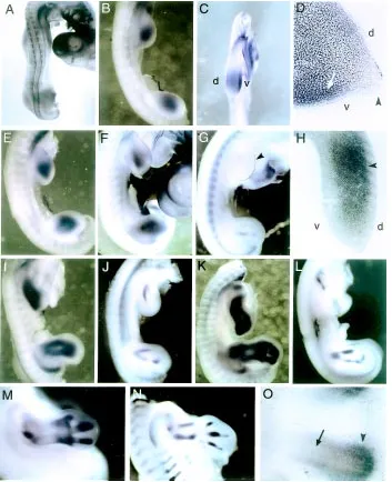
First, we examined the expression pat-tern of Frzb-1 in limb buds between stages 18 to 28. At stage 18, Frzb-1 is faintly expressed throughout mesenchymal cells of the limb field (Fig. 1A). At stage 20, Frzb-1 expression is observed in the core region of the limb bud (Fig. 1B), predomi-nantly in the ventral mesenchyme (Fig. 1C,D). The expression is restricted to the mesenchyme, and no expression is ob-served in the ectoderm, including the api-cal ectodermal ridge (AER) (Fig. 1D). By stage 22, triangular-shaped expression of Frzb-1 is observed, and the signal becomes intense (Fig. 1E). Expression in
Fig. 1. Expression patterns of Frzb-1 and aggrecan in the chick limb bud. (A-F,H,I,K,M,O) Frzb-1 expression at stage 18 (A), stage 20 (B,C,D), stage 22 (E), stage 24 (F,H), stage 25 (I), stage 26 (K), and stage 28 (M,O). (G,J,L,N) Aggrecan expression at stage 24 (G), stage 25 (J), stage 26 (L), and stage 28 (N). (A) Frzb-1 is faintly expressed throughout the mesenchymal cells of the limb bud at stage 18. (B-D) The stage 20 limb bud shows mesenchymal Frzb-1 expression in the central region of the limb bud with dorsoventral polarity (C,D; arrow). No expression is observed in the ectoderm, including the AER (D; arrowhead). (E,F) Triangular-shaped expression is observed in the limb bud at stage 22 (E) and 24 (F). (H) Transverse section of stage 24 hind limb bud. Frzb-1 is expressed at the central domains where the mesenchymal condensation has formed (arrowhead). At this stage, polarized expression along dorsoventral axis has ceased. (I,K) At stage 25 and 26, expression of Frzb-1 marks the cartilage rudiments of the limb. (M,O) At stage 28, expression of Frzb-1 is detected in the perichondrium of differentiated cartilage tissue, predominantly at the epiphyses (arrowheads), and Frzb-1 expression is excluded from the core region (O; arrow). (G,J,L,N) Expression of aggrecan to show the developing skeletal elements of the limb bud. Aggrecan is also expressed in the AER at early stages of limb development (G; arrowhead). Abbreviations: d, dorsal; v, ventral.
expression is localized to perichondrium of differentiated cartilage tissue, predominantly at the epiphyses (Fig. 1M), although weak expression is observed in the central region of the cartilage (Fig. 1O).
We compared the expression pattern of Frzb-1 with that of aggrecan, which is prima-rily expressed by chondrocytes (Oettinger and Pacifici, 1990; Enomoto-Iwamoto et al., 1998). Aggrecan mRNA is not detected in the mesenchyme of the developing limb bud until stage 23, when its expression is re-stricted to the AER (Fig. 1G). At stage 24, weak expression of aggrecan is detectable in the mesenchyme in the proximal-central region of the limb bud in addition to the AER. In the hind limb bud, the mesenchymal ex-pression is already divided into two domains corresponding to the presumptive tibia and fibula (Fig. 1G). By stage 25-26, bifurcated and segmented expression of aggrecan is observed representing presumptive zeugopodal structures (Fig. 1J,L). Aggrecan expression is restricted within the Frzb-1 domain. At stage 28, intense expression of aggrecan is detectable throughout the carti-lage primordia of the limb (Fig. 1N).
Altogether, Frzb-1 expression starts prior to the cartilage marker gene expression, and coincided with the region of mesenchymal condensations in the limb bud between stages 24 to 28, whereas aggrecan expres-sion in the limb mesenchyme represents the pattern of differentiating cartilage.
Expression patterns of Frzb-1 in the re-combinant limb bud
Next, we investigated the expression of Frzb-1 and aggrecan in a recombinant limb bud. During development of recombinant limb buds, cell-cell interactions and position specific gene expressions of mesenchymal cells are re-established, and the cells ac-quire new positional identity in the recombi-nant limb (Ros et al., 1994; Hardy et al., 1995; Wada et al., 1998b). To prepare the recombinant limb bud, we recombined distal mesenchyme of stage 20 wing buds with stage 22-23 leg ectodermal jackets. The recombinant limb bud was grafted onto the
Frzb-1 was clearly expressed in the proximal core region of the recombinant limb bud, and Frzb-1 could not be detected in the peripheral region (3/3 cases) (Fig. 2D). The localized expression of Frzb-1 was maintained during further development of the recombinant limb bud, then the expression in the proximal region became weak after 60 h (3/3 and 2/2 cases at 48 and 60 h, respectively) (Fig. 2F,H; arrowhead).
On the contrary, expression of aggrecan started 36 h after grafting of the recombinant limb buds (3/3 cases) (Fig. 2E). At this
Fig. 2. Expression patterns of Frzb-1 and aggrecan in the recombinant limb bud.(A) An example of cartilage elements which developed from the recombinant limb bud (asterisk). Arrowhead indicates a tungsten pin used to fix the recombinant limb bud. (B,C,D,F,H) Expression patterns of Frzb-1 in the recombinant limb bud. Expression of Frzb-1 was observed within 12 h after grafting(B). The expression was restricted to the central region throughout recombinant development (C,D,F,H). After 60 h grafting, proximal expression became weak (H; arrowhead). (E,G,I) Expression patterns of aggrecan in the recombinants at 36 h (E), 48 h (G) and 60 h (I) after grafting. Abbreviations: g, grafted recombinant limb bud; h, host limb bud.
rostral end of the host limb bud, and allowed to develop further. They formed well-shaped cartilage structures with several digits at the distal end, although these structures lost anteroposterior polarity (Fig. 2A).
stage, aggrecan expression was restricted to most proximal region of the recombinant. By 48 h after grafting, aggrecan was expressed in the proximal core region of the recombinant, and weak expression was detected in the distal region (3/3 cases) (Fig. 2G). Unlike Frzb-1, widespread expression of aggrecan was not observed at this stage. After 60 h, aggrecan was strongly expressed in the proximal region, while weak but clear expression was observed in the distal region of the recombinant (2/2 cases) (Fig. 2I).
Expression of Frzb-1 in cultured cells
We next investigated the expression of Frzb-1 and aggrecan in cultured cells to further examine temporal changes of Frzb-1
expression during cartilage differentiation. Stage 22-23 limb bud cells were plated at high density so that they undergo chon-drogenesis. Results are shown in Figure 3. Weak and broad expression of Frzb-1 was already observed 24 h after plating (Fig. 3A,B). At this time, we could not detect cellular condensations using a phase-contrast microscopy. Forty-eight hours after plating, when cellular conden-sations were observed, expression of Frzb-1 was mainly detected in the condensing cells (Fig. 3E,F). Frzb-1 expression was more enhanced at 72 h, and intense ex-pression was more widespread (Fig. 3I,J). On the other hand, aggrecan was faintly expressed in a few cells at 24 h after plating (Fig. 3C,D), and started at least 48 h after plating (Fig. 3G,H). Within 72 h, expression was clearly detected in the cellular condensations (Fig. 3K,L). At this time, Frzb-1 expression was larger than aggrecan expression domain, in agree-ment with the endogenous expression in the limb bud.
Discussion
In this study, we show the expression pattern of Frzb-1 in the chick limb bud, and compare its expression to that of the car-tilage marker, aggrecan. Frzb-1 is first expressed in the center of the limb bud, initially in the ventral mesenchyme, but later localizes midway along the dorsoven-tral axis. During later limb development, Frzb-1 expression shows the prepattern of skeletal elements of the limb, although Frzb-1 expression domain is wider than the region of aggrecan expression. Once the cartilage rudiments have formed, ex-pression of Frzb-1 localizes to the peri-chondrial tissue. In recombinant limb buds, Frzb-1 is also expressed in the proximal core region prior to the aggrecan expres-sion, and no expression of Frzb-1 was
Fig. 3. Expression of Frzb-1 and aggrecan in cultured limb bud cells. Phase contrast (A,C,E,G,I,K) and corresponding bright-field views (B,D,F,H,J,L) are shown for Frzb-1 expression at 24 h (A,B), 48 h (E,F) and 72 h (I,J) after plating, and aggrecan expression at 24 h (C,D), 48 h (G,H) and 72 h (K,L) after plating. At 24 h, weak and broad expression of Frzb-1 was detected (A,B; arrowheads), whereas aggrecan expression was detected in a few cells (C,D; arrowheads). At 48 h, Frzb-1 was expressed in the condensing cells (E,F; arrowheads). At this time point, aggrecan was expressed in a small patches of cells (G,H; arrowheads). At 72 h, strong expression of Frzb-1 was observed (I,J; arrowheads), and aggrecan mRNA was also detected (K,L; arrowheads). Both genes were expressed in the cellular condensations, although expression of Frzb-1 was more intense and extensive than that of aggrecan.
observed near the ectoderm. Moreover, Frzb-1 is expressed in cultured limb mesenchymal cells. Its expression started within 24 h after plating in culture, when aggrecan mRNA was not yet detectable, and later localized to condensing mesenchyme. These expression patterns suggest that Frzb-1 products are involved in the early phase of chondrogenesis in the limb bud.
Frzb-1 starts earlier than the beginning of condensation in regions not associated with chondrogenesis, but later coincides with the area of mesenchymal condensation in vivo and in vitro. The later expression pattern of Frzb-1 in the limb bud is similar to the distribution of the peanut agglutinin lectin binding molecule or the chondroitin sulfate proteoglycan, versican (Aulthouse and Solursh, 1987; Shinomura et al., 1990), although the peanut agglutinin lectin binding region seems to be narrower than the Frzb-1 expression domain. These molecules are predominantly ex-pressed in mesenchymal condensations (Shinomura et al., 1990). Therefore, Frzb-1 may have roles in initiation and maintenance of mesenchymal condensations, eventually promoting cartilage dif-ferentiation in the limb bud.
At later stages of development, Frzb-1 may regulate Wnt activity in the central core region of the limb bud, since Frzb-1 can bind to some Wnts, and antagonize their biological activity (Leyns et al., 1997; Wang et al., 1997). In the limb bud, various Wnt genes are expressed in the mesenchyme (Dealy et al., 1993; Tanda et al., 1995; Kawakami et al., 1999), and Wnts (Wnt-1, Wnt-5a and Wnt-7a ) inhibit chondrogenesis (Rudnicki and Brown, 1997; Kawakami et al., 1999). A similar result was also obtained from the analysis of Wnt-1 transgenic mice (Zakany and Duboule, 1993). Frzb-1 products may promote condensation formation and cartilage differentiation by antagonizing Wnt activity in the limb bud.
On the other hand, Frzb-1 is already expressed at early stages of limb development when mesenchymal condensations have not yet formed. At these stages, expression of Frzb-1 is enhanced in the ventral mesenchyme. Such a polarized expression along the dors-oventral axis has been also reported in the mouse limb bud (Hoang et al., 1998; Leimeister et al., 1998). These results indicate the existence of distinct regulatory pathways of Frzb-1 expression in early limb buds, and possible involvement of Frzb-1 in dorsoventral specification of the limb. Wnt-7a is expressed in the dorsal ectoderm of the early limb bud (Dealy et al., 1993; Riddle et al., 1995), and has a role in dorsal patterning of the limb by activating Lmx-1 in underlying mesenchyme (Riddle et al., 1995; Vogel et al., 1995). On the contrary, En-1 is expressed in ventral ectoderm, and is involved in ventral structure formation (Loomis et al., 1996; Logan et al., 1997). Early expression of Frzb-1 underlies En-1 expression in the ecto-derm. Thus, at least at early stages of limb development, Frzb-1 products may enhance the ventralizing signal of En-1 by antagoniz-ing Wnt-7a activity in the ventral region of the limb bud.
In the recombinant limb bud, Frzb-1 was restricted to the core region, and no expression signal was detected in the mesen-chyme near the ectoderm throughout limb development, possibly owing to the ectodermal stage of the recombinant limb bud. We used stage 22-23 leg bud to prepare ectodermal jacket of the recombinant, and no polarized expression of Frzb-1 was detected in the normal limb bud at these stages. Therefore, Frzb-1 was expressed only in the core region in the recombinant limb.
In the recombinants, Frzb-1 was expressed prior to aggrecan expression, and then their domains of expression overlapped. This indicates that the chondrogenesis in the recombinant limb bud proceeds similarly to that of the normal limb bud. The restricted expression of Frzb-1 was established within 12 h after recombinant grafting, suggesting that Frzb-1 expression responds rapidly to changing environmental signals. Also, in the normal limb bud, Frzb-1 is not expressed in the mesenchyme near the
ectoderm at chondrogenic stages. These results indicate that expression of Frzb-1 may be inhibited by the ectoderm. Because mesenchymal expression of Frzb-1 is observed at a distance from the ectoderm, diffusible factors produced in the ectoderm may inhibit the expression of Frzb-1 in the peripheral region of the limb bud. Since Frzb-1 possibly promotes mesenchymal condensa-tions as discussed above, inhibition of Frzb-1 may in turn sup-press chondrogenesis. It is known that cartilage differentiation in the limb bud is inhibited by the surrounding ectoderm (Solursh and Reiter, 1988; Gregg et al., 1989), hence ectodermal factors may regulate cartilage differentiation in the limb bud by inhibiting Frzb-1 expression in the mesenchyme.
Materials and Methods
Animals
Fertilized White Leghorn chicken eggs were used. Eggs were incu-bated at 38ºC and embryos were staged according to Hamburger and Hamilton (1951).
Medium
To culture mesenchymal cells of the limb bud, we used F-12 medium (Nissui) containing 2% FBS (ICN) as the culture medium (Wada et al., 1998a).
In situ hybridization
Whole-mount in situ hybridization was performed according to the method of our previous report (Kawakami et al., 1996), using the following chicken cRNAs. An antisense cRNA probe for Frzb-1 was synthesized with a 1.2 kb fragment encoding part of the coding region and 3'-noncoding region (Ladher et al., in preparation). Chicken cartilage aggrecan cDNA, obtained from Dr. Iwamoto (Enomoto-Iwamoto et al., 1998), was used as a template for cRNA synthesis. After in situ hybridization, the embryos were re-fixed in 4% paraformaldehyde, embedded in 4% agar-ose (FMC), and sectioned on a vibratome at 50 µm (Logan et al., 1997). In situ hybridization of cultured cells was performed according to the method of whole-mount in situ hybridization with a slight modification. In brief, 2 µg/ml of proteinase K was used in pretreatment of the cells, and hybridization was performed at 50ºC for 16 h.
Preparation of recombinant limb buds
Recombinant limb buds were prepared as reported previously (Wada et al., 1998b). The distal regions of stage wing buds were excised, and mesodermal tissues were dissociated into single cells. The cells were pelleted by centrifugation and packed into an ectodermal jacket prepared from stage 22-23 whole leg buds. The recombinant limb bud was grafted onto the anterior-proximal region of stage 21-23 wing bud with a tungsten pin, allowed to develop, and fixed for either in situ hybridization or skeletal analysis.
Skeletal analysis
For the analysis of the skeletal pattern, the recombinants were allowed to develop for five days. Embryos with a limb-like structure were fixed in 10% formalin, stained with 0.1% Alcian green, macerated in 1% potas-sium hydroxide, and cleared in 50% glycerol solution.
Micromass culture of mesenchymal cells
2 ml of culture medium was added. After incubation, cultured cells were fixed with 4% paraformaldehyde for in situ hybridization.
Acknowledgments
We thank Dr. M. Iwamoto for providing the aggrecan cDNA, Dr. S. Nishimatsu for help in preparation of the manuscript, and C. Komaguchi and R. Nakao for technical assistance. This work was supported by grants-in-aids from the Ministry of Education, Science, Sports and Culture of Japan, Research Project grants from Kawasaki Medical School, and funding from the Arthritis and Rheumatism Campaign and the Wellcome Trust.
References
AHRENS, P.B., SOLURSH, M. and REITER, R.S. (1977). Stage-related capacity for limb chondrogenesis in cell culture. Dev. Biol. 60: 69-82.
AULTHOUSE, A.L. and SOLURSH, M. (1987). The detection of a precartilage, blastema-specific marker. Dev. Biol. 120: 377-384.
CHUONG, C.-M., WIDELITZ, R.B., JIANG, T.-X., ABBOTT, U.K., LEE, Y.-S. and CHEN, H.-M. (1993). Roles of adhesion molecules NCAM and tenascin in limb skeletogenesis: Analysis with antibody perturbation, exogenous gene expres-sion, talpid2 mutants and activin stimulation. Prog. Clin. Biol. Res. 383B: 465-474.
DEALY, C.N., ROTH, A., FERRARI, D., BROWN, A.M. and KOSHER, R.A. (1993). Wnt-5a and Wnt-7a are expressed in the developing chick limb bud in a manner suggesting roles in pattern formation along the proximodistal and dorsoventral axes. Mech. Dev. 43: 175-186.
EDE, D.A. (1983). Cellular condensations and chondrogenesis. In Cartilage Vol. 2 (Ed. Hall, B.K.), Academic Press, San Diego, pp. 143-186.
ENOMOTO-IWAMOTO, M., IWAMOTO, M., MUKUDAI, Y., KAWAKAMI, Y., NOHNO, T., HIGUCHI, Y., TAKEMOTO, S., OHUCHI, H., NOJI, S. and KURISU, K. (1998). Bone morphogenetic protein signaling is required for maintenance of differentiated phenotype, control of proliferation, and hypertrophy in chondrocytes. J. Cell Biol. 140: 409-418.
GREGG, B.C., ROWE, A., BRICKELL, P.M. and WOLPERT, L. (1989). Ectodermal inhibition of cartilage differentiation in micromass culture of chick limb bud mesenchyme in relation to gene expression and cell shape. Development 105: 769-777.
HALL, B.K. and MIYAKE, T. (1992). The membranous skeleton: the role of cell condensations in vertebrate skeletogenesis. Anat. Embryol. 186: 107-124.
HALL, B.K. and MIYAKE, T. (1995). Divide, accumulate, differentiate: cell conden-sation in skeletal development revisited. Int. J. Dev. Biol. 39: 881-893.
HAMBURGER, V. and HAMILTON, H. (1951). A series of normal stages in the development of the chick embryo. J. Morphol. 88: 49-92.
HARDY, A., RICHARDSON, M.K., FRANCIS-WEST, P.H., RODRIGUEZ, C., IZPISUA-BELMONTE, J.-C., DUPREZ, D. and WOLPERT, L. (1995). Gene expression, polarizing activity and skeletal patterning in reaggregated hindlimb mesenchyme. Development 121: 4329-4337.
HOANG, B., MOOS, M., Jr., VUKICEVIC, S. and LUYTEN, F.P. (1996). Primary structure and tissue distribution of FRZB, a novel protein related to Drosophila Frizzled suggest a role in skeletal morphogenesis. J. Biol. Chem. 271: 26131-26137.
HOANG, B.H., THOMAS, J.T., ABDUL-KAMIN, F.W., CORREIA, K.M., CONLON, R.A., LUYTEN , F.P. and TRACY BALLOCK, R. (1998). Expression pattern of two Frizzled-related genes, Frzb-1 and Sfrp-1, during mouse embryogenesis suggests a role for modulating action of Wnt family members. Dev. Dyn. 212: 364-372.
KAWAKAMI, Y., ISHIKAWA, T., SHIMABARA, M., TANDA, N., ENOMOTO-IWAMOTO, M., ENOMOTO-IWAMOTO, M., KUWANA, T., UEKI, A., NOJI, S. and NOHNO, T. (1996). BMP signaling during bone pattern determination in the developing limb. Development 122: 3557-3566.
KAWAKAMI, Y., WADA, N., NISHIMATSU, S., ISHIKAWA, T., NOJI, S. and NOHNO, T. (1999). Involvement of Wnt-5a in chondrogenic pattern formation in the chick limb bud. Dev. Growth Differ. 41: 29-40.
KENGAKU, M., CAPDEVILA, J., RODRIGUEZ-ESTEBAN, C., DE LA PENA, J., JOHNSON, R.L., IZPISUA-BELMONTE, J.-C. and TABIN, J.C. (1998). Distinct WNT pathways regulating AER formation and dorsoventral polarity in the chick limb bud. Science 280: 1274-1277.
LEIMEISTER, C., BACH, A. and GESSLER, M. (1998). Developmental expression patterns of mouse sFRP genes encoding members of the secreted frizzled related protein family. Mech. Dev. 75: 29-42.
LEYNS, L., BOUWMEESTER, T., KIM, S.H., PICCOLO, S. and DE ROBERTIS, E.M. (1997). Frzb-1 is a secreted antagonist of Wnt signaling expressed in the Spemann organizes. Cell 88: 747-756.
LOGAN, C., HORNBRUCH, A., CAMPBELL, I. and LUMSDEN, A. (1997). The role of Engrailed in establishing the dorsoventral axis of the chick limb. Development 124: 2317-2324.
LOOMIS, C.A., HARRIS, E., MICHAUD, J., WURST, W., HANKS, M. and JOYNER, A.L. (1996). The mouse Engrailed-1 gene and ventral limb patterning. Nature 382: 360-363.
OBERLENDER, S.A. and TUAN, R.S. (1994). Expression and functional involve-ment of N-cadherin in embryonic limb chondrogenesis. Developinvolve-ment 120: 177-187.
OETTINGER, H.F. and PACIFICI, M. (1990). Type X collagen gene expression is transiently up-regulated by retinoic acid treatment in chick chondrocyte cul-tures. Exp. Cell Res. 191: 292-298.
RIDDLE, R.D., ENSINI, M., NELSON, C., TSUCHIDA, T., JESSELL, T.M. and TABIN, C.J. (1995). Induction of the LIM homeobox gene Lmx1 by WNT7a establishes dorsoventral pattern in the vertebrate limb. Cell 83: 631-640.
ROS, M.A., LYONS, G.E., MACKEM, S. and FALLON, J.F. (1994). Recombinant limbs as a model to study homeobox gene regulation during limb development. Dev. Biol. 166: 59-72.
RUDNICKI, J.A. and BROWN, A.M.C. (1997). Inhibition of chondrogenesis by Wnt gene expression in vivo and in vitro. Dev. Biol. 185: 104-118.
SHINOMURA, T., JENSEN, K.L., YAMAGATA, M., KIMATA, K. and SOLURSH, M. (1990). The distribution of mesenchyme proteoglycan (PG-M) during wing bud outgrowth. Anat. Embryol. (Berl.) 181: 227-233.
SOLURSH, M. and REITER, R.S. (1988). Inhibitory and stimulatory effects of limb ectoderm on in vitro chondrogenesis. J. Exp. Zool. 248: 147-154.
TANDA, N., OHUCHI, H., YOSHIOKA, H., NOJI, S. and NOHNO, T. (1995). A chicken Wnt gene, Wnt-11, is involved in dermal development. Biochem. Biophys. Res. Commun. 211: 123-129.
VOGEL, A., RODRIGUEZ, C., WARNKEN, W. and IZPISUA-BELMONTE, J.-C. (1995). Dorsal cell fate specified by chick Lmx1 during vertebrate limb develop-ment. Nature 378: 716-720.
WADA, N., KIMURA, I., TANAKA, H., IDE, H. and NOHNO, T. (1998a). Glycosylphosphatidylinositol-anchored cell surface proteins regulate position-specific cell affinity in the limb bud. Dev. Biol. 202: 244-252.
WADA, N., OHSUGI, K., YOKOUCHI, Y., KUROIWA, A. and IDE, H. (1998b). Hox gene expression, AV-1 antigen expression, and cartilage pattern formation in chick recombinant limb bud. J. Exp. Zool. 281: 26-35.
WANG, S., KRINKS, M., LIN, K., LUYTEN, F.P. and MOOS, M., Jr. (1997). Frzb, a secreted protein expressed in the Spemann organizer, binds and inhibits Wnt-8. Cell 88: 757-766.
ZAKANY, J. and DUBOULE, D. (1993). Correlation of expression of Wnt-1 in developing limbs with abnormalities in growth and skeletal patterning. Nature 362: 546-549.