A treasure house of comparative embryology
0214-6282/98/$15.00 © UBC Press
Printed in Spain
www.lg.ehu.es/ijdb
*Address for reprints: Department of Anatomy and Developmental Biology, St. Georges Hospital Medical School, Cranmer Terrace, London SW17 ORE, United Kingdom. FAX: 44-0181-725 3326. e-mail: m.richardson@sghms.ac.uk
MICHAEL K. RICHARDSON
1*and
JENNIFER NARRAWAY
21Department of Anatomy and Developmental Biology, St. George’s
Hospital Medical School, London, United Kingdom and 2Hubrecht
Laboratory, Netherlands Institute for Developmental Biology (NIOB), Utrecht, The Netherlands
Introduction
The Netherlands Institute for Developmental Biology (NIOB) in Utrecht (Hubrecht Laboratory) is the most important centre for the study of comparative vertebrate embryology in the world. In addition to its active research programmes in molecular aspects of development, the Laboratory houses an incomparable set of resources in comparative embryology, available for study by all interested scholars. There is a large and important collection of historical and modern reprints, covering all fields of developmen-tal biology. There is also a superb embryology library containing such landmarks in the history of comparative embryology as von Baer’s Entwicklungsgeschichte der Thiere (von Baer, 1828), described as ‘one of the greatest works in the whole history of biological science’ (Minot, 1906); and Keibel’s Normentafeln, which contain tabulated data on the timing of organogenesis in a wide range of vertebrates (Keibel, 1895; Keibel, 1897).
But perhaps the most important resource of the NIOB is the Embryo Collection –by far the largest and most valuable of its kind in existence. Significant collections of animal embryos exist elsewhere, but none can match the range, quality and rarity of material in Utrecht. The Embryo Collection was founded on the material collected by A.A.W. Hubrecht. It has since been enriched by donations, the largest of which is the Hill collection. For many years Hubrecht’s collection was the core resource of the Hubrecht
Laboratory, which was founded in 1916 for the study of the comparative embryology of vertebrates (Bangma, 1986).
The research emphasis of the lab has changed with time, and is now concentrated on experimental embryology and molecular genetics. The status of the collection has changed accordingly. However, unlike many other great embryo collections which were neglected, dispersed or destroyed, the one at Utrecht has been carefully preserved. Embryo collections are vulnerable to changes in scientific fashion. Personal communications with other scien-tists suggest to us that many collections have been disposed of since comparative embryology fell out of favour with biologists earlier this century. Other collections were consigned to storage, and have suffered accordingly.
when examined by one of the authors (MKR) in December 1998. Very few wet specimens at Utrecht appear to have been lost in this way.
Another reason why few embryo collections survive is that taxonomy is based principally on the description of adult type specimens. Museums of natural history therefore tend to collect adult material only. For example in The Natural History Museum in London the few embryonic and fetal specimens in the collection are not separately catalogued, and a laborious search of the general catalogues is therefore required to locate them. Embryo collections are held mainly in university institutions, such as departments of anatomy in medical or veterinary schools. They are often based on the personal collections of embryologists and may survive only as long as their founder. But as we shall see, a personal collection may occasionally become the nucleus of something more lasting, as is the case with the Hubrecht, Hill, Minot and Mall collections.
The scope of the Utrecht collection
The Utrecht Collection1 of embryos consists of wet specimens,
histological sections and material blocked out in paraffin but not
yet sectioned. Much of this material was assembled in the late nineteenth and early twentieth centuries. The archives contain photographs and original drawings, as well as many notebooks of the scientists who contributed to the Collection. These often give details of the source, fixation and processing of specimens. There is a printed catalogue of the Lemuridae and Dermoptera (1921) and two Concise Catalogues (Nieuwkoop,1953 and Boterenbrood, 1977). A partial computerised catalogue can be consulted in the lab. The Utrecht Collection includes reprints of around 200 papers based on its material (Bangma, 1988). Stained paraffin sections of embryos, mounted on glass slides, are stored in purpose-built wooden cabinets. Many of the microscope slides are of the old fashioned large-format types, and a compound microscope with a large stage is available for viewing these. Estimates2,3 of the
number of slides range from 30,000 to 80,000.
The 2,000 wet specimens3 (embryos, uteri –even whole
ani-mals at postnatal stages) are stored in 80% alcohol4. The smaller
wet specimens are in glass vials, each with a paper label and a plug of cotton wool inside. These small vials are gathered together into domestic preserving jars filled with alcohol. A similar arrange-ment was used in the Minot collection at Harvard (Minot, 1905). Great care is needed when handling the smaller wet specimens –the paper label can crush the embryo when the plug of cotton is pushed back into the vial. A few specimens appear to have been damaged in this way. Because 80% alcohol is flammable, the collection is housed in a specially designed fireproof vault.
The Utrecht Collection is estimated to represent around 600 species of chordate, in 175 families and 10 classes4. The range
of species represented has been influenced by several factors. These include the research interests of the founder and donors, the ease of obtaining embryos from particular species, and even the colonial possessions of the collector’s home country. While the collection is remarkably wide in its coverage, there are gaps; cetaceans and humans are poorly represented for example. Furthermore the earlier stages of development tend to be more extensively covered than later stages5. This rather random,
piecemeal representation contrasts with the Harvard Embryologi-cal Collection, which was formed by Minot along systematic lines, so as to represent vertebrate development through ‘a certain number of carefully selected types’.6
The philosophy of the NIOB is that the embryo collection is there to be used, and may be consulted on request. Loans can be arranged, except in the case of rare or historically important material. If there are enough specimens, whole embryos in alcohol can be borrowed and even sectioned by the borrower, provided proficiency in histological technique is demonstrated. The borrower signs a loan agreement and promises to return the material, including that which has been sectioned, after a speci-fied time. Sadly, not all scientists honour this agreement. Some material has recently been returned 25 years after it was bor-rowed.
Collecting embryos
To study comparative embryology, species need to be chosen so as to give some kind of phylogenetic context to the comparison (Richardson et al., 1999). As Hubrecht noted in his discussion of mammalian placentation: ‘As in all other attempts at comparative analysis, so in this case the selection of material that is to furnish
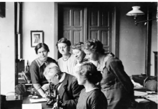
Fig. 1. Hubrecht teaching a group of students on an embryology course (dated 17th November–17th December 1913). From the archive of the Embryo
Collection (folder number: “A.A.W Hubrecht, 1876”).
1 In this article, ‘Utrecht Collection’ refers to the embryo collection of the Hubrecht
Laboratory, (NIOB).
2 Bangma (1988) p. 14.
3 Royal Netherlands Academy of Arts and Sciences, Hubrecht Laboratory, Netherlands
Institute for Developmental Biology (NIOB). Report, 1986.
4 One of us (MR) has prepared paraffin sections from embryos from stored in alcohol
for many decades in the Utrecht Collection; the resulting histology is excellent.
5 Hubrecht Laboratory Netherlands Institute for Developmental Biology. Progress
Report, 1993. Koninklijke Nederlandse Akademie van Wetenschappen, Amsterdam (p. 59).
6 Minot (1905) p. 499. 7 Balfour (1876) p.175.
8 C.S. Minot, in the introduction to Scammon (1911, p.1). 9 Wilson (1889) p. 209.
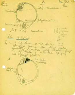
Fig. 2. The Spemann and Mangold experiment. In addition to a historical collection of embryos, the Hubrecht collection contains slides and laboratory notes from pioneers in embryology. These include a collection made by Otto Mangold containing the lab notebooks from Hilde Proescholdt, later his wife, Hilde Mangold. We show here her careful analysis of two sections from the famous Um 132, the most completely developed of five salamander larvae derived from embryos which had previously received a transplant of an upper blastoporal lip at the gastrula stage. The drawings illustrate that the transplant induces a second embryonic axis and the lower drawing shows clearly that donor cells from the implanted blastopore (drawn in ink, and recognisable because they were derived from Trituris cristatus, which has unpigmented eggs, while the host was Trituris taeniatus, which has pigmented eggs) gave rise to somite, notochord and the floorplate of the neural tube. These findings (H. Spemann and H. Mangold, W. Roux’ Arch. Entwmech. Org. 100: 599-638, 1924) helped win a Nobel prize.
the bases of comparison is most important’ (Hubrecht, 1894b). Embryos also need to be in a good state of preservation, and at the right stages of development. There are essentially two ways to meet these various requirements. One is to set up a captive breeding population, and the other is to collect embryos in the wild. To the modern biologist, collecting in the wild poses ethical problems if large numbers of animals have to be killed, or if the species in question is endangered. Before considering the Hubrecht Collection in detail, we shall look more generally at how Hubrecht’s contemporaries developed their embryo col-lections.
Francis Balfour records that he collected shark and ray em-bryos at the Zoological Station in Naples, and also obtained specimens from animals kept in the Brighton Aquarium7. Charles
Minot purchased dogfish embryos (Squalus sp.) from fishermen: ‘As Squalus acanthias is abundant on the New England coast, it is caught in large numbers by the fishermen, who are glad to bring the fish in for a small price, as they have no market value. It is therefore possible to obtain during the summer an almost unlimited supply of “candles” as the fisher-men call the egg-cases, with ova in all stages of development from segmentation up to those with embryos of about 50 cm. in length.’8
Henry Wilson describes how he obtained material for the study of the Sea Bass (Serranus atrarius): ‘The fish is one of several (mackerel, scup, tautog, etc.), which at this season [May-July] are reared in the hatchery of the Wood’s Holl Station, and I was therefore able to obtain, with the least amount of trouble, as complete a set of material as could be desired.’9 (Our note in
brackets [ ]). Material collected in the wild can be fixed on the spot, or returned to the lab and grown until it reaches the desired stage. Hubrecht reminds us that fixation needs to be very soon after death in order to get good histology: “Preparations made from animals that had been dead even for only a very short time have already undergone so considerable an alteration that they are of very inferior value for comparative and especially for histological research.”10
Some embryologists relied on the services of “collectors” to provide material. Parker acknowledged his debt to a collector for procuring specimens of the kiwi (Apteryx sp.): “For some time I only succeeded in obtaining two or three specimens of advanced stages, and it was only when I was fortunate enough to secure the services of Mr. Richard Henry, of Lake Te Anua, as collector, that my material became copious enough to be worth working up.”11
Mr. Henry was paid for his services. Parker’s experiences with Henry also highlight another problem with collectors: they could not always be relied upon to deliver embryos in good condition. Henry removed the kiwi embryos from the egg and preserved them in alcohol. No fixative was used, so the histology was poor. Parker complains about one specimen: “This embryo was unfor-tunately much damaged by the collector during removal from the egg. The head was severed from the body, the surface was considerably abraded, and, worst of all, both fore-limbs were destroyed.”12
Arthur Dendy gives a fascinating account of how he obtained eggs of the Tuatara a reptile living on Stephens Island, Cook
Straits, New Zealand (Dendy, 1899). This was a protected re-serve, and Dendy had to seek permission from the authorities. He used the services of the lighthouse keeper on the island who collected eggs, and sent them by boat to the mainland, packed in moist sand. Dendy then opened the eggs and fixed the embryos on receipt. The main problem encountered was not in the collec-tion itself, but in getting the eggs to Christchurch before they perished.
succeeds in obtaining a variety of stages under the present precarious means of collecting in relatively deep water.”13 For
some species, it has proven possible to collect fertilised eggs in the wild, and then rear them in the laboratory. This technique has been used for example for the Queensland lungfish (Kemp, 1982). W.E. Agar, on his expedition to Gran Chaco in 1907, collected embryos and larvae of the eel-like fish Symbranchus marmoratus. Some specimens were fixed immediately on re-moval from the nest, while others were reared further in dishes of water (Taylor, 1913).
Many collectors, including Hubrecht, circulated pamphlets ap-pealing for the donation of embryos. These appeals were especially necessary for collectors of human embryos, who depended on the
co-operation of physicians and surgeons (Mall, 1898, 1903, 1910, 1911, 1913a). A printed flyer in the archive of the Utrecht Collection (dated by the archivist ‘March 1980’) appears to have been circulated by Cornell University Anatomy Department. It asks for donations of domestic animal embryos and includes the statement that: ‘We desperately need new material, as there are many gaps in the collection. Especially "needed" are specimens representing early stages of domestic animal development...please save these speci-mens. Simply place them in 10% formalin or any other suitable fixative and send them [to Cornell]...Please include any information concerning the fetus and mother which you have’. (Our note in brackets [ ]).
The Hubrecht collection
The Utrecht Collection is based on the personal collection of Ambrosius Arnold Willem Hubrecht (1853-1915)14. Following his
appointment to the chair of zoology and comparative embryology in Utrecht (1882), Hubrecht became increasingly focused on comparative embryology, and its importance in revealing evolu-tionary relationships (Anonymous, 1972; Assheton, 1915; Keibel, 1915). He was concerned at the threat, posed by European settlement and expansion in the New World, to wild animal populations (Hubrecht, 1910). This was an important drive behind his collecting activities. Hubrecht was instrumental in establishing the Institute International d’Embryologie15. In 1911 he wrote to
James Hill, of University College London, describing what he had in mind16:
My Dear Hill,
have you a mind to join a meeting of diverse vertebrate embryologists which is going to take place in Utrecht on the Friday or Saturday before Whitsuntide this year with the object of coming to an agreement on certain disputed points and to prepare [this/the] road for the work of the Nomen-clature Commission.
I expect to see here Keibel, Bonnet, Spee, Peter, Brachet and perhaps Henneguy, Prenant and others. Nothing however is settled or definite yet. But your presence would be immensely appre-ciated. It need only last a very few days and bringing preparations might simplify matters. It might be the nucleus of an international embryo-logical society of only very small size, few mem-bers...
Let me know your decision soon, Ever yours, Hubrecht
After the meeting, Hubrecht wrote again to Hill. He made it clear that the principle aim of the Institute was the collection and study of rare embryological material:
“A result of the meeting was the formation of an international embryological Institute which takes for its primary object the collecting of unbroken series of embryological preparations of those gen-era of mammals which are in imminent danger of disappearing from the earth’s surface within calcu-lable time”17
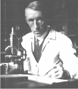
Fig. 3. James Peter Hill (1873-1954). From an undated photo in the Collection.
13 Dean (1899) p. 221.
14 A list of Hubrecht’s publications is given in Keibel (1915).
15 Keibel et al. (1912). Note that the l’Institut International d’Embryologie (IIE) is now
the International Society of Developmental Biologists (ISDB). By contrast the International Embryological Institute (IEI) is the former name for The Netherlands Institute for Developmental Biology (abbreviated in Dutch to NIOB and also called the Hubrecht Laboratory.
16 Hubrecht to Hill, Utrecht, May 2, 1911, letter in archive of the Utrecht Collection. 17 Hubrecht to Hill, undated letter in archive of Utrecht Collection.
18 Hubrecht (1910) p. 18.
19 He discussed these expeditions with Charles Darwin, whom he visited on May 24th,
1880 at Down House. The visit to Darwin is described in Hubrecht’s diary (typescript translation in archive of Utrecht Collection).
20 Hubrecht (1894b) p. 80.
21 Hubrecht (1894b), p. 81; van Oordt (1921); Hubrecht (1894) p. 80.
22 Hubrecht (undated pamphlet in the archive of the Utrecht Collection) Instructions
for aiding Professor Hubrecht’s embryological researches.
23 Hubrecht (1890). Open letter to the Natural History Society of the Dutch East-Indies
1929). At first Hubrecht studied European insectivores such as such as the mole (Talpa europaea), shrew (Sorex araneus) and hedgehog (Erinaceus europaeus). There are large numbers of hedgehog embryos in the Collection, because Hubrecht initiated a hedgehog hunt in the environs of Utrecht. According to Historia Medicinae in Nederland he offered to pay 25 cents for any live hedgehog brought to him (Bangma, 1986). In summer, as many as 40 a day were being brought in.
Around 1,500 specimens were acquired in this way between 1884 and 1890. In 1888 he used this material in his first publication on mammalian embryology (Hubrecht, 1888), a study of hedgehog placentation, in which he introduced the term “trophoblast”. After this he turned his collecting attention to shrews (Hubrecht, 1894a) harvested, like the hedgehogs, in the environs of Utrecht. Over several summers, shrews were collected at harvest time when the females were pregnant, and were driven out of their nests by the harvest.
Later, Hubrecht studied placentation in eutherian mammals from the tropics. At the invitation of the Royal Physical Society in Jakarta (Batavia), he made a visit to Indonesia, the former Dutch East Indies19. He arrived in November 1890 and set about collecting a
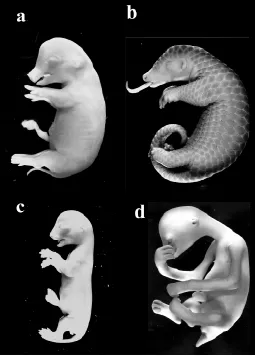
large series of embryos of the pangolin or scaly anteater (Manis javanica), the treeshrew (Tupaia javanica), the tarsier (Tarsius bancanus) the slow loris (Nycticebus coucang) and the flying lemur (Cynoceaphlaus varieagatus). Hubrecht wrote an account of his expedition under the title Spolia nemoris (spoils of the forest) (Hubrecht, 1894b).
His rich harvest of material was based on remarkably skilful preparation (Hubrecht, 1894b). Before his arrival in Indonesia, he
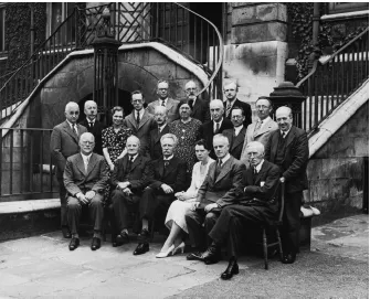
Fig. 4. Meeting of the IIE 2nd-5th August 1938, London. Back row (left to right) H.M.W. Woerdeman, H. Bluntschli, J. Florian. Middle row (left to right) G.L. Streeter, K. Peter, K. Jones Hill, J.T. Flynn, E.S. Goodrich, E.A. Fraser, W.H. Lewis, A. Celestino da Costa, P. Gérard, H. Woollard. Front row (seated, left to right) O. Grosser, J.F. Wilson, J. Boeke, H.B. Fell, D. de Lange, J.P. Hill. From a photograph in the collection.
arranged for the Physical Society in Jakarta to distribute coloured drawings of the animals he was interested in. This attracted correspondence from interested locals. The correspondence was collected by the Librarian of the Physical Society and passed on to Hubrecht when he arrived. He therefore established a network of collectors. As he records in Spolia Nemoris: “...I left behind me, wherever I had succeeded in enlisting co-operators, printed instructions, chemicals, glass tubes, etc., as well as cash for the payment of premiums to the natives by whom the collecting of live material was to be done.”20. There was a payment of 50 cents per
embryo.
Hubrecht’s printed instructions survive in the archive; in them, Hubrecht places great stress on the importance of fixing the embryos while the mother is still warm22,23. In an open letter23 to
the Royal Natural History Society of the Dutch East Indies he includes the following instructions:
1. The principal point is that 'only absolutely fresh material' be used. Animals obtained by the gun and transported home are of no value for conservation.
2. The animals especially required are: Aardvarks (Orycteropus), Klipdassies or Rock Rabbits (Hyrax), Golden Moles (Chrysochloris), Rock and Elephant Shrews (Macroscelides), Bushbabies (Galago), and Springhares (Pedetes). The two first-named can hardly be obtained alive. Specimens obtained by the gun can in this case perfectly serve if the sportsman who kills them is provided with means of operating on them on the spot, by extracting uterus and immersing same immediately in one of the fluids for preserving it23.
Later in the same letter Hubrecht acknowl-edged the practical difficulties involved in study-ing comparative embryology:
“Besides this the Institute will promote the unification with respect to scientific nomenclature and will take prelimi-nary steps towards a method of gen-eral cooperation by which the study of embryological material that is often very difficult to collect can be facili-tated and secured”17
He believed that the funding for the Institute would come from private sources, and viewed Carnegie Institution (Washington, USA) as a model for what he had in mind (Hubrecht, 1910). He envisaged a series of collecting stations all over the world, especially Madagascar, South America, South Africa and Australia, for the pro-curement of embryos from wild animals (Hubrecht, 1910). He wanted to harvest embryos to “prevent the danger that numerous mammal species should become extinct before revealing their secrets to Man.”18
In the 15 years of collecting, around 4000 specimens were obtained. Hubrecht was only able to publish findings on a small part of this vast collection. Although he published major works on the early development and placentation of mammals, later orga-nogenetic stages were neglected24. It was therefore left to others
to make full use of the Hubrecht’s Indonesian material. As von Oordt commented in 1921 for Manis:
“These Manis-specimens, not yet being investi-gated, (Hubrecht himself published himself only some short communications on Manis) Dan. De Lange Jr., director of the Hubrecht-Laboratory at Utrecht, put a part of the collection of Manis-embryos at my disposal in order to examine the early developmental stages.”25
De Lange published Normal Tables of Tupaia with H.F. Nierstrasz (De Lange and Nierstrasz, 1932) and Manis with F.J. Huisman (Huisman and De Lange, 1937). They were published under the auspices of the IIE and, according to Raven26, they
were mainly De Lange’s work. Interestingly, these normal tables were not officially part of Keibel’s series of Normal Tables. After
Keibel’s death, the publisher (Fischer) indicated that he had no intention of publishing further Normal Tables unless the authors made a substantial contribution to the publishing costs27. Equally
he declined to allow the use the Normal Tables title by other publishers. De Lange and the IIE were therefore forced to turn to Dutch publishers Oosthoek to publish the studies of Manis and Tupaia. De Lange also wrote up Hubrecht’s notes on Galeopithecus after Hubrecht’s death (De Lange, 1919).
The Hill collection
By far the largest addition to the Hubrecht Collection was the material collected by James Peter Hill (1873-1954). This consists of about 3,000 bottles of material in alcohol28; 28,000 microscope
slides27; specimens blocked out in wax; detailed field and
labora-tory notebooks, and other documentation; and photographs, in-cluding pairs of stereomicrographs of platypus and other embryos, to be viewed with the special pair of viewing glasses which survive in the collection. In 1966, Hill’s collection was deposited on perma-nent loan at Utrecht by University College London. This doubled the size of the holdings in the Hubrecht Laboratory, and was the initiative of Hill’s daughter Catherine (Katie) Kirkham Jones, who spent 10 years cataloguing her father’s collection. She was an embryologist and had published, with her father, a paper on monotreme development (Hill and Hill, 1933; Watson, 1955). In 1966, she wrote to Nieuwkoop, director of the Hubrecht Laboratory:
You will have heard from Mr. Tattersall, Secretary to the College, that the College Committee has agreed to the transference of the whole of the Hill Collection to the International Institute of Embryol-ogy -I am delighted to think that it will be safely housed under your ca and be available to re-search workers in a way which would not be pos-sible here. I shall be finished with my work on the Collection by the beginning of September [1966] -so that any time after that it could be sent over to you28. (Our note in brackets [ ]).
From 1892 Hill worked at the University of Sydney, where he made important studies on the early embryology of marsupials and monotremes including the duck-billed platypus, Ornithorhynchus anatinus (de Beer, 1948). In 1906, he was appointed professor of Comparative Anatomy and Embryology in University College, London. In 1910 he published a description of the early stages of marsupial development (Hill, 1910). This was based on the material he had collected over a period of 8 years in Australia29. After taking up his appointment in London, he
ex-panded his collection through purchases from hunter-collectors. These later additions included South American marsupials and African Mammals.
Hill’s “camp notes” (Hill notebook 17) detail his expeditions in 1895 and 1896 where he hunted platypus, rock wallabies and possums. In a paper with J.T. Wilson, Professor of Anatomy at Sydney, he describes the difficulties he faced in obtaining platypus embryos: “The animal is extremely shy and difficult of approach. They are occasionally, but rarely, captured as an incident in net-fishing in the larger rivers: otherwise they are practically only obtainable with the gun. During the breeding season, however, the pregnant female appears to keep much more closely to the burrow,
May 30, 1923 My Dear Hill,
We have received a lot of stuff from South America from a man named Ehrhardt who has been collecting, more or less for the Museum, in the
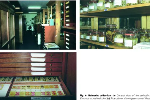
Fig. 6. Hubrecht collection. (a) General view of the collection. (b) Embryos stored in alcohol. (c) Slide cabinet showing sections of Macaque.
b
a
c
24 His Normal Tables for Tarsius and Nycticebus, with Keibel, is a notable exception.
See Hubrecht and Keibel (1907).
25 van Oordt (1921) p. 1.
26 Raven (1966). The Hubrecht laboratory during the past fifty years, a retrospect.
Typescript in the archive of the Hubrecht Laboratory.
27 although further editions of Keibel’s Normal Tables were published in the 1930s by
Fischer (Henneberg, 1937; Kudo, 1938).
28 Kirkham-Jones to Nieuwkoop, 25th July, 1966. Letter in archive of Utrecht Collection. 29 Hill (1910) p. 2.
30 Wilson and Hill (1907) p. 33-34. 31 Watson (1955) p. 113.
32 Kenny, presumably to J.P. Hill, 9th November, 1899. Letter in Hill notebook 4, In
archive of Embryo Collection.
33 Clout to ?Hill 21st November, 1899. Transcription, in Hill notebook 26, of a letter. In
archive of Embryo Collection.
34 Hinton to Hill, May 30, 1923. Letter in Hill notebook 10, in archive of Utrecht
Collection. Martin Hinton (1883-1961) was a mammalogist, appointed as an assistant in the Department of Zoology at the British Museum (Natural History) in 1921, and Keeper of Zoology, 1936-1945. Information kindly supplied by the Natural History Museum. We have been unable to trace the current whereabouts of the embryo collection in Giessen mentioned in this letter.
35 Ehrhardt, presumably to Hill, though unaddressed, 8th July, 1923. Letter in Hill
notebook 10, in archive of Utrecht Collection.
36 Flynn (1998) p. 19.
so that one may then commonly enough shoot five or six males to one female.”30 Watson31 states that Hill himself took an active part
in expeditions in Australia and Brazil.
The Hill notebooks contain a number of letters and receipts which make it clear that hunter-collectors were employed to help secure platypus embryos. For example, Patrick Kenny wrote: “I received the cheque and your letter safely to hand. ... The nest where the young Platypus where (sic) is about 20 feet long, and, at the end where the nest was, is about twelve inches wide. The young ones where (sic) in the nest with her, and the egg shells, I got them in the first week of August...”32. Wombats also proved to
be difficult to collect, and Hill’s notebook no. 26 contains a transcription of a letter from a James Clout:
Out of 29 sent, only shot one –all the rest dug out. It takes three of us all we can do to pull one of those animals out of the hole with hooks after digging three or four hours, perhaps six or seven feet deep and then very often it turns out to be a male. Killed 77 wombats since we commenced getting for you. There has only been 29 females out of that lot. So you will understand it is not a very easy matter to get them.33
island of Marajo. His material includes a good many embryos (mostly in the uterus) of the Three-toed Sloth, about a dozen embryos and a good many pouch young of Didelphis, Pera?? etc.
Ehrhardt used to supply the Embryologischen Institut in Giessen with material and has been collecting regularly since 1898. He says that before the war he used to get £1 each for Bradypus embryos, and for the others a number of prices ranging from 2/- to 10/- a piece. I expect he’ll want a little more today –especially as he seems to have had a hell of a time in Marajo. Perhaps you will kindly let me know whether you would like anything from this consignment or not.
Yours Sincerely,
Martin A.C. Hinton, British Museum (Natural History) This letter demonstrates the lack of interest which museums may have in embryonic material. Ehrhardt states in a letter to Hill that he fixes the embryos in utero by injecting alcohol containing formalin35. Some of his specimens, including Didelphis, Marmosa,
and Carulomys, remain in the collection. Hill also collaborated with Theodore Flynn, father of the actor Errol Flynn. The young Errol recalls of his father:
He bought all the kangaroo rats he could get hold of for Hobart University. I learned to set box traps in the hills of near-by Mount Wellington. He paid a shilling a head. Occasionally I went with him on a trip in quest of one of the rare Tasmanian animals. We headed for the Western Coast, a difficult ter-rain, where there were huge fossilised trees. We hunted the Tasmanian tiger, an animal so rare it took father four years to trap one.36
The Hill collection is famed for its series of monotreme and marsupial embryos, but includes embryos from many other verte-brate groups. Hill, like Hubrecht, was principally interested in the evolutionary implications of early development. According to Watson37, Hill ‘admired and respected Hubrecht, although he
steadily corrected his conclusions until nothing was left...’. It is fairly clear that Hill had little interest in organogenesis. Thus it is a feature of both Hill and Hubrecht that they made relatively little use of their valuable material covering post-gastrulation stages. This is unfortunate because there is still no table of organogenesis for any monotreme or marsupial, apart from McCrady’s inad-equate staging series (McCrady, 1938).
Other comparative embryological collections in the
Hubrecht lab
We only have space here to mention a few of the later additions to the Utrecht Collection (see Nieuwkoop, 1953 for a summary). After Hubrecht’s death, the IIE decided to spend money on the purchase of embryological material, especially in areas where the collection was deficient. At the meeting of 29th December 1919 it was reported that “Due to the buying-in of embryological material there is nothing left in the coffers of the IIE”38.
In 1916 the IIE wrote to the widows of A. Dohrn and E. Selenka stating that it would like to retain the specimens left in the Collection by their husbands39. Mrs Selenka replied that she was
in difficult circumstances and wanted payment of Hfl. 1000. Keibel pointed out that the slides made by Selenka were from material collected by Hubrecht, and that Mrs Selenka therefore has no legal rights to them40. She eventually abandoned her request and
at least some of the Selenka material is now listed in the catalogue of the NIOB. Dohrn’s material is still in the collection and consists of slides of reptile, fish and amphibian embryos. In 1922, the IIE paid fl. 700 for Kohlbrugge’s bat material41.
In 1985 the Department of Anatomy and Embryology at the University of Amsterdam transferred to the Hubrecht Laboratory the Louis Bolk (1866-1930) collection of 15,000 slides covering the normal development of 135 species of vertebrate42. Bolk was
professor at the University of Amsterdam and developed theories on the evolution of humans from the juvenile stage of a primate ancestor (Gould, 1977). The collection, dating from 1900-1920, came with a list of material, but no lab notes. It consists mainly of primates and marsupials. Other donations include those of R. Hartman (Raritan, USA; slides of Didelphis), J. Pasteels (Brus-sels; Mabuya, Chamaeleo, Camaesura), T. Boveri (slides on the development of Amphioxus), and F.R. Kopsch (Berlin; slides of Rana temporaria43).
After the Second World War, the emphasis of developmental biology shifted from comparative morphology to experimental studies. The nature of material entering the Hubrecht Laboratory reflected this trend. The Mangold collection consists of 115 boxes of microscope slides, 12 bottles of specimens in alcohol and 54 file boxes with protocols44. This material represents experimental
work with Spemann on amphibian development as does that prepared by Raven and Rotmann. The H. Grüneberg collection consists of 138 boxes of microscope slides45 and accompanying
documentation, transferred to Utrecht from the Department of Animal Genetics, University College London, in 1973. The sec-tions are of mutant mouse embryos at different stages of devel-opment.
Value of the collection to modern biologists
In the age of molecular embryology, the Utrecht Collection may easily be seen as little more than a historical relic. Indeed, as recently as 1993, financial constraints led to the abolition of the curator’s post45. Fortunately the collection is now maintained by
one of the authors (J.N.). The embryo collection is irreplaceable; it would be virtually impossible to assemble a comparable collec-tion today. There are several reasons for this. The serial-seccollec-tion- serial-section-ing of embryos is a time consumserial-section-ing process. Indeed the collection must represent thousands of hours of work by technicians and scientists. Technical assistance on this scale is simply not avail-able to the modern biologist, and it would be difficult or impossible to obtain grant funding for such work.
The collection of embryos from wild animals, on the scale practised by Hubrecht and Hill, would be difficult to contemplate today. There is growing concern over the decline of wild animal populations, and some species are protected by law. Monotremes are reluctant to breed in captivity, and are generally protected in the wild. It is therefore unlikely at the present time that significant numbers of embryos from monotremes will become available to scientists. The nearest rival to the Hubrecht Collection, in terms of species diversity, is the material held at the Tornblad Institute (see below). The uncertain future of the Tornblad Collection, and its ruined state, makes the Hubrecht collection all the more important. Interest in the collection has led to a number of recent newspaper reports46.
The scientific importance of the Utrecht Collection lies in the current resurgence of interest in comparative embryology (Richardson, 1995, 1999; Richardson et al., 1997). It is now important to interpret findings from molecular embryology in terms of classical morphology. In particular, there is a need to give studies of evolution and development a phylogenetic context. This is not always possible when studies are restricted to a few laboratory species. So, the wide diversity of species covered in the collection at Utrecht makes it particularly important. This is clearly seen when a comparison is made with other great collec-tions, such as the Minot collection, which was based on a few ‘representative’ species.
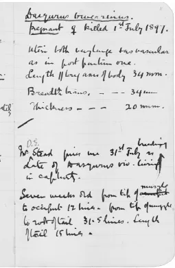
Fig. 8. Copy of page from Hill’s “Camp Notes” (notebook 16). “Dasyurus viverrinus pregnant killed 1st July, 1897. Uteri both very large + as vascular
as in post partum one. Length/long axis of body 34 mm. Breadth brains, -34 mm, thickness- 20 mm. / Mr. Stead gives me 31st July as breeding date of Dasyurus viv. living in captivity. Seven weeks old from tip of occiput muzzle to occiput 12 lines. from tip of muzzle to root of tail 31.5 lines. Length of tail 15 lines.”
37 Watson (1955) p. 114.
38 Entry for 29 December, 1919, in the Notebooks of meetings of the International
Embryological Institute, in the archive of the Hubrecht Lab. Translation from Dutch.
39 Entries for 24 August and 28 October 1916 and 28 December, 1917. Notebooks
of meetings of the International Embryological Institute, in the archive of the Embryo Collection.
40 Ibid 29 December, 1919. 41 ibid. 24 June, 1922; 28 June, 1922. 42 Bangma (1986).
43 Presumably the basis of his normal table for this species (Kopsch, 1952). 44 Customs declalartion for Mangold Collection, in the archive of the Utrecht
Collec-tion. 23 Sept. 1974.
45 Truslove to Boterenbrood, 12th October, 1973. Letter in the archive of the Utrecht
Collection.
46 Radford, T. (27.12.1997). The Guardian, London. p. 7. “Old, Old Embryos Offer
New Insights for Scientists”. aan de Brugh M. 17.8.1998, NRC Handelsblad, The Netherlands. p. 20 “Ongeboren wonderen”. 9.9.1998, Frankfurter Allgemeine Zeitung, Germany. Natur und Wissenschaft section. “Streit um Hackels Thesen entschärft — Moderne Version der Embryonen-Tafel”.
47 Information kindly provided by the Director, Professor Bengt Källén. Specimens
may be consulted in the Institute.
48 Information kindly provided by Dr. Franz Wachtler who acts as custodian and
should be contacted for further details at: Institute for Anatomy, Währingerstraße 13, A-1090, Wien, Austria. <Franz.Wachtler@univie.ac.at>
Hill and Hubrecht devoted much of their attention to early development and placentation. There is therefore still a great deal of work to be done in understanding how patterns of organogen-esis vary among species (Richardson, 1995). The recent devel-opment of techniques for analysing heterochrony in vertebrate development (Nunn and Smith, 1998) could usefully be applied to the rich material held in Utrecht.
The future
and computerised 3D reconstructions. Eventually it is hoped to link, via the Internet, with similar collections throughout the world. This will provide a central search facility for specimens, together with a Newsletter and an Electronic Guest Book for questions, comments and information. One of us (JN) plans to visit a number of institutes and museums in various countries to assess the feasibility of this project.
There have been suggestions that the collection should be expanded to include more experimental material, as well as cell lines and other resources. However, limitations of staffing, space and funding mean that new acquisitions will have to be carefully considered.
Other embryo collections
The collection in Utrecht is by far the largest and most valuable collection of vertebrate comparative embryology in the world. Nonetheless, there are important collections of human and ani-mal embryos elsewhere. We describe here some of those which are available for study by scholars. The nearest equivalent to the Utrecht collection is at The Tornblad Institute47 (University of
Lund, Sweden), which has a remarkable and valuable collection of sectioned chordate embryos. Some unique specimens are also stored in alcohol. Wet specimens include fetuses of bears, seals, badgers, pangolins, many bird and reptile species, and a large number of human fetuses. Tragically, because of a lack of funds, about four fifths of these have dried out. A conservation operation was carried out in 1999 by the two authors in conjunction with the Director Professor Bengt Källén. There is a card catalogue in Swedish, with species names in Latin.
The Tornblad Institute was built in 1934 for the embryo collec-tion and research activities of Ivar Broman. In 1929 Broman made a field trip to South Africa, and there are some important survivals from this trip in the collection. Tornblad and Broman went to the Carnegie Institution in Baltimore in 1931 to exchange surplus African embryos for possum embryos.
The Anatomy Department of the University of Vienna, Austria houses a substantial embryo collection48, including the Hochstetter
collection of human embryos. A catalogue is available, and the collection is open to scholars on application. The collection consists of around 420 human embryos ranging from 0.4 mm to 230 mm length. The largest entire embryos present as serial
sections are around 50 mm in length. The UK Human Embryo Database, details human embryos in British university collec-tions49. Human embryos are collected for molecular studies by the
Newcastle Human Embryo Group, School of Biochemistry and Genetics, University of Newcastle upon Tyne, NE2 4AA. UK, (Strachan et al., 1997). Parts of the Boyd collection of human embryos are in the anatomy departments at Cambridge Univer-sity, UK, and St. George's Hospital, London, UK.
The most important collection of embryos in the USA is housed in the National Museum of Science and Medicine, part of the Armed Forces Institute for Pathology (AFIP) in Washington DC50.
A partial catalogue of some of the AFIP embryo collection can be searched on the Internet51. The primate and insectivore material
includes the Bluntschli and Hartman collections. Perhaps the best-known series in the AFIP is the Carnegie Collection of human embryos52, which have formed the basis of numerous
studies of human developmental anatomy53. They form one of the
major resources of the Human Developmental Anatomy Centre (HDAC) at the AFIP.
The Carnegie collection was founded by Franklin P. Mall, who worked in Leipzig with His and became director of the Department of Embryology at the Carnegie Institution in 1914 (Mall, 1898, 1903, 1910, 1911, 1913a; see also note54). The Carnegie
Institu-tion sent the collecInstitu-tion to the University of California, Davis in 1973, before it was finally transferred to the AFIP in 1991. There are case records for 10,000 embryos in the Carnegie collection; the total number of specimens remaining (for all of the collections in the HDAC) is around 7000, sectioned or whole. Mall described the motivation behind his collecting as follows:
“We must thank His [Wilhelm His Sr.] for the first attempt to study carefully the anatomy of human embryos, but his work was planned on so large a scale that he never completed it. Gradually he lost courage to go ahead single-handed, as may be observed in his numerous publications in favor of cooperation in embryologic research. Thus we may trace back to him the incentive for Keibel’s Normentafeln, Minot’s great collection of verte-brate embryos and mine of human embryos”55 (Our
note in brackets [ ]).
The Minot collection mentioned by Mall was assembled by the comparative embryologist Charles Sedgwick Minot (Minot, 1905, 1906). His collection was formerly housed in the Warren Anatomi-cal Museum at Harvard MediAnatomi-cal School but has now been split into two groups. Around 100 human specimens are housed at the AFIP. The remainder are in the Museum of Comparative Zoology (Cambridge, USA). They include the spiny dogfish embryos used to prepare Scammon’s Normal Table (Scammon, 1911).
The Patten Embryology Research Facility56 at the University of
Michigan in Ann Arbor includes serial sections of human, chick, pig, mouse, opossum and guinea pig embryos. There is a collec-tion of embryology reprints. The colleccollec-tion has historical links with important figures in the history of twentieth century embryology, including Huber, Streeter, and Patten. The human material at Michigan includes over 2,500 human prenates chiefly of the second trimester. The majority are completely serially –sectioned specimens. Intact preserved specimens are also available for specific staining uses. Approximately 1500 of these specimens
49 Contact: Dr John Maclachlan; see website at: http://embryos.st-andrews.ac.uk/
default.htm
50 Information kindly supplied by Elizabeth Lockett, Human Developmental Anatomy
Centre, National Museum of Science and Medicine, Armed Forces Institute of Pathology, Walter Reed Army Medical Center, Washington, DC 20306-6000. See also (http://bubba.afip.org/embryo/intro.html).
51 (http://www.bubba.afip.org/cgi-bin/php/embryo/dbase/search.phtml). 52 (http://www.ciwemb.edu/links/emb.html).
53 reviewed by (O’Rahilly and Müller, 1987).
54 (http://www.pandora.med.yale.edu/caim/c_embryol/ce_site/mm/history/us.htm) 55 Mall (1913b).
56 Information kindly provided by Dr. Alphonse R. Burdi, director. 57 Evans and Sack (1973) p.11-12.
58 Information kindly provided by Dr. D.M. Noden, Department of Biomedical
Sciences, College of Veterinary Medicine, Cornell University, Ithaca, NY. Dr Noden should be contact for further details <dmn2@cornell.edu>. Dr Evans has deposited material relating to the collection in the Cornell Archives.
59 Information kindly supplied by the director, Professor Kohei Shiota, Department of
are with “coded” medical and familial histories. The collection has sizeable groups of prenates with syndromic and non-syndromic abnormalities, e.g., trisomies, cleft lip and palate, fetal alcohol. The Cornell Embryo Collection57,58 (Veterinary College, Cornell
University, USA) consists of serial sections of embryos of domes-tic animals (including cat, horse, cow, pig, and chicken, as well as the Howard Evans collection of serially-sectioned sheep and dog embryos). In total there are about 3500 sectioned mammalian and 1000 avian embryos. The collection also contains whole-mounts of congenital malformations in these species. One hundred and seventeen human embryos, and many more rodent embryos, were transferred to the AFIP from Cornell in 1997.
The Kyoto Collection of Human Embryos59 is housed in the
Congenital Anomaly Research Centre, Kyoto University Faculty of Medicine, Japan. The collection was started in 1961 by Profes-sor Hideo Nishimura, and now consists of around 44,000 speci-mens (Nishimura, 1975; Shiota, 1991). Normal and malformed embryos are included, and around 500 of the normal embryos have been serially-sectioned. The embryos were collected from induced abortions.
The archive contains catalogues of certain other collections which have not been examined in detail by us, but will be mentioned briefly as follows. The Anatomical Supplement to the China Medical Journal of February 1927 contains a catalogue of human embryos in the Department of Anatomy, Peking Union Medical College. A catalogue (from the Anat. Anz. 1936 pp. 415-436) lists animal and human embryos in the Anatomisches Institut, Marburg (Germany) and the Anatomisches Institut, Jena (Germany). There is a 1974 catalogue by V.W.D. Schenk and D.T. Kiers describing human embryos in the Anatomy Lab at Rotterdam, The Netherlands.
Summary
The Embryo Collection of the Hubrecht Laboratory is a trea-sure house of comparative embryology. It is the largest and most important collection of its kind in the world, and consists of thousands of vertebrate embryos stored in alcohol, or prepared as histological sections. Many elusive species are included in the collection, some represented by complete developmental series. The accompanying archives offer a remarkable insight into the methods used to collect embryos form wild animals, as well as the motives behind the founders of the collection. Carefully main-tained, documented and catalogued, the collection is available for study by all interested scientists. We argue that this collection is one of the greatest biodiversity resources in existence.
Acknowledgements
We are grateful to colleagues John Bluemink and Ferdinand Vervoordeldonk for help and information. In addition to the contacts listed for the embryo collections world-wide, we thank the following for informa-tion: A. Altemus, R. Bellairs, P. Craig, H.-R. Duncker, S. Emmonds, R. Falkner, J-G. Forsberg, S. Gilbert, E. Gilland, M. Halpern, L. McClure, G. Müller, R. Raff, C. Rose, S. Sullivan.
References
ANONYMOUS (1972). Hubrecht, Ambrosius Arnold Willem. In Dictionary of scien-tific bibliography. (ed.C.C. Gillispie). New York.
ASSHETON, R. (1915). Dr Ambrosius Arnold Willem Hubrecht. Proc. Linn. Soc. 127: 28-31.
BALFOUR, F.M. (1876). On the development of the spinal nerves in elasmobranch fishes. Philos. Trans. Roy. Soc. Lond. (Biol.) 166: 175-195.
BANGMA, G.C. (1986). De embryologische collectie van het Hubrecht Laboratorium. Vakbl. Biol. 66: 455-458.
BANGMA, G.C. (1988). The Embryological Collection of the Hubrecht Laboratory. Collection Forum 4: 14-15.
BOTERENBROOD, E.C. (1977), Concise Catalogue of the Central Embryological Collection of the Hubrecht Laboratory. Hubrecht Laboratory, Utrecht. Nether-lands.
DE BEER, G.R. (1948). J.P. Hill: A biographical sketch of his career and an appreciation of his work, together with a bibliography of his published writings. J. Anat. 82: 3-8.
DE LANGE, D. (1919). Früheste entwicklungstadien und placentation von Galeopithecus. Verh Akad Wet Amst (tw sect ) 16: 1-39.
DE LANGE, D. and NIERSTRASZ, H.F. (1932). Tabellarische uebersicht der entwicklung von Tupaia javanica . Oosthoek Verlag, Utrecht.
DEAN, B. (1899). On the embryology of Bdellostoma stouti. In Fetschrift zum siebenzigsten Geburtstag von Carl von Kupffer. Fischer-Verlag, Jena, pp 221-276.
DENDY, A. (1899). Outlines of the development of the tuatara, Sphenodon (Hatteria) punctatus. Q.J. Microsc. Sci. 42: 1-87.
EVANS, H.E. and SACK, W.O. (1973). Prenatal development of domestic and laboratory mammals: growth curves, external features, and selected refer-ences. Anat. Histol. Embryol. 2: 11-45.
FLYNN, E. (1998). My Wicked, Wicked Ways. Arrow, London, -386.
GOULD, S.J. (1977). Ontogeny and Phylogeny. Belknap Press, Cambridge, Mas-sachusetts.
HENNEBERG, B. (1937). Normentafeln zur entwicklungsgeschichte der Wanderratte (Rattus norvegicus Erxleben). In Normentafeln zur entwicklungsgeschichte der Wirbelthiere. (ed. F. Keibel). Verlag von Gustav Fischer, Jena.
HILL, C.J. and HILL, J.P. (1933). The develompent of the monotremata. Trans. Zool. Soc. Lond. 21: 413-476.
HILL, J.P. (1910). The early development of the marsupialia, with special reference to the native cat (Dasyurus Viverrinus). Q.J. Micrsoc. Sci. 56: 1-134.
HUBRECHT, A.A.W. (1888). Keimblätterbildung und Placentation des Igels. Anat. Anz. 3: 510-515.
HUBRECHT, A.A.W. (1894a). Studies in mammalian embryology. III. The placen-tation of the shrew (Sorex vulgaris). Q.J. Microsc. Sci. 35: 481-537.
HUBRECHT, A.A.W. (1894b). Studies from the Zoological Laboratory in the University of Utrecht IV: Spolia nemoris. Q.J. Microsc. Sci. 36: 77-125.
HUBRECHT, A.A.W. (1910). De plaats der vergelijkende embryologie in het Hooger Onderwijs. P.N. van Kampen & zoon, Amsterdam.
HUBRECHT, A.A. and KEIBEL, F. (1907). Normentafeln zur entwicklungsgeschichte des Koboldmaki (Tarsius spectrum) und des Plumplori (Nycticebus tardigradus). In Normentafeln zur entwicklungsgeschichte der Wirbelthiere. (ed. F. Keibel). Verlag von Gustav Fischer, Jena.
HUISMAN, F.J. and DE LANGE, D. (1937). Tabellarische uebersicht der entwicklung von Manis javanica Desm. Oosthoek Verlag, Utrecht.
KEIBEL, F. (1895). Normentafeln zur Entwickelungsgeschichte der Wirbeltiere. Anat. Anz. 11: 225-234.
KEIBEL, F. (1897). Normentafeln zur Entwicklungsgeschichte der Wirbelthiere. (ed. F. Keibel). Verlag von Gustav Fischer, Jena.
KEIBEL, F. (1915). A. A. W. Hubrecht. Anat. Anz. 48: 201-208.
KEIBEL, F., ASSHETON, R. and HUBRECHT, A.A.W. (1912). Institut International d’Embryologie, Session de 1912. Bibl. Anat. 22: 1-12.
KEMP, A. (1982). The embryological development of the Queensland lungfish, Neoceratodus forsteri, (Krefft). Mem. Qd. Mus. 20: 553-597.
KOPSCH, F.R. (1952). Die entwicklung des braunen grasfrosches Rana fusca Roesel. Georg Thieme Verlag, Stuttgart.
KUDO, T. (1938). Normentafeln zur entwicklungsgeschichte des Japanischen Riesensalamanders (Megalobatrachus japonicus Temminck). In Normentafeln zur entwicklungsgeschichte der Wirbelthiere. (ed. F. Keibel). Verlag von Gustav Fischer, Jena.
MALL, F.P. (1903). Note on the collection of human embryos in the Anatomical Laboratory of Johns Hopkins University. Johns Hopkins Hosp. Bull.14: 1-12.
MALL, F.P. (1910). A list of normal human embryos which have been cut into serial sections. Anat. Rec. 4: 355-367.
MALL, F.P. (1911). Report on the collection of human embryos at the Johns Hopkins University. Anat. Rec. 5: 343-357.
MALL, F.P. (1913a). Embryological Research. In Year Book No. 12 of the Carnegie Institution of Washington (for 1913). Carnegie Institution of Washington, Wash-ington, pp 290-291.
MALL, F.P. (1913b). A plea for an Institute of human embryology. J. Am. Med. Assoc. 60: 1599-1601.
McCRADY, E. (1938). Embryology of the opossum. Am. Anat. Mem. 16: 12-33.
MINOT, C.S. (1905). The Harvard embryological Collection. J. Med. Res. 13: 499-522.
MINOT, C.S. (1906). The relations of embryology to medical progress. Popular Science Monthly: 5-20.
NIEUWKOOP, P.D. (1953). Concise Catalogue of the Central Embryological Collections of the Hubrecht Laboratory. Hubrecht Laboratory, Utrecht, Nether-lands.
NISHIMURA, H. (1975). Prenatal versus postnatal malformations based on the Japanese experience on induced abortions in the human being. In Aging Gametes. (R.J. Blandau ed.) S. Karger, Basel, pp 349-368.
NORDENSKIÖLD, E. (1929). The History of Biology: A Survey. Alfred A. Knopf, London.
NUNN, C.L. and SMITH, K.K. (1998). Statistical analyses of developmental se-quences: The craniofacial region in marsupial and placental mammals. Am. Nat. 152: 82-101.
O’RAHILLY, R. and MÜLLER, F. (1987). Developmental Stages in Human Em-bryos. Washington.
PARKER, T.J. (1891). Observations on the anatomy and development of Apteryx. Philos. Trans. Roy. Soc. Lond. (Biol.) 182: 25-134.
RICHARDSON, M.K. (1995). Heterochrony and the phylotypic period. Dev. Biol. 172: 412-421.
RICHARDSON, M.K. (1999). Vertebrate evolution: the developmental origins of adult variation. BioEssays: 21: 604-613.
RICHARDSON, M.K., HANKEN, J., GOONERATNE, M.L., PIEAU, C., RAYNAUD, A., SELWOOD, L. and WRIGHT, G.M. (1997). There is no highly conserved embryonic stage in the vertebrates: implications for current theories of evolution and development. Anat. Embryol. 196: 91-106.
RICHARDSON, M.K., MINELLI, A. and COATES, M. (1999). Some Problems with Typological Thinking in Evolution and Development. Evol. Dev. 1: 5-7.
SCAMMON, R.E. (1911). Normal plates of the development of Squalus acnathias. In Normentafeln zur entwicklungsgeschichte der Wirbelthiere. (ed. F. Keibel). Verlag von Gustav Fischer, Jena.
SHIOTA, K. (1991). Development and intrauterine fate of normal and abnormal human conceptuses. Cong. Anom. 31: 67-80.
STRACHAN, T., LINDSAY, S. and WILSON, D.I. [eds] (1997). Molecular Genetics of Early Human Development. Bios, Oxford.
TAYLOR, M. (1913). The development of Symbranchus marmoratus. Q. J. Microsc. Sci. 59: 1-51.
VAN OORDT, G.J. (1921). Early developmental stages of Manis Javanica Desm. In Mededeelingen uit het Embryologisch Instituut van het Hubrecht-Fonds. Johannes Müller, Amsterdam.
VON BAER, K.E. (1828). Entwicklungsgeschichte der Thiere: Beobachtung und Reflexion. Borntrager, Kornigsberg.
WATSON, D.M.S. (1955). James Peter Hill. Bibliographic Memoirs of Fellows of the Royal Society 1: 101-117.
WILSON, H.V. (1889). The embryology of the sea bass (Serranus atrarius). Bull. US Fish. Comm. 9: 209-277.