CSEIT1831128 | Received : 12 Jan 2018 | Accepted : 29 Jan 2018 | January-February-2018 [(3) 1 : 623-628]
International Journal of Scientific Research in Computer Science, Engineering and Information Technology © 2018 IJSRCSEIT | Volume 3 | Issue 1 | ISSN : 2456-3307
623
Segmentation and Classification Techniques of Acute Myeloid
Leukemia using Image Processing: A Survey
Kishan Patel, Rashmin B. Prajapati
Department of Computer Engineering, S.V.I.T. Vasad, Gujarat, India
ABSTRACT
The White blood cells or Leukocytes plays a major role in the diagnosis of different diseases (including leukemia). Leukemia is a type of blood disease or so-called cancer of the blood that begins in the bone marrow and usually caused by an excessive alterations in the production of malignant and immature white blood cells. Acute Myeloid leukemia (AML) is a subtype of acute leukemia, which is characterized by the accumulation of myeloid blasts in the bone marrow. Subtypes of AML i.e., M0–M7. AML is also called as Acute Myelogenous Leukemia, Acute Meroblastic Leukemia, Acute Granulocytic Leukemia and Acute No lymphocytic Leukemia. This paper provides a survey of several segmentation and classification techniques for detection of acute myeloid leukemia using image processing.
Keywords : Acute Myeloid Leukemia (AML), Segmentation, Feature Extraction, Classification
I.
INTRODUCTION
Leukemia is the eleventh most common cancer worldwide with more than 250,000-300,000 new cases each year. Leukemia is a type of blood cancer that originates in the bone marrow and is characterized by abnormal proliferation of white blood cells. Leukemia customarily occurs when a big portion of abnormal, immature white blood cells, called Leukemia cells(“blasts”) produced in the body by bone marrow.[1] Diagnosing leukemia is based on the fact that WBC count are increased with immature blast cells (lymphoid or myeloid), and neutrophils and platelets are decreased. Therefore, hematologists routinely examine blood smears under microscope for proper identification and classification of blast cells. The presence of the excess number of blast cells in peripheral blood is a significant symptom of leukemia.[6] Leukemia can be classified into two categories: (1) Acute Leukemia which progress quickly; and (2) chronic Leukemia which progress slowly. It can also be classified based on the affected cell type as (1)myeloid
(myelogeneous) leukemia and (2)lymphocytic leukemia.[2] In this paper Acute myeloid leukemia(AML) is only considered.
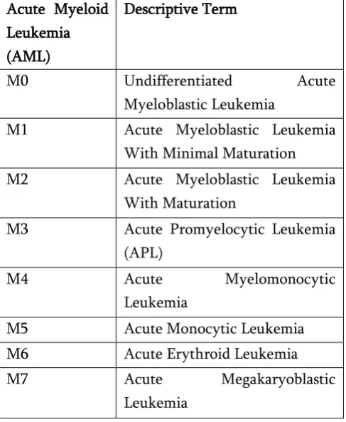
The FAB classification system divides AML into eight subtypes, M0-M7 based on the type of cell from which leukemia developed and degree of maturity [2,7].
Figure 1. Myeloblast from AML patient [6]
Figure 2. Healthy cells from non AML patient [6] Table 1. Subtypes of AML
Acute Myeloid Leukemia (AML)
Descriptive Term
M0 Undifferentiated Acute
Myeloblastic Leukemia
M1 Acute Myeloblastic Leukemia With Minimal Maturation M2 Acute Myeloblastic Leukemia
With Maturation
M3 Acute Promyelocytic Leukemia (APL)
M4 Acute Myelomonocytic
Leukemia
M5 Acute Monocytic Leukemia
M6 Acute Erythroid Leukemia
M7 Acute Megakaryoblastic
Leukemia
II.
RELETED WORKS
This section provides some essential work done by numerous researchers for leukemic cell segmentation and classification in microscopic blood cell images.
Luis H. S. Vogado, et al. [1], proposed a new automatic leukemic cell segmentation technique that uses two-color systems, CMYK and L*a*b and the clustering algorithm K-means. Series of morphological operations are performed on segmented images to remove noise.
Fatemeh Kazmi et al.[2], presents an automatic technique for identification and detection of AML and its prevalent subtypes, i.e., M2-M5. Which performs processing including color correlation, segmentation of the nucleated cells, and effective validation and classification. At first, microscopic images are acquired from blood smears of patients with AML and normal cases. After applying image preprocessing, color segmentation strategy(k-means clustering) is applied for segmenting white blood cells from other blood components and then discriminative features, i.e., irregularity, nucleus-cytoplasm ratio, Hausdorff dimension, shape, color, and texture features are extracted from the entire nucleus in the whole images containing multiple nuclei. Images are classified to cancerous and noncancerous images by binary support vector machine (SVM) classifier with 10-fold cross validation technique.
provided accurate segmentation of WBCs from the blood microscopic images. It is added with Fuzzy C mean classification trained with the extracted features row vector. The proposed system provided accurate segmentation of leukemia from blood sub images.
Huey Nee Lim et al.[4],presents a combination of color and morphological based segmentation techniques for blood cells segmentation on microscopic images. K-means clustering is implemented for color based clustering and primary segmentation of the image. The clustering is cascaded into two layers for cell recognition and background elimination. Morphological based segmentation is conducted using watershed transform based on gradient magnitude and skeleton by influence zone. S.S.Savkare, S.P.Narote [5], presents a methods to segment the blood cells from microscopic thin blood images. This paper describes method for blood cell segmentation utilizing K-Mean clustering. Proposed method utilizes median filter for reduction of noise and laplacian filter for enhancement.
D.Goutam, S.Sailaja [6], proposed a system, which mainly composed of four main stages are preprocessed stage, segmentation stage, feature extraction stage and classification stage respectively. This system framework consists simple and known technique such as K-mean clustering, Local Directional path (LDP), and support vector machine (SVM) respectively.
Jakkrich laosai and Kosin chanongthai [7], proposed a system that takes as input, Color images of stained peripheral blood smears and identifies the class of each of the White Blood Cells (WBC). The process involves segmentation, feature extraction and classification. Their work focuses on classification of Foil of Bretagne (Lymphoid) and Almeida Lloyd (Myeloid).
Monica Madhukar et al. [8], presents a system to a) demonstrate that the classification of peripheral
blood smear images containing multiple nuclei can be fully automated, b) to validate the segmented images using holdout cross validation method.
Reymond Joseph A. Cabrera et al. [9], introduces a system that will apply the different techniques of Image Processing and Genetic Algorithm, specifically in automating the detection of Leukemia. In the pre-processing stage, color extraction will be done by multiplying RGB color planes in order to extract the foreground or region of interest (White Blood Cells) from the background (Red Blood Cells).
Adnan Khashman and Hayder Hassan Abbas [10], presents a novel approach for acute lymphoblastic leukemia (ALL) blood cell identification using pattern averaging of whole cell images and neural network classifiers. The novelty in this work is not only in using a neural model as a classifier, but also in considering normal and abnormal (leukemic) blood cells as a whole without the need to extract local features. This in turn reduces the computational and time costs by avoiding segmentation and local feature extraction from the blood cell images. The proposed system is implemented to identify ALL-infected abnormal cells from normal blood cells.
Sonali Mishra et al. [11], proposed an acute lymphoblastic leukemia detection strategy from the microscopic images. The scheme utilizes all the steps associated with any other classification scheme, but their contribution lies on a marker-based segmentation (MBS), gray level co-occurrence matrix (GLCM) based feature extraction, and probabilistic principal component analysis (PPCA) based feature reduction. A new method of extracting the textural feature from the nucleus and cytoplasm region of the cropped image is described using gray level co-occurrence matrix. The relevant features are used in a random forest (RF) based classifier.
has been reviewed in this paper, the steps of detecting leukemia is divided into three stages, namely Segmentation, Feature Extraction and Classification. At the stage of segmentation,
researchers to obtain results, which have a better classification of leukemia cells, have carried out various methods.
III.
COMPARATIVE ANALYSIS
Table 2. Segmentation Techniques
Technique Advantages Limitations
FCM[ 3,12] -Accurate
-Works well for noise free images.
-96.31% Accuracy[12]
-Apriori specification of no. of clusters. -increase no. of iteration.
-sensitive to noise.
FLICM[13] -enhance clustering performance.
-image detail preservation. -effective and efficient
-Works on fixed distance.
Objective function is defined previously
KWFLICM[14] -Better segmentation
-Better accuracy
-At each iteration it‟s necessary to calculate or update trade of fuzzy factor. - Works on adaptive distance.
K-means[1,2,4,5,6,7,8,12] Easy to implement and interpret clustering results.
Cluster size is manually added. It is not adaptive.
K-means with LAB[2,5,7,8] -Luminosity and Color kept separate.
-Works batter for low intensity images.
-Proper Color and light is must needed to segment image object.
K-means with CMYK -Segment Part into Extreme Level
-Not Work with Noisy images pre-processing should need.
K-means With CMYK-LAB[1 ]
Works with all Light Conditions. -High Accuracy.98.59%
-Less Complex.
-Low Memory and time Consumption.
Post-Processing is Needed after segmentation.
Table 3. Classification Techniques
Classifier Advantages Limitations
1. Naive Bayes [9] - Data set is small then high bias low variance classifier like NB will work well.
-Data set is small than generative class will work well.
-large dataset can‟t use it. 2. Neural Network[10] -High degree of non-linearity
possible.
3. SVM[1,6,7,8] -High accuracy
-Easy to generate rules. -Easy to understand.
-Hard to interpret
-It takes more time to predict the new instance.
4. Random Forest [11] -One of the most accurate learning algorithms available for most data sets
It reduces over fitting and is -therefore more accurate. -Easy to Implement
-works with all types of data. -Multi classification Support.
It may not work if the dependent variables considered in the model are linearly related. Therefore, one has to remove correlated variable by some other technique.
IV.
CONCLUSION
This paper presents various studies that have been conducted on acute leukemia detection using image processing. Segmentation is considered an important step in the automatic diagnosis of different computer systems. It was found that several methods in the literature have shown promising results. From literature review it was found that automatic segmentation technique that uses two-color systems and clustering algorithm k-means provides high segmentation accuracy and SVM and Random Forest classifiers has quite high accuracy results.
V.
REFERENCES
[1]. Luis H. S. Vogado, Rodrigo de M. S. Veras, Alan R. Andrade, Flavio H. D. de Araujo,Romuere R. V. e Silva, Fatima N. S. de Medeiros"Unsupervised Leukemia Cells Segmentation Based on Multi-space Color Channels" 2016 IEEEinternational symposium on multimedia
[2]. Fatemeh Kazmi, Tooraj Abbasian Najafabadi,Babak Nadjar Araabi" Automatic recognition of Acute Myelogenous Leukemia in blood microscopic images using K-means clustering and support vector machine" 2016 Journal of Medical signals and sensors.
[3]. Viswanathan P,"Fuzzy C means Detection of Leukemia based on Morphological Contour
Segmentation", „‟Procedia Computer Science 58 ( 2015 ) 84 – 90‟‟.
[4]. Huey Nee Lim, Mohd Yusoff Mashor, Nadiatun Zawiyah Supardi, Rosline Hassan "Color and Morphological Based Techniques on White Blood Cells Segmentation", "2015 2nd International Conference on Biomedical Engineering (ICoBE), 30-31 March 2015, Penang"
[5]. S.S.Savkare, S.P.Narote "Blood Cell Segmentation from Microscopic Blood Images", „‟2015 InternationalConference on Information Processing (ICIP) Vishwakarma Institute of Technology. Dec 16-19, 2015‟‟ [6]. D. Goutam, S. Sailaja, "Classification of Acute
MyelogenousLeukemia in Blood Images Using Supervised Classifier." International Conference on Engineering and Technology. March 2015.
[7]. Jokkrich Laosai, kosin Chamnongthai"Acute Leukemia Classification by Using SVM and K-Means Clustering,"Proceedings of the International Electrical Engineering Congress 2014"
[8]. Monica Madhuka,Sos Agaian,Anthony T.Chronopoulos" Deterministic Model for Acute Myelogenous Leukemia Classification" 2012,IEEE.
Separation of Overlapping Blood Cells through Image Processing and Genetic Algorithm" 2017 IEEE International Conference on Applied System Innovation IEEE-ICASI 2017 - Meen, Prior & Lam (Eds).
[10]. Adnan Khashman and Hayder Hassan Abbas" Acute Lymphoblastic Leukemia Identification using blood smear images and a neural classifier"2013, springer.
[11]. Sonali Mishra, Banshidhar Majhi, Pankaj Kumar S, Lokesh Sharma " Gray level co-occurrence matrix and random forest based acutelymphoblastic leukemia detection"2016 ELSEVIER Biomedical Signal Processing and Control 33 (2017) 272–280
[12]. R.G Bagasjvara, Ika Candradewi, Sri Hartati, Agus Harjoko " Automated Detection and Classification Techniques of Acute Leukemia using Image Processing: A Review" 2016 2nd International Conference on Science and Technology-Computer (ICST), Yogyakarta, Indonesia.
[13]. Stelios Krinidis and Vassilios Chatzis, "A Robust Fuzzy Local Information C-Means Clustering Algorithm", "IEEE TRANSACTIONS ON IMAGE PROCESSING, VOL. 19, NO. 5, MAY 2010"
[14]. R. Shalini, V. Muralidharan and M. Varatharaj, "MRI Brain Tumor Segmentation using Kernel Weighted Fuzzy Clustering, "International Journal of Engineering Research & Technology (IJERT) Vol. 3 Issue 4, April - 2014"
[15]. Manel Jarrar, Asma Kerkeni, Asma Ben Abdallah And Mohamed Hédi Bedoui, "MLP neural network classifier for medical image segmentation", 2016 13th International Conference Computer Graphics, Imaging and Visualization, pages 88-93.