0214-6282/2005/$25.00
© UBC Press Printed in Spain
www.intjdevbiol.com
Plastids unleashed: their development and their integration
in plant development
ENRIQUE LOPEZ-JUEZ*
,1and KEVIN A. PYKE
21School of Biological Sciences, Royal Holloway, University of London, Egham, Surrey, UK and 2Plant Sciences Division, School of
BioSciences, University of Nottingham, Sutton Bonnington Campus, Loughborough, UK
ABSTRACT Derived by endosymbiosis from ancestral cyanobacteria, chloroplasts integrated seamlessly into the biology of their host cell. That integration involved a massive transfer of genes to the cell’s nucleus, with the modification of pre-existing processes, like plastid division and the operation of the plastid genetic machinery and the emergence of new ones, like the import of proteins translated in the cytoplasm. The uncovering in molecular detail of several of these processes reveals a merger of mechanisms of symbiont and host origin. Chloroplasts acquired roles as part of the biology of land plants by differentiating into a variety of interconvertible plastid forms according to the cell type. How these conversions take place, or how new problems, like the regulation of the plastid population size in cells, have been solved, is barely starting to be understood. Like the whole plant and as a result of the requirements and dangers associated with photosynthetic activity, chloroplasts in particular are under the control of environmental cues. Far from being passive targets of cellular processes, plastids are sources of signals of plastid-nuclear communication, which regulate activities for their own biogenesis. Plastids are also sources of developmental signals, in whose absence tissue architecture or cell differentiation are aberrant, in a cell-autonomous fashion. Over evolutionary time, plastids also contributed many genes for activities that are no longer directly associated with them (like light perception or hormone function). The overall picture is one in which plastids are at both the receiving and the acting ends in plant development, in both ontogenic and evolutionary terms.
KEY WORDS:
chloroplast, plastid, photosynthesis, endosymbiotic, plastid-nuclear communication
Abbreviations used in this paper: GDP, guanosine diphosphate; GFP, green fluorescent protein; GTP, guanosine triphosphate; NEP, nuclear-encoded RNA polymerase; PEP, plastid-encoded RNA polymerase; POR, protochlorophyllide reductase; PS, photosystem; SRP, signal recognition particle; Tic, translocon of the inner membrane of chloroplast; Toc, translocon of the outer envelope of chloroplast.
*Address correspondence to: Dr. Enrique López-Juez. School of Biological Sciences, Royal Holloway, University of London, Egham, Surrey TW20 0EX, U.K. Fax: +44-17-8447-0756. e-mail: [email protected]
Introduction
If there is one feature that distinguishes plant from animal life on our planet, it is not plants being primarily sessile or developing continuously through life, as a few animals share those; rather, it is the reliance of plants on solar energy to generate molecules with energy-rich bonds, the fuel that will be used by almost the entire biosphere (including plants themselves) to build other organised molecules and drive the rest of the processes that we know as life. Chloroplasts are the sites of this wonder-process. If an interstellar traveller arrived on our planet to analyse the organisms in it, it would probably eventually describe plants as ‘living things that fix star photon energy in green corpuscles and produce organs that harbour those corpuscles exposed to light and air, in a protected environment, or bring water and other inorganic substances from the substrate to them’. Photosynthesis was an invention of several, seemingly very early prokaryotes, which eventually associated between themselves to exploit the
chloroplast
proplastid
elaioplast chromoplast
etioplast amyloplast chloroplast
proplastid
elaioplast chromoplast
etioplast amyloplast
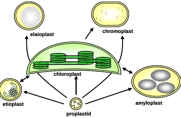
Fig. 1. Diversity of plastid types and their interconversions.Chloroplasts occupy the centre of the figure to signify their evolutionary role as ancestors of all other plastid types, although during ontogeny all plastids derive from embryonic proplastids.
engulfed cyanobacteria had turned into what we know as chloro-plasts. Chloroplasts retained a small degree of genetic autonomy, a large degree of their biochemistry, but lost some of their original functions and also acquired ones they did not possess when free-living (Timmis et al., 2004). They needed to synthesise and accumulate their proteins, now produced in two separate com-partments, within themselves and in their surrounding cytoplasm, locate them to their correct destination, divide and propagate because the cell they were in did and fill that cell to the right extent. The chloroplast’s requirements to carry out photosynthesis would determine the land plant’s development and its need to adapt such development to environmental signals, such as light or the availability of raw materials. The chloroplasts would also diversify into a variety of derivatives, that we now call other plastid types, to carry out other essential or specialised functions in other cells that were no-longer photosynthetic, or merely to be transmitted more easily and economically in young, embryonic or undifferen-tiated cells (Waters and Pyke, 2004). The chloroplasts or their derivatives would therefore become under the control of develop-mental signals that affected the cells harbouring them, or be influenced by the same environmental cues, to insure their function remained possible under a variety of conditions (Rodermel, 2001). As it is now apparent, the chloroplasts themselves would directly relay information to the nucleus of the cells in which they resided, on their own status or on environmental influences upon them, to integrate them fully into the biology of the cell or the organ (Surpin et al., 2002). Finally it is becoming increasingly clear that, as part of this integration, the chloroplasts accepted contributions from their original hosts, in order to build up the new processes that the endosymbiotic relationship required, but they also con-tributed extensively to the toolkit available to the host’s genome in terms of biochemistry of secondary metabolism, or develop-mental or environdevelop-mental perception mechanisms (Martin et al., 2002). This means that plants now possess a range of environ-mental sensors, small chemical regulators and morphogens, cell-surface sensors or signalling mechanisms that play a role in their
development as multicellular organisms, even mechanisms to fight pathogens, that today distinguish plant genomes and were never available to non-photosynthetic eukaryotes.
In this article we will attempt to provide an overview of some of the processes above. The task is arguably overambitious. Fortu-nately there are a number of excellent reviews that address many of the individual aspects and the reader is referred to them (Sugiura, 1992, 2003; McFadden, 1999; Pyke, 1999; Staehelin and Newcomb, 2000; Flügge, 2001; Mache and Lerbs-Mache, 2001; Soll, 2002; Leister, 2003; Osteryoung and Nunnari, 2003; Wakasugi et al., 2001; Timmis et al., 2004; Dyall et al., 2004; Waters and Pyke, 2004; Jarvis and Robinson, 2004).
Chloroplasts and other types of plastid
Chloroplasts are the most noticeable feature of green cells in leaves and, excluding the vacuole, probably constitute the largest compartment within mesophyll cells. These cells appear under the microscope as thin layer cytoplasms, appressed between the vacuole and the cell wall and invariably contain one layer of green lens-shaped organelles, between 5 and 10 µm in diameter and 3-4 µm in thickness (Fig. 2). Depending on species, they number from a few tens to over 100 (Waters and Pyke, 2004). Their obvious primary role is the photosynthesis of carbohydrate. A double membrane, the chloroplast outer and inner envelopes, delimits chloroplasts. Inside, extensive photosynthetic membranes, the thylakoids, extend parallel to the main chloroplast axis, forming flat vesicles, some appearing individually (stromal thyla-koids), some organised into stacks or grana, containing an internal space or lumen. The thylakoids appear as discrete units in transverse sections, but actually form in three dimensions an interlinked compartment, enclosing a single lumen (Staehelin and Newcomb, 2000; Mustardy and Garab, 2003). The thylakoid membranes harbour the four main protein or protein-pigment complexes involved in the light reactions of photosynthesis: photosystems (PS) I and II, the cytochrome b6/f complex and the ATP synthase. The arrangement into grana is important, as it allows for the separation between the two PS, with PSII and its main light harvesting complex being limited to granal membranes not in contact with stroma, while photosystem I is exclu-sively in stroma-exposed thylakoids. This in turn makes it possible to redistribute the harvesting of light according to the prevailing light conditions (Anderson 2000). The ability for this fast redistribu-tion has often been difficult to grasp, in the light of models of the internal three-dimensional structure of membranes inside chloroplasts, with proteins in most granal thylakoids being ‘several membrane layers away’ from the closest stromal thylakoid. Reassuringly, recent detailed analysis shows that many stromal thylakoids surround and often con-nect obliquely multiple layers of thylakoids in grana (Mustardy and Garab, 2003).
and small lipid droplets, called plastoglobuli. The lipid component of chloroplast envelopes and thylakoids is different from the rest of the plant, being based primarily on galactolipids instead of phospholipids; as a result a deficiency in galactolipids has severe consequences on chloroplast development (Jarvis et al., 2000). The envelopes are sites of chloroplast membrane lipid biogenesis and also control the exchange of molecules between the stroma and the cytoplasm. The outer envelope is broadly permeable to molecules up to 10 kDa, while the inner envelope is much more selective, it contains a sophisticated series of dedicated small-molecule transporters that allow, among others, the export of photoassimilates (Flügge, 2001).
Chloroplasts are also central to plant metabolism overall. This is important in many respects since there are abundant examples of mutants identified on the basis of plant- or plastid-developmen-tal phenotypes, which have turned out to be defective in one or another aspect of plastid-localised metabolism, probably for di-rect as well as indidi-rect reasons. Starch synthesis, photoreduction of nitrogen, for all aminoacids and sulphur, for cysteine, biosyn-thesis of fatty acids, of the phenolic group in aromatic ring-containing aminoacids and in their derived secondary metabolites (the shikimate pathway), of the purine and pyrimidine base constituent of nucleic acids, of chlorophyll and other tetrapyrroles (although haem is produced in both the plastid and the mitochon-dria), all take place in chloroplasts (Neuhaus and Emes, 2000). Isoprenoids (also called terpenoids and including carotenoids, steroids and many secondary metabolites), were until recently considered to be produced in chloroplasts from cytoplasmic precursor isoprenoid units, but even these are the product of a major chloroplast anabolic pathway, until recently undetected, the ‘non-mevalonate’ or methylerythritol pathway (Rodríguez-Concepción and Boronat, 2002). Carbohydrate oxidation can also take place, through the oxidative pentose phosphate path-way (Neuhaus and Emes, 2000). Several plant hormones, includ-ing the isoprenoid-derived abscisic acid, gibberellins and brassinosteroids, are additional products of plastid activity.
In light of this central metabolic role, it is evident that non-photosynthetic cells would not be able to survive totally deprived of chloroplasts, unless heavily nourished by close neighbours. Such cells contain non-photosynthetic relatives of chloroplasts, generically called plastids (Fig. 1). Meristematic cells contain colourless proplastids, of between 0.2 and 1 µm and with very limited internal membrane vesicles, which appear as inner enve-lope invaginations (Fig. 2B). There are around 10-20 such pro-plastids per cell (Pyke and Leech, 1992; Waters and Pyke, 2004). The embryo, as well as many cell types not metabolically specialised, also contain proplastids. Plastids with a highly vari-able morphology, larger than proplastids and with more devel-oped internal membranes, both in many root cells and in very young leaf cells that will eventually contain chloroplasts, have been called ameboid plastids.
One main type of differentiated plastid in many root cells is the amyloplast. This plastid is filled with a store of starch granules generated from imported photosynthate and also has a very active oxidative pentose phosphate pathway, that generates energy to assimilate nitrogen (Neuhaus and Emes, 2000). Amy-loplasts are also major constituents of the cells of storage organs, like tubers, cotyledons and seed endosperm (Staehelin and Newcomb, 2000, Waters and Pyke, 2004). Plastids can also
specialise in storing lipid and in this case are called leucoplasts, as those accumulating aromatic oils and produced in secretory hairs (trichomes), or elaioplasts, as those in oil-accumulating storage organs, such as oilseeds.
Plastids have also evolved the capacity to accumulate pig-ments, primarily the isoprenoid carotenoids and xanthophylls and are responsible for the yellows, oranges and reds of many flowers and fruits, the attractants of animals helping transfer pollen or disperse seed. These plastids are then called chromoplasts. Petal cells of flowers with coloured petals convert either proplas-tids or chloroplasts into pigmented chromoplasts (Weston and Pyke, 1999), while tomato fruit pericarp cells contain during development chloroplasts that, upon ethylene-induced ripening, undergo a transition to chromoplast and accumulate vast quanti-ties of the red carotenoid lycopene (Bramley 2002). Many chro-moplast types have been described, which probably reflects the variety of carotenoids, of different solubility and ability to form crystals, which they can accumulate (Waters and Pyke, 2004).
Finally leaf cells, normally containing chloroplasts, need in angiosperms light for the conversion of protochlorophyllide into chlorophyll. When light is unavailable or insufficient, as is often the case in cotyledons of germinating seedlings or exceptionally in young leaf cells, proplastids accumulate large amounts of thylakoid lipids with the complex of protochlorophyllide and a form of the enzyme responsible for its light-driven reduction, protochlorophyllide reductase A (Armstrong et al., 1995; Vinti et al., 2005). Such plastids are called etioplasts, as dark-grown seedlings are said to be etiolated (Fig. 2A). Their internal mem-branes can be seen as a semicrystalline structure called the prolamellar body. Upon illumination, flat membrane sacs will emerge from the prolamellar body that will eventually become thylakoids with their normal photosynthetic complexes.
Plastid genetics
Shortly after the rediscovery of Mendel’s laws, at the start of the 20th century, it was reported that some variegated mutations were transmitted in a way that did not obey such laws, rather the mutation was only maternally inherited (see Sugiura, 1992). This was, interestingly, at approximately the same time that a Russian cell biologist, Mereschkowsky, first proposed that plastids be-haved like and could be, reduced, en-slaved forms of blue green algae, now cyanobacteria, inside plant cells (see Martin and Kowallik, 1999). However, it would be years before the two observations would actually come to reinforce each other through backing up a model of ‘endosymbiosis’ (Dyall et al., 2004).
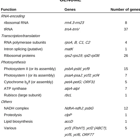
Chloroplast genome
Function Genes Number of genes
RNA-encoding
ribosomal RNA rrn4.3-rrn23 8
tRNA trnA-trnV 37
Transcription/translation
RNA polymerase subunits rpoA, B, C1, C2 4
Intron splicing (putative) matK 1
Ribosomal proteins rps2-rps19; rpl2-rpl36 26
Photosynthesis
Photosystem II (or its assembly) psbA-psbl; ycf9 15
Photosystem I (or its assembly) psaA-psaJ; ycf3; ycf4 7
Cytochrome b6/f (or assembly) petA-petG; ORF31 5
ATP synthase atpA-atpI 7
Rubisco (large subunit) rbcL 1
Others
NADH complex NdhA-ndhJ; psbG 12
Proteolysis clpP 1
Lipid biosynthesis accD 1
Various ycf1 (FtsH?), ycf2 (ABC?), 5
ycf5, ycf6, ORF77
GENE CONTENT OF THE ARABIDOPSIS CHLOROPLAST GENOME
TABLE 1
Note: Many other higher plant chloroplast genomes also encode the translation initiation factor gene infA.
in individual circular molecules, one genome per circle, instead it appears that long, polyploid linear molecules and branched mol-ecules undergoing replication are abundant (Bendich, 2004). To date the sequence of the full chloroplast genome of a total of 45 photosynthetic organisms has been determined, of which 22 are seed plants. A comprehensive list of fully-sequenced chloroplast genomes is currently available at http://megasun.bch.umontreal.ca/ ogmp/projects/other/cp_list.html. DNA exists in plastids in discrete regions, in the form of nucleoids associated to the inner envelope. Nucleoids contain DNA-binding proteins, one of which is plastid envelope DNA-binding and another turns out to be a bifunctional protein with a second role as the enzyme sulphite reductase (Sato et al., 2003). A DNA polymerase, the origins of replication (different between monocots and dicots) and a number of topoisomerases have been identified (Mache and Lerbs-Mache, 2001).
Higher plant chloroplast DNA is very highly conserved. In fact phylogenetic analyses using chloroplast genome sequences have concluded that a single endosymbiotic event between a mitochon-drion-containing eukaryote and an unknown cyanobacteria, took place and gave rise to all existing chloroplasts (Martin et al., 2002; De las Rivas et al., 2002; Timmis et al., 2004). The early event was soon followed by diversification into what are now the chloroplasts of red alga (and, remarkably, those of organisms which have secondarily engulfed red algae, like diatoms) and those of green algae and eventually land and seed plants (Martin et al., 2002; De las Rivas et al., 2002). The genome has a physical peculiarity, the presence of two copies of the same large region, separate and in inverted position (large inverted repeat). The regions outside the repeats are called large single-copy and small single-copy regions. The size of the repeat is variable, this accounting for most of the variation in genome sizes. Chloroplast genomes contain between 120 and 135 genes, 130 in the model species mentioned above, of
which 76 are protein-coding genes, the rest encoding other RNAs (Table 1). The chloroplast genome is concerned mostly with encoding components of the four thylakoid photosynthetic com-plexes, or proteins necessary for their assembly and also encodes part of the genetic machinery necessary to do so, this genetic machinery being of a eubacterial type (Fig. 3A). Through the use of bioinformatics techniques, including comparisons of large scale gene clusters to full cyanobacterial genomes, two large open reading frames of so far unknown function, ycf1 and ycf2, have been recently proposed to encode one of the FtsH group of proteases and an ATP-binding cassette transporter respectively (De las Rivas et al., 2002).
Organisation of chloroplast genes
As in bacteria, many genes are organised in operons and expressed as polycistronic units. In one instance this is more the case than in bacteria themselves: the rpl23 operon of chloroplasts contains genes that in E. coli are encoded in three separate, although contiguous, operons (Sugiura, 1992). Fifty transcriptional units exist overall, giving rise to the need for post-transcriptional processing. Most operons encode subunits of the same molecular complex, although hybrid ones also exist (psbB and pet genes are expressed as a single operon). In some cases it includes intron splicing: contrary to the general case in bacteria, some chloroplast genes contain introns, but these relate to mitochondrial or unusual yeast introns, with a conserved folding pattern, rather than con-served splice sites (Sugiura, 1992).
Plastid genome expression
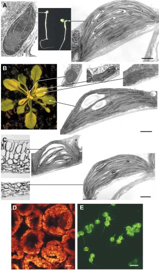
photosynthe-Fig. 2. Developmental or environmental influences on chloroplasts. (A)Cotyledons of seedlings grown in the dark (left) exhibit etioplasts, while those in the light (right) contain chloroplasts. (B) Meristematic cells con-tain proplastids, which quickly differentiate into chloro-plasts with increasingly complex thylakoid structures as cells themselves differentiate with leaf develop-ment. The conversion is slowed down in the ‘virescent’
cue6Arabidopsis mutant, in which the delay reveals the gradient of maturation of leaf mesophyll cells. (C) Chloroplasts of high (top) or low-light (bottom) exposed
Arabidopsis leaf cells (shown in cross section) exhibit a different composition, particularly in the abundance of light-harvesting antenna complexes and as antenna complexes are grana-localised, low-light chloroplasts contain more abundant grana. (D)Mesophyll cells from a ‘high-light’ Arabidopsis leaf (left) develop as a thick palisade layer of highly elongated cells, the elongation being matched by an increase in chloroplast numbers, so that the cells appear as ‘tunnels’ coated internally in one layer of chloroplasts. Chloroplasts are visualised in this confocal Z-axis view of stacked sections as glob-ules of red (chlorophyll) fluorescence. (E)Plastids from the inner mesocarp of mature green tomato fruit, initiating the transition from chloroplast to chromoplast, under confocal imaging. The plastids contain both chlo-rophyll and green fluorescent protein (GFP), which appear yellow when both are present whereas with only GFP appear green. Thin, green, tubular stromules, rare in mesophyll chloroplasts, are evident in most of the plastids. Scale bars (for plastids): 1 µm (A-C) and 10
µm (D, E).
sis-related genes can be transcribed by both poly-merases (Cahoon and Stern, 2001).
Whether transcription is the most important level of regulation of gene expression has been a matter of much debate. Overall that notion is supported by the fact that protein levels, translation activity and mRNA levels correlate (Mullet, 1993). However it is also the case that much evidence for translational regulation exists (Bruick and Mayfield, 1999). In general plastid-encoded mRNAs have a long half-life, of between hours and days and recently a large family of nuclear genes, for pentatricopeptide-re-peat proteins, appear in many cases to encode organellar-targeted RNA-binding proteins (Lurin et al., 2004).
The plastid proteome
Since the plastid genome encodes less than 80 proteins, it is obvious that a much greater number is required for the variety of plastid functions. Many genes, particularly for photosynthetic proteins, have
been individually identified as being encoded in the cell’s nuclear genome. The availability of full genome sequences of plants has revealed the extent and range of plastid-contained proteins. Nuclear-encoded proteins, as discussed below, are translated in the cytoplasm and imported into the plastids, the targeting signal being localised at the N-terminus of the proteins as a transit peptide or signal sequence (Soll, 2002). Algorithms have been developed based on known properties of these signals and further refined
(‘trained’) on experimentally determined sequences to identify transit peptides, the most faithful to date being TargetP (http:// www.cbs.dtu.dk/services/TargetP/). Use of this algorithm on the full Arabidopsis genome sequence and corrections for its experi-mentally-determined specificity and sensitivity, leads to a predic-tion of a total of 3,100 proteins as chloroplast targeted (Abdallah and Leister, 2000; Leister, 2003). A combination of algorithms, on the other hand, has estimated the number in Arabidopsis at around
A
B
C
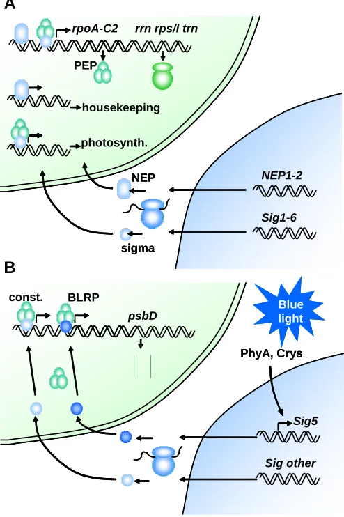
Fig. 3. Plastid genome expression. (A)Plastid genetic machinery and contributions of the nuclear genome towards it. The chloroplast (upper) and nucleo-cytoplasmic (lower) ribosomes are also indicated. PEP: plastid-encoded RNA polymerase. NEP: nuclear-plastid-encoded RNA polymerase. Photosynth.: photosynthesis. (B)Example of regulation of a chloroplast-encoded gene psbD, by an environmental cue, blue light, perceived by extraplastidic sensors, phytochrome A (PhyA) and cryptochromes (Crys). BLRP, blue light-responsive promoter; Const, constitutive promoter.
NEP1-2
Sig1-6 rpoA-C2 rrn rps/l trn
housekeeping
photosynth. PEP
NEP
sigma
NEP1-2
Sig1-6 rpoA-C2 rrn rps/l trn
housekeeping
photosynth. PEP
NEP
sigma
b
Sig5
Sig other psbD
const. BLRP
PhyA, Crys
Blue light
b
Sig5
Sig other psbD
const. BLRP
PhyA, Crys
Blue light
A
B
2,100, while that in rice is 4,500 (Richly and Leister, 2004). Only about half of the predicted Arabidopsis chloroplast proteins are shared by rice and play a role in metabolism, energy-generation and transcription. The low number is surprising and suggests there may be large diversity in the fine detail of plastid functions across groups, although it may also be a consequence of ambiguities derived from the use of bioinformatics tools, both in the prediction and in the detection of homology. Work is intensively being carried out in several laboratories to catalogue every plastid protein directly detected using mass spectrometry-based proteomic tech-niques. This has been done for thylakoids or chloroplast envelopes (Peltier et al., 2002; Ferro et al., 2003), as well as for complete chloroplast and envelope-enriched preparations (Klefmann et al., 2004). The latter authors identified a total of 690 proteins from Arabidopsis. Two databases of plastid proteins have been pro-duced: PLprot currently contains the data of Klefmann and
co-workers from Arabidopsis chloroplasts, but will be supplemented with data from other plastid types (http://www.pb.ipw.biol.ethz.ch/ ~w3pb/index.php?toc=91), while Plastid Proteome Database, PPDB, contains both experimentally determined (Peltier et al., 2004) as well as predicted chloroplast proteins in Arabidopsis and maize (http://ppdb.tc.cornell.edu). The study by Klefmann et al. (2004) identified proteins in many functional categories, including ‘energy’ (primarily photosynthesis) and metabolism, among oth-ers, of amino acids and carbohydrate, but also detected a large number of transporters, particularly envelope-associated, proteins involved in cell defence, gene expression, protein fate (folding and modification), even DNA replication and cell cycle. Interestingly, as a general rule, simultaneous monitoring of global gene expression detected a broad correlation between amount of transcripts and amount of protein, measured as frequency of detection. This suggests a primary role for transcription/transcript accumulation in the regulation of gene expression for chloroplast-targeted proteins. The correlation, however, was pathway-specific. One surprising finding of this study was the fact that over one third of proteins identified did not have a signal peptide predicted by TargetP. It is known that a number of outer envelope plastid proteins posses a secretory pathway-related targeting signal, which nevertheless leads to envelope localisation unless altered (Lee et al., 2001). This suggests that our estimates of chloroplast-targeted proteins may under-represent their true number and that novel pathways and functions may still emerge. Both proteomics and genetics, through the large-scale identification of mutations in essential plastid genes, using Arabidopsis (Leister, 2003) or maize (Stern et al., 2004; see the Photosynthetic Mutant Library at http:// chloroplast.uoregon.edu/) will play a role.
Gene transfer to the nucleus and contribution of the endosym-biont to the host
The global comparisons of a full plant genome with that of cyanobacteria, other representative prokaryotes and yeast (Martin et al., 2002) has provided an estimate of around 4,500 Arabidopsis genes derived from the chloroplast endosymbiont ancestor and has pointed to the cyanobacteria Nostoc as its closest relative among those with fully determined genomes. This highlights the massive scale of gene reduction in the organelle and transfer to the nucleus (Timmis et al., 2004). The complete transfer probably involves the incorporation into the nucleus of copies of the plastid gene, the acquisition of plastid transit peptides, either by integra-tion next to existing ones or by generaintegra-tion of new ones from previous upstream sequences and finally the loss of the organellar copy. The integration of plastid genes into the nuclear genome has been monitored through selection of transfer events with a marker which was only active when expressed in the nucleus and has turned out to be unexpectedly easy (in one out of 16,000 gametes tested; Huang et al., 2003). In fact many instances of recent transfers have been detected, including an almost complete inte-gration of the chloroplast genome into rice chromosome 10 (The Rice Chromosome 10 Sequencing Consortium, 2003).
resident proteins; for example two of the Calvin cycle enzymes, triosephosphate isomerase and fructose bisphosphatase, are of mitochondrial origin (Martin and Herrmann, 1998). Conversely, originally cyanobacterial genes have explored novel functions. Genome-wide estimates of the destinations of ancestral cyanobacterial genes have revealed that less than half the total has been retargeted to the chloroplast, the second most common destination being the secretory pathway (endoplasmic reticulum, Golgi, plasma membrane and cell wall export), but others being predicted as cytoplasmic and mitochondrial (Martin et al., 2002). Even if, as seen above, some of the proteins predicted as targeted to the secretory pathway end up being chloroplast envelope proteins, there is still a wealth of genes contributed by the endo-symbiont to the host’s genome. The most common functional categories among those are biosynthesis and metabolism, signal transduction, ‘cellular responses’ and energy generation (Martin et al., 2002). This has left its mark in the exceptionally large primary and secondary metabolic abilities of plants, as well as in their developmental toolkit. Among many examples, hormonal sensory mechanisms, including the ethylene and cytokinins receptors, use two-component histidine kinases and the same can be said of environmental phytochrome photosensors (see http:// www.bio.unc.edu/research/two-component/default.htm; Fankhauser, 2001). Such kinases are widespread in eubacteria, including cyanobacteria, but exceptionally rare in non-photosyn-thetic eukaryotes (none in metazoans, Stock et al., 2000). It is not just because of their photosynthetic capacity that plastids make plants unique.
Why a plastid genome?
Given the massive reduction in the number of genes, which in cyanobacterial genomes range from 3000 to over 7000 and in chloroplasts are around 100 (Timmis et al. 2004), the question as to why has a genome been retained at all in the organelle appears legitimate. An overview of the proteins whose genes have been transferred and those that have not shows a general pattern: the majority of retained proteins encode thylakoid membrane compo-nents (or genetic machinery proteins necessary to produce those). One explanation would be that it has been primarily highly hydro-phobic membrane proteins, particularly difficult to import and assemble correctly, that have been retained by the organellar genome. This would be consistent with the pattern of retention of mitochondria-encoded proteins (Dyall et al., 2004). An intriguing alternative has been proposed. As we will discuss, chloroplasts are capable of using their redox processes as signals to regulate the expression of plastid-encoded genes, rapidly, in response to sudden changes in environmental circumstances, like changes of light quantity. Rapid regulation following a burst of light in a sunfleck may make the difference between survival and catastrophic oxida-tion for a chloroplast and the cell that harbours it. The need for such a rapid redox regulation may explain the retention of genes by both chloroplasts and mitochondria (Allen, 2003).
The biogenesis of plastids
Plastid protein import machinery
Nuclear-encoded proteins are translated in cytoplasmic ribo-somes and, unless they are targeted to the outer envelope, need to be brought into the chloroplast crossing two plastid envelopes
(Fig. 4). Proteins destined to be imported carry an N-terminal transit peptide, generally between 20 and 80 aminoacids. Transit peptides of different proteins show no obvious sequence conser-vation, their general physical characteristics consisting of an abundance of hydroxylated, positively charged and small aminoacids and a low abundance or absence of acidic or large hydrophobic ones. A common feature of transit peptides appears to be a site for phosphorylation at a Ser or Thr residue, this site being bound by 14-3-3 proteins and a chaperone into what has been termed a guidance complex (May and Soll, 2000). The functional significance of this complex, however, is in question, as the phosphorylation site can be mutated without obvious detrimen-tal effects (see Jarvis and Robinson, 2004).
The import is carried out by protein complexes located in both membranes: translocon of the outer envelope of chloroplast (Toc) and translocon of the inner envelope of chloroplast (Tic) (Soll, 2002). Our understanding of the function of Toc is greater than that of its inner envelope counterpart. The Toc complex, as first purified biochemically from pea chloroplasts, is composed of three subunits, Toc159, Toc75 and Toc34 (Keegstra and Froehlich, 1999), as well as probably Toc64 (see Soll, 2002). Toc75 forms a channel across the outer membrane. It has a predicted structure similar to bacterial porins, with 16 β-strands, into what has been called a β-barrel. The channel is selective to cations, as expected from the nature of transit peptides (Soll, 2002). Toc159 and Toc34 jointly function as receptors and docking sites for the polypeptides to be imported. They both are guanosine triphosphatases (GTPases) with a region of high homology. When Toc34 binds GTP, it shows much higher affinity for substrates and the binding causes the hydrolysis of GTP, followed by the release of gua-nosine diphosphate (GDP) and the substrate. It is possible that the same phenomenon occurs in the associated component, Toc159 and that the conformation change associated with the hydrolysis of GTP gates the import channel (Sun et al., 2002). A preliminary structure of purified Toc complexes shows a ring (presumed formed by Toc75 subunits) leaving four pores cross-ing the membrane, with a middle structure and fcross-ingers protrudcross-ing perpendicular to the membrane, consistent with the soluble receptor domains of Toc159 and Toc34 (Schleiff et al., 2003)
Each subunit of the Toc and Tic complexes is encoded by a small family of genes (Jackson-Constan and Keegstra, 2001). Recent genetic evidence has shown that defects in individual genes are not incompatible with survival, indicating a degree of redundancy among those genes. For example the co-receptor Toc34 is encoded in Arabidopsis by two genes, atTOC33 and atTOC34. The plastid protein import 1 (ppi1 ) mutant contains a disruption of atTOC33 that leads to defective chloroplasts (Jarvis et al., 1998). The mutant ppi3, a knockout of atTOC34, has only a minor phenotype; the loss of both forms, however, is lethal (Constan et al., 2004). A loss in ppi2 of atToc159, the main form of the receptor, leads to chloroplasts with severe loss of thylakoids and plants unable to grow autotrophically (Bauer et al., 2000). Loss of either of the two other family members encoding alterna-tive forms of Toc159, namely at Toc132 and atToc120, leads to no obvious phenotype, but loss of both is severely deleterious (Kubis et al., 2004) or lethal (Ivanova et al., 2004), presumably depend-ing on the growth conditions, while loss of atToc159 and atToc132 is lethal in every case (Kubis et al., 2004).
Tic110, often co-purifies with Toc complexes, making it likely that they both act in tandem, at points in which both plastid envelopes contact each other. Like Toc75, Tic110 can form a channel for cations. Since Tic110 also folds into a number of predicted β -strands, it is possible that it also forms a β-barrel (Heins et al., 2002). This all leads to assume Tic110 forms the protein import pore across the internal membrane. However there is conflicting evidence and claims in the literature, with a small Tic component, Tic20, having also been proposed to play this role (see Jarvis and Robinson, 2004). A number of chaperones associate with the Tic complex and may provide the pulling power that drives the transport of the polypeptide through the two pores (Soll, 2002). Once across the envelope, a stromal processing peptidase cleaves the transit peptide, leaving the mature protein to fold in the stroma or continue its journey.
Proteins destined for the thylakoid membranes or the thylakoid lumen use a second signal sequence for the final leg of their journey. Such proteins, therefore, need a bipartite transit peptide, with two domains, one for ‘envelope-transit’ and one for ‘thyla-koid-targeting’ (Robinson et al., 2001). The thylakoid-targeting signal is comparable to signals used by prokaryotes to export or secrete proteins. Plastids use a system comparable to bacteria, in which the translocation takes place through a SecA protein, but have also evolved an alternative route, in which the pH gradient across the thylakoid is used to drive the translocation (the ∆ pH-dependent pathway, also called Tat as it uses a T ranslocase for domains with a T win-A rginine motif). Integral thylakoid mem-brane proteins are either targeted through a third type of domain, recognised by a bacterial-type signal recognition particle (SRP), or apparently integrate spontaneously, using the polypeptide’s biophysical/solubility properties (Robinson et al., 2001). Proteins that do use a thylakoid-targeting domain have it finally cleaved off in the lumen by a thylakoid processing peptidase.
Plastid division machinery
Plastids only originate from pre-existing plastids. The process during which one leaf primordium cell, containing 20 proplastids, gives rise to several hundred mature leaf cells, each carrying around 100 chloroplasts, must be accompanied by massive plastid division (Fig. 5A). Under the microscope, this appears similar to bacterial fission; a plastid undergoes a constriction, with a ring of electron-dense material appearing at both the cytoplas-mic and stromal sides of the envelopes at the middle of the plastid. Eventually the physical constriction fully separates the two daughter plastids (Pyke, 1999; Marrison et al., 1999; Osteryoung and Nunnari, 2003).
Our understanding of the mechanisms of plastid division has progressed in two parallel fronts. The genetic approach has been based on the identification of mutant ‘accumulation and replica-tion of chloroplasts’ (arc ) plants with altered numbers of plastids (Pyke and Leech, 1994; Pyke et al., 1994). This has been aided by the fact that altered plastid division turns out to not impair the build-up of the total chloroplast compartment of the cell, i.e., in mutants with altered division, at least in leaf cells there is an inverse relationship between the number of chloroplasts in the cell and their size (Marrison et al., 1999). In the extreme arc6 mutant, only one large chloroplast occurs per mesophyll cell. The phenotype of two arc mutants, arc3 and arc5, is consistent with a defect in the accumulation of chloroplasts in mesophyll cells,
without compromising proplastid division in meristematic tissue, therefore leading to the presence of only around 20 chloroplasts per cell (Pyke, 1999). This suggests that there are separate mechanisms, or at least separate gene family members, playing roles in division of plastids at different stages. The genetic approach has also yielded the ARTEMIS protein (Fulgosi et al., 2002). When ARTEMIS is mutated, plastid division is not com-pleted but separate thylakoid systems can be identified. This demonstrates that thylakoid systems, constituting a single, inte-grated structure per chloroplast, are divided by a process which is to some extent distinct from the division of the chloroplast.
A complementary, genomic approach to understanding plastid division was sparked by the identification in Arabidopsis of a homologue of the bacterial cell division gene FtsZ, whose product was targeted to chloroplasts (Osteryoung and Vierling, 1995). Defects in FtsZ in E. coli lead to defects in division without impairing growth at non-permissive temperature, resulting in a filamentous phenotype. The protein is a GTPase, that is, it binds and hydrolyses GTP. It also has a domain with homology to eukaryotic tubulin, can polymerise into filaments and is generally accepted as tubulin’s prokaryotic ancestor. The FtsZ proteins of chloroplasts (FtsZ1 and FtsZ2) polymerise into a ring at the inner envelope and GTP hydrolysis may help generate the constriction force (Osteryoung and Nunnari, 2003) (See Fig. 5B). The plastid division rings themselves, however, are associated to but distinct from FtsZ (Miyagishima et al., 2001). In bacteria, the FtsZ division ring is placed in the middle of the long bacterial rod through the action of Min proteins, mutations in which lead to asymmetric divisions and consequently the appearance of mini-cells. Chloro-plast MinD (Colletti et al., 2000) and MinE (Itoh et al., 2001; Maple et al., 2002) have also been identified. As in some bacteria, MinE could act by excluding FtsZ from the chloroplast poles. MinD would contribute to this process and turns out to be the defective gene in the arc11 mutant (Fujiwara et al., 2004). Its molecular role explains the variable size of chloroplasts in arc11.
The ARC5 gene encodes a dynamin-related protein (Gao et al., 2003). Dynamins are involved in eukaryotes in membrane severing, for example during endocytosis or membrane trafficking and, interestingly, also in mitochondrial division. The other func-tions of dynamins suggest that ARC5 could play a role in complet-ing off the separation of membranes, once the first stages of division have created a small enough constriction and this is consistent with the incomplete-division, dumbbell shape of arc5 chloroplasts. ARC6, on the other hand, encodes a DnaJ related protein, a chaperone partner and is considered to assist in the assembly of the FtsZ ring (Vitha et al., 2003).
Plastid ‘plasticity’ and stromules
Toc 159
Fig. 4. Plastid protein import machinery and its key components. The model for the Toc complex is consistent with the observations from Schleiff
et al. (2003). Both the involvement of 14-3-3 proteins as import guides and the model for Tic are hypothetical; for example conflicting data exist as to whether Tic110 or Tic20 play the role of import pore. Import into the thylakoids occurs through involvement of one of the routes shown, depending on the protein. Some membrane proteins integrate without involvement of any of the mechanisms shown (‘spontaneous route’). SPP: stromal processing peptidase. TPP, thylakoid processing peptidase.
actively transport endogenous protein and that the extent of stromule production is highly dependent on the cell type and type of plastid (Köhler and Hanson, 2000). The nature of stromules in chloroplasts in green tomato fruit, during their transition into chromoplasts in ripe fruit, has been examined in detail (Waters et al., 2004). The basic findings were that stromules increase in frequency as plastids become further apart during cell expansion, that chloroplasts display the lowest number of stromules and that a ripening inhibition mutation causes a reduction in stromule formation, as conversion into chromoplasts is arrested. This suggests that stromules are associated primarily with non-photo-synthetic plastids. One possible function of stromules is to provide an increase in the plastid surface area. A role helping to integrate the total ensemble of plastids into a coordinated single plastid compartment is less likely but cannot be dismissed yet, as occasional pairs of plastids can be seen joined together by stromules (Pyke and Howells, 2002) and exchange of marker proteins is possible (Köhler et al. 1997)
The origin and role of organelle-specific functions
Our current understanding of chloroplast development allows us to address the question of how did the original endosymbiont adapt to become a fully integrated organelle in its host. The organelle, like its ancestor, was still required to divide and had a readily available set of prokaryotic cell division proteins allowing it to do so. Indeed chloroplasts utilise MinD and MinE to position the FtsZ ring, which forms with the assistance of ARC6, a homologue of cyanobacterial Ftn2. All of these proteins retained the basic function they possessed in the free-living cyanobacterium. The organelle, on the other hand, enrolled a dynamin-related protein to complete the constriction of the plastid envelopes, as dynamins were already doing in eukaryotic membrane trafficking and, indeed, in mitochondrial division. The consideration that this final function was recruited from pre-existing eukaryotic machin-ery for the maintenance of mitochondria is attractive. In one known case, ARC3, the origin of the protein itself is dual: it is a chimeric protein with homology to FtsZ plus an additional domain consisting of a portion of a eukaryotic signal transduction protein, phosphatidylinositol phosphate kinase (Shimada et al., 2004).
Meanwhile, plastid protein import was a novel requirement emerged from the symbiotic way of life and the vast degree of migration of genetic control to the cell’s nucleus that it brought. Where did the elements come from? As we have seen, plastids resorted to modifying the function of porins, proteins involved in secretion, basically inverting the direction of their transport. Interestingly, some of the minor components of the Tic complex are also related to aminoacid permeases (Dyall et al., 2004). Pulling power for translocation was also required and cyanobacterial chaperones/heat shock proteins provided this. However the outer pore required gating and for this GTPases of eukaryotic origin and with homology to the RAS oncogene were recruited (Toc159, Toc34). The emerging picture is that the organelle utilised pre-existing cyanobacterial elements for both processes, more so for division, but that in both cases it also resorted to available eukaryotic proteins, as it did to distinguish proteins in need of being translocated.
Another question worth asking is whether the organelle re-quired active division at all. arc6 mutant plants and plants overexpressing almost any component of the plastid division
machinery display one or two giant chloroplasts per mesophyll cell, but macroscopically are remarkably normal under laboratory conditions (Pyke et al., 1994; Fujiwara et al., 2004). Hornworts, bryophytes that form a sister group of vascular plants (Willis and McElwain, 2002), naturally have a single, large chloroplast per cell, in common with many green algae. A key answer may be environmental adaptability. Chloroplasts adjust, among other ways, physically to prevailing light conditions, displaying an accumulation response towards low-intensity light and an avoid-ance of strong, potentially damaging light, both movements being activated by phototropins (Sakai et al., 2001). It has been demon-strated that tobacco plants with altered levels of FtsZ, showing between one and three large chloroplasts per mesophyll cell, perform worse under both low light (reduced photosynthetic performance) and high light environments (greater incidence of photodamage) than wild type plants (Jeong et al., 2002).
Plastids are under the control of developmental signals
From proplastids to chloroplasts
results, with older cells at the tip of the leaf containing a full complement of mature chloroplasts, while at the leaf base cells have undergone division more recently, possess thinner walls and contain a smaller number of developing chloroplasts. The gradient in plastid development manifests itself as very active plastid DNA replication in the meristematic region, followed by high transcriptional activity and eventually the massive build-up of photosynthetic complexes (Baumgartner et al., 1989). Leaves of developing dicotyledons such as pea have been examined in a temporal sequence and show consecutive, partly overlapping stages of plastid DNA replication, transcription of the genetic machinery and transcription for photosynthesis proteins (DuBell and Mullet, 1995a, 1995b). Leaves of dicotyledons like Arabidopsis also show a spatial gradient in cellular differentia-tion, with cells at the distal end and in the vicinity of the mid vein being first to reach full differentiation (Pyke et al., 1991). The gradient is less clear for chloroplasts, but is equivalent and can be uncovered by nuclear mutations that slow down chloroplast development, such as in slow-greening or virescent mutants (López-Juez et al., 1998). For example in the cue6 mutant, chloroplasts at the leaf tips are undistinguishable from wild type, while those at the flanks closer to the base are essentially proplastids, of small size and very limited extent of develop-ment of thylakoids (Fig. 3B).
One spectacular case of interaction between chloroplast biogenesis and leaf development is that of variegated mutants (Aluru et al., 2001; Sakamoto, 2003). Variegation can be caused by defects in core mechanisms in chloroplast biogenesis, due to unstable insertions of transposons in such essential nuclear genes. A more interesting kind of variegation, however, is that caused by stable mutations, which however cause heteroge-neous chloroplast deficiency phenotypes. One such variegated mutation, immutans, is caused by a defect in a plastid targeted alternative oxidase (Aluru et al., 2001; Rodermel, 2001). It is postulated that this protein is involved in a carotenoid biogen-esis step, in a non-essential way (Carol et al., 1999). However the most commonly identified mutations resulting in variegated plants have been found to define VAR1 or VAR2, genes of the FtsH protease family. This protease class is required for the disassembly and turnover of photosystem II reaction centre proteins when damaged by reactive electrons generated by excess light (Sakamoto et al., 2003). Once plastid damage occurs, the sector of leaf formed by the clone of cells derived from the cell that was photodamaged will be devoid of chloro-phyll. An interesting question is why does the damage take place in such a discrete fashion, when the cells are genetically identical and no satisfactory answer has been found (Aluru et al., 2001; Sakamoto et al., 2003). The variegation occurs when the defect in plastids in a cell takes place very early, when leaf primordia are formed by a very small number of cells. Plastid or cell population factors might play a role. It is possible that the function of this FtsH protein class specifically results in a peculiar type of damage, one that takes place rarely, only in very immature plastids, but that once it has happened, leads to the release of free pigment, for example, this having an auto-catalytic effect on the complete damage of that plastid and, if the envelope breaks, possibly of other plastids in that cell. Progress in chloroplast differentiation might reach a point in which the organelle is far more resistant to damage from loss of
the protein, so that no new differential plastid/cell clones emerge.
Other plastid types
Our knowledge of the regulatory networks underlying the con-version of proplastids into other plastid types, like amyloplasts or chromoplasts, is surprisingly limited. We obviously know that plastids differentiate according to the cell type they reside in. For example root plastids in the vicinity of the meristem appear as proplastids, but a few cells in very close proximity, the columella cells of the root cap, accumulate a special type of amyloplast, the statolith, that plays a critical role in gravity sensing (Morita and Tasaka, 2004). Amyloplasts accumulate in potato tubers, proplas-tids being converted to amyloplasts under the influence of kinetin (Mingo Castel et al., 1991), while light induces the conversion of amyloplasts into chloroplasts (Ljubicic et al., 1998). Amyloplast development is also susceptible to external intervention in suspen-sion cultures of tobacco Bright Yellow-2 cells. Cells of such cultures possess proplastids or undifferentiated leucoplasts, but depletion of auxin and exposure to cytokinin triggers amyloplast formation (Miyazawa et al., 1999). This plastid differentiation has been shown to require the transcription of nuclear genes for starch biosynthesis proteins (Miyazawa et al., 1999), while the plastid genome shows a global decrease both in transcription and in the turnover of mRNA, leading to the paradoxical increase in steady state levels for some transcripts (Sakai et al., 1999). In this sense amyloplasts could be interpreted as proplastids that have become semi-passive recipients of specific nuclear-encoded proteins. Another develop-mental transformation studied in some detail is that of chloroplasts in green tomato fruits to chromoplasts in ripe fruits. This conversion is accompanied by the fall in expression of photosynthetic nuclear genes, the active degradation of chloroplast photosynthetic pro-teins and the disassembly of thylakoids (Piechulla et al., 1987). At the same time the expression of carotenoid biogenesis genes increases dramatically (Giovanonni, 2004). Meanwhile, the tran-scriptional activity of the plastid genome remains virtually un-changed (Marano and Carrillo, 1992). Once again it appears that, in this transition, plastids act as acceptors of proteins expressed under the control of regulatory mechanisms in the nucleus.
Differential plastid development within leaf cells
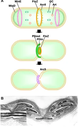
Fig. 5. Plastid division.(A) Plastid division process and role of several known components of the division machinery. PDout, plastid division ring, outer; PDin, plastid division ring, inner; GC, giant chloroplast; Art, Artemis. (B) Chloroplasts arrested in incomplete division by physical interference caused by a long, narrow starch rod. The left inset shows the starch rod apparently being subjected to constriction from the plastid division and associated rings.
Arc5 Arc5 MinD
MinE GC
Art FtsZ
Arc6 MinD
MinE GC
Art FtsZ
Arc6
PDout PDin
FtsZ PDout
PDin FtsZ
A
B
(Bsd2 ) encodes a protein required for the post-translational control of the accumulation of the large subunit of Rubisco, its absence leading to defective bundle-sheath chloroplasts (Brutnell et al., 1999). Golden2 (G2 ), also designated as Bsd1, encodes a nuclear protein with transcriptional regulatory activity and necessary for the differentiation of C4 bundle sheath chloroplasts. A golden2 -like (ZmGlk1 ) homologue exists in maize and interestingly this gene is differentially expressed in C4 mesophyll cells (Rossini et al., 2001). Given the phenotype of the absence of G2 and the molecular function of G2 and Glk1, it is tempting to speculate that these two genes are activators of the respective kinds of chloroplast develop-ment. It is also worth noting that the differentiation of specific chloroplast functions in cells closely associated to vascular bundles has been observed widely, so predating the emergence of C4 species (Hibberd and Quick, 2002).The control of plastid gene transcription
As we have seen, during leaf cell differentiation a specific stage is reached during which massive accumulation of photosynthetic complexes takes place. This stage does not occur in non-photo-synthetic cells. How is the specific transcription of plastid-encoded, photosynthetic genes achieved? The sequential regulation of RNA polymerases plays a key role (Fig. 3B). NEP activity is first required for housekeeping functions. A knock-out mutant of one of three widely-expressed NEP genes in Arabidopsis, RpoT;2, shows not only delayed greening, but also reduced root and hypocotyl growth and altered leaf shape (Baba et al., 2004). Once NEP function has eventually led to the translation of the PEP chloroplast-encoded subunits, these become more important in the expression of photosynthesis-related proteins. The recognition of PEP promot-ers requires the activity of sigma factors. Most higher plants contain multiple sigma factors, the Arabidopsis genome encoding 6 genes (Isono et al., 1997; Fujiwara et al., 2000). Plants defective in genes for these factors are revealing differential specificities. Responses to signals, like light cues, are achieved by phosphorylation of sigma factors (Ogrzewalla et al., 2002) and by their differential expression (see below). A knockout of AtSig2 shows defective greening but no deficiency in the production of most photosynthetic protein tran-scripts. The riddle is explained by the deficiency observed in this mutant in the transcription of tRNA s (Kanamaru et al., 2001). One of those, tRNA-Glu, is the raw material for the production of aminolevulinic acid, the first committed precursor in the biosynthe-sis of chlorophyll and other tetrapyrroles. AtSig2 appears to play a primary role in tRNA transcription. The analysis of a number of other knockout mutants and of the expression patterns of different AtSig genes suggests the following sequence of activities: A general sigma factor, AtSig6, acts on multiple photosynthetic gene promoters in young seedlings (Ishizaki et al., 2004). Its function is gradually taken over by one or more other general sigma factors, probably AtSig3 and AtSig1 (Privat et al., 2003). AtSig5 shows unique properties. It has long been known that the genes rbcL, psbA, 16S rrn, (Chun et al., 2001) and psbD (Thum et al., 2001) respond to blue light signals through increased transcription. psbD is unique in that it can use several promoters, with distinct transcrip-tion initiatranscrip-tion sites and produces multiple transcripts. One of those transcripts is specifically induced by blue light, from a blue-light responsive promoter (Thum et al., 2001), or by both blue and red light of high fluence (Mochizuki et al., 2004). The response to light is of adaptive significance, as psbA and psbD encode the reaction
this factor, targeted to the chloroplast, acts as a co-factor of the plastid RNA polymerase and activates the transcription of a plastid-encoded gene (Fig. 3B).
Differentiation by control of protein import?
It is possible for the variety of plastids in different cells to result not only from the differential expression of each of the various nuclear-encoded genes for plastid proteins, but by differences in the basic mechanisms of plastid build-up, particularly plastid protein import. In pioneering work, Wan and coworkers (1996) demonstrated that seed ‘leucoplasts’ and leaf chloroplasts im-ported individual proteins differentially, the efficiency for each protein being related to the actual in vivo abundance of the protein in each plastid type and that the differences were specified by each protein’s transit peptide. Evidence that this phenomenon is ex-plained by the differential composition of the import machinery is now accumulating. As discussed earlier, most components of the Toc and Tic complexes are encoded by small gene families in Arabidopsis. Individual gene family members may encode proteins with slightly different functions. This is particularly the case for Toc159 and Toc34, which together act as receptors gating the outer envelope pore and therefore control which proteins are or are not allowed passage. Loss of atTOC159 or of atTOC34, both leading to significant or extreme defects in greening, causes no obvious effect in root cells, or even in guard cells (Yu and Li, 2001). Meanwhile Kubis et al. (2004) and Ivanova et al. (2004) have observed that reducing or knocking out expression of atTOC132 and atTOC120, the two other alternative members of the Toc159 family, which result in chloroplast phenotypes only when both are fully knocked-out, bring about a defect also in root plastids and cause altered root growth. Similarly a defect in atTOC34, the homologue of atTOC33, causes deficient root plastids and a 20-30% reduction in root growth, but does not lead to pale leaves, indicating a predominant photosynthetic role for atToc33 and a house-keeping or root-plastid role for atToc34 (Constan et al., 2004). These specificities must help avoid competition for import sites by proteins expressed at very different levels in developing chloroplasts, but are also likely to be part of the various differentia-tion programmes, a nodifferentia-tion that the various patterns of gene expression by tissue type is consistent with. Two interesting sets of observations were those of Kubis et al. (2003, 2004) who monitored protein accumulation and gene expression for a number of photosynthesis-associated or unrelated chloroplast proteins. Defective atToc132, which is devoted to import of non-photosyn-thetic proteins, led to both reduced accumulation of those proteins and to lower expression of their genes, while loss of atToc33, which imports photosynthetic proteins, resulted in decreases in photo-synthetic protein accumulation and gene expression. A conse-quence is that changes initiated by an altered balance of the two forms for each subunit of the receptor will have an autocatalytic impact: as a reduced amount of photosynthetic proteins is im-ported, less transcription of their genes will take place, gradually amplifying the difference.
The regulation of plastid proliferation
The accumulation of a full complement of chloroplasts in mature leaf cells must involve a large, regulated extent of plastid DNA replication and plastid proliferation. This has been observed in expanding Avena leaves (Hashimoto and Possingham, 1989a),
regula-Photosynthetic gene
Antenna/PS gene ?
Chl
Proto IX Mg Proto
?
Glk / master?
+ive signal
Gun5 Gun1
?
Det/ Cop PSII Cyt b PSI
Developmental signals
Photosynthetic gene
Antenna/PS gene ?
Chl
Proto IX Mg Proto
?
Glk / master?
+ive signal
Gun5 Gun1
?
Det/ Cop PSII Cyt b PSI
Developmental signals
Fig. 6. Regulatory networks that influence plastids/chloroplasts and integrate their development into the cell’s biology. Developmental (jigsaw symbol) and environmental (star symbol) influences are indicated. The plastids themselves emit signals that feedback onto those networks, or may in some cases mediate them. For interactions relating to the expression of the plastid genome, see Fig. 3. Where indicated (?), the nature or role of signals or their mode of interaction is hypothetical. The redox-type chloroplast signal (emanating from Cyt b) can have positive or negative effects, depending on the target gene. Evidence exists for other redox-type signals. Target genes (antenna/photosystem genes) are a subset of photosynthetic genes. +ive, positive; Chl, chlorophyll; Cyt b, cytochrome b6f; Mg Proto, magnesium protoporphyrin; ProtoIX, protopor-phyrin IX; PS, photosystem. See text for other details.
tion of Arc5 would be consistent with the accumulation of incom-pletely divided plastids in the dark. In spite of all this, each of these targets play mechanical, rather than regulatory, roles in plastid division and it is unlikely that they explain the global control per se. An ability to intervene in this global regulation and alter the size of the plastid compartment per cell, would have consequences in plant biotechnology that would be difficult to underestimate.
Plastids/chloroplasts are under environmental control
Photoreceptors and chloroplast development
As mentioned earlier, environmental signals can control re-sponses like patterns of chloroplast gene expression, as in the psbD blue light responsive promoter. The most extreme such case is the control by light of the differentiation of distinct plastid types: proplastids are converted into etioplasts in the absence of light, while cotyledon etioplasts (and to some extent tuber amylo-plasts) rapidly differentiate into chloroplasts in the light. Etioplast development allows a degree of membrane build-up and very large accumulation of the chlorophyll precursor protochlorophyllide, in a form, bound to protochlorophyllide reductase (POR), that makes it ready to be photoconverted by light, without causing photooxidative damage. A specific form of POR, encoded by the PORA gene, has evolved for this process (Armstrong et al., 1995). In normal, light-grown leaf tissue, a second form of POR encoded by the PORB gene plays the predominant role and PORB is constitutively expressed. Expression of the PORA gene, on the other hand, is very high in the dark, but is light-repressed and the protein itself, key component of the prolamellar body, is light labile (Armstrong et al., 1995).
The conversion of etioplasts into chloroplasts involves a far greater number of components, in essence as many as are required to build chloroplasts from proplastids. The light signals are perceived by two main classes of plant photoreceptors, phytochromes (of broad spectral sensitivity, but primarily for red light) and cryptochromes (for blue-ultraviolet A light). The function of phytochromes and cryptochromes is described elsewhere in this issue. Upon light sensing by dark-grown seedlings, a vast number of nuclear genes, between a few % and over 10% (depending on threshold) of the total plant genome changes in expression and about half of the genes whose expression is elevated encode chloroplast proteins (Ma et al., 2001; Tepperman et al., 2001). Two long-studied such genes are Lhcb1, for the major granal thylakoid protein associated to photosystem II and RbcS, for the small subunit of Rubisco (Kuno and Furuya, 2000). A large number of components involved in signalling events downstream of photoreceptors are known, as also discussed in detail elsewhere in this issue. One key aspect is the function of repressors of photomorphogenesis, all identified by mutations that allowed photomorphogenesis to occur in the absence of light. These repressors include the De-etiolated 1 gene product (DET1), the Constitutively phomotomorphogenic 1 product (COP1) and the COP9 and other members of the COP9 ‘signalosome’, which either remodel chromatin or target effectors of light responses for proteolysis (Schäfer and Bowler, 2002; Wang and Deng, 2003). DET1 and COPs mediate global light responses, so are not specific for genes for chloroplast proteins. The fact that it is possible to prevent the light induction of Lhcb1 or RbcS without altering other light responses, including the induction of genes for
non-chloroplast proteins (López-Juez et al., 1996; Lin and Cheng, 1997; López-Juez et al., 1998; Vinti et al., 2005) suggests that mechanisms that act upon light perception and specifically acti-vate chloroplast development should exist, but no such mecha-nism has been identified to date.