Developmental Expression Pattern
Tbx1 is expressed at multiple sites of epithelial-mesenchymal
interaction during early development of the facial complex
MARIA ZOUPA
1, MAISA SEPPALA
1, THIMIOS MITSIADIS
1and MARTYN T. COBOURNE*
,21Department of Craniofacial Development and 2Department of Orthodontics and Craniofacial Development, King’s College London,
GKT Dental Institute, Guy’s Hospital, London, UK
ABSTRACT TBX1 encodes a T-box-containing transcription factor, which is thought to be a key player in the aetiology of the DiGeorge and Velocardiofacial syndromes (DGS/VCFS). In addition to defects affecting structures derived from the pharyngeal pouches, these patients exhibit varying degrees of facial dysmorphology and cleft palate. We have analysed the expression of murine Tbx1 during early facial development and found transcripts at sites of known epithelial-mesenchymal interaction. In particular, Tbx1 was expressed in epithelium of the early facial processes, including the fronto-nasal, medial and lateral nasal and palatine. Transcripts were also localised to the epithelium of developing tooth germs and hair follicles at several stages during their early development. Together, these expression domains suggest a role for Tbx1 in mediating epithelial-mesenchymal signalling in regions of the developing face, a finding which is consistent with the spectrum of facial deformity encountered amongst subjects affected by DGS/VCFS.
KEY WORDS:
Tbx1, facial development, tooth morphogenesis, hair follicle morphogenesis
The vertebrate face arises from the interaction of several embry-onic cell lineages; ectoderm, endoderm, mesoderm and cranial neural crest. These cell populations construct the face via co-ordinated growth and differentiation, in the form of a series of simple swellings. Principal amongst these, are the maxillary and mandibular processes of the first pharyngeal arch and the fronto-nasal process. The complex molecular mechanisms underlying development of these processes are mediated by an array of signalling molecules and transcription factors, generating interac-tion between the epithelium and underlying mesenchyme (Francis-West et al., 2003). Any perturbation of these mechanisms can lead to developmental anomalies of the craniofacial region, which can include varying degrees of facial dysmorphology and oro-facial clefting.
T-box genes encode a large group of transcription factors characterized by a 180-amino-acid DNA-binding domain homolo-gous to the murine Brachyury (T) gene product (Herrmann et al., 1990). TBX1 encodes one member of the vertebrate family of T-box-containing transcription factors and is thought to be a key player in the aetiology of the DiGeorge and Velocardiofacial syndromes (DGS/VCFS) (Jerome and Papaioannou, 2001, Lind-say et al., 2001, Merscher et al., 2001, Yagi et al., 2003). These syndromes form part of a group of related human dysmorphic disorders that result from deletion or rearrangement of a 3Mb region of chromosome 22q11 (Baldini, 2002, Baldini, 2005,
*Address correspondence to: Dr. Martyn T. Cobourne. Department of Orthodontics and Craniofacial Development, King’s College London, GKT Dental Institute, Floor 28, Guy’s Hospital, London SE19RT, U.K. Fax: +44-(0)20-7188-1795. e-mail: [email protected]
Abbreviations used in this paper: DGS, DiGeorge syndrome; Tbx, T-box; VFCS, Velocardiofacial syndrome.
0214-6282/2006/$25.00
© UBC Press Printed in Spain www.intjdevbiol.com
Scambler, 2000). DGS/VCFS subjects are characterised by de-fects in the cardiac outflow tract and aortic arch, thymic and parathyroid aplasia or hypoplasia, learning and/or behavioural difficulties and anomalies of the craniofacial region (Klewer et al., 2004). The principle clinical phenotypes present within these syndromes are thought to result from an absence of normal pharyngeal pouch signalling and localised disruption of neural crest migration within the pharyngeal arch system (Kochilas et al., 2002, Vitelli et al., 2002a). The candidacy of TBX1 for DGS/VCFS has been based upon its chromosomal location (Chieffo et al., 1997), expression domain in the endoderm and mesodermal core of the developing pharyngeal arches (Chapman et al., 1996, Kochilas et al., 2003, Sauka-Spengler et al., 2002) and the finding that mice generated with a targeted deletion in murine Tbx1 (Jerome and Papaioannou, 2001, Lindsay et al., 2001, Merscher et al., 2001) or hemizygous deletion of chromosome 16 (Lindsay et al., 2001, Merscher et al., 2001) exhibit a spectrum of pheno-typic effects encompassing most of the common DGS/VCFS malformations. More recently, mutation in TBX1 has been identi-fied in one case of sporadic DGS (Yagi et al., 2003).
palate; whilst VCFS involves a more severe facial dysmorphogenesis (pear-shaped nose, long face, malar flatten-ing and micrognathia) in addition to cleft palate (Klewer et al., 2004) and hypodontia (Oberoi and Vargervik, 2005). We present a detailed analysis of Tbx1 expression in the developing facial region of mice and report the presence of transcripts in epithelial tissues that play a crucial role in normal development of the face and its appendages. Tbx1 expression is restricted to epithelial regions of the developing facial processes, palatal shelves, tooth germs and epidermal invaginations of developing hair follicles. Together, these expression domains are consistent with the phenotypic consequences of a loss of TBX1 function in human subjects and suggest the possibility of an important additional role for this transcription factor during development of regions beyond the pharyngeal pouches.
Results and Discussion
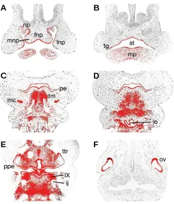
At embryonic (E) day 11.5, Tbx1 was expressed in the epithe-lium of the early facial primordia; the frontonasal process in the midline and the medial and lateral nasal processes more periph-erally, this expression being confluent with the epithelium at the base of the nasal pits (Fig. 1A). In the early stomatodium, Tbx1 was present throughout the epithelium, but was upregulated in the localised thickenings of presumptive tooth germs (Fig. 1B). More proximally, strong expression existed in the pharyngeal endo-derm and pre-muscular masses of the lateral lingual swellings and tuberculum impar of the developing tongue. Interestingly, expression was also present bilaterally in the primordial conden-sations of Meckel’s cartilage, within the first pharyngeal arch (Fig. 1C). In the more proximal regions of the early face, Tbx1 was expressed in epithelium of the early laryngeal orifice and epiglotic swelling (Fig. 1D). Expression was also strong in ectoderm of the tubotympanic recess and the pharyngeal endoderm, in addition to pharyngeal mesoderm surrounding the internal jugular vein (Fig. 1E). Further proximally, Tbx1 transcripts were strongly localised to the lateral ectoderm of the otic vesicle (Fig. 1F).
From E12.5-13.5, Tbx1 was expressed in the epithelium at the
Fig. 1. Tbx1 expression in the craniofacial region of the developing mouse embryo at E11.5 (A-F). Tbx1 is expressed in the epithelium of the developing facial processes and early oral cavity (A,B), whilst more proximally, transcripts are localised in the endoderm and mesoderm of the pharyngeal region (C-E). In the otic vesicle, expression is intense within the lateral ectoderm (F). fnp, fronto-nasal process; ij, internal jugular vein; lnp, lateral nasal process; lo, laryngeal orifice; mc, Meckel’s cartilage; mnp, medial nasal process; mp, mandibular process; np, nasal pit; ov, otic vesicle; pe, pharyngeal endoderm; ppe, pharyngeal pouch endoderm; st, stomatodeum; tg, tooth germ; tm, tongue mesoderm; ttr, tubotympanic recess; IX, inferior ganglion of glossopharyngeal nerve.
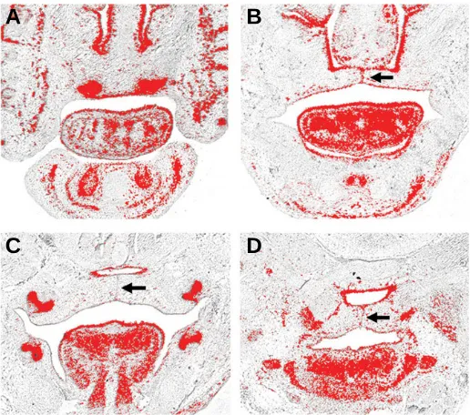
Fig. 2. Tbx1 expression in the craniofacial region of the developing mouse embryo at E12.5 (A-D) and E13.5 (E-H).Tbx1 is expressed in the epithelial component of the incisor tooth germs (A,E). In the developing palate, transcripts are expressed throughout the epithelium of the palatine shelves, with intense expression anteriorly (B,F) and on the buccal sides more proximally (C,G). In the pharyngeal regions, Tbx1 is expressed throughout the endoderm and within the mesodermal mass of the developing tongue (D,H). itg, incisor tooth germ; aps, anterior palatal shelf; mps, mid palatal shelf; tm, tongue mesoderm.
A
B
C
D
E
F
A
B
C
D
floor of the nasal cavities and became restricted to the odontoge-nic epithelium of the early tooth buds (Fig. 2A, E). In the maxilla, Tbx1 transcripts were present throughout the epithelium of the early palatine processes of the maxilla anteriorly (Fig. 2B, F). These palatine processes lie bilaterally adjacent to the develop-ing tongue and are destined to elevate and fuse in the midline with their counterpart on the opposite side. Tbx1 demonstrated low-level transcription throughout the epithelium of these shelves in the mid-portion of the palate, but was upregulated markedly on the buccal sides (Fig. 2C, G). More proximally, Tbx1 remained strong in the pharyngeal endoderm and mesoderm, with tran-scription also present in the epithelium and intrinsic musculature of the developing tongue (Fig. 2D, H).
By E14.5 the palatine shelves have elevated above the tongue and are fusing with each other in the midline and with the nasal septum superiorly. Tbx1 was expressed in the epithelium of the nasal cavity, septum and palatal shelves (Fig. 3A-D); and interest-ingly, in the mid-epithelial seam of the palate where fusion is mediated (Fig. 3B-D, arrowed). In the tongue, expression contin-ued in the epithelium and within both the intrinsic and extrinsic musculature (Fig. 3A-C), whilst strong expression was main-tained in the pharyngeal mesoderm (Fig. 3D).
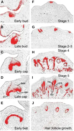
During the initiation of tooth development at E11.5, Tbx1 was strongly expressed in the localised thickenings of odontogenic epithelium that form the presumptive tooth germs (see Fig. 1B) and this expression continued in the epithelium of the early tooth bud, being marked particularly on the lingual side (Fig. 4A). At the late bud stage, Tbx1 remained strong in the lingual and basal regions of the invaginating tooth bud (Fig. 4B). The transition from bud to cap stage is marked by complex morphological changes in the tooth germ as the essential crown shape of the tooth is delineated. At the early cap stage, Tbx1 transcripts were localised to the peripheral and basal regions of the epithelial cap (Fig. 4C). During the late cap stage, differentiation of distinct cell popula-tions within the epithelial component of the tooth germ begins. Within these distinct epithelial cell populations, Tbx1 was present peripherally in the external and internal enamel epithelia and enamel knot. In contrast, internally within the stellate reticulum of the enamel organ, Tbx1 transcripts were absent (Fig. 4D). By the early bell stage, Tbx1 was restricted to the internal enamel epithelium, which is destined to form the ameloblasts responsible for enamel production (Fig. 4E).
Prior to the onset of hair follicle development in the facial region, Tbx1 expression was absent from the embryonic epider-mis, but during initiation, expression upregulated in localised regions of invaginating epithelium that form the early stage 1 epidermal placode (Fig. 4F). During stages 2-3 of hair follicle morphogenesis, as invagination of the epidermis forms the hair germ and then plug, Tbx1 expression remained strong throughout these structures (Fig. 4G). Further ingrowth of the follicular epidermis during stage 4 was accompanied by strong Tbx1 expression in the epidermal component of the early hair follicle (Fig. 4H). At stage 5, differentiation of cellular components within the hair follicle is complete and Tbx1 expression was restricted to cells of the outer root sheath that surround the future hair shaft and dermal papilla. No transcripts were detectable in the hair matrix or dermal papilla itself, but interestingly Tbx1 was upregulated in mesenchymal cells situated between the develop-ing hair follicles (Fig. 4I). Durdevelop-ing early growth of the hair follicle,
Tbx1 expression continued in the outer root sheath but downregu-lated in the surrounding mesenchyme (Fig. 4J).
DGS/VCFS subjects have a spectrum of congenital malforma-tions that are thought to arise from anomalies in development of the pharyngeal pouches (Kochilas et al., 2002, Vitelli et al., 2002a). To date, most investigations into the developmental basis of these syndromes have concentrated upon defects within the cardiovascular system (Baldini, 2002). DGS/VCFS are also asso-ciated with anomalies in the craniofacial region, principally vary-ing degrees of facial dysmorphology and cleft palate. The aetiol-ogy of these disruptions is less well understood, but the expres-sion of Tbx1 is consistent with an important role in mediating some of the complex interactions that govern early development of the facial region and its appendages. A key question is whether Tbx1 integrates into some of the currently known signalling pathways that regulate facial development.
The epithelium of the early face expresses members of several signalling molecule families, including Bone morphogenetic pro-teins (Bmp), Fibroblast growth factors (Fgf), Hedgehog, Retinoid and Wnt, which mediate many of the interactions that govern outgrowth, patterning and differentiation of the facial primordia and pharyngeal arches (Francis-West et al., 2003). Genetic links between Tbx1 transcription and signaling from several members of these families have been elucidated in several model organ-isms. Retinoic acid is important for patterning the pharyngeal arches and fronto-nasal region of the embryonic face and endog-enous levels appear to be important for normal regulation of Tbx1 (Roberts et al., 2005), a finding consistent with DGS-type defects in embryos exposed to increased (Mulder et al., 2000, Mulder et al., 1998) or reduced (Niederreither et al., 2003, Vermot et al., 2003) retinoid signaling. Fgf8 is also important during early morphogenesis and outgrowth of the facial processes and first
Fig. 3. Tbx1 expression in the craniofacial region of the developing mouse embryo at E14.5 (A-D).Tbx1 is expressed in the incisor and molar tooth germs (A,C), epithelium of the nasal cavity (A,B), epithelium of the fused palatal shelves (B-D) including the midpalatal seam (arrowed) and the pharyngeal endoderm and mesoderm (D).
A
B
pharyngeal arch (Abzhanov and Tabin, 2004, Firnberg and Neubuser, 2002, Trumpp et al., 1999). In the pharyngeal endo-derm and cardiac outflow tract, Fgf8 expression overlaps with and
Fig. 4. Tbx1 expression in the developing tooth (A-E) and hair follicle (F-J). Transcripts are present within the epithelial component of the tooth germ at the early (A) and late bud (B) stages. During the early (C) and late cap stages (D) expression is present in the external and internal enamel epithelia, being continuous at the cervical loop and enamel knot. By the early bell stage (E) expression has localised to the internal enamel epithelium. In the developing hair follicle, Tbx1 is expressed in the epithelium of the early stage 1 epidermal placode (F) and hair plug, i.e. stage 2-3 (G). During stages 4 (H) and 5 (I) of hair follicle morphogenesis, Tbx1 progressively localises to the outer root sheath of the epithelial component, with expression in the surrounding mesenchyme. In the early hair follicle (J), Tbx1 is present in the outer root sheath but has downregu-lated in the surrounding mesenchyme. cl, cervical loop; dp, dermal papilla; ek, enamel knot; hs, hair shaft; eee, external enamel epithelium; iee, internal enamel epithelium; ors, outer root sheath.
A
B
C
D
E
F
G
H
I
J
lies downstream of Tbx1, (Hu et al., 2004, Vitelli et al., 2002b); with reduced Fgf8 activity in mice leading to cardiovascular and pharyngeal defects characteristic of DGS/VCFS (Abu-Issa et al., 2002, Frank et al., 2002). The co-expression of Tbx1 with Fgf8 in the facial epithelium of the fronto-nasal process, medial and lateral nasal processes and first pharyngeal arch suggests that in DGS/VCFS subjects, facial dysmorphogenesis may be associ-ated with altered levels of Fgf8 activity in these regions.
Shh signalling is also important for facial development. Shh mutants have disruption in early patterning of the neural plate, resulting in a failure of forebrain cleavage and holoprosencephaly (Chiang et al., 1996). During later stages of facial development, Shh is essential for morphogenesis of the fronto-nasal and maxillary processes; loss of Shh signalling in these region inhibits growth, resulting in hypotelorism and cleft lip/palate, whilst ex-cess signal leads to a mediolateral widening or hypertelorism (Hu and Helms, 1999). Tbx1 expression coincides with that of Shh in the epithelium of these early facial primordia, in addition to several other regions of epithelial-mesenchymal interaction; notably the palatine processes of the maxilla, developing tooth germs and hair follicles.
Tbx1 expression is dependent upon Shh signalling in certain discreet regions of the craniofacial complex, being downregulated in the mesodermal core and lost in the pharyngeal endoderm of the pharyngeal arch system in Shh -/- mutant mice (Garg et al., 2001). In addition, in vivo evidence exists for the molecular regulation of Tbx1 in these regions via Shh-induced activity of Foxa and c-class transcription factors (Yamagishi et al., 2003). In the anterior palate, a network of genetic interaction regulates Shh transcription within the medial edge epithelium (MEE) via Msx1-maintained expression of Bmp4 within the underlying mesen-chyme and in turn, Shh-induced mesenchymal expression of Bmp2; functioning to induce cell proliferation within the palatal shelves (Zhang et al., 2002). Shh is a downstream target of Fgf10-Fgfr2b signalling within this region (Rice et al., 2004) and Fgf10 lies downstream of Tbx1 in the embryonic heart field (Hu et al., 2004, Xu et al., 2004). The expression of Tbx1 in the MEE of the anterior palatine shelves and findings that Tbx1 -/- mutant mice exhibit cleft palate (Jerome and Papaioannou, 2001, Lindsay et al., 2001, Merscher et al., 2001) provide strong indirect evidence of a role for Tbx1 during these stages of palatogenesis, possibly mediated by Shh signalling.
The highly localised expression domains of Tbx1 in the facial regions of the early embryo suggest a role in epithelial-mesenchy-mal interaction during development of the facial prominences and appendages. Existing genetic evidence from other regions of the embryo and the domains of considerable co-expression make it likely that interaction exists with Hedgehog and Fgf family mem-bers.
Experimental Procedures
Animals
CD-1 mice were time-mated and pregnant females sacrificed with cervical dislocation. Matings were set up such that noon of the day on which vaginal plugs were detected was considered as E0.5. Embryonic heads were collected at different stages (E11.5-E15.5) and fixed in 4% (w/v) paraformaldehyde at 4oC overnight. Following this, heads were washed in PBS, dehydrated through a graded series of ethanols, embedded in paraffin wax and sectioned at 7µm in preparation for in situ hybridisation.
In situ hybridisation
Radioactive section in situ hybridisation using 35S-UTP radio-labelled riboprobes was performed as previously described (Wilkinson, 1992). Tbx1 antisense riboprobes were generated from a mouse cDNA clone, a kind gift of Prof Pete Scambler (ICH, UCL, UK). Both light and dark-field images were photographed using a Zeiss Axioscope microscope and merged using Adobe Photoshop CS.
Acknowledgements
This work was funded by a European Orthodontic Society Research Fellowship (MTC) and a GKT Dental Institute Studentship (MZ).
References
ABU-ISSA, R., SMYTH, G., SMOAK, I., YAMAMURA, K. and MEYERS, E.N. (2002). Fgf8 is required for pharyngeal arch and cardiovascular development in the mouse. Development 129: 4613-25.
ABZHANOV, A. and TABIN, C.J. (2004). Shh and fgf8 act synergistically to drive cartilage outgrowth during cranial development. Dev Biol 273: 134-48.
BALDINI, A. (2002). Digeorge syndrome: The use of model organisms to dissect complex genetics. Hum Mol Genet 11: 2363-9.
BALDINI, A. (2005). Dissecting contiguous gene defects: Tbx1. Curr Op Genet Dev
15: 279-284.
CHAPMAN, D.L., GARVEY, N., HANCOCK, S., ALEXIOU, M., AGULNIK, S.I., GIBSON-BROWN, J.J., CEBRA-THOMAS, J., BOLLAG, R.J., SILVER, L.M. and PAPAIOANNOU, V.E. (1996). Expression of the t-box family genes, tbx1-tbx5, during early mouse development. Dev Dyn 206: 379-90.
CHIANG, C., LITINGTUNG, Y., LEE, E., YOUNG, K.E., CORDEN, J.L., WESTPHAL, H. and BEACHY, P.A. (1996). Cyclopia and defective axial patterning in mice lacking sonic hedgehog gene function. Nature 383: 407-13.
CHIANG, C., SWAN, R.Z., GRACHTCHOUK, M., BOLINGER, M., LITINGTUNG, Y., ROBERTSON, E.K., COOPER, M.K., GAFFIELD, W., WESTPHAL, H., BEACHY, P.A. et al.,. (1999). Essential role for sonic hedgehog during hair
follicle morphogenesis. Dev Biol 205: 1-9.
CHIEFFO, C., GARVEY, N., GONG, W., ROE, B., ZHANG, G., SILVER, L., EMANUEL, B.S. and BUDARF, M.L. (1997). Isolation and characterization of a gene from the digeorge chromosomal region homologous to the mouse tbx1 gene. Genomics 43: 267-77.
COBOURNE, M.T., HARDCASTLE, Z. and SHARPE, P.T. (2001). Sonic hedgehog regulates epithelial proliferation and cell survival in the developing tooth germ.
J Dent Res 80: 1974-9.
DASSULE, H.R., LEWIS, P., BEI, M., MAAS, R. and MCMAHON, A.P. (2000). Sonic hedgehog regulates growth and morphogenesis of the tooth. Development 127:
4775-4785.
FIRNBERG, N. and NEUBUSER, A. (2002). Fgf signaling regulates expression of tbx2, erm, pea3 and pax3 in the early nasal region. Dev Biol 247: 237-50.
FRANCIS-WEST, P.H., ROBSON, L. and EVANS, D.J.R. (2003). Craniofacial development: The tissue and molecular interactions that control development of the head. Adv Anat Emb Cell Biol 169: 1-144.
FRANK, D.U., FOTHERINGHAM, L.K., BREWER, J.A., MUGLIA, L.J., TRISTANI-FIROUZI, M., CAPECCHI, M.R. and MOON, A.M. (2002). An fgf8 mouse mutant phenocopies human 22q11 deletion syndrome. Development 129: 4591-603.
GARAVELLI, L., ZANACCA, C., CASELLI, G., BANCHINI, G., DUBOURG, C., DAVID, V., ODENT, S., GURRIERI, F. and NERI, G. (2004). Solitary median maxillary central incisor syndrome: Clinical case with a novel mutation of sonic hedgehog. Am J Med Genet A 127: 93-5.
GARG, V., YAMAGISHI, C., HU, T., KATHIRIYA, I.S., YAMAGISHI, H. and SRIVASTAVA, D. (2001). Tbx1, a digeorge syndrome candidate gene, is regulated by sonic hedgehog during pharyngeal arch development. Dev Biol
235: 62-73.
GRITLI-LINDE, A., BEI, M., MAAS, R., ZHANG, X.M., LINDE, A. and MCMAHON, A.P. (2002). Shh signalling within the dental epithelium is necessary for cell proliferation, growth and polarization. Development 129: 5323-5337.
HERRMANN, B.G., LABEIT, S., POUSTKA, A., KING, T.R. and LEHRACH, H. (1990). Cloning of the t gene required in mesoderm formation in the mouse.
Nature 343: 617-22.
HU, D. and HELMS, J.A. (1999). The role of sonic hedgehog in normal and abnormal craniofacial morphogenesis. Development 126: 4873-84.
HU, T., YAMAGISHI, H., MAEDA, J., MCANALLY, J., YAMAGISHI, C. and SRIVASTAVA, D. (2004). Tbx1 regulates fibroblast growth factors in the anterior heart field through a reinforcing autoregulatory loop involving forkhead transcription factors. Development 131: 5491-502.
JEROME, L.A. and PAPAIOANNOU, V.E. (2001). Digeorge syndrome phenotype in mice mutant for the t-box gene, tbx1. Nat Genet 27: 286-91.
KARLSSON, L., BONDJERS, C. and BETSHOLTZ, C. (1999). Roles for pdgf-a and sonic hedgehog in development of mesenchymal components of the hair follicle. Development 126: 2611-21.
KLEWER, S.E., RUNYAN, R.B. and ERICKSON, R.P. (2004). Tbx1 and the digeorge syndrome critical region. Oxford University Press, New York.
KOCHILAS, L., MERSCHER-GOMEZ, S., LU, M.M., POTLURI, V., LIAO, J., KUCHERLAPATI, R., MORROW, B. and EPSTEIN, J.A. (2002). The role of neural crest during cardiac development in a mouse model of digeorge syn-drome. Dev Biol 251: 157-66.
KOCHILAS, L.K., POTLURI, V., GITLER, A., BALASUBRAMANIAN, K. and CHIN, A.J. (2003). Cloning and characterization of zebrafish tbx1. Gene Expr Patterns
3: 645-51.
LINDSAY, E.A., VITELLI, F., SU, H., MORISHIMA, M., HUYNH, T., PRAMPARO, T., JURECIC, V., OGUNRINU, G., SUTHERLAND, H.F., SCAMBLER, P.J. et al.,. (2001). Tbx1 haploinsufficieny in the digeorge syndrome region causes
aortic arch defects in mice. Nature 410: 97-101.
MERSCHER, S., FUNKE, B., EPSTEIN, J.A., HEYER, J., PUECH, A., LU, M.M., XAVIER, R.J., DEMAY, M.B., RUSSELL, R.G., FACTOR, S. et al.,. (2001). Tbx1
is responsible for cardiovascular defects in velo-cardio-facial/digeorge syn-drome. Cell 104: 619-29.
MULDER, G.B., MANLEY, N., GRANT, J., SCHMIDT, K., ZENG, W., ECKHOFF, C. and MAGGIO-PRICE, L. (2000). Effects of excess vitamin a on development of cranial neural crest-derived structures: A neonatal and embryologic study.
Teratology 62: 214-26.
MULDER, G.B., MANLEY, N. and MAGGIO-PRICE, L. (1998). Retinoic acid-induced thymic abnormalities in the mouse are associated with altered pharyn-geal morphology, thymocyte maturation defects and altered expression of hoxa3 and pax1. Teratology 58: 263-75.
NANNI, L., MING, J.E., DU, Y., HALL, R.K., ALDRED, M., BANKIER, A. and MUENKE, M. (2001). Shh mutation is associated with solitary median maxillary central incisor: A study of 13 patients and review of the literature. Am J Med Genet 102: 1-10.
P. and DOLLE, P. (2003). The regional pattern of retinoic acid synthesis by raldh2 is essential for the development of posterior pharyngeal arches and the enteric nervous system. Development 130: 2525-34.
OBEROI, S. and VARGERVIK, K. (2005). Velocardiofacial syndrome with a single central incisor. Am J Med Genet 132: 194-197.
RICE, R., SPENCER-DENE, B., CONNOR, E.C., GRITLI-LINDE, A., MCMAHON, A.P., DICKSON, C., THESLEFF, I. and RICE, D.P. (2004). Disruption of fgf10/ fgfr2b-coordinated epithelial-mesenchymal interactions causes cleft palate. J Clin Invest 113: 1692-700.
ROBERTS, C., IVINS, S.M., JAMES, C.T. and SCAMBLER, P.J. (2005). Retinoic acid down-regulates tbx1 expression in vivo and in vitro. Dev Dyn 232: 928-38.
SARKAR, L., COBOURNE, M., NAYLOR, S., SMALLEY, M., DALE, T. and SHARPE, P.T. (2000). Wnt/shh interactions regulate ectodermal boundary formation during mammalian tooth development. Proc Natl Acad Sci USA 97:
4520-4.
SAUKA-SPENGLER, T., LE MENTEC, C., LEPAGE, M. and MAZAN, S. (2002). Embryonic expression of tbx1, a digeorge syndrome candidate gene, in the lamprey lampetra fluviatilis. Gene Expr Patterns 2: 99-103.
SCAMBLER, P.J. (2000). The 22q11 deletion syndromes. Hum Mol Genet 9:
2421-2426.
ST-JACQUES, B., DASSULE, H.R., KARAVANOVA, I., BOTCHKAREV, V.A., LI, J., DANIELIAN, P.S., MCMAHON, J.A., LEWIS, P.M., PAUS, R. and MCMAHON, A.P. (1998). Sonic hedgehog signaling is essential for hair development. Curr Biol 8: 1058-68.
TRUMPP, A., DEPEW, M.J., RUBENSTEIN, J.L., BISHOP, J.M. and MARTIN, G.R. (1999). Cre-mediated gene inactivation demonstrates that fgf8 is required for cell survival and patterning of the first branchial arch. Genes Dev 13: 3136-48.
VERMOT, J., NIEDERREITHER, K., GARNIER, J.M., CHAMBON, P. and DOLLE, P. (2003). Decreased embryonic retinoic acid synthesis results in a digeorge
syndrome phenotype in newborn mice. Proc Natl Acad Sci USA 100: 1763-8.
VITELLI, F., MORISHIMA, M., TADDEI, I., LINDSAY, E.A. and BALDINI, A. (2002a). Tbx1 mutation causes multiple cardiovascular defects and disrupts neural crest and cranial nerve migratory pathways. Hum Mol Genet 11: 915-22.
VITELLI, F., TADDEI, I., MORISHIMA, M., MEYERS, E.N., LINDSAY, E.A. and BALDINI, A. (2002b). A genetic link between tbx1 and fibroblast growth factor signaling. Development 129: 4605-11.
WILKINSON, D.G. (1992). In situ hybridisation: A practical approach. IRL Press,
Oxford. UK.
XU, H., MORISHIMA, M., WYLIE, J.N., SCHWARTZ, R.J., BRUNEAU, B.G., LINDSAY, E.A. and BALDINI, A. (2004). Tbx1 has a dual role in the morphogen-esis of the cardiac outflow tract. Development 131: 3217-27.
YAGI, H., FURUTANI, Y., HAMADA, H., SASAKI, T., ASAKAWA, S., MINOSHIMA, S., ICHIDA, F., JOO, K., KIMURA, M., IMAMURA, S. et al., (2003). Role of tbx1
in human del22q11.2 syndrome. Lancet 362: 1366-73.
YAMAGISHI, H., MAEDA, J., HU, T., MCANALLY, J., CONWAY, S.J., KUME, T., MEYERS, E.N., YAMAGISHI, C. and SRIVASTAVA, D. (2003). Tbx1 is regu-lated by tissue-specific forkhead proteins through a common sonic hedgehog-responsive enhancer. Genes Dev 17: 269-81.
ZHANG, Z., SONG, Y., ZHAO, X., ZHANG, X., FERMIN, C. and CHEN, Y. (2002). Rescue of cleft palate in msx1-deficient mice by transgenic bmp4 reveals a network of bmp and shh signaling in the regulation of mammalian palatogenesis.
Development 129: 4135-46.
Received: October 2005 Reviewed by Referees: November 2005