Original Article
Expression of regulatory genes for pancreas development
during murine embryonic stem cell differentiation
JOSUÉ K. MFOPOU
1, ERIK
WILLEMS
2, LUC LEYNS
2and LUC BOUWENS*
,11Cell Differentiation Unit (DIFF) and 2Laboratory of Cell Genetics (CEGE), Vrije Universiteit Brussel (VUB), Brussels, Belgium
ABSTRACT Insulin-producing cells derived from embryonic stem cells could be surrogates for beta cells in diabetes therapy. However, their derivation remains hard to achieve with current protocols which rely on initial embryoid body formation. We assume that factors known to inhibit pancreas development contribute to this limitation in vitro. To evaluate this hypothesis, embryoid bodies were examined after different culture periods by real time RT-PCR to profile the expression of genes known to regulate embryonic pancreas development. Our data indicate that transcripts for pancreas markers (insulin, glucagon and amylase ) were expressed during differentiation, but the highest levels achieved were at least 105 times lower than in the adult mouse pancreas. Notch signalling was activated as suggested by Delta, Jagged, Ngn3 and NeuroD1 profiles. However, Sonic hedgehog, a known inhibitor of pancreas induction in vivo drastically increased in day 6 embryoid bodies, while Inhibin βA and βB were down-regulated and follistatin up-regulated. Members of the Fibroblast- and Transforming Growth Factor families which pattern the endoderm were expressed at low levels, while those that inhibit pancreas development were highly tran-scribed. The profile of pancreas regulators expressed in embryoid bodies is therefore not compat-ible with differentiation of pancreatic and insulin-producing cells. These findings provide an explanation for the limited derivation of such cells to date, in addition to basic information for establishing novel differentiation protocols.
KEY WORDS: embryonic stem cell, pancreas development, differentiation factors, Beta cells, insulin.
Introduction
Since the establishment of the first embryonic stem cell line in 1981, the potential of their derivatives as a cell replacement in degenerative diseases is still under investigation (Evans and Kaufman, 1981; Martin, 1981). ES cell-derived insulin-producing cells are regarded as a possible means of treating diabetes and overcoming limitations imposed by scarcity of islet donors. Soria et al. (2000) investigated the potential of ES cells to develop into
insulin-producing cells and other studies claimed similar findings in mouse and human ES cells (Assady et al., 2001; Lumelsky et al.,
2001; Shiroi et al., 2002; Kahan et al., 2003). However, there is no
generally accepted and efficient paradigm for derivation of beta cells from ES cells and current protocols yield a limited fraction of immature and/or apoptotic insulin-positive cells within a heteroge-neous population (Kahan et al., 2003; Rajagopal et al., 2003;
Hansson et al., 2004; Sipione et al., 2004). Actually, such cells
have been proposed to represent immature neuron-like cells rather than beta cells. Therefore, strategies to differentiate ES cells towards a beta cell-like phenotype, preferably without the need of
*Address correspondence to: Dr. Luc Bouwens. Cell Differentiation Unit (DIFF), Diabetes Research Centre, Faculty of Medicine and Pharmacy, Vrije Universiteit Brussel (VUB). Laarbeeklaan 103, 1090 Brussels, Belgium. Fax: +32-2-477-4405. e-mail: lucbo@expa.vub.ac.be
Abbreviations used in this paper: bHLH, basic helix loop helix; EBs, embryoid bodies; ES, embryonic stem; TF, transcription factor.
0214-6282/2005/$25.00
© UBC Press Printed in Spain www.intjdevbiol.com
genetic modifications, are yet to be developed. These will rely mainly on the knowledge acquired from embryo development studies.
Pancreas develops from the foregut endoderm following three major steps: endoderm formation regulated by forkhead box fac-tors, pancreatic morphogenesis regulated by homeobox factors and differentiation of endocrine versus exocrine cells regulated by basic helix-loop-helix factors (reviewed in Chakrabarti and Mirmira, 2003). Molecules influencing endoderm layer establishment are progressively deciphered, highlighting to some extent the well-organised and complex process of pancreas morphogenesis. For instance, expression of Hedgehog factors is not compatible with pancreas initiation. Their exclusion from the presumptive pancre-atic region by notochord-derived Activin βB and Fibroblast Growth Factor 2 (Fgf2) allows initiation of a pancreatic program (Apelqvist
et al., 1997; Hebrok et al., 1998). By regulating neurogenin 3 (Ngn3
modulate a TFs cascade that control differentiation of exocrine and endocrine subsets (Gradwohl et al., 2000; Schwitzgebel et al.,
2000; reviewed in Jensen, 2004).
Expression of several pancreas/endocrine-related TFs and markers has been extensively studied during ES cell differentiation (Assady et al., 2001; Lumelsky et al., 2001; Shiroi et al., 2002;
Kahan et al., 2003; Rajagopal et al., 2003; Hansson et al., 2004;
Sipione et al., 2004). Forced expression of key endocrine TFs
(Ngn3, Pax4 ) in teratocarcinoma or ES cell lines didn’t entirely
reinforce endocrine differentiation (Blyszczuk et al., 2003; Vetere et al., 2003). However, very little is known of the expression of
extracellular molecules that also regulate pancreas development. Nonetheless, a recent examination of the rare population of pan-creatic lineages spontaneously differentiated from ES cells showed reproduction of many features of normal islet differentiation (Kahan
et al., 2003).
It is undoubtedly that the gene expression program activated early after EBs initiation plays a critical role in the selection of particular developmental pathways by ES cells. Therefore, soluble molecules known to regulate pancreas development in vivo might
be able to induce or to suppress islet cell differentiation when expressed in vitro by differentiating ES cells. To our knowledge, no
previous study using the in vitro system of ES cell differentiation
thoroughly examined the components of major pathways that (negatively) influence early pancreas formation. To test our hy-pothesis that pancreas inhibitors limit acquisition of pancreatic fate in early EBs, we cultured EBs in non insulin-supplemented condi-tions and screened for mRNA profiles of genes regulating early pancreas development.
Results
On morphological examination, early stage EBs showed an outer endoderm and an underlying primitive ectoderm cell layer (Martin, 1981) surrounding a cavity with some apoptotic cells (Fig. 1A: image of a whole EB on upper panel, section of paraffin embedded EB on lower panel). Late EBs displayed cells with various phenotypes representing – as expected – the three germ layers of mammalian development (not shown).
Expression of endoderm and pancreas specific markers
We sought to confirm expression of marker genes for endoderm and more specifically pancreatic tissues. Transcripts of alpha-fetoprotein were absent in ES cells and early EBs, but were
identified later on (peak around day 22) and the protein was localized in the endoderm layer (Figs. 1B, 2A). Similarly, albumin
mRNA was detected in EBs and the protein was located in a subset of endoderm cells (Figs. 1B, 2B). Contrary to EBs, undifferentiated
ES cells were negative or showed low mRNA levels of alpha-1 antitrypsin, intestinal fatty acid binding protein and caudal homeobox gene 2. The epithelial and gut endoderm marker indian hedgehog
(Ihh ) was present in ES cells at a moderate level and increased
thereafter. In contrast, sonic hedgehog (Shh ) was detected in EBs,
but not in undifferentiated ES cells. Similarly, the endoderm marker
Sox17 was not expressed by ES cells, but only low levels could be
detected in EBs (Fig. 1B and data not shown). Furthermore, transcripts for MyoR, a bHLH TF repressor of definitive endoderm
differentiation were detected at high levels (Fig. 1B), suggesting that only a limited definitive endoderm population arose from these cultures.
The beta cell specific marker insulin1 was detected only at 3
weeks culture, whereas insulin2 was expressed from the second
week and increased towards day 29. Amylase, glucagon and islet amyloid polypeptide (Iapp ) were detected in ES cells and showed
a substantial increase afterwards (Fig. 1C). Maximal expression levels of these markers were very low compared to adult mouse pancreas levels, namely 10-8 to 10-5 fold by real time PCR. By immunocytochemistry, few insulin-positive cells (<0.1%) were detected by the end of week 3, but glucagon-immunoreactive cells were more frequently identified, consistent with the higher signal in RT-PCR (Figs. 1C, 2C-D). Since we were not interested in showing
Fig. 1. EB morphology and expression of endoderm and pancreas markers. Four to 6 days old EBs show well organised endoderm and primitive ectoderm layers surrounding a cavity both on whole EB (upper panel) and on paraffin section (lower panel). Original magnifications x200
(A). Several markers expressed by primitive and definitive endoderm are absent in ES cells and abundant in EBs, except for the definitive endoderm specific marker Sox17 (B). Pancreas markers are expressed at various levels and stages of differentiation, but remain very low compared to adult mouse pancreas (10-8 to 10-5 fold by real time RT-PCR; (C)). N, no template
control; P, adult mouse pancreas.
A
B
that true beta cells differentiate from ES cells but that insulin-producing cells are very rare, we didn’t further characterise these cells by C-peptide detection for instance.
Expression profiling of regulatory genes for pancreas devel-opment
The restricted differentiation of pancreatic cells in this system is concordant with the literature and prompted us to critically analyze the profile of major regulatory pathways for pancreas development known from mouse embryo.
Hedgehog pathway
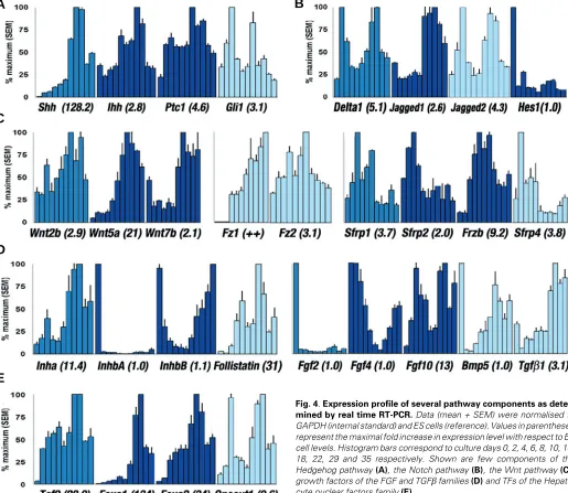
We detected no Shh transcripts in ES cells and early EBs. As
from day 6, Shh was expressed with gradual increase to a pinnacle
at day 18 (128 fold increase). In contrast, Ihh was expressed in ES
cells and increased further up to day 18 (2.8 fold). The hedgehog receptors Ptc1 and Ptc2, as well as the membrane protein Smo
were all expressed in ES cells and EBs at high levels. Intracellular effectors were present in ES cells and remained so (Gli2 ) or
increased (Gli1 ; 3.1 fold at day 4) after EBs initiation. The
fluctuations of these components paralleled those of the hedgehog molecules (Figs. 3A, 4A).
Considering the very early in vivo requirement for Shh
repres-sion during embryonic pancreas initiation, we sought to confirm the expression of this pancreas inhibitor at the protein level. Indeed, we found a widespread Shh expression in embryoid bodies, encom-passing both the outer and the inner cells in many of them (Fig. 2 E-F).
Notch pathway
Several components of the Notch pathway were detected in ES cells, of which some further increased in EBs. Notch1, -2 and -3
transcripts showed minor increase from day 2 (2.9 fold for Notch1) and did not significantly change thereafter. Notch4, expressed at
lower levels than other receptors displayed the major variation with down regulation through week 3 (Fig. 3B). Notch ligands Delta1, Jagged1 and Jagged2, which are known regulators of pancreas
development were all induced in EBs (2.6 to 5 folds). The relative level of Delta3 remained low compared to other ligands and the
initial increase was followed by down regulation through weeks 3 to 5 (Figs. 3B, 4B). We also found a sustained expression of Notch antagonists Hes6 and Sel -1l (Fig. 3B).
Wnt pathway
We measured the expression level of a set of Wnt members
recently described in the developing pancreas. Our data showed the expression of the Wnt receptor Fz2 at all stages of
differentia-tion with a peak at day 14 (3.1 fold), while Fz1 was absent from ES
or early EBs up to day 6. The three members of the Wnt family (Fz ligands) described in mouse embryonic pancreas were low in early phases of culture and increased substantially only after day 14 (Wnt2b, 2.9 fold; Wnt5a, 21 fold; Wnt7b, 2.1 fold). In contrast, most
of the secreted antagonists (Sfrp1, Sfrp2, Frzb, Sfrp4 ) showed
higher levels in the earlier phases of cultures. These patterns suggest a reduced activity of Wnt signals described in mouse embyonic pancreas during the initiation of EBs cultures. (Fig. 4C).
Growth factors of the FGF and TGFβββββ families
During embryonic development, notochord-derived growth
fac-tors (Activin βB and FGF2) repress Shh in the dorsal pancreatic
anlagen; in vitro they activate Pdx1 expression from the isolated
dorsal pancreatic epithelium. Since Activins are homo- or het-erodimers of inhibin α, βA and βB (Inha, InhbA, InhbB ), we
examined the expression of the inhibins to deduce the Activin profile. ES cells showed minute amounts of Inha and InhbA
subunits, but high levels of InhbB, suggesting a higher amount of
Activin βB compared to Activin βA, Inhibin A or B. Upon EBs formation, Inha followed a biphasic pattern with peaks at days 4
and 22, corresponding to a 3.8 and 11.4 fold respectively. InhbA,
which was already low in ES cells was further down regulated (40 to 50 times) and showed a slight increase during the last 3 weeks.
InhbB gradually decreased to a barely detectable level at days 8
to 10 and was then up-regulated to its initial levels in late EBs (Figs. 3C, 4D). Follistatin is a potent inhibitor of Activin/TGFβ family members. ES cells expressed its transcripts, which increased
Fig. 2. Immunodetection of endoderm proteins, pancreatic hormones and Shh. Alpha-fetoprotein was confined to the endoderm (A) as well as Albumin detected in subsets of endodermal cells (B). Though very rare, Insulin-positive cells were mainly detected after 3 weeks of culture (C) as well as Glucagon-positive cells (D). The signaling protein Shh was identi-fied in the endoderm and ectoderm layers of many EBs (E). (F) Negative control. Scale bar, 100 µm (A-D) and 200 µm (E-F).
A
B
C
D
A
B
C
D
akin to that of Pdx1. The pattern of Pax6 and Mist1 mimicked that
of Nkx6.1, whereas Ptf1a-p48 was only barely detected from day
14 (Fig. 3D).
Discussion
The limited derivation of insulin-producing cells from ES cells in the framework of published protocols is progressively docu-mented (Kahan et al., 2003; Rajagopal et al., 2003; Hansson et al., 2004; Sipione et al., 2004). Although in EBs definitive
endo-derm lineages are overwhelmed by preferential ecto- and meso-dermal differentiation, most studies relied on the initial formation of these embryo-like structures. Only recently, enhanced deriva-tion of definitive endoderm from Activin-treated embryoid bodies was reported (Kubo et al., 2004). Expression of the analysed
endoderm markers followed trends extensively described in lit-erature, but they are not exclusively expressed by definitive endoderm that still lacks a specific marker not expressed else-where (Abe et al., 1996; Hamazaki et al., 2001; Kubo et al., 2004).
Nevertheless, the patterns of Sox17 and MyoR point to a limited
population of definitive endoderm cells contributing to EBs (Kubo
et al., 2004; Yu et al., 2004). In the first part of this study, we
confirmed EBs expression of pancreatic markers by quantitative RT-PCR and immunocytochemistry. As expected, very few cells express insulin or glucagon and the maximum transcripts levels
achieved were 105 times lower compared to normal mouse pancreas, suggesting that a meaningful differentiation strategy
Fig. 3. Expression of genes thought to control pancreas develop-ment. Analysis focussed on the signalling components and TFs involved
upon differentiation up to 31 fold by day 18.
Among the bone morphogenetic proteins evaluated in our system, Bmp6 and -7 were highly expressed in ES cells and did
not considerably change thereafter. Bmp5 showed a transient
shut-off at days 2-4 (30 fold decrease; Figs. 3C, 4D).
Fgf2 mRNA was detected in ES cells and substantially
decreased upon differentiation with a nadir at day 8 (50 fold lower), whereas intermediate levels of Fgf4 transcripts were
identified with a profile similar to InhbB. Fgf10 increased from its
high level in ES cells, showing two peaks at days 4 (11.7 fold) and 22 (13.1 fold; Fig. 4D).
TFs expressed by pancreatic progenitor cells
To complete the analysis of the aforementioned extrinsic regulators, we further dissected the profile of TFs expressed by pancreatic progenitors. Of the 4 hepatocyte nuclear factors tested, the expression of Tcf2, Foxa1 and Foxa2 gradually increased to
a pinnacle at day 18-22 representing 28, 134 and 34 fold respec-tively (Figs. 3D, 4E). Onecut1 (α and β) profile was biphasic with peaks at days 4 and 22 (9 and 9.6 fold). The pro-endocrine gene
Ngn3 was expressed in ES cells at hardly detectable levels. An
increase in its expression was noticed early after EBs initiation (day 2 to 6), but was rapidly followed by a gradual decrease. Likewise, its target NeuroD1 showed the same pattern with a
slower kinetic (Fig. 3D). ES cells expressed high levels of Nkx6.1,
which was gradually repressed during the initial two weeks. In contrast, Nkx2.2 increased gradually, showing an overall profile
A
B
C
D
E
Fig. 4. Expression profile of several pathway components as deter-mined by real time RT-PCR. Data (mean + SEM) were normalised to GAPDH (internal standard) and ES cells (reference). Values in parentheses represent the maximal fold increase in expression level with respect to ES cell levels. Histogram bars correspond to culture days 0, 2, 4, 6, 8, 10, 14, 18, 22, 29 and 35 respectively. Shown are few components of the Hedgehog pathway (A), the Notch pathway (B), the Wnt pathway (C), growth factors of the FGF and TGFβ families (D) and TFs of the Hepato-cyte nuclear factors family (E).
should increase these markers by at least 1000 fold to represent 1% of pancreas levels. Selection of EBs harbouring dithizon-positive clusters (Shiroi et al., 2002) did not significantly enrich for insulin mRNA (unpublished observations).
Following the hypothesis that inhibitory pancreas regulators are expressed during EBs cultures, we aimed at establishing a comprehensive and descriptive profile of those pathways so as to build the ground for further work towards pancreatic cell differen-tiation from ES cells. We therefore screened the expression profile of components of major pathways known to influence embryonic pancreas development over a 5 week-period. In this screening, we focused on extracellular molecules, most of which are soluble and known to have effects at long distance from their site of expression. Transcripts of Shh were absent from ES cells,
but were highly induced in the first week of culture. As Shh is a
diffusible potent inhibitor of pancreas induction, it is very likely that
its high expression by EBs limits the acquisition of pancreatic fate in ES cell differentiation (Apelqvist et al., 1997, Hebrok et al.,
1998). On the other hand, hedgehog signals favour the develop-ment of neurons, dermomyotome, sclerotome and liver at the expense of pancreas and the pattern of its components explains the relative abundance of neuroectodermal cell types as men-tioned before (Wichterle et al., 2002; Loebel et al., 2003; Maye et al., 2004). This suggests that the mandatory Shh repression in
embryonic endoderm should be reproduced in EBs to allow patterning to pancreatic lineages.
Induction of Notch ligands in the presence of their receptors points to a functional Notch pathway in EBs. Indeed, the Notch ligands known to participate in pancreas development (Delta1, Jagged1, Jagged2 ) where found up-regulated between 2 to 5 fold
and the Notch target Hes1 was initially repressed 8-fold at day
biphasic profile that somewhat followed Delta1, Delta3 and Jagged2
patterns. Although Hes1 shows a small response to Notch signals
in some tissues, the initial down-regulation suggests a tight control of the pathway by Notch antagonists (Notch3, Hes6 and Sel-1l)
(Donoviel et al., 1998; Bae et al., 2000). The secondary
down-regulation of Ngn3 and NeuroD1 was likely coupled to Hes1
activity, revealing a prolonged Notch signalling in EBs. In vivo,
Notch signalling defects accelerate endocrine differentiation while constitutively active Notch1 represses both endocrine and
exo-crine development (Apelqvist et al., 1999; Hald et al., 2003; Hart et al., 2003; Norgaard et al., 2003). As demonstrated in vivo, a
sustained Notch activity in EBs would impede the differentiation of tissues such as pancreas that rely on the attenuation of this pathway.
Wnt signalling was recently suggested to participate in
pan-creas development, based on the expression of several Wnt
genes, receptors and antagonists in the developing and adult pancreas (Heller et al., 2002, Heller et al., 2003). Although
convinc-ing data are still lackconvinc-ing on the requirement of Wnt signals during
initiation of pancreatic buds in vivo and on its exact role in pancreas
development, our expression profiling suggests that if those Wnt genes are required for the pancreatic cells to develop, this role is not achieved in EBs system as they are not consistently expressed during the major patterning events.
Activin βB and Fgf2 repress endodermal Shh, although this
exclusion is not sufficient to induce pancreatic genes outside the presumptive anlagen. Patterning of the early endoderm by Fgf4 is necessary to define a window of competence to pancreatic fate (Hebrok et al., 1998; Wells and Melton, 2000; Kumar et al., 2003).
Expression patterns of Inhibin in our EBs compare to that found by
Albano et al. (1993) and illustrate a decrease of Activins that would
otherwise repress Shh. Indeed, Shh expression in EBs suggests
that its ordinary repression by Fgf2/Activin βB signals was not effective and this is consistent with the observed profiles of both molecules in our study. Furthermore, the follistatin profile was
compatible with the complete inhibition of the low levels of Activins achieved in these cultures (de Winter et al., 1996). Activin was
recently reported to induce definitive endoderm from ES cells; therefore the observed down regulation of InhbA and InhbB and
up-regulation of follistatin upon EBs formation also explains the
limited differentiation into endoderm tissues (Kubo et al., 2004).
Considering the profiles and effects of FGF and TGFβ family members in endoderm/pancreas development, we presume that their combined supplementation would be one option for improving the pancreatic fate in differentiating ES cells; especially because EBs treatment with Fgf2 alone induced meso- and ectodermal derivatives, while exposure to cyclopamine or Activin A increased insulin by only 2-4 fold (Schuldiner et al., 2000; Skoudy et al.,
2004). Similarly, the blocking of Bmp6 could be associated in this
strategy as its expression under Pdx1 promoter leads to complete
pancreas agenesis (Dichmann et al., 2003). The call for this
concomitant modulation of several pathways is even made impera-tive by data from Léon-Quinto et al. (2004) showing significant
increase in insulin-producing cells (19%) only after EBs co-culture with pancreatic rudiments, but not upon application of anti-Shh antibody.
The majority of TFs expressed by pancreas progenitors were detected, however the expression of these factors is not a bona fide
illustration of pancreatic differentiation within heterogeneous EBs
since many of them are shared with neuroectoderm. The selection of the mesendoderm progenitor from early EBs might help to resolve this issue (Kubo et al., 2004). During development, islet
progenitors arise from Ptf1a-p48 +/Pdx1 + cells through removal of
repressive and stimulation of inductive pathways, a phenomenon unlikely to occur in EBs considering the low Ptf1a-p48 expression
profile (at least 105 times lower compared to the mouse pancreas). As ES cell lines and the differentiation protocols used by several groups may differ considerably, it remains unclear whether ob-served insulin-positive cells arise from such progenitors, because of the contradictory data on p48 expression (barely detected in this
study; not detected by Kahan et al. (2003); easily detected by
Moritoh et al. (2003) and by Skoudy et al. (2004). Nevertheless,
early pancreatic epithelium bears insulin and glucagon cells in the absence of Pdx1 (Ahlgren et al., 1996), a phenomenon that might
characterize immature pancreatic cells and that cannot be ruled out in the case of Ptf1a-p48 on the basis of present knowledge.
In conclusion, we examined the mRNA profile of extracellular pancreas regulatory factors in embryoid bodies in order to assess the actual limited ES cell differentiation into insulin-producing cells. Our data indicate that pancreas fate acquisition is blocked at several stages within EBs starting from definitive endoderm. Al-though we used a crude but highly sensitive approach that provides no information on cell types expressing the mRNA, or their actual translation into proteins, it first of all allows screening for useful targets that can be further confirmed and modulated in cultures. The generation of beta cells from ES cells might benefit from these observations through implementation of strategies that integrate, as we are currently testing, the inhibition of suppressive pathways (Shh, Notch, Bmp6, Fgf10) and supplementation of defective inductive factors (Activin, Fgf2, Fgf4, Wnt) with the ultimate goal of reproducing the features of normal development.
Materials and Methods
ES cells and EB cultures
The E14 ES cell line derived from 129P2/Ola mice was used in this study. Mitomycin C (Sigma-Aldrich, St Louis - USA) inactivated STO cells were plated on gelatine-coated tissue culture dishes and used as feeder layers. ES cells were expanded on feeders and passaged every 2 to 3 days (following trypsin dissociation and separation from feeder cells by a short passage on gelatine-coated dishes). Expansion medium consisted of knockout DMEM (Gibco, California - USA) supplemented with 15% knock-out serum replacement (Gibco, California - USA), L-glutamine (Sigma-Aldrich, St Louis - USA), non-essential amino acids (Sigma-Aldrich), beta-mercaptoethanol (Sigma-Aldrich), penicillin-streptomycin (Sigma-Aldrich) and 1000U/ml leukaemia inhibitory factor (LIF, Sigma-Aldrich).
EBs were initiated by dissociating ES cells and removing feeders, then plating in suspension culture dishes in the absence of LIF. The differentia-tion medium consisted of DMEM supplemented with 15% FCS (PAA, Pasching - Austria), L-glutamine, non-essential amino acids, beta-mercap-toethanol and penicillin-streptomycin. At day 4 of culture, EBs were transferred to the medium of same composition, but lower glucose concen-tration (6.25mM). As from day 8, cultures were supplemented with 10mM nicotinamide (Merck, Darmstadt - Germany) and maintained for 4 more weeks, the medium being renewed every other day.
Immunocytochemistry and immunofluorescence
Embryoid bodies were harvested at several time points (day 8, 14, 22, 29 and 35) and fixed in 4% formol for 90 min, then rinsed in PBS and
processed for paraffin embedding. Staining was carried out on 5 µm
incubated overnight at 4°C with a primary antibody. The primary antibodies used were monoclonal mouse anti-insulin (1/2000, Sigma-Aldrich, St Louis, USA), rabbit anti-glucagon (1/3000, Dr. C. Van Schravendijk, Vrije Universiteit Brussel, Belgium), rabbit anti-alpha fetoprotein (1/200, Dako, Glostrup, Denmark), sheep anti-albumin (1/100, Biogenesis, Poole, En-gland) and goat anti-sonic hedgehog (1/200, Santa Cruz, California, USA). After rinsing in PBS, slides were incubated at room temperature for 1h with the appropriate secondary antibody and further developed when appli-cable. For negative control slides, the primary antibody was omitted. Slides were examined on a Leica microscope and pictures taken with an Axiocam cold camera using AxioVision software.
Classical RT-PCR analysis
Total RNA was extracted from EBs using TRIzol (Gibco, California -USA) and following the manufacturer’s recommendations. cDNA was
synthesised from 1 µg DNase-treated RNA with random hexamers and an
80ng equivalent used in each PCR set-up. All cDNA synthesis and PCR reaction components were from Invitrogen (California - USA). Whenever possible, intron-spanning primers were designed from published mouse sequences (EMBL) and were BLASTed against the nucleotide databank to check for their specificity. The complete list and sequences of primers are available upon request. All reactions underwent a standard amplifica-tion program for 28-35 cycles. PCR products were analysed on 1.2% agarose gel and images acquired with a UV sensitive camera. Three independent experiments were carried out from three different ES cell aliquots and the expression profile of target genes found to be reproduc-ible.
Real time PCR analysis
Selected target genes from each pathway that already indicated sharp and interesting differences upon classical PCR were further analyzed by real time PCR in order to quantify their expression level. For this purpose, cDNA was prepared from 500 ng of total RNA following DNase treatment and 10 ng RNA equivalent used for PCR with specific primers (previously tested and confirmed on gel) in the presence of SYBR Green I. A melt curve analysis was performed at the end of each reaction. Data (mean + SEM) are from triplicate runs of one samples set representative of the three independent experiments. Expression levels were normalized to individual GAPDH (internal control) and to undifferentiated ES cells (reference). The profile was obtained by plotting relative expression levels as a percentage of the maximum value. For pancreatic gene expression analysis, 150 ng RNA equivalent was used (Fig.1C, Ptf1a-p48 in Fig. 3D) and adult mouse pancreas taken as reference.
Acknowledgements
MKJ and WE are supported by the Horizontale Onderzoekactie of the VUB. Special thanks to De Blay E. and Guns Y. for their technical skills and to Pipeleers D.G. for logistic support. Our gratitude also goes to Rooman I., Lardon J., De Breuck S. and Baeyens L. for their comments and criticisms. Grants from VUB Horizontale Onderzoekactie, Fonds voor Wetenschappelijk Onderzoek - Vlaanderen and European Union.
References
ABE, K., NIWA, H., IWASE, K., TAKIGUCHI, M., MORI, M., ABÉ, S.I., ABE, K. and YAMAMURA, K.I. (1996). Endoderm-specific gene expression in embryonic stem cells differentiated to embryoid bodies. Exp. Cell Res. 229: 27-34.
AHLGREN, U., JONSSON, J. and EDLUND, H. (1996). The morphogenesis of the pancreatic mesenchyme is uncoupled from that of the pancreatic epithelium in IPF1/PDX1-deficient mice. Development 122: 1409-1416.
ALBANO, R.M., GROOME, N. and SMITH, J.C. (1993). Activins are expressed in preimplantation mouse embryos and in ES and EC cells and are regulated on their differentiation. Development 117: 711-723.
APELQVIST, A., AHLGREN, U. and EDLUND, H. (1997). Sonic hedgehog directs specialised mesoderm differentiation in the intestine and pancreas. Curr. Biol.
7(10): 801-804.
APELQVIST, A., LI, H., SOMMER, L., BEATUS, P. ANDERSON, D.J., HONJO, T., HRABE DE ANGELIS, M., LENDAHL, U. and EDLUND, H. (1999). Notch signalling controls pancreatic cell differentiation. Nature 400: 877-881.
ASSADY, S., MAOR, G., AMIT, M., ITSKOVITZ-ELDOR, J., SKORECKI, K.L. and TZUKERMAN, M. (2001). Insulin production by human embryonic stem cells.
Diabetes 50: 1691-1697.
BAE, S.K., BESSHO, Y., HOJO, M. AND KAGEYAMA, R. (2000). The bHLH gene Hes6, an inhibitor of Hes1, promotes neuronal differentiation. Development
127: 2933-2943
BLYSZCZUK, P., CZYZ, J., KANIA, G., WAGNER, M., ROLL, U., ST-ONGE, L. and WOBUS, A.M. (2003). Expression of Pax4 in embryonic stem cells promotes differentiation of nestin-positive progenitor and insulin-producing cells. Proc. Natl. Acad. Sci. USA 100: 998-1003.
CHAKRABARTI, S.K. and MIRMIRA, R.G. (2003). Transcription factors direct the development and function of pancreatic β cells. Trends Endocrinol. Metab.
14(2): 78-84.
DE WINTER, J.P., TEN DIJKE, P., DE VRIES, C.J., VAN ACHTERBERG, T.A., SUGINO, H., DE WAELE, P., HUYLEBROECK, D., VERSCHUEREN, K. and VAN DEN EIJNDEN-VAN RAAIJ, A.J. (1996) Follistatins neutralize activin bioactivity by inhibition of activin binding to its type II receptors. Mol. Cell. Endocrinol.116: 105-114.
DICHMANN, D.S., MILLER, C.P., JENSEN, J., HELLER, R.S. and SERUP, P. (2003). Expression and misexpression of members of the FGF and TGFβ families of growth factors in the developing mouse pancreas. Dev. Dyn. 226:
663-674.
DONOVIEL, D.B., DONOVIEL, M.S., FAN, E., HADJANTONAKIS, A. and BERNSTEIN A. (1998). Cloning and characterization of Sel-1l, a murine homolog of the C. elegans Sel-1 gene. Mech. Dev. 78: 203-207.
EVANS, M.J. and KAUFMAN, M.H. (1981). Establishment in culture of pluripotential cells from mouse embryos. Nature 292:154-156.
GRADWOHL, G., DIERICH, A., LEMEUR, M. and GUILLEMOT, F. (2000). Neurogenin3 is required for the development of the four endocrine cell lineages of the pancreas. Proc. Natl. Acad. Sci. USA 97: 1607-1611.
HALD, J., HJORTH, J.P., GERMAN, M.S., MADSEN, O.D., SERUP, P. and JENSEN, J. (2003). Activated Notch1 prevents differentiation of pancreatic acinar cells and attenuate endocrine development. Dev. Biol. 260(2): 426-437.
HAMAZAKI, T., IIBOSHI, Y., OKA, M., PAPST, P.J., MEACHAM, A.M., ZON, L.I. and TERADA, N. (2001). Hepatic maturation in differentiating embryonic stem cells in vitro. FEBS Lett. 497: 15-19.
HANSSON, M., TONNING, A., FRANDSEN, U., PETRI, A., RAJAGOPAL, J., ENGLUND, M.C., HELLER, R.S., HAKANSSON, J., FLECKNER, J., SKOLD, H.N., MELTON, D., SEMB, H. and SERUP P. (2004). Artifactual insulin release from differentiated embryonic stem cells. Diabetes 53: 2603-2609.
HART, A., PAPADOPOULOU, S. and EDLUND, H. (2003). FGF10 maintains notch activation, stimulates proliferation and blocks differentiation of pancreatic epithelial cells. Dev. Dyn. 228: 185-193.
HEBROK, M., KIM, S.K. and MELTON, D.A. (1998). Notochord repression of endodermal sonic hedgehog permits pancreas development. Genes Dev. 12:
1705-1713.
HELLER, R.S., DICHMANN, D.S., JENSEN, J., MILLER, C., WONG, G., MADSEN, O.D. and SERUP, P. (2002). Expression patterns of Wnts, Frizzleds, sFRPs and
misexpression in transgenic mice suggesting a role for Wnts in pancreas and foregut pattern formation. Dev. Dyn. 225: 260-270.
HELLER, R.S., KLEIN, T., LING, Z., HEIMBERG, H., KATOH, M., MADSEN, O.D. and SERUP, P. (2003). Expression of Wnt, frizzled, sFRP and DKK genes in adult human pancreas. Gene Expr. 11: 141-147.
JENSEN, J. (2004). Gene regulatory factors in pancreas development. Dev. Dyn.
229: 176-200.
KAHAN, B.W., JACOBSON, L.M., HULLETT, D.A., OCHOADA, J.L., OBERLEY, T.D., LANG, K.M. and ODORICO, J.S. (2003). Pancreatic precursors and differentiated islet cell types from murine embryonic stem cells: an in vitro model to study islet differentiation. Diabetes 52: 2016–2024.
KUMAR, M., JORDAN, N., MELTON, D.A. and GRAPIN-BOTTON, A. (2003). Signals from lateral plate mesoderm instruct endoderm toward a pancreatic fate. Dev. Biol. 259: 109-122.
LÉON-QUINTO, T., JONES, J., SKOUDY, A., BURCIN, M. and SORIA, B. (2004). In vitro directed differentiation of mouse embryonic stem cells into insulin-producing cells. Diabetologia 47: 1442-1451.
LOEBEL, D.A.F., WATSON, C.M., DE YOUNG, R.A. and TAM, P.P.L. (2003). Lineage choice and differentiation in mouse embryos and embryonic stem cells.
Dev. Biol. 264: 1-14.
LUMELSKY, N., BLONDEL, O., LAENG, P., VELASCO, I., RAVIN, R. and MCKAY, R. (2001). Differentiation of embryonic stem cells to insulin-secreting structures similar to pancreatic islets. Science 292: 1389-1394.
MARTIN, G.R. (1981). Isolation of a pluripotent cell line from early mouse embryos cultured in medium conditioned by teratocarcinoma stem cells. Proc. Natl. Acad. Sci. USA 78: 7634-7638.
MAYE, P., BECKER, S., SIEMEN, H., THORNE, J., BYRD, N., CARPENTINO, J. and GRABEL, L. (2004). Hedgehog signaling is required for the differentiation of ES cells into neuroectoderm. Dev. Biol. 265(1): 276-290.
MORITOH, Y., YAMATO, E., YASUI, Y., MIYAZAKI, S. and MIYAZAKI, J. (2003). Analysis of insulin-producing cells during in vitro differentiation from feeder-free embryonic stem cells. Diabetes 52: 1163-1168.
NORGAARD, G.A., JENSEN, J.N. and JENSEN, J. (2003). FGF10 signaling maintains the pancreatic progenitor cell state revealing a novel role for Notch in organ development. Dev. Biol. 264: 323-338.
RAJAGOPAL, J. ANDERSON, W.J., KUME, S., MARTINEZ, O.L. and MELTON, D.A. (2003). Insulin staining of ES cells progeny from insulin uptake. Science
299: 363.
SCHULDINER, M., YANUKA, O., ITSKOVITZ-ELDOR, J., MELTON, D.A. and BENVENISTY, N. (2000). Effects of eight growth factors on the differentiation of cells derived from human embryonic stem cells. Proc. Natl. Acad. Sci. USA
97(21): 11307-11312.
SCHWITZGEBEL, V.M., SCHEEL, D.W., CONNERS, J.R., KALAMARAS, J., LEE, J.E. ANDERSON, D.J., SUSSEL, L., JOHNSON, J.D. and GERMAN, M.S.
(2000). Expression of neurogenin3 reveals an islet cell precursor population in the pancreas. Development 127: 3533-3542.
SHIROI, A., YOSHIKAWA, M., YOKOTA, H., FUKUI, H., ISHIZAKA, S., TATSUMI, K. and TAKAHASHI, Y. (2002). Identification of insulin-producing cells derived from embryonic stem cells by zinc-chelating dithizone. Stem Cells 20: 284-292.
SIPIONE, S., ESHPETER, A., LYON, J.G., KORBUTT, G.S. and BLEACKLEY, R.C. (2004). Insulin expressing cells from differentiated embryonic stem cells are not beta cells. Diabetologia 47: 499-508.
SKOUDY, A., ROVIRA, M., SAVATIER, P., MARTIN, F., LEÓN-QUINTO, T., SORIA, B. and REAL, F.X. (2004). Transforming growth factor (TGF)β, fibro-blast growth factor (FGF) and retinoid signalling pathways promote pancreatic exocrine gene expression in mouse embryonic stem cells. Biochem. J. 379:
749-756.
SORIA, B., ROCHE, E., BERNÁ, G., LEÓN-QUINTO, T., REIG, J.A. and MARTÍN, F. (2000). Insulin-secreting cells derived from embryonic stem cells normalize glycemia in streptozotocin-induced diabetic mice. Diabetes 49: 157-162.
VETERE, A., MARSICH, E., DI PIAZZA, M., KONCAN, R., MICALI, F. and PAOLETTI, S. (2003). Neurogenin3 triggers β-cell differentiation of retinoic acid-derived endoderm cells. Biochem. J. 371: 831–841.
WELLS, J.M. and MELTON, D.A. (2000). Early mouse endoderm is patterned by soluble factors from adjacent germ layers. Development 127: 1563-1572.
WICHTERLE, H., LIEBERAM, I., PORTER, J.A. and JESSELL, T.M. (2002). Directed differentiation of embryonic stem cells into motor neurons. Cell 110(3):
385-397.
YU, L., SANGSTER, N., PEREZ, A. and MCCORMICK, P.J. (2004). The bHLH protein MyoR inhibits the differentiation of early embryonic endoderm. Differen-tiation 72(7): 341-347.
Received: March 2005
Reviewed by Referees: May 2005