Conservation and non-conservation of genetic pathways in
eye specification
AMY L. DONNER and RICHARD L. MAAS*
Division of Genetics, Department of Medicine, BWH and HMS, Boston, MA, USA
ABSTRACT In this review we highlight two genetic pathways important for eye morphogenesis that are partially conserved between flies and vertebrates. Initially we focus on the ey paradigm and establish which aspects of this genetic hierarchy are conserved in vertebrates. We discuss experiments that evaluate the non-linear relationship amongst the genes of the hierarchy with a concentration on vertebrate functional genetics. We specifically consider the Six genes and their relationship to sine oculis, as tremendous amounts of new data have emerged on this topic. Finally, we highlight similarities between Shh/Hh directed morphogenesis mediated by basic Helix-Loop-Helix factors in vertebrate retinal cell specification and in specification of fly photoreceptors.
KEY WORDS:
bHLH gene, eyeless, lens, neural retina, Pax6
0214-6282/2004/$25.00 © UBC Press
Printed in Spain www.ijdb.ehu.es
*Address correspondence to: Dr. Richard L. Maas. Harvard Medical School-NRB 458H, 77 Louis Pasteur Avenue, Boston, MA 02115, USA. Fax: +1-617-525-4751. e-mail: [email protected]
Abbreviations used in this paper: AEL, anterior epithelial layer; Ato, atonal; bHLH, basic helix-loop-helix transcriptional regulator; dac, dachshund; Dpp, decapentaplegic; E, embryonic day; ey, eyeless; eyg, eyegone; eya eyes absent; GCL, ganglion cell layer; Hh, hedgehog; INL, inner nuclear layer; LP, lens placode; MF, morphogenetic furrow; NR, neural retina; OC, optic cup; ON, optic nerve; ONL, outer nuclear layer; OS, optic stalk; OV, optic vesicle; P, postnatal day; PN, proneural; PPN, pre-proneural; RGC, retinal ganglion cell; RPE, retinal pigmented epithelium; Shh, Sonic hedgehog; so, sine oculis; toy, twin of eyeless.
Introduction
Developmental and evolutionary biologists have identified nu-merous protein families that maintain high sequence conservation across metazoan phyla. Analysis of these gene families reveals a striking conservation of both gene function and of the relationships among gene families in the patterning of analogous structures in evolutionarily distant organisms. In this review we will focus on the genetic hierarchies that control morphogenesis of the vertebrate eye, and we will compare them to the genetic pathways that control patterning of the compound eye of the fruit fly, Drosophila. In particular, we will review the Drosophilaeyeless paradigm that is involved in patterning the fly eye disc and we will assess the conservation of this pathway in the vertebrate lens and retina. Finally, we will review the hedgehog (hh) dependent regulation of bHLH transcription factors that specify the Drosophila R8 photore-ceptor and we will discuss the similarity to the specification of retinal cell fate by Sonic hedgehog (Shh) and bHLH transcription factors.
Definition of the eye fields
The fly eye field
In Drosophila, the eye-antennal disc invaginates from the em-bryonic ectoderm and for most of three larval stages these epithe-lial cells proliferate without differentiating. At the end of the third instar larval period, however, a transition from a monolayer of ectoderm to a highly organized compound eye begins with the formation of the morphogenetic furrow (MF) at the posterior edge of the eye imaginal disc (see Fig. 1). Subsequently, a wave of
differentiation sweeps across the disc as the MF moves from posterior to anterior. In the anterior compartment the cells are unpatterned and proliferate asynchronously. Just prior to entering the MF, cells become synchronized in the G1 phase of the cell cycle. In the wake of the MF, differentiation of photoreceptor cells begins with the specification of R8, which is necessary for all subsequent cells to be specified and recruited. R8 quickly recruits R2, R3, R4, and R5 to form a pre-cluster. The remaining unspeci-fied cells undergo a second mitotic division prior to specification of the final 14 precursor cells, including four cells that secrete the lens and crystalline cone and six pigment cells that optically isolate each ommatidium. The 19th founder cell divides twice to form the 4-cell
The vertebrate eye field
In vertebrates, the eye field develops mainly from two separate but interactive tissues, the anterior neurectoderm and the head surface ectoderm. The retinal anlage is specified at the end of gastrulation in the anterior neurectoderm. This eye field splits into two symmetric retinal primordia that evaginate from the forebrain as optic vesicles (OV; Fig. 2). Each OV closely approaches the overlying surface ectoderm of the head. The close apposition between the vesicle and the head ectoderm results in the induction of the lens placode (LP), a thickened layer of ectoderm composed of a pseudo-stratified columnar epithelium. The LP and the OV remain closely apposed as development proceeds. Invagination of the OV results in a bi-layered structure that is patterned along a proximal-distal axis into optic stalk (OS), retinal pigmented epithe-lium (RPE), and neural retina (NR). Invagination of the LP results in the formation of the lens vesicle, which pinches off from the surface ectoderm. Cells in the posterior half of the lens vesicle elongate through the vesicle and differentiate into primary fiber cells. The anterior epithelial layer (AEL) in the lens remains proliferative and cells produced in the AEL migrate laterally to the equatorial region of the lens where they differentiate into second-ary fiber cells (reviewed in Ogino and Yasuda, 2000; Ashery-Padan and Gruss, 2001).
Differentiation in the mouse NR begins at the OS, extends to the central retina, and spreads as a wave to the peripheral retina (McCabe et al., 1999). Retinal cell fate determination in the mouse occurs over a broad period of time (E12 to P21) and involves the cessation of mitosis (birth), commitment to one of seven major cell fates, migration from the ventricular portion of the retina to the appropriate cell layer in the laminate retina, and differentiation (Cepko et al., 1996). The first neurons born in the vertebrate eye are always retinal ganglion cells (RGCs), while the birth order of the other retinal cell types varies among species (Cepko et al., 1996). In mice, the birth order for mature retinal cells begins with RGCs and cone photoreceptors, followed by amacrine and horizontal cells, and lastly, rod photoreceptors, bipolar cells, and Müller glia are specified. There is tremendous overlap in the timing of
speci-fication owing to the acquisition of properties such as competence and bias which are not tightly defined temporally (reviewed in Cepko et al., 1996; Marquardt and Gruss, 2002). In this review, we will focus on genetic aspects of the Drosophila eye morphogenetic process that are conserved, at least in part, in vertebrate lens and retinal development.
The eyeless paradigm in Drosophila
Studies on the paired domain containing transcription factor encoded by the eyeless (ey) gene have been central to our understanding of eye morphogenesis in Drosophila. Ey was coined the “master regulator” of Drosophila eye development since re-moval of ey from the eye disc abolishes eye formation (Quiring et al., 1994), and ectopic ey expression initiates ectopic eye formation (Halder et al., 1995). We now know that ey is one of several genes (ey; twin of eyeless (toy); sine oculis (so); eyes absent (eya); and dachshund (dac); see Fig. 1 and Table I) that form a nonlinear network of regulatory interactions essential for fly eye morphogen-esis. This pathway has been reviewed extensively elsewhere (Desplan, 1997; Gehring and Ikeo, 1999; Wawersik and Maas, 2000). For the purposes of this review, we will refer to this genetic network as the ey paradigm.
Conservation and non-conservation of the eyeless
paradigm
Since the elucidation of the ey paradigm in the Drosophila eye and the identification of highly related genes in vertebrates, the extent to which the paradigm has been conserved during verte-brate eye morphogenesis has been of considerable interest (Table I). However, comparison of the corresponding genetic networks between Drosophila and vertebrates has been complicated by the existence of multiple members of the respective gene families. Ey was originally placed at the top of the genetic hierarchy required for Drosophila eye specification. Subsequently, two Drosophilaey -related genes have been found, twin of eyeless (toy) and eyegone (eyg), see Czerny et al., 1999 and Jang et al., 2003. Both of these genes are required for eye formation and function in unique capacities. Toy acts upstream of ey, directly inducing ey expres-sion in the eye disc (Hauck et al., 1999; Czerny et al., 1999), and is dependent upon ey for its function (Czerny et al., 1999). Toy is not regulated by ey,eya, so, or dac (Czerny et al., 1999). Eyg, on the other hand, acts in a pathway independent of ey (Jang et al., 2003), and plays an entirely separate role during eye development (Dominguez et al., 2004). Eyg promotes growth of the eye disc and acts downstream of Notch (Dominguez et al., 2004).
In Drosophila, two additional so family members, optix and D-six4, have also been found (Kawakami et al., 2000; Table I). Like eyg, optix is essential for eye development but is not involved in the same signaling network as so (Seimiya and Gehring, 2000). D-six4, on the other hand, is not expressed in the fly eye (Kawakami et al., 2000). In vertebrates, the gene families are for the most part larger, and it is therefore difficult to define orthologues. There exists one ey/toy/eyg homologue (Pax6 ), six so/optix/D-six4 homo-logues (Six1-6 ) (Kawakami et al., 2000), four eya homologues (Eya1-4) (Xu et al., 1997; Borsani et al., 1999), and two dac homologues (Dach1 and Dach2) (Hammond et al., 1998; Caubit et al., 1999; Davis et al., 1999; Heanue et al., 1999).
TABLE I
DROSOPHILA EYE SPECIFICATION GENES AND THEIR VERTEBRATE COUNTERPARTS
Drosophila Vertebrate Eye Phenotype (loss of function) References Ey Pax6 small eyes, anophthalmia, Aniridia Hill et al., 1991;
Glaser et al., 1994 Eya Eya1-3 Eya1: open eyelids Xu et al., 1997;
EYA1: anterior segment anomalies Azuma et al., 2000
So Six1/2 none Laclef et al., 2003
Optix Six3/6 holoprosencephaly, anophthalmia Gallardo et al., 1999; Wallis et al., 1999; Pasquier et al., 2000; Li et al., 2002; D-six4 Six4/5 Six5: adult onset cataracts Sarkar et al., 2000;
Klesert et al., 2000; Winchester et al., 1999
Dac Dach1 none Davis et al., 2001
Hh Shh holoprosencephaly, cyclopia Chiang et al., 1996 Atonal Math5 > 80% loss of RGCs Brown et al., 2001; (Xath5, ath5) Wang et al., 2001 hairy Hes1 premature retinal neurogenesis Tomita et al., 1996
the embryonic craniofacial region, but not in the developing eye (Borsani et al., 1999). Thus, we will focus on the comparison of eya to Eya1, 2, and 3, the vertebrate Eya genes that are ex-pressed during vertebrate eye development. Lastly, Dach1 is expressed in the retina in a pattern overlapping with, albeit delayed from, Pax6 (Hammond et al., 1998; Caubit et al., 1999; Heanue et al., 2002). Dach2, on the other hand, is not expressed in the eye (Heanue et al., 1999), and thus, we will compare and contrast dac with Dach1.
The activity of Pax6 and eyeless is conserved
Similar to ey (Quiring et al., 1994), Pax6 expression is found in the eye as soon as the eye field can be identified. This is true, both in the retinal anlage specified in the anterior neurectoderm and in the presumptive head ectoderm that becomes the lens placode (Walther and Gruss, 1991). As seen with ey mutations in the fly, disruption of Pax6 severely affects vertebrate eye formation.
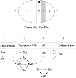
Fig. 1. Schematic representation of Drosophila eye development. Differentiation of the Drosophila eye is controlled by a complex series of signaling events that produce precise compartmentalization of transcription factor activity. The MF, marked by hatched lines, is a wave of differentiation that moves from posterior (P) towards anterior (A) across the eye field during the third instar larvae. Compartments of the eye disc are divided by a dotted line. Compartment 1 represents the majority of cells anterior to the MF. The pre-proneural (PPN) region, represented by compartment 2, is just anterior to the MF. Compartment 3 represents the MF. The arrow indicates the direction of furrow progression. In the compartment 4, posterior to the MF, photoreceptor differentiation and ommatidia assembly occur. Below both the major cellular events and the expression domains of ey paradigm genes are indicated. Ey and toy are only expressed anterior to the MF. In the PPN region, ey and toy induce expression of so, while ey and dpp induce expression of eya. Together ey, so, and eya activate dac. The nonlinear regulatory relationship amongst these genes is illustrated and is herein referred to as the eyeless paradigm. Just posterior to the MF furrow so, eya, and dac continue to be expressed in the absence of eye.
Defining vertebrate orthologues
Pax6 is more closely related to toy and ey than to eyg (Gehring and Ikeo, 1999), and may have taken on the functional role of both ey and toy in vertebrate eye specification (Plaza et al., 1993). Eyg, on the other hand, shares both sequence and functional homol-ogy and with the Pax6 isoform Pax6(5a) (Dominguez et al., 2004). The central role that Pax6 plays in vertebrate eye formation and its remarkable similarity to ey has been reviewed extensively (Gehring and Ikeo, 1999; Wawersik and Maas, 2000; Ashery-Padan and Gruss, 2001; Hansen, 2001; van Heyningen and Williamson, 2002; Simpson and Price, 2002), and will be only briefly reviewed here.
The Six genes fall into three gene families: Six1/Six2/so, Six3/ Six6/optix, and Six4/Six5/D-six4. This classification is based upon molecular phylogeny, chromosomal arrangement, DNA binding specificity, and the ability to interact with Eya proteins (Kawakami et al., 2000). Surprisingly, one of the so orthologues, Six1, is not expressed during vertebrate eye morphogenesis, and homozygous deletion of Six1 has no affect on eye development (Oliver et al., 1995; Laclef et al., 2003). Six2 is expressed in the inner and outer nuclear layers (INL and ONL) and the ganglion cell layer (GCL) of the adult mouse retina (Kawakami et al., 1996), but ectopic expression of Six2 in developing medaka fish has no affect on eye morphogenesis (Loosli et al., 1999). Thus, it is unlikely that Six1 or Six2 is an essential part of a signaling network in vertebrate eye morphogenesis.
Both optix orthologues, Six3 and Six6, are expressed through-out eye morphogenesis (Jean et al., 1999), and homozygous mutation of Six6 in mice deletes the optic chiasm and ON and causes retinal hypoplasia (Li et al., 2002). Deletion of Six3 expression in medaka fish with a morpholino completely deletes all eye and forebrain structures (Carl et al., 2002). These results indicate that Six3 and Six6 are required for vertebrate eye formation, and we will therefore discuss the likelihood that one or both of these genes plays a role comparable to that of so in the Drosophilaey paradigm.
Homozygous or compound heterozygous mutation of the Pax6 gene results in anophthalmia in both humans (Glaser et al., 1994) and Pax6Sey mice (Hill et al., 1991).
Similarly, in the zebrafish mutant cyclops, which lacks Shh expression, the Pax6 expression domain is expanded and retina specification occurs at the expense of the optic nerve (Macdonald et al., 1995). Ectopic expression of Shh, on the other hand, restricts Pax6 expression, and nearly abolishes the retinal field (Macdonald et al., 1995). Thus, alterations in Shh alter Pax6 expression. This is reminiscent of the fly, where down-regulation of ey in the MF coincides with cells receiving Hh signals (Halder et al., 1998).
Ectopic Pax6 expression in Xenopus embryos can produce various eye related phenotypes including the induction of well-organized, ectopic eyes in the head anterior to the hindbrain-spinal cord junction (Chow et al., 1999). This observation is reminiscent of the ectopic eye forming activity of ey in Drosophila imaginal discs (Halder et al., 1995). Collectively, these experi-ments show that specification of the eye in vertebrates is tightly linked to Pax6 expression. Clearly, Pax6 in the vertebrate eye has retained some striking functional similarities with ey.
Do non-orthologous genes in the vertebrate eye fulfill the role of so?
The ey paradigm is used during vertebrate organogenesis in tissues unrelated to the eye. In two such examples, a gene family member has been substituted into the hierarchy. In the developing kidney, homozygous deletion of Eya1, Pax2, or Six1 disrupts early kidney morphogenesis (Torres et al., 1996; Xu et al., 1999; Xu et al., 2003). In addition, compound heterozygous mutants of Eya1
and Six1 have hypoplastic kidneys (Xu et al., 2003, Li et al., 2003) demonstrating an interaction between these genes. While all of the ey paradigm interactions from the Drosophila eye are not con-served in the vertebrate kidney, enough parallels exist to establish the importance of the Pax2/Eya1/Six1 genetic hierarchy for orga-nogenesis (Torres et al., 1996; Xu et al., 1999; Xu et al., 2003). Pax2, however, is substituted for Pax6. In the developing somites, Pax3, Six1, Eya2, and Dach2 synergize to promote myogenesis (Heanue et al., 1999). In this example, Pax6 is replaced by the
TABLE II
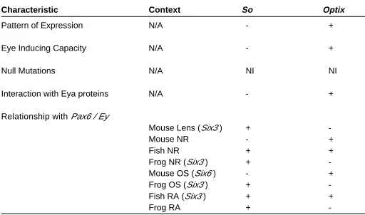
QUALITATIVE AND FUNCTIONAL COMPARISON OF SIX3 AND SIX6 TO SO AND OPTIX
Characteristic Context So Optix
Pattern of Expression N/A - +
Eye Inducing Capacity N/A - +
Null Mutations N/A NI NI
Interaction with Eya proteins N/A - +
Relationship with Pax6 / Ey
Mouse Lens (Six3 ) +
-Mouse NR - +
Fish NR + +
Frog NR (Six3 ) + -Mouse OS (Six6 ) - + Frog OS (Six3 ) + -Fish RA (Six3 ) + +
Frog RA +
-In the vertebrate eye it is unclear if Six3 and Six6 function more like optix or like so. We assessed their role in vertebrate eye formation and have classified them as behaving either like optix or so. In some instances Six3 and Six6 have characteristics of both Drosophila genes. These instances are indicated by a (+) for each so and optix. Abbreviations; N/A, not applicable; NI, not informative; (+) similar; (-) not similar; NR, neural retina; OS, optic stalk; RA, retinal anlage
Fig. 2. Vertebrate eye formation. Four key stages of embryonic mouse eye development are shown. (A E9.5; B E11.5; C E12.5; and D E15.5). Each panel shows a representative DAPI stained section through the eye of a paraffin embedded embryo. The vertebrate eye is formed from two separate tissues the neurectoderm and the head surface ectoderm.
D
A
B
C
The retinal anlage, specified in the anterior neurectoderm, is divided into two distinct fields (not shown). From each field an optic vesicle (OV) evaginates laterally and opposes the overlying surface ectoderm (SE) (A). In the mouse, the surface ectoderm is induced to form the lens placode (LP) at roughly E9. The OV is patterned proximal-distally into optic stalk (OS) and optic cup (OC, not shown), which is subse-quently divided into retinal pigmented epithelium (RPE) and neural retina (NR) (B). The OS matures to form the optic nerve (ON, D). The LP invaginates and forms a hollow lens vesicle (LV; panel B), which is subsequently filled with differentiating primary fiber cells (1o FC) that
Pax3 gene. Thus, there are clear examples in which the Drosophila ey paradigm has been utilized in vertebrate organogenesis, but the hierarchy has been modified by substituting tissue-appropriate family members.
Is it possible that a non-orthologous Six gene, Six3 or Six6, replaces the function of so in the vertebrate eye? As noted earlier, two Six genes are broadly expressed in the vertebrate eye during morphogenesis, Six3 and Six6 (Jean et al., 1999). These genes, however are orthologous to optix (Kawakami et al., 2000), which is expressed during and is important for eye development in Droso-phila (Seimiya and Gehring, 2000), and not to so. To determine if Six3 or Six6 might replace so in the lens or NR, we would like to know whether these Six genes behave more like so or more like optix during vertebrate eye development. For both genes we will assess their qualitative and functional similarity to so and to optix by considering their temporal and spatial expression patterns, their ability to induce ectopic eyes, their homozygous null phenotypes, their regulatory relationships with Pax6, and their ability to interact with Eya proteins (Table II).
Patterns of expression
Optix and so have different patterns of expression in Drosophila eye development (Seimiya and Gehring, 2000). Optix has an expression pattern similar to ey in the eye primordium and anterior to the MF in the differentiating disc, while so has an expression pattern comparable to eya, and is found in cells adjacent to and including the MF (Seimiya and Gehring, 2000). Six3 has an expression pattern nearly identical to that of Pax6, as both genes are found in the retinal anlage, the LP, and throughout the devel-oping lens vesicle and OC (Walther and Gruss, 1991; Oliver et al., 1995). Six6 expression overlaps that of Pax6 in derivatives of the retinal anlage (optic stalk and neural retina) but is absent from the actual anlage (Toy et al., 1998; Toy and Sundin, 1999; Jean et al., 1999; Bernier et al., 2000). Six6 is completely absent from the head surface ectoderm and its derivatives (Oliver et al., 1995; Toy et al., 1998; Toy and Sundin, 1999; Jean et al., 1999). Six3 and Six6 are expressed earlier than and more broadly than Eya1, Eya2 or Eya3 (Xu et al., 1997). Thus, Six3 and Six6 are expressed in patterns similar to that of Pax6, and therefore in this regard more closely resemble optix than so.
Ectopic eye inducing capacity
In the fly, ectopic expression of optix alone in the antennal disc induces eyes, whereas ectopic so expression does not (Seimiya and Gehring, 2000; Pignoni et al., 1997). Similar to optix, ectopic expres-sion of Six3 in medaka fish induces ectopic retinal primordia in competent locations within the brain, and at a much lower frequency ectopic lenses in the head ectoderm near the otic vesicle (Loosli et al., 1999). In Xenopus ectopic expression of either Six3 or Six6 converts anterior neural plate to retina (Bernier et al., 2000.) In these experiments low concentrations of Six3 of Six6 expand the size of the retina, while high concentrations of either gene transform the midbrain to retina and delete the normal eye (Bernier et al., 2000). Thus, Six3 and Six6 resemble optix in their ability to induce an eye-specific developmental program in non-ocular tissue.
Homozygous null phenotypes
So is required for all aspects of visual system development in Drosophila (Cheyette et al., 1994; Serikaku and O’Tousa et al.,
1994), while an optix fly mutant has not been described. Six6 null mice exhibit retinal hypoplasia and often lack both an optic nerve and an optic chiasm (Li et al., 2002). In addition, heterozygous mutation of SIX6 in humans correlates with anophthalmia (Gallardo et al., 1999). Mutation of SIX3 in humans causes holoprosencephaly, with phenotypes ranging from cyclopia to hypotelorism (Wallis et al., 1999; Pasquier et al., 2000). Morpholino inhibition of Six3 expression in medaka fish deletes both forebrain and eye tissue (Carl et al., 2002). The absence of an optix fly mutant makes it unclear whether Six3 and Six6 mutation in vertebrates are more reminiscent of so or optix, although given the severity of each of the reported null phenotypes it is unlikely that this feature would provide a definitive distinction.
Regulatory relationship to ey/Pax6
In the fly, optix expression is truly independent of ey, as eyes ectopically induced by optix do not express ey, and optix can induce eyes in ey deficient flies (Seimiya and Gehring, 2000). In contrast, so expression is dependent upon ey (Pignoni et al., 1997; Halder et al., 1998; Niimi et al., 1999; Michaut et al., 2003). Moreover so acts in conjunction with eya to induce ey expression in ectopic eyes (Pignoni et al., 1997; Bonini et al., 1997).
In vertebrates, the relationship of Six3 or Six6 to Pax6 depends upon both the species in question and the particular compartment
of developing eye (Table II). In mice, Six6 is expressed in the optic stalk and in the OV remnant in homozygous Pax6Sey mice at E9.5 (Jean et al., 1999), demonstrating that Six6 expression is indepen-dent of Pax6. Furthermore, Six6 null mice have normal Pax6 expression in both the lens and retina (Li et al., 2002). Since the expression of Six6 and Pax6 are independent in mice, their relationship is reminiscent of that between ey and optix. Six3 and Pax6, however, regulate each other’s expression in mouse surface ectoderm derivatives (Goudreau et al., 2002). In Pax6LacZ mice, expression of Six3 is unchanged in the retina, but is reduced in the AEL of the lens (Goudreau et al., 2002). Ectopic expression of either Pax6 or Six3 in lens fiber cells induces the expression of the other gene (Goudreau et al., 2002). Thus, while Six3 expression is independent of Pax6 in the retina, the relationship between Six3 and Pax6 in the mouse lens is interdependent, and therefore, resembles the ey-so relationship.
In Xenopus, ectopic Pax6 expression expands Six3 expression in the OV at the midline (presumptive optic stalk) and reduces Six3 expression distally (presumptive NR; Chow et al., 1999). In addition, ectopic expression of either Six3 or Six6 induces ectopic retinas coincident with the expansion of Pax6 expression (Bernier et al., 2000). Likewise, in medaka fish, ectopic expression of either Six3 or Six6 expands Pax6 expression and induces ectopic retinas (Loosli et al., 1999). In each of the aforementioned examples misexpression is achieved by injection of high concentrations of RNA into blastomeres during early stages of development. Thus, expression of the injected gene is not necessarily at a physiological concentration and neither the time nor the place of expression is regulated. Therefore, such overexpression experiments define relationships that may occur in normal development but do not prove that they do occur. The relationship between Pax6 and Six3 predicted by overexpression experiments is, however, supported by complementary reduced expression data. Down-regulation of Six3 expression by morpholino interferes with Pax6 expression, whereas interference with Pax6 expression does not interfere with Six3 (Carl et al., 2002). Thus, in both Xenopus and medaka fish Pax6 and Six3/Six6 are at least partly inter-dependent and, therefore, resemble ey and so.
Synergism with eya
Lastly, in Drosophila, so acts synergistically with eya, both by direct protein-protein interaction and in the cooperative induction of ectopic eyes (Pignoni et al., 1997). Optix, however, does not form a protein-protein complex with Eya (Ohto et al., 1999), and co-expression with eya does not effect the frequency of ectopic eye induction by optix (Seimiya and Gehring, 2000). Six3 also does not interact strongly with Eya proteins in biochemical assays in vitro (Ohto et al., 1999; Purcell, 2002). Six6, however, acts as a co-repressor with Dach proteins both in vitro and in chromatin immu-noprecipitations, suggesting that this protein-protein interaction occurs in vivo (Li et al., 2002). No direct interaction between either So or Optix and Dac proteins have been reported in the fly. This Six-Dach interaction may, therefore, represent a feature unique to vertebrates. However, due to their inability to interact strongly with Eya proteins, Six3 and 6 may be more similar to Optix than to So. Thus, based on the aforementioned criteria (summarized in Table II), Six3 and Six6 in the eye do not behave exactly like so. They have some functional characteristics of both optix and so. Overall, however, they have more in common with optix than they have with so.
Eyes absent related genes are not critical for vertebrate eye formation
As stated above, Eya1 and Six3 do not interact strongly at the protein level (Ohto et al., 1999; Purcell, 2002). What then becomes of the eya component of the ey paradigm in vertebrates? Are other aspects of eya function conserved?
Disruption of eya activity in the fly prevents eye formation (Bonini et al., 1997), and the collective expression of Eya1, Eya2, and Eya3 encompass most tissues of the developing mouse eye. Eya1 is expressed in the lens, OS, and NR (Xu et al., 1997). Eya2 is absent from the lens, but is expressed in the NR in a pattern complementary to Eya1 (Xu et al., 1997). In the retina, Eya1 is in the ONL and the peripheral retina, while Eya2 is in the posterior region and the INL (Xu et al., 1997). Eya3 is present in the OV and the periocular mesen-chyme, but is absent from the lens (Xu et al., 1997).
These expression patterns suggest that Eya genes may play a role in vertebrate eye morphogenesis and that each Eya gene may have taken on a different component of Drosophilaeya activity. Homozygous mutation of Eya1, however, results only in a mild, extrinsic eye phenotype, open eyelids at birth (Xu et al., 1999), and mice with compound homozygous mutations in both Eya1 and Eya2 also retain morphologically normal eyes (P.Y. Xu and R.L. Maas, unpublished; Purcell, 2002). The null mutation of Eya3 in mouse has not yet been reported. Thus, in the mouse, Eya1 and Eya2 are not required for eye morphogenesis and, therefore, cannot play a role comparable to the requisite role of eya in Drosophila. Three human cases, however, have been identified in which heterozygous mutation of EYA1 correlates with anterior segment defects (Azuma et al., 2000). Additional insight into the mechanism for this human defect is needed, however, since many other mutations in EYA1 have been reported that have no effect on eye development (Vervoot et al., 2002). Hopefully these mutations will ultimately provide insight into the function of Eya genes in the vertebrate eye.
Drosophilaeya is downstream of ey and critical for its function (Pignoni et al., 1997; Bonini et al., 1997). Indeed, both the frequency and size of ectopic eye induction are enhanced when eya and ey are co-expressed (Bonini et al., 1997). In mice, neither Eya1 nor Eya2 expression is changed in the early eyes of Pax6LacZ heterozygous mice (Goudreau et al., 2002). Unfortu-nately, expression of vertebrate Eya genes begins too late to reliably assess expression patterns in homozygous Pax6Seymice since the tissues in which these genes are expressed fail to form (Purcell, 2002). Clearly, however, if Eya1 and Eya2 genes can be disrupted with no effect on eye morphogenesis (Xu et al., 1999; Purcell, 2002), they cannot be essential downstream mediators of Pax6 function.
Dach1 is not essential for vertebrate eye formation
In the fly, the pattern of dac expression coincides with that of eya (Chen et al., 1997). In the vertebrate eye, Dach1 is expressed in the lens and the periphery of the retina (Caubit et al., 1999; Purcell, 2002), similar to the expression of Eya1 (Xu et al., 1997). Thus vertebrate Dach1 is similar to dac in its ocular co-localization with Eya1. Dach1 expression also overlaps with that of Pax6, but is significantly delayed in its time of appearance (Hammond et al., 1998; Caubit et al., 1999; Davis et al., 1999; Heanue et al., 2002). However, while loss of dac expression disrupts Drosophila eye formation (Shen and Mardon, 1997; Chen et al., 1997), homozy-gous mutation of Dach1 in mice has no affect on eye morphogen-esis (Davis et al., 2001). Thus, while dac is essential in the fly, Dach1 is not essential for vertebrate eye morphogenesis.
In Drosophila, dac acts downstream of ey, eya and so (Shen and Mardon, 1997; Chen et al., 1997; Michaut et al., 2003). In mouse, expression of Dach1 in the OC is not dependent upon Pax6 as its expression is maintained in the presumptive NR of homozygous Pax6Sey mice (Heanue et al., 2002). Expression of Dach1 is, however, disrupted in the lens ectoderm of homozygous Pax6Sey mice (Purcell, 2002). This loss of Dach1 expression is not the result of global loss of gene expression in a quiescent tissue because other genes are still expressed (Purcell, 2002). In the fly, dac expression is involved in the maintenance of ey, eya, and so expression (Chen et al., 1997). In Dach1 mutant mice, neither Pax6 nor Six3 expression is altered in the developing eyes (Purcell, 2002). Thus, although Dach1 is downstream of Pax6 in the lens ectoderm, most aspects of the ey-dac relationship are missing in the vertebrate eye.
As observed for the rescue of Drosophilaeya mutants with Eya1, 2 and 3, Dach2 expression in dac mutant flies rescues the eye phenotype (Heanue et al., 1999). This is not surprising since Dach2 and its relationship to Pax3, Six1, and Eya2 during somitogenesis is comparable to the relationship in the fly ey paradigm (Heanue et al., 1999). Thus, the ability of vertebrate Dach2 to rescue the Drosophila dac eye phenotype reflects the conservation of the ey paradigm in vertebrate organogenesis rather than a specific conservation of Dach function in the vertebrate eye.
The potential conservation of the ey paradigm in vertebrate oculogenesis has received tremendous attention. In the preceding sections, we have compared each genetic component of the ey paradigm to equivalent vertebrate genetics. While homologues of all of the genes from the fly ey paradigm are expressed during development of the vertebrate eye, the function of each of these genes has not been strictly preserved. The most notable example of non-conservation is the failure of mutations in Eya1 and Eya2 to produce an embryonic eye phenotype. It is intriguing to note, however, that the vertebrate genes are capable of many of the interactions present in the Drosophila eye, as evident from the vertebrate genes either rescuing Drosophila mutants or inducing ectopic eyes in the fly. This suggests that the orthologous verte-brate genes have maintained their molecular function but that the components have, to some extent, become uncoupled. In addition it is important to note that some aspects of the ey paradigm are well conserved. In particular, Pax6 is highly reminiscent of ey, while Six3 and Six6 have some characteristics of so. Thus, despite the lack of strict conservation of the ey paradigm, it is significant that several critical eye regulator genes have been preserved between the morphologically divergent fly and vertebrate eye.
Parallel genetic hierarchies control retinal
differentia-tion in the fly and vertebrates
Interestingly, another parallel genetic pathway between Droso-phila and vertebrates has also emerged between R8 photorecep-tor differentiation and vertebrate RGC specification. Once again this conservation is found in analogous structures that are morpho-logically quite different. Below, we briefly highlight the genetic similarities between the retinal developmental pathways in Droso-phila and vertebrates. In both systems, the transition from a naive, progenitor state to a proneural state is controlled in part by hedgehog signaling, and by the antagonistic relationship between a proneural transcriptional activator and a proneural repressor of the basic helix-loop-helix (bHLH) class.
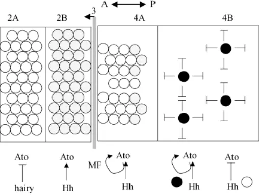
In the Drosophila eye disc, the MF spatially and temporally precedes a wave of differentiation (Figs. 1,3). Initiation and pro-gression of the MF depend in part upon signaling by Hh, which is secreted from differentiating photoreceptors posterior to the MF (Dominguez and Hafen, 1997; reviewed in Treisman and Heberlein, 1998). Hh initiates photoreceptor differentiation through two dis-tinct signals, one long-range and one short-range (Greenwood and Struhl, 1999; Kango-Singh et al., 2003). Decapentaplegic, Dpp, mediates the long-range signal within the MF (Greenwood and Struhl, 1999; Fig. 3) and facilitates the shift from a naive cell to a pre-proneural (PPN) cell. The shift to a PPN state is marked by the upregulation of the bHLH transcription factor hairy (Greenwood and Struhl, 1999; Fig. 3). Hairy is a proneural repressor and marks the PPN state (Greenwood and Struhl, 1999). In the PPN compart-ment, cells exit the cell cycle and prepare for neuronal differentia-tion (Greenwood and Struhl, 1999).
The mediator of the second, short-range signal downstream of Hh is unknown but uses the Raf pathway (Greenwood and Struhl, 1999). The result of this short-range signal is the expression of Atonal, a bHLH transcription factor that induces a proneural state (Jarman et al., 1994). Hairy and atonal share a sharp expression boundary at the border between the PPN and the proneural (PN) compartments (Fig. 3; Greenwood and Struhl, 1999). Cells that do not pass through the PPN (hairy +) to PN (atonal +) transition do not differentiate into R8 photoreceptors (Greenwood and Struhl, 1999). Once beyond the MF, the expression of atonal becomes gradually restricted from all of the cells in the MF to one per cluster, the R8 founder cell (reviewed in Treisman and Heberlein, 1998; Frankfort and Mardon, 2002). This restriction is partially dependent on Hh signaling (Dominguez and Hafen, 1997; Greenwood and Struhl, 1999).
A wave of Shh signaling also marks retinal differentiation in vertebrates
2000). During the wave of RGC specification, the number of differentiating RGCs can be increased or decreased by altering Shh levels (Zhang and Yang, 2001). Low concentrations of Shh induce an increase in the number of differentiating RGCs, while high concentrations of Shh inhibit RGC differentiation and reduce their numbers (Zhang and Yang, 2001). This is reminiscent of photoreceptor differentiation in Drosophila where Hh induces the expression of the R8 neuronal precursor marker, atonal, in all cells at the MF, but interferes with expression of atonal in cells dese-lected as the R8 neuronal precursor, posterior to the MF (Fig. 3). Alteration of the concentration of Hh in Drosophila alters the number of R8 neuronal precursors (Dominguez and Hafen, 1997). Thus, a wave of expression of both Hh and Shh drive differentia-tion, and their concentrations are critical for specification of appro-priate numbers of either R8 founders or RGCs, respectively.
In mice, complete abolition of Shh expression produces a single retinal anlage that fails to divide into symmetric retinal primordia, and therefore results in the formation of a single, centrally located OC (Chiang, et al., 1996). Due to the timing and severity of the phenotype resulting from mutation of Shh in mice, assessing the role of Shh in retinal specification is complicated. However, the single OC in Shh mutant mice is severely dysmorphic: the two-layered structure is inappropriately patterned along the proximal-distal axis (Chiang et al., 1996). Specifically, NR is lost at the expense of RPE, suggesting that Shh may play an important role in NR specification. Additionally, Shh expression has been ob-served in the NR of mice at the time of RGC specification (Jensen and Wallace, 1997). Definitive evidence of a role for Shh in mouse retinal specification awaits the generation of a conditional knock out.
A family of bHLH genes specifies neuronal identity
The mouse RGC marker, Math5, is the orthologue of the fly R8 progenitor cell marker, atonal (Brown et al., 1998). Expression of Math5 precedes that of other bHLH PN genes (Ngn2, NeuroD, and Mash1) in mice (Brown et al., 1998). Math5 is expressed initially in the mouse retina at E11.5 in the central OC, and correlates with the appearance of early neurons (Brown et al., 2001; Wang et al., 2001). Math5 expression expands throughout the retina in a wave and peaks at E13.5 (Brown et al., 2001;Wang et al., 2001). During RGC birth at E15.5, Math5 is expressed in the periphery of the retina where neuronal specification occurs (Brown et al., 2001; Wang et al., 2001). Similarly, the zebrafish and Xenopusatonal orthologues, ath5 and Xath5 respectively, predict the pattern of neuronal differentiation with their expression (Kaneker et al., 1997; Masai et al., 2000). Thus, the dynamic expression patterns of the vertebrate proneural genes Math5/ath5/Xath5 resemble the ex-pression of atonal that moves across the fly eye disc in front of the MF as photoreceptor differentiation proceeds.
Mice bearing a homozygous deletion of Math5 lose up to 80% of cells expressing RGC markers. This leaves an excess of progenitor cells in the proliferative layer of the retina (Brown et al., 2001; Wang et al., 2001). The presence of these excess retinal precursor cells after the first wave of neurogenesis leads to the specification of large numbers of amacrine cells (Brown et al., 2001; Wang et al., 2001). In zebrafish, mutation of ath5 (lak) abolishes the first wave of differentiation in the retina, which produces RGCs (Kay et al., 2001). Likewise, cells in the Drosophila eye disc that do not transition through and ato+ state, cannot differentiate into photoreceptors (Greenwood and Struhl, 1999).
In the developing vertebrate retina, the neuronal repressor Hes1, a homologue of the fly repressor hairy, is expressed in the ventricular zone and is absent from the GCL (Tomita et al., 1996). Hes1 positive cells remain in the proliferative layer and do not differentiate into neurons (Tomita et al., 1996). In Hes1 deficient mice, the waves of differentiation that produce the different neu-rons of the retina are greatly accelerated (Tomita et al., 1996), and neurogenesis proceeds at the expense of proliferation in RPCs. Loss of both proneural repressor proteins (Hairy and Extramacrochaete) in Drosophila also results in premature differ-entiation of photoreceptors (Brown et al., 1995). In Hes1-/- mice, fewer progenitor cells are available for specification at each stage so the resulting retinas have very few neurons (Tomita et al., 1996). Based on these data, Hes1 is believed to repress neurogenesis in proliferative cells, thereby preventing premature differentiation. Consistent with this idea, overexpression of Hes1 in postnatal rat retinal progenitors increases the number of Müller glia at the expense of neurons (Furukawa et al., 2000).
Hes1 and Math1 have an antagonistic relationship in vertebrate retinal neurogenesis. Hes1 is expressed in progenitor cells and represses premature differentiation, while Math5 is expressed in early neural precursors and promotes RGC differentiation. Addi-tionally, in heterozygous Pax6Seymice, fewer Math5 expressing cells are present, while the domain of Hes1 expression is ex-panded (Brown et al., 1998). In homozygous Pax6Seymice, Math5 expression is abolished (Brown et al., 1998). Since the loss of Math5 expression correlates with an expansion of Hes1, these data provide additional evidence for the antagonistic relationship between these genes in vertebrates. This antagonistic relationship is highly reminiscent of that observed between hairy and atonal in the fly.
Summary
In conclusion, we have reviewed the ey paradigm as character-ized in Drosophila, and we have evaluated its potential conserva-tion in vertebrates. The evidence to date does not support the idea that the entire ey genetic hierarchy is conserved in the vertebrate eye. On the other hand, some genetic parallels do exist, as Pax6 activity is highly reminiscent of ey, and Six3 and Six6 have some characteristics of so. Nonetheless, it is the overall epistatic relation-ship amongst the vertebrate homologues of ey, so, eya, and dac that appears to be specifically absent in the vertebrate eye.
We have also briefly reviewed some aspects of neuronal speci-fication in the vertebrate retina, as the control of both cell prolifera-tion and RGC identity by bHLH transcripprolifera-tion factors is highly reminiscent of R8 photoreceptor differentiation in the fly. Thus in both cases, specific features of developmental regulatory cas-settes have been retained and re-deployed in vertebrate ocular organogenesis.
multiple organogenic processes, and that they have been partially maintained in the eye, despite the significant divergence of verte-brate and inverteverte-brate eyes.
Acknowledgements
The authors would like to acknowledge Fang Ko for preparation of DAPI stained sections and Robert Moy for critical reading of the manuscript.
References
ASHERY-PADAN, R., and GRUSS, P. (2001). Pax6 lights the way for eye develop-ment. Curr. Opin. Cell Biol. 13:706-714.
AZUMA, M., HIRAKIYAMA, A., INOUE, T., ASAKA, A., and YAMADA, M. (2000). Mutations of a human homologue of the Drosophilaeyes absent gene (EYA1 ) detected in patients with congenital cataracts and ocular anterior segment anomalies. Hum. Mol. Genet. 9:3636-366.
BAKER, N.C. (2001). Cell proliferation, survival, and death in the Drosophila eye. Semin. Cell Dev. Biol. 12:499-507.
BERNIER, G., PANITZ, F., ZHOU, X., HOLLEMANN, T., GRUSS, P., and PIELER, T. (2000). Expanded retina territory by midbrain transformation upon overexpression of Six6 (Optx2) in Xenopus embryos. Mech. Dev. 93:59-69.
BONINI, N.M., BUI, W.T., GRAY-BOARD, G.L., and WARRICK, J.M. (1997) The Drosophilaeyes absent gene directs ectopic eye formation in a pathway con-served between flies and vertebrates. Development 124:4819-4826.
BORSANI, G., DEGRANDI, A., BALLABIO, A., BULLFONE, A., BERNARD, L., BANFI, S., GATTUSO, C., MARIANI, M., DIXON, M., DONNAI, D., METCALFE, K., WINTER, R., ROBERTSON, M., AXTON, R., BROWN, A., VAN HEYNINGEN, V., and HANSEN, I. (1999). EYA4, a novel vertebrate gene related to Drosophila eyes absent. Hum. Mol. Genet. 8:11-23.
BROWN, N.L., SATTLER, C., PADDOCK, S.W., and CARROLL, S.B. (1995). Hairy and Emc negatively regulate morphogenetic furrow progression in the Drosophila eye. Cell 80:879-887.
BROWN, N.L., KANEKAR, S., VETTER, M.L., TUCKER, P.K., GEMZA, D.L., and GLASER, T. (1998). Math5 encodes a murine basic helix-loop-helix transcription factor expressed during early stages of retinal neurogenesis. Development 125:4821-4833.
BROWN, N.L., PATEL, S., BRZEZINSKI, and GLASER, T. (2001). Math5 is required for retinal ganglion cell and optic nerve formation. Development 128:2497-2508.
BUI, Q.T., ZIMMERMAN, J.E., LIU, H., and BONINI, N. M. (2000). Molecular analysis of Drosophila eyes absent mutants reveals features of the conserved Eya domain. Genetics 155:709-720.
CARL, M., LOOSLI, F., and WITTBRODT, J. (2002) Six3 inactivation reveals its essential role for the formation and patterning of the vertebrate eye. Development 129:4057-4063.
CAUBIT, X. THANGARAJAH, R., THEIL, T., WIRTH, J., NOTHWANG, H-G., RÜTHER, U., and KRAUSS, S. (1999). Mouse Dac, a novel nuclear factor with homology to Drosophiladachshund shows a dynamic expression in the neural crest, the eye, the neocortex, and the limb bud. Dev. Dyn. 214:66-80.
CEPKO, C.L., AUSTIN, C.P., YANG, X., ALEXIADES, M., and EZZEDDINE, D. (1996). Cell fate determination in the vertebrate retina. Proc. Natl. Acad. Sci. USA 93:589-595.
CHEN, R., AMOUI, M., ZHANG, Z., and MARDON, G. (1997). Dachshund and eyes absent proteins form a complex and function synergistically to induce ectopic eye development in Drosophila. Cell 91:898-903.
CHEYETTE, B. N. R., GREEN, P. J., MARTIN, K., GARREN, H., HARTENSTEIN, V. and ZIPURSKY, S. L (1994). The Drosophila sine oculis locus encodes a homeodomain-containing protein required for the development of the entire visual system. Neuron 12: 977-996
CHIANG, C., LITINGTUNG, Y., LEE, E., YOUNG, K.E., CORDEN, J.L., WESTPHAL, H., and BEACHY, P.A. (1996) Cyclopia and defective axial patterning in mice lacking Sonic Hedgehog gene function. Nature 383:407-413.
CHOW, R.L., ALTMANN, C.R., LANG, R.A., and HAMMATI-BRIVANLOU, A. (1999). Pax6 induces ectopic eyes in a vertebrate. Development 126:4213-4222. CZERNY, T., HALDER, G., KLOTER, U., SOUABNI, GEHRING, W.J., and
BUSSLINGER, M. (1999). Twin of ey, a second Pax6 gene of Drosophila, acts
upstream of eyeless in the control of eye development. Mol. Cell 3:297-307.
DAVIS, R.J., SHEN, W., HEANUE, T.A. and MARDON, G. (1999). Mouse Dach, a homologue of Drosophila dachshund, is expressed in the developing retina, brain, and limbs. Dev. Genes Evol. 209:526-536.
DAVIS, R.J., SHEN, W., SANDLER, Y.I., AMOUI, M., PURCELL, P., MAAS, R., OU, C.N., VOGEL, H., BEAUDET, A.L., and MARDON, G. (2001) Dach1 mutant mice bear no gross abnormalities in eye, limb, and brain development and exhibit postnatal lethality. Mol. Cell Biol. 21:1484-90.
DESPLAN, C. (1997). Eye development: governed by a dictator or a junta? Cell 91:861-864.
DOMINGUEZ, M. and HAFEN, E. (1997) Hedgehog directly controls initiation and propagation of retinal differentiation in the Drosophila eye. Genes Dev. 11:3254-3264.
DOMINGUEZ, M., FERRES-MARCO, D., GUTIERREZ-AVINO, F.J., SPEICHER, S.A., and BENEYTO, M. (2004). Growth and specification of the eye are controlled independently by Eyegone and Eyeless in Drosophila melanogaster. Nat. Genet. 36: 31-39.
FRANKFORT, B.J. and MARDON, G. (2002) R8 development in the Drosophila eye: a paradigm for neural selection and differentiation. Development 129:1295-1306.
FURUKAWA, T., MUKHERJEE, S., BAO, Z.Z., MORROW, E.M., and CEPKO, C.L. (2000). Rax, Hes1, and Notch1 promote the formation of Müller glia by postnatal progenitor cells. Neuron 26:383-394.
GALLARDO, M.E., LOPEZ-RIOS, J., FERNAUD-ESPINOSA, I., GRANADINO, B., SANZ, R., RAMOS, C., AYUSO, C., SELLER, M.J., BRUNNER, H.G., BOVOLENTA, P., and RODRIGUEZ DE CÓRDOBA, S. (1999). Genomic cloning and character-ization of the human homeobox gene SIX6 reveals a cluster of SIX genes in chromosome 14 and associates SIX6 hemizygosity with bilateral anophthalmia and pituitary anomalies. Genomics 61:82-91.
GEHRING, W.J. and IKEO, K. (1999). Pax6 mastering eye morphogenesis and eye evolution. Trends Genet. 15: 371-377.
GHANBARI, H., SEO, H-C., FJOSE, A. and BRÄNDLI, A.W. (2001). Molecular cloning and embryonic expression of Xenopus Six homeobox genes. Mech. Dev. 101:271-277.
GOUDREAU, G., PETROU, P., RENEKER, L.W., GRAW, J., LOSTER, J., and GRUSS, P. (2002) Mutually regulated expression of Pax6 and Six3 and its implications for the Pax6 haploinsufficient lens phenotype. Proc. Natl. Acad. Sci. USA 99:8719-8724.
GLASER, T., JEPEAL, L., EDWARDS, J.G., YOUNG, S.R., FAVOR, J. and MAAS, R.L. (1994). PAX6 gene dosage effect in a family with congenital cataracts, aniridia, anophthalmia and central nervous system defects. Nat. Genet. 7:463-471.
GREENWOOD, S. and STRUHL, G. (1999). Progress of the morphogenetic furrow in the Drosophila eye: the roles of Hedgehog, Decapentaplegic, and the Raf pathway. Development 126:5795-5808.
HALDER, G., CALLAERTS, P., and GEHRING, W.J. (1995). Induction of ectopic eyes by targeted expression of the eyeless gene in Drosophila. Science 267:1788-92.
HALDER, G., CALLAERTS, P., FLISTER, S., WALLDORF, U., KLOTER, U., and GEHRING, W.J. (1998). Eyeless initiates the expression of both sine oculis and eyes absent during Drosophila compound eye development. Development 125:2181-2191.
HAMMOND, K. L., HANSON, I. M., BROWN, A. G., LETTICE, L.A., and HILL, R.E. (1998). Mammalian and Drosophila dachshund genes are related to the Ski proto-oncogene and are expressed in eye and limb. Mech. Dev. 74:121-131.
HANSON, I.M. (2001). Mammalian homologues of the Drosophila eye specification genes. Semin. Cell Dev. Biol. 12:475-484.
HAUCK, B., GEHRING, W. J., and WALLDORF, U. (1999). Functional analysis of an eye specific enhancer of the eyeless gene in Drosophila. Proc. Natl. Acad. Sci. USA 96:564-569.
HEANUE, T.A., RESHEF, R. DAVIS, R.J., MARDON, G., OLIVER, G., TOMAREV, S., LASSAR, A.B., and TABIN, C.J. (1999) Synergistic regulation of vertebrate muscle development by Dach2, Eya2, and Six1, homologs of genes required for Drosophila eye formation. Genes Dev. 15:3231-43.
HEATH, S. K., CARNE, S., HOYLE, C., JOHNSON, K. J., and WELLS, D.J. (1997). Characterization of expression of mDMAHP, a homeodomain encoding gene at the DM locus. Hum. Mol. Genet. 6:651-657.
HILL, R.E., FAVOR, J. HOGAN, B.L., TON, C.C., SAUNDERS, G.F., HANSON, I.M., PROSSER, J., JORDAN, T. et al. (1991). Mouse Small eye results from mutations in a paired-like homeobox containing gene. Nature 354:522-525.
HSIUNG, F. and MOSES, K. (2002). Retinal development in Drosophila: specifying the first neuron. Hum. Mol. Genet. 11:1207-1214.
JANG, C.C., CHAO, J.L., JONES, N., YAO, L.C., BESSARAB, D.A., KUO, Y.M., JUN, S., DESPLAN, C., BECKENDORF, S.K., and SUN, Y.H. (2003). Two Pax genes, eye gone and eyeless, act cooperatively in promoting Drosophila eye develop-ment. Development 130:2939-51.
JARMAN, A.P., GRELL, E.H., ACKERMAN, L., JAN, L.Y., and JAN, Y.N. (1994). Atonal is the proneural gene for Drosophila photoreceptors. Nature 369:398-400. JEAN, D., BEIMER, G., and GRUSS, P. (1999). Six6 (optx2) is a novel murine Six3 related homeobox gene that demarcates the presumptive pituitary/hypothalamic axis and ventral optic stalk. Mech. Dev. 84:31-40.
JENSEN, A.M. and WALLACE, V.A. (1997) Expression of Sonic hedgehog and it putative role as a precursor cell mitogen in developing mouse retina. Development 124:363-371.
KANEKER, S., PERRON, M., DORSKY, R., HARRIS, W.A., JAN, L.Y., JAN, Y.N., and VETTER M.L. (1997). Xath5 participates in a network of bHLH genes in the developing Xenopus retina. Neuron 19:981-994.
KANGO-SINGH, M., SINGH, A., and SUN, H. (2003) Eyeless collaborates with Hedgehog and Decapentaplegic signaling in Drosophila eye induction. Dev. Biol. 256:49-60.
KAY, J.N., FINGER-BAIER, K.C., ROESER, T., STAUB, W., and BAIER, H. (2001) Retinal ganglion cell genesis requires lakritz, a zebrafish atonal homologue. Neuron 30:725-736.
KAWAKAMI, K., OHTO, H., TAKIZAWA, T., and SAITO, T. (1996). Identification and expression of six family genes in mouse retina. FEBS Lett.393:259-263.
KAWAKAMI, K., OHTO, H., IKEDA, K., and ROEDER, R.G. (1996b). Structure, function, and expression of a murine homeobox protein AREC3, a homologue of Drosophilasine oculis gene product, and implication in development. Nucleic Acids Res. 24:303-310.
KAWAKAMI, K. SATO, S., and IKEDA, K. (2000) Six family genes – structure and function as transcription factors and their roles in development. BioEssays 22:616-626.
KLESERT, T. R., CHO, D.H., CLARK, J. I., MAYLIE, J., ADELMAN, J., SNIDER, L., YUEN, E. C., SORIANO, P., and TAPSCOTT, S. J. (2000). Mice deficient in Six5 develop cataracts: implications for myotonic dystrophy. Nat. Genet. 25:105-109.
KOBAYASHI, M.,OSANAI, H., KAWAKAMI, K., and YAMAMOTO, M. (2000). Expres-sion of three zebrafish Six4 genes in cranial sensory placodes and developing somites. Mech. Dev. 98:151-155.
LACLEF, C., SOUIL, E., DEMIGNON, J., and MAIRE, P. (2003). Thymus, kidney, and craniofacial abnormalities in Six1 deficient mice. Mech. Dev. 120: 669-679.
LI, X., PERISSI, V., LIU, F., ROSE, D.W., and ROSENFELD, M.G. (2002) Tissue-specific regulation of retinal and pituitary precursor cell proliferation. Science 297:1180-1183.
LI, X., OGHI, K.A., ZHANG, J., KRONES, A., BUSH, K.T., GLASS, C.K., NIGAM, S.K., AGGARWAL, A.K., MAAS, R., ROSE, R.W., and ROSENFELD, M.G. (2003). Eya protein phosphatase activity regulates Six1-Dach-Eya transcriptional effects in mammalian organogenesis. Nature 426:247-254.
LOOSLI, F., WINKLER, S., and WITTBRODT, J. (1999) Six3 overexpression initiates the formation of ectopic retina. Genes Dev. 13:649-654.
MACDONALD, R., BARTH, K.A., XU, Q., HOLDER, N., MIKKOLA, I., and WILSON, S.W. (1995). Midline signaling is required for Pax gene regulation and patterning of the eyes. Development 121:3267-3278.
MASAI, I., STEMPLE, D.L., OKAMOTO, H., and WILSON, S.W. (2000). Midline signals regulate retinal neurogenesis in zebrafish. Neuron 27:251-263.
MARQUARDT, T. and GRUSS, P. (2002) Generating neuronal diversity in the retina: one for nearly all. Trends Neurosci. 25:32-38.
MCCABE, K.L, GUNTER, E.C., and REH, T.A. (1999). The development of pattern of retinal ganglion cells in the chick retina: mechanisms that control differentiation. Development 126:5713-5724.
MICHAUT, L., FLISTER, S., NEEB, M., WHITE, K.P., CERTA, U., and GEHRING, W.J. (2003). Analysis of the eye developmental pathway in Drosophila using DNA microarrays. Proc. Natl. Acad. Sci. USA 100:4024-4029.
NEUMANN, C.J., and NUESSLEIN-VOLHARD, C. (2000). Patterning of the zebrafish retina by a wave of sonic hedgehog activity. Science 289:2137-2139.
NIIMI, T. SEIMIYA, M., KLOTER, U., FLISTER, S., and GEHRING, W.J. (1999). Direct regulatory interaction of the eyeless protein with an eye-specific en-hancer in the sine oculis gene during eye induction in Drosophila. Development 126:2253-2260.
OGINO, H., and YASUDA, K. (2000). Sequential activation of transcription factors in lens induction. Dev. Growth Diff. 42:437-448.
OHTO, H., KAMADA, S., TAGO, K., TOMINAGA, S.I., OZAKI, H., SATO, S., and KAWAKAMI, K. (1999). Cooperation of Six and Eya in activation of their target genes through nuclear translocation of Eya. Mol. Cell Biol. 19:6815-6824.
OLIVER, G., MAILHOS, A., WEHR, R., COPELAND, N.G., JENKINS, N.A., and GRUSS, P. (1995). Six3, a murine homologue of the sine oculis gene, demarcates the most anterior border of the developing neural plate and is expressed during eye development. Development 121: 4045-4055.
OLIVER, G., WEHR, R., JENKINS, N.A., COPELAND, N. G. CHEYETTE, B. N. R., HARTENSTEIN, V., ZIPURSKY, L., and GRUSS P. (1995b). Homeobox genes and connective tissue patterning. Development 121:693-705.
OZAKI, H., WANTABE, Y., TAKAHASHI, K., KITAMURA, K., TANAKA, A., URASE, K., MOMOI, T., SUDO, K., SAKAGAMI, J., ASANO, M., IWAKURA, Y., and KAWAKAMI, K. (2001). Six4 a putative myogenin gene regulator, is not essential for mouse embryonal development. Mol. Cell Biol. 21:3343-3350.
PASQUIER, L., DUBOURG, C., BLAYAU, M., LAZARO, L., LE MAREC, B., DAVID, V., and ODENT, S. (2000). A new mutation in the six-domain of SIX3 gene causes holoprosencephaly. Eur. J. Hum. Genet. 8:797-800
PIGNONI, F., HU, B., ZAVITZ, K.H., XIAO, J., GARRITY, P.A., and ZIPURSKY, S.L. (1997). The eye specification proteins So and Eya form a complex and regulate multiple steps in Drosophila eye development. Cell 91:881-891.
PLAZA, S. DOZIER, C., and SAULE, S. (1993). Quail Pax-6 (Pax-QNR) encodes a transcription factor able to bind and trans-activate its own promoter. Cell Growth Differ. 4:1041-1050.
PURCELL, P. (2002) Genes involved in early development of the mouse sensory placodes. Thesis, Harvard University.
QUIRING, R., WALLDORF, U., KLOTER, U., and GEHRING, W.J. (1994). Homology of the eyeless gene of Drosophila to the small eye gene in mice and Aniridia in humans. Science 265:783.
SARKAR, P. S., APPUKUTTAN, B., HAN, J., ITO, S., AI, C., TSAI, W., CHAI, Y., STOUT, J. T., and REDDY, S. (2000). Heterozygous loss of Six5 in mice is sufficient to cause ocular cataracts. Nat. Genet. 25:110-114.
SEIMIYA, M. and GEHRING, W.J. (2000). The Drosophila homeobox gene optix is capable of inducing ectopic eyes by an eyeless-independent mechanism. Devel-opment 127:1879-1886.
SERIKAKU, M. A. and O’TOUSA, J. E (1994) Sine oculis is a homeobox gene required for Drosophila visual system development. Genetics 138: 1137-1150.
SHEN, W. and MARDON, G. (1997). Ectopic eye development in Drosophila induced by directed dachshund expression. Development 124:45-52.
SIMPSON, T. A. and PRICE, D. J. (2002). Pax6; a pleiotropic player in development. BioEssays 24:1041-1051.
TOMITA, K, ISHIBASHI, M. NAKARHARA, K., ANG, S.L., NAKANISHI, S., GUILLEMOT, F., and KAGEYAMA, R. (1996) Mammalian hairy and Enhancer of splithomologue 1 regulates differentiation of retinal neurons and is essential for eye morphogenesis. Neuron 16:723-34.
TORRES, M., GOMEZ-PARDO, E., and GRUSS, P. (1996). Pax2 contributes to inner ear patterning and optic nerve trajectory. Development 122:3381-3391.
TOY, J., YANG, J-M., LEPPERT, G.S., and SUNDIN, O.H. (1998). The optx2 homeobox gene is expressed in early precursors of the eye and activates retina-specific genes. Proc. Natl. Acad. Sci. USA 95:10643-10648.
TOY, J. and SUNDIN, O.H. (1999). Expression of the Optx2 homeobox gene during mouse development. Mech. Dev. 83:183-186.
VAN HEYNINGAN, V. and WILLIAMSON, K. A. (2002) PAX6 in sensory development. Hum. Mol. Genet. 11:1161-1167.
VERVOOT, V.S., SMITH, R.J., O’BRIEN, J., SCHROET, R., ABBOTT, A., STEVENSON, R.E., and SCHWARTZ, C.E. (2002) Genomic rearrangements of EYA1 account for a large fraction of families with BOR syndrome. Eur. J. Hum. Genet. 10:757-766.
WALTHER, C. and GRUSS, P. (1991). Pax6, a murine paired box gene, is expressed in the developing CNS. Development 113:1435-1449.
WALLIS, D. E., ROESSLER, E., HEHR, U., NANNI, L., WILTSHIRE, T., RICHIERI-COSTA, A., GILLESEN-KAESBACH, G., ZACKAI, E. H., ROMMENS, J., and MUENKE, M. (1999). Mutations in the homeodomain of the human SIX3 gene cause holoprosencephaly. Nat. Genet. 22:196-198.
WINCHESTER, C. L., FERRIER, R. K., SERMONI, A., CLARK, B.J., and JOHNSON, K. J. (1999). Characterization of the expression of DMPK and SIX5 in the human eye and implications for pathogenesis in myotonic dystrophy. Hum. Mol. Genet. 8:481-492.
WANG, S.W., KIM, B.S., DING, K., WANG, H., SUN, D., JOHNSON, R.L., KLEIN, W.H., and GAN, L. (2001). Requirement for Math5 in the development of retinal ganglion cells. Genes Dev. 15:24-29.
WAWERSIK, S., and MAAS, R.L. (2000). Vertebrate eye development as modeled in Drosophila. Hum. Mol. Genet. 9:917-925.
XU, P., WOO, I., HER, H., BEIER, D.R., and MAAS, R.L. (1997). Mouse Eya homologues of the Drosophila eyes absent gene require Pax6 for expression in lens and nasal placode. Development 124:219-231.
XU, P., ADAMS, J., PETERS, H., BROWN, M.C., HEANEY, S. and MAAS, R.L. (1999). Eya1 deficient mice lack ears and kidneys and show abnormal apoptosis of organ primordial. Nat. Genet. 23:113-117.
XU, P., ZHENG, W.M., HUANG, L., MAIRE, P., LACLEF, C., and SILVIUS, D. (2003). Six1 is required for the early organogenesis of mammalian kidney. Development 130:3085-3094.