Defining the cell lineages of the islets of langerhans
using transgenic mice
PEDRO L. HERRERA*
Department of Morphology, University of Geneva Medical School. 1 Rue Michel-Servet, CH-1211 Geneva 4, Switzerland
ABSTRACT In this Special Issue of the Int. J. Dev. Biol., we summarize our own studies on the development of the mouse endocrine pancreas, with special emphasis on the cell lineage relation-ships between the four islet cell types. Considerable knowledge concerning the ontogeny of the endocrine pancreas has been gained in recent years, mainly through the use of two complementary genetic approaches in mice: gene inactivation and genetic labelling of precursor cells. However, neither gene inactivation in KO mice nor co-localisation of hormones in single cells during development can be taken as evidence for cell lineage relationships among different cell types. The
β-cell lineage analysis was started by selectively ablating specific islet cell types in transgenic mice. We used the diphtheria toxin A subunit coding region under the control of insulin, glucagon or pancreatic polypeptide (PP) promoters, in order to eliminate insulin-, glucagon- or PP-expressing cells, respectively. Contrary to the common view, we demonstrated that glucagon cells are not precursors of insulin-producing cells. These results were in addition the first evidence of a close ontogenetic relationship between insulin and somatostatin cells. We pursued these analyses using a novel, more subtle approach: progenitor cell labelling through the expression of Cre recombinase in doubly transgenic mice. We were able to unequivocally establish that 1) adult glucagon- and insulin-producing cells derive from precursors which have never transcribed insulin or glucagon, respectively; 2) insulin cell progenitors, but not glucagon cell progenitors transcribe the PP gene and 3) adult glucagon cells derive from progenitors which do express pdx1.
KEY WORDS:
pancreas, islet, transgenic, mouse, lineage
0214-6282/2002/$25.00
© UBC Press Printed in Spain
www.ijdb.ehu.es
*Address correspondence to: Pedro L. Herrera, Department of Morphology, University of Geneva Medical School. 1, rue Michel-Servet. CH-1211 Geneva 4, Switzerland. Fax: +41 22 702 5260. e-mail: [email protected]
Abbreviations used in this paper: KO, knock-out; PP, pancreatic polypeptide;
TGF, Transforming Growth Factor;
Introduction
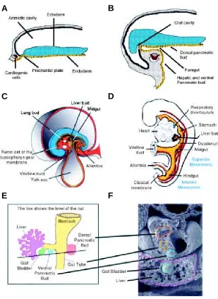
The mammalian pancreas is not a bilateral organ, yet it originates from two separate endodermal outgrowths (Fig. 1). The islets of Langerhans are well-organized micro organs that consti-tute the endocrine portion of the pancreas. They are scattered amidst the exocrine tissue (the secretory acini and excretory ducts) (Fig. 2A). Islet endocrine cells are of four different types, each producing one hormone: insulin (β-cells), glucagon (α -cells), somatostatin (δ-cells), or pancreatic polypeptide (PP-cells) (Fig. 2B) (Orci, 1982; Orci and Unger, 1975). Contrary to what is observed in adult islets, cells containing simultaneously more than one hormone are frequent in the embryonic pancreas; this has suggested the existence of a common precursor cell for all four islet cell types (Alpert et al., 1988; Rall et al., 1973; Teitelman et al., 1993).
The pancreas forms as a double evagination of the duodenal entoblast, through a complex pathway of cell differentiation and organogenesis (Fig. 2C) (Pictet et al., 1972). Islet endocrine cells
Several different strains of genetically modified mice, i.e. lacking or on the contrary overexpressing different transcription factors, have added insights into the ontogeny of the pancreas (Table 1). The notochord induces dorsal pancreas development through the inhibition of Shh (sonic hedgehog) in pre-pancreatic endoderm (Wells and Melton, 1999). This repression is sufficient for expres-sion of pancreatic genes such as the homeodomain gene Pdx1 (Pancreatic and duodenal homeobox gene-1) (Hebrok et al., 1998). PDX1 is the earliest marker, found in the duodenal wall, of pancreatic bud cells (Edlund, 1998; Larsson, 1998; Sander and German, 1997; St-Onge et al., 1999; Yamaoka and Itakura, 1999). Pdx1 is necessary for the development of the pancreas, as Pdx1-deficient mice lack pancreas at birth. However, insulin and gluca-gon cells are present in early embryonic buds of Pdx1-/- mice, and their pancreatic mesenchyme develops normally (Ahlgren et al.,
A
B
C
D
E
F
Fig. 1. The mammalian pancreas originates from the embryonic endoderm.
Different buds formed in separate areas of the endoderm (A,B) are brought together into the presumptive duodenal region as a consequence of the cephalic folding of the embryo (B-D). The growing ventral pancreatic bud represents a third diverticulum in the liver primordium (E,F).
1996). Isl1 (LIM homeodomain protein islet-1)-null mice are not viable and lack the dorsal pancreatic mesenchyme; pancreatic buds from -/- embryos cul-tured for 7 days showed that ductal Isl1 expression is required for the differentiation of islet cells (Ahlgren et al., 1997). Pax-4 (paired-box domain 4) -/- mice have no insulin nor somatostatin cells, whereas Pax-6-deficient mice do not have glucagon-producing cells (Sander and German, 1997; Sosa-Pineda et al., 1997; St-Onge et al., 1997); together, the two genes regu-late the differentiation of all proximal gastrointestinal endocrine cells and reflect common pathways for pan-creatic and gastrointestinal endocrine cell differentia-tion (Larsson et al., 1998). Homeodomain transcripdifferentia-tion factor Nkx2.2-deficient mice have incompletely differ-entiated β-cells and become diabetic (Sussel et al., 1998). Inactivation of the winged helix factor Hnf3-α (hepatocyte nuclear factor 3-α) in mice greatly impairs islet glucagon gene expression (Kaestner et al., 1999). Mice bearing the homeobox gene Hlxb9 null mutation fail to develop the dorsal pancreas, and in the ventral part have reduced numbers of insulin cells (Harrison et al., 1999; Li et al., 1999). Cell adhesion molecules are also important regulators of pancreas organogenesis: N-cadherin-deficient mice have selective agenesis of the dorsal mesenchyme and pancreas, like Isl1-KO embryos (Esni et al., 2001).
Two basic helix-loop-helix (bHLH) transcription fac-tors seem to be implicated in determining whether an early pancreas cell is to become exocrine or endocrine: PTF1-p48 and neurogenin3 (ngn3). Mice in which PTF1-p48 is mutated have no exocrine pancreas (Krapp et al., 1998), whereas inactivation of ngn3 results in agenesis of endocrine pancreas (Gradwohl et al., 2000). ngn3 is a pro-neural gene, and appears to be required for the specification of the four pancreatic endocrine cell types. Furthermore, overexpression of ngn3 under control of the Pdx1 promoter in transgenic mice is sufficient to drive early and ectopic differentiation of islet cells, mostly α-cells (Apelqvist et al., 1999; Schwitzgebel et al., 2000). In the early pancreatic bud, ngn3 is detected in proliferating cells expressing Pdx1 but not in hormone-containing cells (Schwitzgebel et al., 2000). This strongly suggests that ngn3+ cells could represent an islet cell precursor population, although it remains to be proven by a strict cell lineage analysis (Herrera, 2000).
The study of cell lineages is hampered by the fact that a precursor cell may express a gene at one developmental stage and turn it off later; in addition, as cells can move, their location at the end of morphogenetic movements is rarely the position of origin. Therefore, despite the important findings obtained using a gene KO approach, without the ability of tracking the fate of a cell over time, the problem of islet cell lineages would likely remain largely unresolved; in particular, in vivo cell labeling techniques (in transgenic mice) are indispensable to discriminate between paracrine/endocrine and lineage relationships.
Fig. 2. The pancreas is both an exocrine and an endocrine gland.(A) Whole mount adult mouse pancreas perfused with dithizone, which specifically stains insulin cells. Islets appear as red dots scattered in the exocrine tissue. (B) Mouse pancreas paraffin section stained so that insulin-containing cells are purple (aldehyde-fuchsin method). In islets, large proportions of centrally located endocrine cells do produce insulin. Exocrine acini and ducts constitute the pink tissue surrounding the islet. (C) The two pancreatic buds (dorsal and ventral) embedded in mesenchyme can be easily distinguished in 11.5-day old mouse fetuses (arrows); s, primordium of the stomach; d, duodenum. (D) Epon semi-thin section of a 17.5-day old mouse fetal pancreas. An insulin-containing epithelial cell can be seen using a fluorescein-conjugated antibody.
Timing of the Appearance of Differentiated Endocrine
Phenotypes
By immunofluorescence on cytospin preparations of trypsinized total mouse pancreatic buds and on semi-thin sections of epon-embedded mouse pancreatic buds, we have found glucagon and pancreatic polypeptide (PP) family-containing cells at embryonic day 10.5 (E10.5) in dorsal buds and at E11.5 in ventral buds (Herrera et al., 1991). Insulin-containing cells appear in dorsal buds at E11.5, and one to two days later in ventral buds. Soma-tostatin-containing cells are detectable from E13.5 in both dorsal and ventral buds. A quantitative analysis showed that up to E15.5, PP family (mostly PYY and PP)-containing cells are relatively abundant in both buds. By PCR amplification of cDNAs prepared from total pancreatic RNA, we also detected PP mRNA from E10.5 onwards, thus confirming the early expression of the PP gene in the developing mouse pancreas.
Analysis of endocrine cells in situ suggested three major patterns of cell distribution in embryonic pancreas. First, individual hormone-containing cells are located within the epithelium of pancreatic ducts. In both dorsal and ventral buds, the majority of these endocrine cells contain PP family peptides, but many also contain glucagon, insulin or somatostatin. Secondly, clusters of endocrine cells are found in the
pancreatic interstitium. Many of these cells contain simultaneously peptides of the PP family and glucagon, which, by immunogold labeling of consecutive thin sections, can be shown to coexist within individual secretory granules. Finally, starting on E18.5, typical islets are formed with centrally located β-cells and with the adult “one cell-one hormcell-one” phenotype. These results suggested that PP family-containing cells might occupy an unexpected place in the lineage of endocrine islet cells.
Role of Diffusible Factors in Pancreatic Morphogenesis
We cultured pancreatic rudiments from E12.5 mouse embryos in three-dimensional gels of extra cellular matrix proteins and examined their development and differentiation for up to 12 days (Sanvito et al., 1994). Addition of cytokines to cultures of pancreatic buds in collagen gels modified the relative proportions of the epithelial components of the gland. In the presence of EGF the proportion of the tissue occupied by ducts overrided that of acinar structures, whereas the endocrine portion of the tissue was not significantly modified. TGF-β1 inhibited the development of acinar tissue without decreasing the amount of ducts, mesenchyme, and endocrine cells. These results showed that cytokines can modu-late the development of the pancreas and suggested a role for TGF-β1 in regulating the balance between the acinar and endo-crine portions of the gland in vivo. This hypothesis was further explored by generating transgenic mice over expressing TGF-β1 in islet β-cells (Sanvito et al., 1995), which resulted in massive fibrosis of both islets and exocrine pancreas. The number of β-cells was not decreased and the mice were normoglycemic. We con-cluded that the lesions developed in the TGF-β1 transgenic mice are reminiscent, in many aspects, of those found in chronic pancreatitis, suggesting that the cytokine may be involved in this disease. These animals thus represent a good experimental model to study the fibrotic lesions characteristic of this disorder.
Ablation of Islet Endocrine Cells by Targeted Expression
of Hormone-Promoter-Driven Toxigenes
We performed a study (Herrera et al., 1994) of the ontogenetic relationships between the different types of endocrine cells in the islets of Langerhans by generating transgenic mouse embryos in which cells transcribing the glucagon, insulin, or PP genes were destroyed through the promoter-targeted expression of the
diphthe-TABLE 1
SOME FACTORS INVOLVED IN PANCREAS DEVELOPMENT AS DETERMINED USING KO MICE
Targeted gene Dorsal pancreas Ventral pancreas Reference
Pdx1 Aplasia Aplasia (Ahlgren et al., 1996; Jonsson et al., 1994; Offield et al., 1996) Ngn3 No islets No islets (Gradwohl et al., 2000) PTF1-p48 No exocrine cells No exocrine cells (Krapp et al., 1998) Pax4 No β- nor δ-cells No β- nor δ-cells (Sosa-Pineda et al., 1997) Pax6 No α-cells No α-cells (St-Onge et al., 1997) Isl1 Agenesis; no mesenchyme No endocrine cells (Ahlgren et al., 1997) Hlxb9 Agenesis Fewer β-cells (Harrison et al., 1999;
Li et al., 1999) Hes1 Hypoplasia Hypoplasia (Jensen et al., 2000) N-cadherin Agenesis; no mesenchyme Normal (Esni et al., 2001) Beta2/NeuroD Fewer β-cells; Fewer β-cells; (Naya et al., 1997)
distorted islets distorted islets
Nkx2.2 Immature β-cells Immature β-cells (Sussel et al., 1998)
C
D
Fig. 4. General Cre/loxP-based strategy to genetically label islet endocrine cell precursors (from Herrera, 2000; see text). For simplicity, only one example is illustrated: the experiment designed to assess whether adult insulin-producing β-cells descend from glucagon- and/or PP-producing progenitors. If Cre is made in the precursor cell, then mature insulin cells will produce hGH (Schwitzgebel et al., 2000).
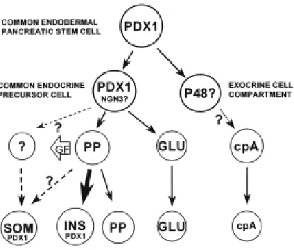
ria toxin A chain (DT)-coding region. These results unambiguously demonstrated that neither glucagon nor insulin gene-expressing cells are essential for the differentiation of the other islet endocrine-cell types; they are thus incompatible with a model of islet develop-ment according to which cells co expressing glucagon and insulin would be precursors to all islet endocrine cells. They also suggested that PP gene-expressing cells are indispensable for the differentia-tion of islet β and δ cells, either because they produce a necessary paracrine or endocrine factor and/or because of a cell-lineage relationship (Fig. 3).
Islet Cell Lineage Analyses
In vivo cell ablation is one approach to depict ontogenic relation-ships, but it does not allow to discriminate between lineage and paracrine/endocrine relationships. Similarly, a gene KO approach is not suitable for demonstrating a cell lineage connection. Only the ability of irreversibly labeling a cell permits to follow its fate over time. Developments in gene recombination technology allowed only recently setting up an approach to “mark” irreversibly cells at one developmental stage using a given promoter, and therefore to identify all their descendants. In the context of our work, this involved the genetic labeling of cells that transcribe the insulin, glucagon, PP or PDX1 genes early during development, and the analysis of islet α- and β-cells later in development or in the adult, to determine whether they are derived from these early insulin, glucagon, PP or PDX1 gene-expressing cells.
To study the cell lineages of the endocrine pancreas we have used a version of the Cre/loxP system (Barinaga, 1994; Ramirez-Solis and Bradley, 1994; Sauer, 1993; Sauer, 1994; Zou et al., 1994), whereby a reporter transgene is “activated” in a tissue-specific manner (Jacob and Baltimore, 1999). The experimental design consists in generating mice bearing two transgenes (sum-marized in Fig. 4). One transgene, “reporter”, placed under the control of either the insulin (β-specific) or the glucagon (α -specific) gene promoter, contains a hGH coding region placed downstream of a “floxed” (i.e., loxP-flanked) transcription
termi-nation site (STOP sequence) (Lakso et al., 1992; Sauer, 1993; Tsien et al., 1996). The native reporter transgene cannot be expressed in β- or α-cells because of the STOP sequence; its expression requires the deletion of this sequence, which can be obtained in the presence of Cre recombinase. hGH is a good marker for immunohistochemical detection of gene expression, and has been used by others in a number of transgenic studies (Artavanis-Tsakonas et al., 1995; Lardelli et al., 1995; Tsien et al., 1996; Weinmaster, 1997).
A second, or “tagger” transgene, consists of the Cre recombinase gene placed under the control of a promoter active in putative progenitor cells; its expression results in the deletion of the STOP site of the reporter transgenes, thus allowing hGH synthesis. This approach has been used by others to activate the transcription of either the SV40 T antigen, the LacZ, or the placental alkaline phosphatase genes in transgenic mice (Jacob and Baltimore, 1999; Lakso et al., 1992; Tsien et al., 1996), but not to analyze cell lineages.
Using this genetic approach allowing to follow cell lineages through the expression of Cre recombinase, we showed that: 1) adult α- and β-cells derive from cells that have never transcribed insulin or glucagon, respectively, 2) β-cell progenitors but not α-cell progenitors transcribe the PP gene, and 3) the PDX1 gene, whose expression characterizes the earliest pancreatic stem cells, and which is expressed by adult β-cells, is also expressed by α-cell progenitors (Herrera, 2000). Thus the islet α- and β-cell lineages appear to arise independently during ontogeny, probably from a common precursor (Fig. 5).
The power and elegance of this innovative approach also resides in the versatility of the transgenic mice encoding Cre recombinase in different subsets of pancreatic cell types. Thus, after these mice were employed to determine cell lineage relation-ships, they are now unique tools to inactivate specific genes in these same cell lineages (“conditional” KO mice).
Role of Notch1 in Pancreas Differentiation: Generation
and Study of Pancreas-Specific Notch1 Knock-Out Mice
The generation of a multicellular organism, or of a single organ, results from a cascade of coordinate developmental decisions taken by cells as a consequence of the interactions between them and their neighbors. Many regulative events of this nature are mediated by Notch, a family of transmembrane proteins (Artavanis-Tsakonas et al., 1995; Lardelli et al., 1995; Weinmaster, 1997); these receptors are components of a highly conserved signaling mechanism present in the whole Animal Kingdom. As a conse-quence of such signaling interactions, specific transcription factors are then induced to achieve the expression of cell-type specific products, such as the different islet hormones. Inactivation of Notch1 in mice by homologous recombination results in embryo death early after implantation (at about E9 - E9.5) (Swiatek et al., 1994). Notch receptors are broadly expressed in embryos, and continue to be expressed in uncommitted and proliferative cells throughout life (Fehon et al., 1991; Xu et al., 1992). The Notch ligands belong to the Delta family of transmembrane proteins: activation of Notch receptors by Delta-like (Dlk) proteins either prevents or influences the differentiation program of a cell by modifying its ability to reply to other instructive signals (Weinmaster, 1997). When Notch is inactivated by Delta (expressed by differen-tiating neighboring cells), it triggers the expression of bHLH genes such as Hes-1, inhibitors of the expression of proneural genes. Notch1, 2 and 3 are expressed in developping pancreas, as well as the ligands Jag1, 2, Dll1, 3, and Hes-1 (Jensen et al., 2000). It appears that the Notch signalling pathway is involved in pancreatic differentiation (Apelqvist et al., 1999), so that mice lacking either Dll1 or RBP-jk (in the Notch cascade), which are not viable, have an accelerated differentiation of endocrine cells in pancreatic buds (Apelqvist et al., 1999). Mice having a Hes-1 null mutation have a
severe pancreatic hypoplasia due to a loss of epithelial precursor cells: in the absence of Hes-1 there is an accelerated differentiation of glucagon-expressing cells (Jensen et al., 2000).
To precisely study the role of Notch1 in the ontogeny of the endocrine pancreas we have devised the pancreas-specific inac-tivation of Notch1, in doubly transgenic mice, using the Cre/LoxP system. This study is ongoing, in collaboration with M. Aguet and F. Radtke (Lausanne, Switzerland), which provided the Notch1 floxed (i.e. loxP-flanked) mice. We are using the Pdx1 promoter-Cre, Insulin promoter-promoter-Cre, and Glucagon promoter-Cre mice described earlier, as well as the Pax6 promoter-Cre animals, in which the recombinase is made in all islet cell types (Ashery-Padan et al., 2000). The preliminary results suggest that the conditional inactivation of Notch1 specifically in the precursors of the endocrine pancreas results in a reduction of the proportion of endocrine tissue, and that the silencing of Notch1 in cells already transcribing the insulin or glucagon genes has no further conse-quences in their viability or function (manuscript in preparation).
Concluding Remarks
Besides the fact that embryonic stem (ES) cells might be in the future a source of differentiated cell types, one should not forget that the potential to form new such differentiated cells is not limited to ES cells. In particular, as an example, formation of pancreatic endocrine tissue is not restricted to the period of embryonic development. Interestingly, it is thought that islet neogenesis in adults is probably similar in its intrinsic mechanisms to that of the embryonic development. Future experiments should be directed at determining the extent of such similarities. In this context, there are efforts put on trying to obtain pancreatic pluripotent cell lines that should allow developing an in vitro model of β-cell development for transplantation in diabetes.
As a final consideration, we would like to suggest that the ontogenic relationship described here between different islet endocrine cells (Herrera et al., 1994; Herrera, 2000) may be viewed in an evolutionary perspective. In particular, the relation between PP promoter activity and formation of new insulin-secreting cells. The endocrine pancreas has taken up, in the course of evolution, the task of participating in the control of nutrients’ metabolism. In early Protostomians, this might have been achieved by subsets of nerve cells and mediated by the secretion of neuropeptides, for instance from the PP-fold family such as NPY (Falkmer et al., 1985). In late Protostomians, like Insects, the four islet hormones are present in brain neurons (Falkmer et al., 1985): these may be the evolutionary descen-dants of the putative earlier PP-family neuropeptide secreting cells. In this view, expression of the PP gene family would be a hallmark of the precursors to brain insulin cells during evolution, as it appears to be in the pancreas for islet β-cells during ontogeny (Herrera et al., 1994; Herrera, 2000).
References
AHLGREN, U., JONSSON, J. and EDLUND, H. (1996). The morphogenesis of the pancreatic mesenchyme is uncoupled from that of the pancreatic epithelium in IPF1/PDX1-deficient mice. Development 122: 1409-16.
AHLGREN, U., PFAFF, S. L., JESSELL, T. M., EDLUND, T. and EDLUND, H. (1997). Independent requirement for ISL1 in formation of pancreatic mesenchyme and islet cells. Nature 385: 257-60.
ALPERT, S., HANAHAN, D. and TEITELMAN, G. (1988). Hybrid insulin genes reveal a developmental lineage for pancreatic endocrine cells and imply a relationship with neurons. Cell 53: 295-308.
APELQVIST, A., LI, H., SOMMER, L., BEATUS, P., ANDERSON, D. J., HONJO, T., HRABE DE ANGELIS, M., LENDAHL, U. and EDLUND, H. (1999). Notch signalling controls pancreatic cell differentiation. Nature 400: 877-81.
ARTAVANIS-TSAKONAS, S., MATSUNO, K. and FORTINI, M. E. (1995). Notch signaling. Science 268: 225-32.
ASHERY-PADAN, R., MARQUARDT, T., ZHOU, X. and GRUSS, P. (2000). Pax6 activity in the lens primordium is required for lens formation and for correct placement of a single retina in the eye. Genes Dev 14: 2701-11.
BARINAGA, M. (1994). Knockout mice: round two [published erratum appears in Science 1994 265: 855]. Science 265: 26-8.
BONNER-WEIR, S., BAXTER, L. A., SCHUPPIN, G. T. and SMITH, F. E. (1993). A second pathway for regeneration of adult exocrine and endocrine pancreas. A possible recapitulation of embryonic development. Diabetes 42: 1715-20.
BONNER-WEIR, S., TRENT, D. F. and WEIR, G. C. (1983). Partial pancreatectomy in the rat and subsequent defect in glucose- induced insulin release. J Clin Invest 71: 1544-53.
BROCKENBROUGH, J. S., WEIR, G. C. and BONNER-WEIR, S. (1988). Discor-dance of exocrine and endocrine growth after 90% pancreatectomy in rats. Diabetes 37: 232-6.
EDLUND, H. (1998). Transcribing pancreas. Diabetes 47: 1817-23.
ESNI, F., JOHANSSON, B. R., RADICe, G. L. and SEMB, H. (2001). Dorsal Pancreas Agenesis in N-Cadherin-Deficient Mice. Dev Biol 23:, 202-212.
FALKMER, S., DAFGARD, E., EL-SALHY, M., ENGSTROM, W., GRIMELIUS, L. and ZETTERBERG, A. (1985). Phylogenetical aspects on islet hormone families: a minireview with particular reference to insulin as a growth factor and to the phylogeny of PYY and NPY immunoreactive cells and nerves in the endocrine and exocrine pancreas. Peptides 6 Suppl 3: 315-20.
FEHON, R. G., JOHANSEN, K., REBAY, I. and ARTAVANIS-TSAKONAS, S. (1991). Complex cellular and subcellular regulation of notch expression during embryonic and imaginal development of Drosophila: implications for notch function. J Cell Biol 113: 657-69.
GRADWOHL, G., DIERICH, A., LEMEUR, M. and GUILLEMOT, F. (2000). neurogenin3 is required for the development of the four endocrine cell lineages of the pancreas. Proc Natl Acad Sci USA 97: 1607-11.
HARRISON, K. A., THALER, J., PFAFF, S. L., Gu, H. and KEHRL, J. H. (1999). Pancreas dorsal lobe agenesis and abnormal islets of Langerhans in Hlxb9-deficient mice. Nat Genet 23: 71-5.
HEBROK, M., KIM, S. K. and MELTON, D. A. (1998). Notochord repression of endodermal Sonic hedgehog permits pancreas development. Genes Dev 12: 1705-13.
HERRERA, P. L. (2000). Adult insulin- and glucagon-producing cells differentiate from two independent cell lineages. Development 127: 2317-22.
HERRERA, P. L., HUARTE, J., SANVITO, F., MEDA, P., ORCI, L. and VASSALLI, J. D. (1991). Embryogenesis of the murine endocrine pancreas; early expression of pancreatic polypeptide gene. Development 113: 1257-65.
HERRERA, P. L., HUARTE, J., ZUFFEREY, R., NICHOLS, A., MERMILLOD, B., PHILIPPE, J., MUNIESA, P., SANVITO, F., ORCI, L. and VASSALLI, J. D. (1994). Ablation of islet endocrine cells by targeted expression of hormone- promoter-driven toxigenes. Proc Natl Acad Sci USA 91: 12999-3003.
JACOB, J. and BALTIMORE, D. (1999). Modelling T-cell memory by genetic marking of memory T cells in vivo. Nature 399: 593-7.
JENSEN, J., PEDERSEN, E. E., GALANTE, P., HALD, J., HELLER, R. S., ISHIBASHI, M., KAGEYAMA, R., GUILLEMOT, F., SERUP, P. and MADSEN, O. D. (2000). Control of endodermal endocrine development by hes-1. Nat Genet 24: 36-44.
JONSSON, J., CARLSSON, L., EDLUND, T. and EDLUND, H. (1994). Insulin-promoter-factor 1 is required for pancreas development in mice. Nature 371: 606-9.
KAESTNER, K. H., KATZ, J., LIU, Y., DRUCKER, D. J. and SCHUTZ, G. (1999). Inactivation of the winged helix transcription factor HNF3alpha affects glucose homeostasis and islet glucagon gene expression in vivo. Genes Dev 13: 495-504.
KRAPP, A., KNOFLER, M., LEDERMANN, B., BURKI, K., BERNEY, C., ZOERKLER, N., HAGENBUCHLE, O. and WELLAUER, P. K. (1998). The bHLH protein PTF1-p48 is essential for the formation of the exocrine and the correct spatial
organiza-tion of the endocrine pancreas. Genes Dev 12: 3752-63.
LaksO, M., SAUER, B., MOSINGER, B., JR., LEE, E. J., MANNING, R. W., YU, S. H., MULDER, K. L. and WESTPHAL, H. (1992). Targeted oncogene activation by site-specific recombination in transgenic mice. Proc Natl Acad Sci USA 89: 6232-6.
LARDELLI, M., WILLIAMS, R. and LENDAHL, U. (1995). Notch-related genes in animal development. Int J Dev Biol 39: 769-80.
LARSSON, L. I. (1998). On the development of the islets of Langerhans. Microsc Res Tech 43: 284-91.
LARSSON, L. I., ST-ONGE, L., HOUGAARD, D. M., SOSA-PINEDA, B. and GRUSS, P. (1998). Pax 4 and 6 regulate gastrointestinal endocrine cell development. Mech Dev 79: 153-9.
LE DOUARIN, N. M. (1988). On the origin of pancreatic endocrine cells. Cell 53: 169-171.
LI, H., ARBER, S., JESSELL, T. M. and EDLUND, H. (1999). Selective agenesis of the dorsal pancreas in mice lacking homeobox gene Hlxb9. Nat Genet 23: 67-70.
NAYA, F. J., HUANG, H. P., QIU, Y., MUTOH, H., DEMAYO, F. J., LEITER, A. B. and TSAI, M. J. (1997). Diabetes, defective pancreatic morphogenesis, and abnormal enteroendocrine differentiation in BETA2/neuroD-deficient mice. Genes Dev 11: 2323-34.
OFFIELD, M. F., JETTON, T. L., LABOSKY, P. A., RAY, M., STEIN, R. W., MAGNUSON, M. A., HOGAN, B. L. and WRIGHT, C. V. (1996). PDX-1 is required for pancreatic outgrowth and differentiation of the rostral duodenum. Development 122: 983-95.
ORCI, L. (1982). Macro- and micro-domains in the endocrine pancreas. Diabetes 31: 538-65.
ORCI, L. and UNGER, R. H. (1975). Functional subdivision of islets of Langerhans and possible role of D cells. Lancet 2: 1243-1244.
PETERS, J., JURGENSEN, A. and KLOPPEL, G. (2000). Ontogeny, differentiation and growth of the endocrine pancreas. Virchows Arch 436: 527-38.
PICTET, R. L., CLARK, W. R., WILLIAMS, R. H. and RUTTER, W. J. (1972). An ultrastructural analysis of the developing embryonic pancreas. Dev.Biol. 29: 436-467.
RALL, L. B., PICTET, R. L., WILLIAMS, R. H. and RUTTER, W. J. (1973). Early differentiation of glucagon-producing cells in embryonic pancreas: a possible developmental role for glucagon. Proc.Natl.Acad.Sci.USA 70: 3478-3482.
RAMIREZ-SOLIS, R. and BRADLEY, A. (1994). Advances in the use of embryonic stem cell technology. Curr Opin Biotechnol 5: 528-33.
SANDER, M. and GERMAN, M. S. (1997). The beta cell transcription factors and development of the pancreas. J Mol Med 75: 327-40.
SANVITO, F., HERRERA, P. L., HUARTE, J., NICHOLS, A., MONTESANO, R., ORCI, L. and VASSALLI, J. D. (1994). TGF-beta 1 influences the relative development of the exocrine and endocrine pancreas in vitro. Development 120: 3451-62.
SANVITO, F., NICHOLS, A., HERRERA, P. L., HUARTE, J., WOHLWEND, A., VASSALLI, J. D. and ORCI, L. (1995). TGF-beta 1 overexpression in murine pancreas induces chronic pancreatitis and, together with TNF-alpha, triggers insulin-dependent diabetes. Biochem Biophys Res Commun 217: 1279-86.
SAUER, B. (1993). Manipulation of transgenes by site-specific recombination: use of Cre recombinase. Methods Enzymol 225: 890-900.
SAUER, B. (1994). Site-specific recombination: developments and applications. Curr Opin Biotechnol 5: 521-7.
SCHWITZGEBEL, V. M., SCHEEL, D. W., CONNERS, J. R., KALAMARAS, J., LEE, J. E., ANDERSON, D. J., SUSSEL, L., JOHNSON, J. D. and GERMAN, M. S. (2000). Expression of neurogenin3 reveals an islet cell precursor population in the pancreas. Development 127: 3533-42.
SLACK, J. M. (1995). Developmental biology of the pancreas. Development 121: 1569-80.
SOSA-PINEDA, B., CHOWDHURY, K., TORRES, M., OLIVER, G. and GRUSS, P. (1997). The Pax4 gene is essential for differentiation of insulin-producing beta cells in the mammalian pancreas. Nature 386: 399-402.
St-ONGE, L., WEHR, R. and GRUSS, P. (1999). Pancreas development and diabetes. Curr Opin Genet Dev 9: 295-300.
SUMI, S. and TAMURA, K. (2000). Frontiers of pancreas regeneration. J Hepatobiliary Pancreat Surg 7: 286-94.
SUSSEL, L., KALAMARAS, J., HARTIGAN-O’CONNOR, D. J., MENESES, J. J., PEDERSEN, R. A., RUBENSTEIN, J. L. and GERMAN, M. S. (1998). Mice lacking the homeodomain transcription factor Nkx2.2 have diabetes due to arrested differentiation of pancreatic beta cells. Development 125: 2213-21.
SWIATEK, P. J., LINDSELL, C. E., DEL AMO, F. F., WEINMASTER, G. and GRIDLEY, T. (1994). Notch1 is essential for postimplantation development in mice. Genes Dev 8: 707-19.
TEITELMAN, G., ALPERT, S., POLAK, J. M., MARTINEZ, A. and HANAHAN, D. (1993). Precursor cells of mouse endocrine pancreas coexpress insulin, gluca-gon and the neuronal proteins tyrosine hydroxylase and neuropeptide Y, but not pancreatic polypeptide. Development 118: 1031-9.
TSIEN, J. Z., CHEN, D. F., GERBER, D., TOM, C., MERCER, E. H., ANDERSON, D. J., MAYFORD, M., KANDEL, E. R. and TONEGAWA, S. (1996). Subregion-and cell type-restricted gene knockout in mouse brain. Cell 87: 1317-26.
WANG, R. N., KLOPPEL, G. and BOUWENS, L. (1995). Duct- to islet-cell differen-tiation and islet growth in the pancreas of duct-ligated adult rats. Diabetologia 38: 1405-11.
WEINMASTER, G. (1997). The ins and outs of notch signaling. Mol Cell Neurosci 9: 91-102.
WELLS, J. M. and MELTON, D. A. [1999]. Vertebrate endoderm development. Annu Rev Cell Dev Biol 15: 393-410.
XU, G., STOFFERS, D. A., HABENER, J. F. and BONNER-WEIR, S. (1999). Exendin-4 stimulates both beta-cell replication and neogenesis, resulting in increased beta-cell mass and improved glucose tolerance in diabetic rats. Diabetes 48: 2270-6.
XU, T., CARON, L. A., FEHON, R. G. and ARTAVANIS-TSAKONAS, S. (1992). The involvement of the Notch locus in Drosophila oogenesis. Development 115: 913-22.
YAMAOKA, T. and ITAKURA, M. (1999). Development of pancreatic islets (Re-view). Int J Mol Med 3: 247-261.