YEASTBOOK
GENOME ORGANIZATION & INTEGRITY
Structure and Function in the Budding
Yeast Nucleus
Angela Taddei* and Susan M. Gasser†,1
*Unité Mixte de Recherche 218, Centre National de la Recherche Scientifique/Institut Curie-Section de Recherche, 75231 Paris Cedex 05, France, andyFriedrich Miescher Institute for Biomedical Research, Maulbeerstrasse 66, CH-4058 Basel, Switzerland
ABSTRACTBudding yeast, like other eukaryotes, carries its genetic information on chromosomes that are sequestered from other cellular constituents by a double membrane, which forms the nucleus. An elaborate molecular machinery forms large pores that span the double membrane and regulate the traffic of macromolecules into and out of the nucleus. In multicellular eukaryotes, an intermediatefilament meshwork formed of lamin proteins bridges from pore to pore and helps the nucleus reform after mitosis. Yeast, however, lacks lamins, and the nuclear envelope is not disrupted during yeast mitosis. The mitotic spindle nucleates from the nucleoplasmic face of the spindle pole body, which is embedded in the nuclear envelope. Surprisingly, the kinetochores remain attached to short microtubules throughout interphase, influencing the position of centromeres in the interphase nucleus, and telomeres are found clustered in foci at the nuclear periphery. In addition to this chromosomal organization, the yeast nucleus is functionally compartmentalized to allow efficient gene expression, repression, RNA processing, genomic replication, and repair. The formation of functional subcompartments is achieved in the nucleus without intranuclear membranes and depends instead on sequence elements, protein–protein interactions, specific anchorage sites at the nuclear envelope or at pores, and long-range contacts between specific chromosomal loci, such as telomeres. Here we review the spatial organization of the budding yeast nucleus, the proteins involved in forming nuclear subcompartments, and evidence suggesting that the spatial organization of the nucleus is important for nuclear function.
TABLE OF CONTENTS
Abstract 107
Introduction 108
Features of the Yeast Nucleus 108
Unique and conserved characteristics 108
Nuclear envelope and nuclear pore complex 109
Long-range chromosome organization 111
Chromatin dynamics 112
DNA-based compartments 112
Nucleolus: 112
Telomere foci—assemblies of repetitive DNA and silencing factors: 113
tRNA genes: 114
Replication foci: 114
Continued
Copyright © 2012 by the Genetics Society of America doi: 10.1534/genetics.112.140608
Manuscript received March 19, 2012; accepted for publication May 29, 2012
CONTENTS,continued
Sites of DNA repair: 115
Mechanisms Underlying Nuclear Compartmentation 115
Redundancy in telomere-anchoring pathways 115
Minimal anchoring assay and the validation of genetic screens 116
Formation of chromatin compartments: association in trans 117
Mechanisms of chromatin movement 118
Functional Consequences of Nuclear Organization 119
Gene silencing 119
Gene activation 119
Boundaries between active and inactive domains 120
Effects on genome stability 121
Mobility of DSBs: 121
Nuclear pores and Mps3 anchorage in noncanonical repair: 122
Regulation of recombination at the rDNA locus: 122
Concluding Remarks 123
T
HE cell nucleus not only harbors and expresses an organ-ism’s essential genetic blueprint, but also ensures the proper expression, duplication, repair, and segregation of chromosomes while ensuring proper processing and export of messenger and ribosomal RNA (Spector 2003; Taddei et al.2004b). The dense packing of highly charged molecules (DNA, RNA, histones, nonhistone proteins) in a limited nuclear space was once thought to constrain molecular dynamics, yet we now know that the nucleus is neither grid-locked nor a ran-dom jumble (Rouquetteet al.2010). Large rings of chromatin diffuse freely through the nuclear volume in a random diffusive walk (Gartenberget al.2004; Neumannet al.2012), yet the nucleus can maintain functional subcompartments enriched for specific enzymes and chromatin states. Understanding this di-chotomy is key to understanding how nuclear organization facilitates nuclear function (reviewed in Taddeiet al.2004b; Mekhail and Moazed 2010; Egecioglu and Brickner 2011; Rajapakse and Groudine 2011; Zimmer and Fabre 2011).Chromosomes, and the nucleosomal fibers within them, can be thought of as basic structural elements of the nucleus. Long-range chromosome folding is constrained by the physics of polymer dynamics (Kleninet al.1998; Dekkeret al.2002; Gehlenet al.2006; Neumannet al.2012), and the character-istics of the chromosome polymers themselves depend on the folding of the nucleosomalfiber (Rosa and Everaers 2008). Yet chromatin in interphase nuclei is not regularly compacted and is subject to reversible covalent modifications on both DNA and histones within the nucleosomal fiber. Therefore, crucial biophysical properties of long-range chromatin dy-namics, such as persistence length, mass density, and dif-fusion rate, are variable and subject to changes induced by nucleosome remodelers and histone modifiers. Thus, the post-translational modification of histones contributes not only to local chromatin folding, but to the three-dimensional organization of the genome (van Steensel 2011).
A second major factor contributing to nuclear organization is the interaction between chromatin and stable structural elements of the nucleus. In budding yeast, the key structural elements are the nuclear envelope (NE), the nuclear pore complex (NPC), and the nucleolus. The NE encompasses different types of chromatin anchorage sites, including the spindle pole body (SPB), and unique protein components of the inner nuclear membrane that tether heterochromatin, the ribosomal DNA (rDNA), or different types of DNA damage (Akhtar and Gasser 2007; Mekhail and Moazed 2010). The NPC also plays a role in the transient anchoring of activated genes or of DNA damage that cannot be readily repaired by homologous recombination. Finally, long-range interaction of loci intrans, such as the clustering of telomeres or of trans-fer RNA (tRNA) genes, influences nuclear order. The com-bination of physical constraints on chromatin movement and protein–protein interactions helps generate nuclear subcom-partments that are enriched for specific DNA sequences, factors, and enzymatic activities (Gasser et al. 2004; Rosa and Everaers 2008). How these subcompartments affect nuclear function remains a central topic of research.
The basic principles of nuclear organization can be ob-served in all eukaryotes from yeast to humans. This allows us to test the functional implications of nuclear organization in a single-celled organism, despite there being species- and tissue-specific nuclear features. With facile genetics, live mi-croscopy, and genome-wide mapping approaches, budding yeast has proven to be extremely useful for testing the functional roles of nuclear structure, as reviewed below.
Features of the Yeast Nucleus
Unique and conserved characteristics
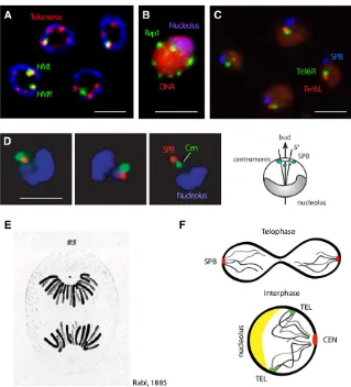
described above, the SPB is embedded in the nuclear envelope and nucleates both intranuclear microtubules in interphase and the mitotic spindle. The SPB has a position strictly determined by the site of new bud emergence (Lee et al. 1999), which itself is opposite the nucleolus (Yang et al. 1989; Bystricky et al. 2004, 2005). The position of bud emergence is determined by landmark proteins at the cell cortex and Cdc42, which are positioned through asso-ciation with the previous bud site. These proteins nucleate actinfilaments and determine the orientation of cytoplasmic actin. The nucleus, in turn, is oriented by the cooperative action of SPB-microtubule-Kar9-Myo2and actin interactions in a manner dependent on the actin filament orientation (Gundersen and Bretscher 2003; Slaughter et al.2009). In other organisms, the link between the cytoskeleton and the nucleus stems from cytoskeletal interactions with Nesprins, KASH-domain and SUN-domain proteins that span from the outer through the inner nuclear membrane (INM) (Fridkin et al.2009; Razafsky and Hodzic 2009). There is one SUN-domain protein in yeast called monopolar spindle protein 3 (Mps3), which is an important anchor of the INM, but whose link to the cytoskeleton is not yet known.
As mentioned above, the centromeres of yeast chromo-somes remain attached to the SPB by short interphase microtubules, and this interaction strongly orients chromo-somes within the interphase nucleus by keeping centro-meres clustered (Guacci et al.1997; Jin et al. 1998; Heun et al.2001b; Bystrickyet al.2004). Opposite this landmark is the nucleolus, which is generated around a single rDNA locus on chromosome XII (Chr XII) that contains200 tan-dem copies of a 9.1-kb repeat. The positioning of this left arm of Chr XII, and the nucleolus that forms around it, strongly influences nuclear order. A subset of rDNA repeats are tethered to the NE to suppress recombination (Mekhail et al. 2008), keeping the nucleolus tightly associated with the nuclear periphery and lending a striking polarity to the yeast nucleus by restricting diffusion of this large chromo-some (Figure 1, B and D).
Apart from the rDNA repeat unit, budding yeast chro-mosomes have little repetitive DNA, and—most notably—no simple satellite repeat DNA at centromeres. This eliminates centric heterochromatin, a major chromosomal structural fea-ture that in other organisms impacts neighboring sequences both in cis and in trans(reviewed in Akhtar and Gasser 2007). The organizational role of the transcriptionally inert, compacted chromatin of mammalian centromeres is ful-filled, at least in part, by the TG-repeat DNA found at yeast telomeres. These TG repeats generate repressive subtelomeric chromatin domains, which spread for several kilobases from the chromosomal ends, silencing nearby promoters (Gottschling et al. 1990). This phenomenon is called telomere position effect (TPE), in analogy to the position effect variegation (PEV) that spreads from satellite-containing centromeres in other species. The repressive chromatin formed at telomeres requires the binding of the silent information regulatory (SIR) proteins to nucleosomes, which also occurs at the two silent
mating-type loci, HML andHMR (reviewed in Rusche et al. 2003). Interestingly, native subtelomeric genes are transcrip-tionally inert under standard growth conditions independent of SIR factor binding, as they carry genes that are expressed only under restrictive nutrient conditions (Fabreet al.2005). Budding yeast lacks the histone H3 K9 methylation that typifies centromeric heterochromatin in most other eukary-otes, as well as the major protein ligand that recognizes this modification (heterochromatin protein 1, or HP1) and the RNA interference machinery that facilitates PEV in fission yeast (reviewed in Buhler and Gasser 2009). Instead, a tri-meric complex ofSir2,Sir3, andSir4recognizes unmodified nucleosomes to repress transcription and reduce endonucle-ase accessibility. Importantly, like PEV, yeast silent chroma-tin can be propagated through mitosis in a heritable manner thanks to its continual nucleation by silencer elements or telomeric repeats (reviewed in Rusche et al. 2003). As in higher eukaryotes, this heterochromatic state is late replicat-ing (Raghuramanet al. 2001) and is found adjacent to the NE, sequestered away from nuclear pores (Palladino et al. 1993; Taddeiet al.2004a) (Figure 1B).
In addition to lacking repressive methylation marks, the yeast genome is also unique in that it lacks canonical linker histones (e.g., H1 and H5) and has a shorter nucleosomal repeat length (165 bp rather than 200 bp; see Woodcock et al.2006). While the yeast genome encodes an H1-related protein, Hho1, this protein binds nucleosomal linker DNA only in rare instances and is not a core component of yeast chromatin (Freidkin and Katcoff 2001). Yeast also lacks the histone H3 subvariant H3.1 and macroH2A, while yeast his-tone H3 is equivalent to vertebrate H3.3, and the yeast H2A serves as the damage-associated, phospho-accepting variant H2AX of other species (Kusch and Workman 2007). Finally, like most organisms with a closed mitosis, yeast lacks the nuclear intermediate filament protein lamin. Nonetheless, yeast expresses other structural proteins of the INM orthol-ogous to Man1 and emerin (Mekhail and Moazed 2010), which are lamin-associated proteins that contribute to chro-matin anchoring in higher eukaryotes (Fridkinet al.2009). Importantly, yeast has allowed one to test the functional impact of chromatin sequestration by the NE by mutating these structural proteins and monitoring effects on tran-scription and genome stability.
Nuclear envelope and nuclear pore complex
messenger RNA (mRNA) transcription and quality control, as well as its export, and tether a subpopulation of inducible genes after activation, both through the mRNA and a quality-control export complex called Tho-Trex (Dieppois and Stutz 2010).
NPCs are massive assemblies of50 MDa containing 456 nucleoporins of 30 different types (D’Angelo and Hetzer 2008). They form a doughnut-shaped structure with an eightfold symmetry around a central channel, with flexible protein filaments emanating from the core into both the cytoplasm and the nucleoplasm. These provide binding sites for the transport of proteins, mRNA, and chromatin. A de-tailed map for the relative position of each nucleoporin was calculated on the basis of multiple molecular, biochemical, and structural data revealing a strongly modular structure (Alberet al.2007).
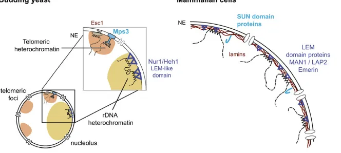
Several conserved proteins, which in other species associate with lamins, are found at the NE in yeast (Huh et al.2003). Of particular interest are the integral proteins of the INM (Lusk et al. 2007), including Doa10, a RING do-main containing proteins that targets nuclear proteins for degradation; Mps3, a member of the SUN (Sad1, UNC-84) family that is a shared component of the INM and the SPB (Jaspersen et al. 2002); and helix–extension–helix-1 and -2 (Heh1 and Heh2) (M. C. King et al. 2006; Fridkin
et al. 2009), which are orthologs of the mammalian lamin-associated protein MAN1 (Figure 2). While these proteins are thought to bridge from the INM or lamina to chromatin in multicellular organisms, in most cases the ligand on the chromatin side is still unknown (Fridkin et al. 2009; Razafsky and Hodzic 2009). The yeast SUN-domain protein Mps3 and the two MAN1 homologs (Heh1 or Src1 and Heh2) serve a similar function in yeast, despite the ab-sence of lamins (Grund et al. 2008; Mekhail and Moazed 2010). In addition to these conserved components, a yeast-specific protein called Esc1 (establishes silent chromatin) (Andruliset al.2002) associates tightly with the inner face of the INM where it anchors silent chromatin through its high affinity forSir4 (Andruliset al. 2002; Gartenberg et al.2004; Taddeiet al.2004a).Esc1may have other func-tions, as its overexpression induces INM expansion (Hattier et al. 2007). Not surprisingly, functional cross talk exists between INM proteins (e.g., Esc1 and Mps3) and the nu-clear pore basket proteins (e.g., Nup60, Mlp1, and Mlp2) (Therizols et al.2006; Lewis et al.2007; Palancade et al. 2007) in that loss of either a INM or a pore protein can interfere with the function of the other, even though they define spatially distinct domains of the NE when localized at high resolution (Taddei et al. 2004a; Horigome et al. 2011).
Long-range chromosome organization
Budding yeast chromosomes assume a Rabl-like conforma-tion throughout the vegetative cell cycle (Figure 1F). The Rabl orientation reflects the spatial orientation of anaphase chromosomes, which means that yeast chromosome arms extend away from the centromeres that are held by the SPB (Rabl 1885; Yang et al. 1989; Dekkeret al.2002; Jin et al.1998, 2000). Telomeres are generally found in clusters around the nuclear periphery (Figure 1B) (Palladino et al. 1993). This organization was demonstrated using fl uores-cence insituhybridization (FISH), as well as by immunofl u-orescence (IF) for centromeric and telomeric proteins (Gotta et al.1996; Jin et al.1998). Later these perinuclear telo-mere foci were confirmed and tracked through the cell cycle using live imaging of GFP-tagged telomeres (Schober et al.2008; Therizolset al.2010). Whereas a polarized chro-mosomal organization exists transiently after telophase in most metazoan cells, it persists through interphase in bud-ding yeast due to the persistent attachment of centromeres that remain linked by short intranuclear microtubules to the SPB (Guacciet al.1997; Jinet al.1998; Heunet al.2001b; Bystricky et al. 2004). Treatment of interphase cells with nocodazole allows centromeres to move away from the SPB (Jin et al. 1998; Heun et al. 2001b; Bystricky et al. 2004).
In 2002, the Kleckner laboratory developed a molecular approach to monitor and model chromosome conformation [chromosome conformation capture (3C)] (Dekker et al. 2002) based on the detection of long-range interactions within and between chromosomes. A population-averaged three-dimensional model of Chr III was thus determined. In this model, Chr III appears to fold as a contorted ring, with a strong bend near the centromere and the telomeres in close proximity to each other (Dekker et al. 2002). This was confirmed byfluorescent tagging of right and left telo-meres of Chr III and for a second short, metacentric chro-mosome (Chr VI) (Bystrickyet al.2005). Later, it was shown that telomere-mediated chromosome looping does not occur in chromosomes that have arms of unequal lengths (Schober et al. 2008; Therizols et al. 2010). The impact of chromo-some arm length on selective telomere–telomere interaction
was demonstrated in an elegant experiment in which long and short arms of different chromosomes were swapped. This converted a chromosome with unequal arm lengths to one with equal arm lengths, which enhanced intrachromo-somal telomere–telomere interaction (Schoberet al.2008). Genome-wide conformation capture approaches (Hi-C), inspired from the 3C method of Dekker et al.(2002), con-firmed the clustering of centromeres in interphase cells and the widespread associations between pairs of telomeres on different chromosomes that had been observed by fl uo-rescence microscopy (Duan et al.2010). The Hi-C analysis also confirmed that two unlinked telomeres positioned at similar distances from their corresponding centromeres are more likely to interact than telomeres on arms of different lengths (Duan et al.2010; Therizols et al.2010), again il-lustrating the impact of the Rabl organization on long-range interactions (Figure 1). Interchromosomal contacts were also detected among clustered tRNA genes, early origins of DNA replication, and at sites of chromosomal breakage (Duanet al.2010).
Importantly, interactions within a chromosome (such as those formed by right and left telomeres) occur far more frequently than interactions between different chromosomes (Rodleyet al.2009; Duanet al.2010). This applies even to large chromosomes and chromosomes with unequal arms, arguing that a yeast chromosome defines a spatial unit or “territory.”Territory positioning is, of course, subject to ar-chitectural constraints imposed by centromere attachment to the SPB and telomere anchoring to the NE (Berger et al.2008; Therizolset al.2010), but territories could also be modeledin silicoon the basis of polymer diffusion kinet-ics, without need for a proteinaceous scaffold or matrix (Rosa and Everaers 2008). In yeast, nonetheless, territories can be significantly remodeled by transcriptional activation (Bergeret al.2008), which is different from the situation in differentiated cells of multicellular organisms, where chro-mosome territories play a more dominant role in nuclear organization (Rouquette et al. 2010). This dominance can be explained both by the sheer size of mammalian chromo-somes, each of which is larger than the entire yeast genome, and by the abundance of repetitive, noncoding DNA sequences in larger genomes.
Chromatin dynamics
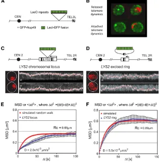
Although chromosomes show a recognizable pattern of posi-tioning, chromatin in all living cells is subjected to constant motion, which has been described as a constrained random walk (Marshallet al.1997; Gasser 2002). Rapid time-lapse imaging led to the distinction of at least two types of motion in yeast: small random movements (,0.2mm within 1.5 sec) that occur constantly, as well as larger steps over relatively short time intervals (i.e., .0.5mm in a 10.5-sec interval) (Heun et al.2001b). The movement of chromatin is ATP-dependent and varies between G1- and S-phase cells, yet can be quite accurately quantified by a mean squared dis-placement analysis as a constrained random walk (Figure 3). Not unexpectedly, different loci move within domains of dif-ferent size within the nucleus, which are defined as radii of constraint (Rc) (Marshall et al. 1997; Heun et al. 2001b; Gasser 2002; Neumannet al.2012). Indeed, telomeres, cen-tromeres, and silent chromatin, which are tethered through protein–protein interactions at the NE, move within smaller radii of constraint than coding and noncoding regions along the longer chromosome arms (telomeric Rc,0.4vs.0.6mm for active loci) (Hedigeret al.2002; Gartenberget al.2004; Cabal et al.2006; Dion et al.2012; Neumannet al.2012). Recent work shows that the targeting of ATP-dependent nu-cleosome remodelers leads to increased mobility, suggesting that the shifting or removal of nucleosomes changes the
flexibility of the chromatin fiber and, in turn, its mobility (Gehlenet al.2006; Neumannet al.2012). This is consistent with the notion that a chromosome can be modeled as afl ex-ible polymer chain subject to random forces and tethering effects (Klenin et al. 1998; Gehlen et al. 2006; Rosa and Everaers 2008; Neumannet al.2012).
DNA-based compartments
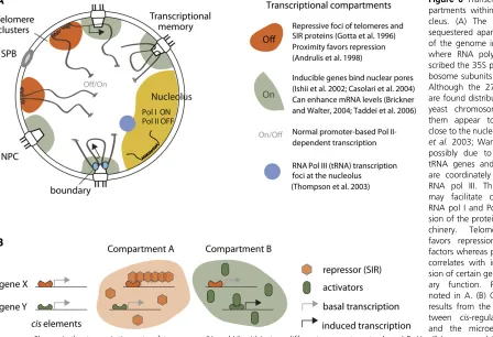
One of the least understood yet most pervasive features of nuclear organization is the existence of subnuclear compart-ments, in which specific DNA sequences and proteins accumulate. These are thought to create microenvironments that favor or impede a particular DNA- or RNA-based activity. The most obvious and well-characterized of such compartments is the nucleolus, the site of RNA polymerase I-mediated rDNA transcription and ribosome subunit assem-bly. We describe this in some detail as it illustrates the self-organizing character of such compartments.
as a result of adapting the rate of ribosome production to the needs of the cell (Oakes et al. 1993; Powers and Walter 1999).
The budding yeast rRNA stems from a 9.1-kb repeat locus that is transcribed as a 35S precursor rRNA and a 5S rRNA by RNA polymerases I and III, respectively. Intriguingly, different yeast strains have different numbers of tandem repeats, extending from 100 to 200 (Pasero and Marilley 1993). Assembly of the nucleolus is thought to be a self-driven process, initiated by production of rRNA (Trumtel et al. 2000; Hernandez-Verdunet al.2002). This argument stems in part from studies in which the rDNA repeat was transcribed by RNA Pol II, rather than by the endogenous RNA Pol I. This led to massive alterations in nucleolar struc-ture, arguing that factors associated with RNA Pol I play a key role in proper nucleolar assembly (Oakes et al. 1993; Trumtelet al.2000).
A recent study (Albertet al.2011) showed that two Pol I-specific subunits,Rpa34and Rpa49, are essential for the high polymerase-loading rate on active copies of rDNA and for nucleolar assembly. Intriguingly, nucleolar assembly can be restored in the absence ofRpa49by decreasing the ribo-somal gene copy number from 190 to 25, with a concomitant increase in the number of RNA Pol I complexes per tran-scribed unit. These authors argue that the spatial constraint of RNA Pol I transcription is a critical feature of nucleolar assembly (Albertet al.2011). Exactly how the 2 Mb of rDNA is folded within the nucleolus is not known, although both cohesin, condensin, andSir2affect rDNA compaction during the mitotic cell cycle (Guacciet al.1994; Lavoieet al.2002, 2004).
The extended array of tandem repeats found in the rDNA locus serves as an ideal template for homologous recombi-nation (HR), the favored pathway for double-strand break (DSB) repair in budding yeast. At the same time, it is clear that rDNA stability is critical for growth and survival of yeast, since the generation of extrachromosomal rDNA circles by inappropriate recombination provokes replicative senescence (Sinclair and Guarente 1997). Thus, budding yeast has evolved several mechanisms to suppress recombi-nation within the rDNA array. Two mechanisms involve the silent information regulator,Sir2, which is a NAD-dependent deacetylase (Imai et al. 2000; Smith et al. 2000; Tanner et al. 2000). The first mechanism correlates with a Sir2 -dependent local nucleosomal organization (Gottlieb and Esposito 1989; Bryk et al.1997; Fritze et al.1997; Smith and Boeke 1997) and the second involves longer-range chromatin tethering to the nuclear envelope through the CLIP (chromosome linkage INM proteins) complex. This complex includes two NE proteins, Heh1 (homologous to the human Man1 protein) andNur1 (Figure 2). The rDNA is connected to the CLIP complex through cohibin, a V-shaped complex of two Lrs4 proteins and two Csm1 homodimers (Mekhail et al. 2008; Chan et al.2011). Loss of eitherSir2or the cohibin-Heh1-anchoring pathway leads to instability of the rDNA repeat, followed by cell cycle
ar-rest or premature senescence. Premature senescence can also be provoked by the loss ofSgs1(Sinclair and Guarente 1997), a RecQ helicase that counteracts recombination be-tween rDNA repeats.
Telomere foci—assemblies of repetitive DNA and silencing factors:The clustering of the 32 yeast telomeres into three to six foci at the NE provides a second prominent feature of yeast nuclear organization (Palladino et al.1993). At these clusters of telomeres, the silent regulatory factorsSir3and Sir4 and the telomere repeat-binding factor repressor acti-vator protein 1 (Rap1) accumulate (Gottaet al.1996), while Sir2 is found both at telomeric foci and in the nucleolus (Gotta et al. 1997). These perinuclear telomeres are late replicating (Raghuraman et al.2001) and generate a zone that favors SIR-mediated repression of silencer-flanked genes (Andrulis et al. 1998). The clustering of telomere repeats also ensures that SIR proteins do not bind promis-cuously to repress other sites in the genome (Mailletet al. 1996; Taddei et al. 2009). Finally, telomere anchorage in S phase contributes to proper telomerase control and sup-presses recombination among telomere repeats (Schober et al.2009; Ferreiraet al.2011).
Intriguingly, telomere-containing foci are dynamic, mov-ing with a constant random motion that is more constrained than that of a nontelomeric locus (Schoberet al.2008; Therizols et al. 2010). These foci fuse and divide in interphase and dissociate at least partially in metaphase (Laroche et al. 2000; Smith et al. 2003) to reform in early G1. With the exception of the intrachromosomal loops formed by short metacentric chromosomes, no strong preferences for telomere– telomere pairing were detected in interphase cells (Schober et al. 2008). Rather, telomeres on ends of chromosome arms of roughly equal length tend to interact, presumably due to the geometry imposed by the Rabl conformation in anaphase (Jin et al. 2000; Schober et al. 2008; Therizols et al. 2010), showing how that the linear architecture of a chromosome can impact its long-range contacts in the nucleus.
At the molecular level, budding yeast telomeres consist of 250–300 bp of irregular tandem repeats with the consensus sequence TG1–3(Shampayet al.1984).Rap1(Rap1) (Shore
telomere clustering is thought to compete for limited amounts of SIR proteins. In this way, multiple weak, but closely juxtaposed, binding sites create an effective sink for this histone-binding complex (Gasser et al.2004). Con-firming this, the dispersion of SIR proteins from telomeres by the mutation of telomere anchorage sites reproducibly reduced the expression of15 nontelomeric genes (Taddei et al.2009).
SIR-mediated silencing is generated in a two-step pro-cess. First, the heterodimer formed between Sir4 andSir2 catalyzes the NAD-dependent deacetylation of lysines in the histone H4 N-terminal tail. The deacetylation of histone H4 K16, in particular, generates a preferred binding site forSir3 (Oppikoferet al.2011), allowingSir3to bind and spread to the adjacent unacetylated nucleosomes (reviewed by Norris and Boeke 2010; Rusche et al.2003).Sir3appears to bind nucleosomal arrays in a stable, stoichiometric complex with Sir2andSir4(Cubizolleset al.2006), which renders linker DNA less accessible to transcription factors or other media-tors of RNA pol II engagement at the transcriptional start site (Aparicioet al.1991; Strahl-Bolsingeret al.1997; Hecht and Grunstein 1999; Chen and Widom 2005; Martinoet al. 2009).
As mentioned above, by disrupting telomere anchors but leaving SIR factors intact, it was shown that the sequestra-tion of SIR proteins in telomere foci favors subtelomeric repression and prevents the promiscuous binding of SIR proteins at a distinct subset of promoters elsewhere in the genome. This mechanism may be usurped and regulated by environmental conditions to control physiological response. For example, the subtelomeric positioning of genes may help coordinate both the repression and the activation programs that permit growth under adverse conditions (Turakainen et al.1993a,b; Halme et al.2004; Fabre et al.2005). Over time this could ensure an evolutionary advantage, which would explain why alternative carbon source genes accumu-late in subtelomeric domains. As discussed below, the im-paired tethering of telomeres also correlates with altered rates of recombinational repair in subtelomeric zones (Louis 1994; Therizols et al. 2006; Schoberet al. 2009) and im-paired control of telomerase (Hediger et al.2006; Ferreira et al. 2011). We discuss how telomere clustering affects gene expression and genome stability in more detail below.
tRNA genes: Although the 274 tRNA genes are found distributed along all 16 yeast chromosomes, many of these genes, when probed by FISH or Hi-C technologies, appear to be clustered in nuclear space (Thompsonet al.2003; Wang et al. 2005; Duan et al. 2010). Intriguingly, at least some tRNA clusters are found close to the nucleolus (Thompson et al. 2003; Wang et al.2005), possibly reflecting the fact that tRNA genes and the 5S rDNA are coordinately tran-scribed by RNA pol III. This juxtaposition may favor coordi-nated activation of RNA polymerase I and polymerase III for expression of the protein synthesis machinery. Interestingly, the clustering of tRNA genes depends on condensin, while
their association with the nucleolus depends on microtubule integrity (Haeusler et al. 2008). It is clear from Hi-C data and GFP-LacO tagging that not all RNA polymerase III genes show similar localization, and it is not yet clear why some tRNA genes shift toward the nucleolus, while others do not (E. Varela, personal communication; Duan et al. 2010). Nonetheless, the broad distribution of tRNA genes means that the association of even a subset of them with the nu-cleolus would impact global chromosome positioning.
It is noteworthy that RNA polymerase II genes found in the vicinity of actively transcribed tRNA genes become silenced through a phenomenon called tRNA-gene-mediated gene silencing (Wang et al. 2005). Although condensin mutations that affect tRNA gene clustering do not affect tRNA transcription, they do abolish the repression of nearby RNA pol II promoters, suggesting that tRNA clustering and tRNA-gene-mediated gene silencing are linked (Haeusler et al.2008). Thus, tRNA genes can be transcribed away from the nucleolus, but interactions in transand the long-range effect of this on chromatin status correlate with relocation.
Replication foci: Eukaryotic genomes initiate DNA replica-tion at multiple sites or origins that are dispersed along each linear chromosomal arm. In budding yeast, origin function is coincident with short cis-acting sequences called autono-mously replicating sequences (ARS), which support both autonomous plasmid replication and genomic initiation events. In budding yeast, DNA synthesis initiates uniquely at these sites, yet the ARS sequences are not sufficient to ensure efficientfiring. Indeed, sites that are competent for initiation on plasmids do not necessaryfire in the genome, and even the most efficient origins do not fire every cell cycle (Raghuramanet al.2001).
The clustering of yeast replication forks in foci wasfirst revealed in an in vitro replication assay that used isolated yeast nuclei as a template for the incorporation of fl uo-rescently labeled nucleotide. Between 15 and 20 discrete foci were detected that were Origin Recognition Complex (ORC), S phase-, and origin-specific (Pasero et al. 1997). The same type of foci were later observed by visualizing replisome components such asPCNAand DNA polymerase a or e by immunostaining or fluorescent tagging in living cells (Ohya et al.2002; Hiragaet al.2005; Kitamuraet al. 2006). Because the number of these foci is far smaller than the number of replication forks, they are thought to corre-spond to replication factories in which several elongating forks are grouped. Consistent with this notion, neighboring origins along a chromosome initiate replication with similar timing and respond in a similar fashion to the loss of an S-phase cyclin (Yabukiet al.2002; McCuneet al.2008).
might be coregulated by juxtaposition in a single focus, it is interesting to note that Hi-C data do score increased inter-action between earlyfiring origins (Duanet al.2010). Such clustering could facilitate initiation by concentrating limiting factors (Mantieroet al.2011) to favorfiring in early S phase. Although late-replicating regions are found mainly at the nuclear periphery, a subnuclear position is neither necessary nor sufficient for the control of late originfiring (Heunet al. 2001a; Ebrahimiet al.2010). The timing of telomere repli-cation is instead determined by telomere length (Bianchi and Shore 2007; Lianet al.2011). The clustering of replica-tion forks could nonetheless increase replicareplica-tion efficiency by concentrating replisome components and deoxy-nucleotides to ensure efficient elongation after recovery from replicative stress. There has been no genetic test of this model to date, since the proteins that ensure origin clustering are unknown. Indeed, the only protein known to influence replication focus formation in vertebrates is lamin (Spannet al.1997), which yeast lack.
Sites of DNA repair: DNA DSB repair by homologous re-combination is also organized into foci called DNA damage response foci (Lisby and Rothstein 2009). These foci contain high local concentrations of repair factors and are called repair foci or factories. The spatially restricted structures may serve to restrict the modification of chromatin to the subdomain that is relevant for repair or to restrict end-joining to the proper chromosome end. Other models argue that different repair foci may prevent telomere addition at certain DSBs by sequestering the break away from telome-rase. Alternatively, a focus found near other telomeres may facilitate TG addition at breaks. Intriguingly, multiple DSBs incurred on different chromosomes were shown to assemble into a single repair focus in yeast (Lisby et al. 2003). This topic will be reviewed elsewhere; thus, only damage mobil-ity and its function are discussed here.
Mechanisms Underlying Nuclear Compartmentation
Redundancy in telomere-anchoring pathways
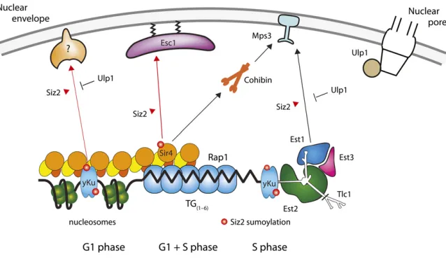
As described above, the positioning of chromatin within the nucleus depends on reversible interactions of chromosomal elements with structural components of the nuclear enve-lope, namely proteins of the INM, the SPB, and, in some cases, nuclear pores. The anchoring of telomeres is achieved through several different protein–protein contacts, which fluctuate during the cell cycle and which have different lev-els of importance for different telomeres (Figure 4) (Hediger et al.2002; Taddeiet al.2004a; Hiragaet al.2008; Schober et al. 2009). The fluctuation can be attributed in part to post-translational modifications; several pathways show a re-quirement for sumoylation by the E3 ligase Siz2 (Ferreira et al.2011)
One major pathway of telomere anchoring requiresSir4 and is therefore amplified by the formation of silent chro-matin (Hedigeret al. 2002; Gartenberget al.2004; Taddei
et al.2004a). This was most clearly shown by tracking the position and mobility of an excised ring of yeast chromatin, which either did or did not contain silencers. The chromatin ring could bind autonomously to the NE when silenced, in a SIR-dependent manner, and did not associate when SIR protein could not bind (Gartenberget al.2004).Sir4anchors repressed chromatin to the NE through a 312-aa domain in its C-terminal half that specifically binds an acidic protein asso-ciated with NE, Esc1 (Andrulis et al. 2002; Taddei et al. 2004a). In parallel, it was shown that Sir4can mediate an-choring through the nucleoplasmic N-terminal acidic domain ofMps3(Buppet al.2007). This may not be a direct interac-tion, however, and a recent study suggests that Sir4/Mps3 association is mediated byLrs4, a component of the cohibin complex (Chanet al.2011). Moreover, contrary to theEsc1– Sir4 interaction,Mps3-Sir4anchoring requires an intact SIR complex and is completely dependent on silencing. Both Mps3 and Esc1 define NE-binding sites that can be distin-guished by microscopy from nuclear pores, and their position around the NE is independent of nuclear pore distribution (Taddeiet al.2004a; Horigomeet al.2011).
Mps3 not only participates in SIR-dependent anchorage, but also mediates a silencing-independent pathway for telo-mere anchoring through its interaction with Est1 (Ever
shortertelomeres) (Lundblad and Blackburn 1990), an
ac-cessory component of telomerase (Antoniacci et al. 2007; Schober et al. 2009).Tlc1, the RNA moiety of telomerase, is linked to telomeric chromatin through the yeast Ku70/ Ku80 heterodimer, which also binds the end of chromo-somes (Martin et al. 1999). It is thought to help recruit the telomerase either directly or indirectly (Peterson et al. 2001; Stellwagen et al.2003; Fisheret al.2004; Pfingsten et al.2012) since the Ku-telomerase and Ku-DNA interaction may be mutually exclusive (Petersonet al.2001; Stellwagen et al.2003; Pfingstenet al.2012). Whereas the intramem-brane portions of Mps3 play a major role in SPB organiza-tion (Jaspersenet al.2002; Nishikawaet al.2003), its role in telomere anchoring requires its acidic N-terminal domain, which extends into the nucleoplasm (Bupp et al. 2007). The use of N-terminal deletions allows one to distinguish the roles of Mps3 in chromatin anchoring and in SPB for-mation and function. Nonetheless, the various telomere-anchoring pathways are extensively interlinked, since the yeast Ku dimer, which mediatesMps3anchorage in S-phase cells, also directly bindsSir4(Royet al.2004; Taddeiet al. 2004a).
mechanism for switching off Yku-mediated peripheral posi-tioning once the majority of the genome has been replicated, allowing telomeres to dislodge from the NE either during or just after replication (Ebrahimi and Donaldson 2008), possibly as a consequence of desumoylation. Sir4 is also phosphorylated in a cell-cycle-dependent manner (Kueng et al. 2012), which may contribute further to the release of telomeres observed during mitotic division.
Antagonism of the yeast Ku–telomerase–Mps3interaction leads to a hyper-recombination phenotype among the short-ened telomeres found in cells lackingTel1, the ATM kinase homolog in yeast (Schober et al.2009). This suggests that the sequestration of telomeres by the NE proteinMps3could protect telomere ends from recombination and deleterious unequal strand exchange. Consistently, the release of telo-meres by the deletion ofSIZ2in both G1- and S-phase cells correlates with abnormally long telomeres (Ferreira et al. 2011). Another telomere-bound protein, namely Cdc13, was also found to be sumoylated in aSiz2-dependent man-ner (Hang et al. 2011). Sumoylated Cdc13 contributes in an independent manner to telomerase activity, synergizing with the effects mediated by yeast Ku andSir4. Thus,Siz2 -mediated sumoylation limits telomere elongation by more than one mechanism, one of which correlates with telomere sequestration at the NE (Figures 4 and 5). Importantly, it was shown that critically short telomeres shift away from the nuclear periphery when they elongate in newly formed wild-type zygotes (Ferreira et al. 2011). If natural elonga-tion coincides with telomere release, it follows that telomere sequestration by sumoylated forms of Sir4 and Yku might indeed attenuate telomerase activity.
Minimal anchoring assay and the validation of genetic screens
Given the redundancy in the pathways of telomere anchor-ing, it was difficult to dissect the requirements of anchoring by scoring the position of a LacO-tagged telomere in single mutants. To solve this problem, a“minimal anchoring assay” was developed in which proteins fused to LexA were
tar-geted to an internal LacO-tagged locus (ARS607). The abil-ity of the fusion protein to shift a randomly distributed locus to the periphery was scored and analyzed statistically (Taddei et al.2004a). The assay is performed in a strain expressing GFP-Nup49to allow an accurate quantitation of position in different genetic contexts. The minimal anchoring assay al-lows one to score for sufficiency and not only for depen-dence on a given factor, which is necessary if one wants to define the molecular machinery of chromatin anchoring.
In addition to these targeted approaches, a genetic screen for genes that influence telomere anchoring was carried out. This study identified the histone acetyl transferase Rtt109 and the acetylation of histone H3K56 as key modulators of telomere anchoring (Hiraga et al. 2008). This probably reflects impaired nucleosomal assembly after DNA replica-tion. Other factors showing partial or cell-cycle-dependent loss of anchoring include Esc2, Ctf18, Tel1, Mre11, Elp4, andRrm3(Hiragaet al.2008). Since these may affect telo-mere replication and the spread or assembly of silent chro-matin, an effect on telomere tethering is not sufficient to consider them as direct anchors. Mutations in proteins of the nuclear pore such asNup133also affect telomere posi-tioning, but these interfere with both mRNA export and cell cycle progression, suggesting that their effects are indirect (Doye et al.1994; Therizolset al.2006; Lewis et al.2007; Palancade et al. 2007). Palmitoylation of telomere-bound proteins could also contribute to the anchoring of silent chromatin as recently shown for Rif1, although long telo-meres in rif1D cells remain peripherally attached through Sir4(Cockellet al.1995; Parket al.2011). Still, redundant roles ofRif1andRif2in telomere anchoring remains are not excluded.
Khadarooet al.2009; Ozaet al.2009). These anchoring ways are less well understood than telomere-anchoring path-ways, but have a clear impact on chromosome stability and are discussed in this context below.
Formation of chromatin compartments: association in trans
There are two basic models for the formation of nuclear compartments. The first reflects the localized tethering of similar sequences to a subnuclear structure, or a variant of this in which intermolecular interactions lead to domain clustering. In the second model, compartments arise pas-sively due to volume exclusion effects. This is favored under conditions of macromolecular crowding, and the high concentration of macromolecules within the nucleoplasm (between 100 and 400 mg/ml) is thought to be sufficient to enhance attractive interactions between macromolecules (Richteret al.2007). Most likely, subnuclear compartments arise from a combination of mechanisms. A few structural
proteins appear to be involved in more than one mechanism, such as the cohesin, condensin, and the more recently char-acterized cohibin complexes (Poon and Mekhail 2011).
chromatin to the NE by bridging between the chromatin-bound RENT/SIR complex and either CLIP or Mps3. The complex is proposed to link rDNA repeats to each other by clustering interspersed IGS1 regions (Mekhail et al. 2008; Chan et al. 2011), a mechanism thought to contribute to SIR-dependent telomere clustering as well.
Yeast telomere clusters and their associated concentration of silent information regulators provide a well-characterized example of a functional compartment generated by the juxtaposition of sequence-defined chromosomal elements. Interactions between subtelomeric zones were proposed to be governed by physical constraints, including chromosome arm length, centromere attachment to the SPB, and nuclear crowding (Therizolset al.2010). On the other hand, interac-tion between HM loci was shown to depend on correctly assembled heterochromatin at these loci (Mieleet al.2009). Consistently, Sir3 has been shown to promote telomere– telomere association (Ruaultet al.2011).Sir3overexpression triggered the clustering of telomeric foci into even larger aggregates positioned away from the NE, arguing that clus-tering can occur independently of anchoring.
Although this “hyper-clustering” phenotype correlates with more stable silencing in subtelomeric regions, silencing is not absolutely required for telomere cluster formation. Notably, the overexpression of aSir3mutant lacking silencing activity induces large telomere clusters, even in the absence ofSir2andSir4(Ruaultet al.2011). TheSir3-mediated clus-tering is likely to be driven bySir3self-oligomerization, likely mediated by its C terminus (Georgelet al. 2001; D. A. King et al. 2006; Liaw and Lustig 2006; McBryant et al. 2008; Adkinset al.2009).
Although silencing is not required for clustering, the in-volvement of Sir3in both processes provides a mechanism for the self-propagation of a silent compartment. Specifi -cally, given that Sir3binding promotes telomere clustering, this leads inevitably to an increased local concentration of SIR factors. Given thatSir3is limiting for the spread of silent chromatin, clustering would favor SIR complex spread into flanking chromatin, extending repression. With more Sir3 bound, more clustering would occur. Other factors also affect telomere clustering, namely the other SIR proteins, the yeast Ku heterodimer, Asf1, Rtt109, Esc2, the cohibin complex, and the two conserved factorsEbp2andRrs1that moonlight in ribosome biogenesis (Gottaet al.1996; Laroche et al.2000; Gehlenet al.2006; Hiragaet al.2008; Mieleet al. 2009; Chan et al. 2011; Horigome et al. 2011). However, because loss of these factors cause replicative stress and im-pair heterochromatin formation, a direct role in clustering is not established.
This raises a chicken-and-egg question: Do silent com-partments persist simply because they are self-forming, or is there an initial triggering event that promotes the function that will be favored by the compartment? The bifunctional role ofSir4as a mediator of repression and as a silencing-independent anchor for chromatin allows it to both partici-pate in the self-organization of these compartments and
trigger entry into the cycle of repression–sequestration– repression for a nonsilenced telomere. Importantly, chromatin immunoprecipitation showed thatSir4can bind telomeres in the absence ofSir3, while the opposite was not true (Strahl-Bolsinger et al.1997). Thus, a nonsilent telomere might be more likely to bind the NE through Esc1–Sir4or telomere-bound Yku–Mps3interactions than to cluster. This would shift a naive telomere into a zone enriched for telomere-bound Rap1and its ligandsSir3andSir4. Proximity to pools of SIR proteins would enhance telomere repression, and, once silenc-ing is established, perinuclear localization would be reinforced by theSir4-anchoring pathway, while clustering would be pro-moted bySir3. In this way, the formation of silent chromatin foci could be self-generating, and not only self-reinforcing. Such a mechanism may apply to other chromatin-based com-partments with other functions and may also allow the nu-cleus to segregate factors away from sites at which they might do harm.
Mechanisms of chromatin movement
To generate compartments, chromatin must be able to move within the nucleus. An excised ring of chromatin explores the entire nuclear volume in random walk (Rc = 0.9 mm) (Figure 3) while a chromosomal locus moves in a constant, near-random walk within a radius of 0.5–0.7 mm (re-viewed in Gasser 2002; Marshall 2002; Hubner and Spector 2010). For a yeast nucleus of2mm in diameter, this means that a chromosomal locus can explore30% of the nuclear volume (Gartenberg et al.2004), whereas the same move-ment in a mammalian nucleus corresponds to,1% of nu-clear volume (Figure 3). In mammalian cells, the shift of a chromatin domain from a repressive to an active compart-ment may involve directional movecompart-ment (Vazquez et al. 2001; Chuang et al. 2006). A similar shift was observed upon targeting of the viral transactivator VP16 to a yeast telomere: the activation displaced the telomeric locus from the NE (Taddei et al. 2004a; Hediger et al. 2006). This suggested that transcription might correlate with at least some degree of chromatin movement, consistent with the sensitivity of movement to glucose levels in the media as well as plasma and mitochondrial membrane potentials (Marshall et al.1997; Heunet al.2001b; Gartenberget al. 2004).
of nucleosomes and rapid gene induction (Neumann et al. 2012). Together, these observations suggest that chromatin movement in the yeast interphase nucleus may reflect the nucleosome packing into a higher-order fiber and be infl u-enced by remodeler-dependent removal of nucleosomes lo-cally. On the level of a chromatinfiber, changes in movement may reflect changes in the persistence length of the polymer brought about by nucleosome displacement.
Both the nucleosome remodeler INO80and the related complex SWR1 not only are recruited to promoters to en-hance transcription, but also are recruited to DSBs to facil-itate early steps in repair (reviewed in van Attikum and Gasser 2009; Lans et al.2012). Intriguingly, DSBs also dis-play enhanced mobility relative to the movement of undam-aged loci (Dion et al. 2012; Miné-Hattab and Rothstein 2012). This increase in mobility required the strand-annealing factorRad51and theSnf2-related ATPase activity ofRad54 (Dion et al. 2012). These two proteins are necessary for repair by HR in an epistatic manner. TheRad9protein and the ATR kinase,Mec1, which are both involved in the check-point response, also are required for increased movement of DSBs (Dion et al.2012), and, consistently, only DSBs—and not protein-DNA adducts or nicks that do not activate the DNA damage checkpoint—increase locus mobility (Dion et al.2012). It was also reported that undamaged sites in-crease mobility coincident with inin-creased movement at the primary lesion, suggesting that there is a genome-wide response to DSBs (Miné-Hattab and Rothstein 2012). To-gether, these results suggest that locus movement not only is an inherent feature of chromatin, but also may play an active role in promoting the formation of compartments, shifting DNA from one compartment to another, or in facil-itating DNA interactions during repair.
Functional Consequences of Nuclear Organization
Gene silencing
The sequestration of SIR proteins at telomeres and silentHM loci due to telomere clustering has two functional consequen-ces. First, the enrichment of SIR proteins favors subtelomeric repression, and second, it prevents promiscuous repression of a distinct subset of promoters elsewhere in the genome (Taddei et al. 2009). Sir3 protein abundance is relatively low (Ghaemmaghami et al. 2003) and is limiting for the spread of silent chromatin (Renauldet al.1993). Therefore, the DNA sequence-dependent enrichment of SIR recruit-ment factors at telomeres probably accounts for the strong enrichment of SIR proteins in foci (Gotta et al. 1996). In-deed, the loss of telomere anchoring upon deletion of either subunit of yeast Ku leads to SIR protein delocalization throughout the nucleoplasm (Larocheet al.1998).
The abundance of tandem RAP1-binding sites found in telomeric sequences competes with silencers for SIR fac-tors, as monitored by functional assays. Early on, silencer-nucleated repression was found to be highly sensitive to the distance of the reporter from a telomere end (Renauldet al.
1993; Stavenhagen and Zakian 1994; Thompsonet al.1994; Maillet et al. 1996; Marcand et al. 1996), and telomere clustering favored the SIR-mediated silencing of endoge-nous subtelomeric genes (Mondoux et al. 2007; Taddei et al.2009). Still, the association of subtelomeric loci with peripheral telomere clusters (visualized as Rap1-GFP foci) did not always correlate with silencing efficiency (Mondoux et al. 2007). This may be due to the dynamic behavior of individual telomeres, which move into and out of telomere foci, without loss of their epigenetic state (Schober et al. 2008). Most strikingly, however, was the demonstration that, by actively tethering a HMRreporter construct with a weakened silencer to the NE, transcriptional repression in-creased (Andrulis et al. 1998) in a manner strictly depen-dent on the presence of telomere clusters and SIR foci (Taddeiet al.2009). This argues that transcriptional repres-sion does not reflect positionper se, but rather access to local high concentrations of SIRs (Mondouxet al.2007). Consis-tently, when telomere anchoring is impaired by deletion of YKU70and ESC1, transcription of genes at internal loci (Mailletet al.2001; Taddeiet al.2009) or at an excised ring of chromatin flanked by silencers (Gartenberget al.2004) showed enhanced repression, while telomere-proximal genes were derepressed.
Interestingly, changes in the spatial distribution of re-petitive sequences can regulate patterns of gene expression genome-wide. Indeed, SIR protein dispersion from perinu-clear foci was shown to specifically affect internal promoters carrying binding sites forAbf1and the PAC-binding factors (Pbf1andPbf2), which regulate genes involved in ribosome biogenesis (Mondouxet al.2007; Taddei et al.2009). The cell may exploit such mechanisms by regulating SIR com-plex affinity at telomeres in a rapid response to environmen-tal insult (Martinet al. 1999; Aiet al.2002). Consistently, SIR dispersion, or modulation of TPE, has been observed under various forms of stress (Stone and Pillus 1996; Martin et al.1999; McAinshet al.1999; Millset al.1999; Rayet al. 2003; Biet al.2004; Mercieret al.2005; Smithet al.2011). Stress-induced redistribution of SIR proteins may derepress subtelomeric genes required for growth on alternative car-bon sources and simultaneously contribute to the down-regulation of genes involved in ribosome biogenesis as a global attempt to suppress growth and favor a survival pathway.
Gene activation
et al.2006; Rubenet al.2011), the tagging and localization of specific inducible genes confirmed that some genes in-deed shift to NPCs upon activation (for reviews, see Akhtar and Gasser 2007; Taddei 2007; Dieppois and Stutz 2010; Egecioglu and Brickner 2011) (Figure 6).
What moves active genes to the NPC is still unclear; indeed, each of the nuclear steps associated with gene expression, including initiation, elongation, mRNA process-ing, proofreadprocess-ing, and export, have been implicated. For example, the accumulation of a stalled intermediate in the mRNP export pathway in THO/sub2 mutants leads to the persistent NPC association of some active genes (Brickner and Walter 2004; Rougemailleet al.2008), yet other data argue that promoter activation without transcription is sufficient for the shift (Casolariet al. 2004, 2005; Schmid et al. 2006). We suggest that multiple different steps of transcription contribute independently to the stable associ-ation of active genes with NPCs and that the importance of any individual step in this process may well be locus-specific. It is argued that the presence of multiple anchoring part-ners favors the formation of active gene loops, with pro-moters linking to 39sequences to improve the recycling of polymerases and elongation efficiency (O’Sullivan et al. 2004).
The targeting of genes to pores can also be mediated by sequence elements. Small DNA sequences called gene recruitment sequences (GRS I and II) were first identified in the promoter ofINO1and were proposed to function as a DNA “zip code” that confers perinuclear localization in-dependently of the chromosomal context (Ahmed et al. 2010). Interestingly, the GRS I element is over-represented in the promoters of genes interacting with the NPC and in the promoters of stress-inducible genes.
NPC association is thought to be particularly important for inducible genes with galactose- or heat-shock-controlled promoters, as these genes require a rapid and high level of expression and efficient mRNA export. This could be facilitated by positioning the activated gene at pores. It was proposed in the“gene gating”hypothesis (Blobel 1985) that the NPC serves as a scaffold to build an assembly line that coordinates transcription, processing, and export of mRNA. Moreover, because of their eightfold symmetry, each pore could accommodate multiple genes, forming a factory that supports highly efficient transcription. Although“gene gat-ing”might optimize the induction of some genes, pore asso-ciation is clearly not obligatory for induced expression; mutations affecting gene-NPC association, for example, showed normal expression levels for induced GAL1 (Cabal et al. 2006). Moreover, gene repression did not generally lead to release from pores (Casolari et al. 2004). Finally, activation of the same promoter by different pathways or with different 39UTRs led to different positions of the gene within the nucleus (Abruzziet al.2006; Taddeiet al.2006). Nonetheless, it remains possible that the kinetics of induc-tion was altered in these mutants, as associainduc-tion with the NPC was correlated with enhanced activation of at least
some genes (Brickner and Walter 2004; Menon et al. 2005; Taddei et al. 2006). Thus, whether it acts by the formation of specialized transcription factories (Eskiw et al.2010) or regulation of mRNA elongation, processing, and export, the NPC seems to efficiently fine-tune gene expression.
A second proposal suggests that the NPC–gene associa-tion is an epigenetic mark that enables a previous inducassocia-tion event to be “remembered” in subsequent generations, thereby facilitating re-induction (Brickneret al.2007). Both GAL1 and INO1 genes are more rapidly reactivated after short-term repression, and this correlates with their persis-tence at the NPC after the inducing agent was removed. Although mechanisms involving cytoplasmic factors have also been implicated in rapid gene reactivation (Zacharioudakis et al. 2007), GALgene transcriptional memory was none-theless correlated with the formation of persistent loops between the 59 and 39 ends of the gene and association with the NPC (Laine et al. 2009; Tan-Wong et al.2009). In the case ofINO1, an 11-bp sequence appears to control both peripheral targeting and H2A.Z incorporation after repression (Brickneret al.2007; Lightet al.2010). Finally, in cells lacking this sequence or a nuclear pore component, Nup100, transcriptional memory, and perinuclear associa-tion ofINO1 were lost (Lightet al.2010). Thus, rapid re-induction may provide the rationale for NPC association of inducible genes.
Boundaries between active and inactive domains
Given the role of telomeric foci in the creation of repressive compartments and the ability of the NPC to support induced gene expression, one can conclude that the nuclear periph-ery has at least two functions in the control of gene ex-pression. Consistent with this duality, proteins involved in gene activation or repression have distinct staining patterns at the nuclear periphery when localized at sufficiently high resolution (Taddeiet al.2004a; Horigomeet al.2011). Such juxtaposition of repressive (telomere foci) and activating compartments (NPCs) could favor efficient reversals of gene expression status, which may be especially relevant for sub-telomeric genes, since, as mentioned above, subsub-telomeric zones are enriched for genes involved in alternative carbon source usage that are needed rapidly, but only under specific growth conditions (Fabreet al.2005).
simply pore or position dependent, but depends on the bind-ing of transactivators or repressors to cis-acting elements (Rubenet al.2011). Consistent with this notion, increasing the association of theHXK1subtelomeric gene with the nu-clear periphery through a neutral anchor improved both its repression of glucose medium and its activation in the ab-sence of glucose (Taddeiet al.2006).
Effects on genome stability
In contrast to the extensive studies on the function of nu-clear organization in gene regulation and DNA replication, relatively little is known about how chromatin dynamics and higher-order nuclear organization contribute to genome stability. Until recently, many basic questions remained un-addressed, such as whether or not DSBs are mobile in the nucleus, whether DSBs are recruited to scaffolds for repair, or whether, instead, repair factors are recruited to sites of damage. It was also unclear whether specific pathways of repair occur preferentially in specific subcompartments of the nucleus, and if so, how chromatin dynamics contribute to the process of repair. With a combination of genetics and real-time live imaging, recent studies show that chromatin dynamics and nuclear organization do play a major role in the repair of DNA lesions and the maintenance of chromo-some integrity in yeast.
Mobility of DSBs:The introduction of two DSBs on different chromosomes can cause reciprocal translocations as fre-quently as they produce intrachromosomal deletions, argu-ing that DSBs are dynamic and can roam throughout the nucleus to find their appropriate template for repair (Lisby et al. 2003). Indeed, the subdiffusive movement of yeast chromatin increases four- or fivefold within 30 min after induction of a DSB, such that the lesion can explore50% of the nuclear volume (Dion et al.2012; Miné-Hattab and Rothstein 2012). Consistently, Houston and Broach have shown in living yeast cells that a homology search for a cleaved MATlocus is efficient, such that pairing with the homologous donor on the distal arm of the same chromo-some can occur within 90 min after DSB induction (Houston and Broach 2006; Hickset al.2011). Given that a homology search takes longer for ectopic donor sequences, it can be predicted that DSBs require enhanced mobility (over that of an undamaged locus) tofind the donor sequence for HR.
fibroblasts (Dimitrova et al. 2008; Krawczyk et al. 2012). This latter was dependent on 53BP1, a repair factor that helps limit resection in favor of end-joining. Some of the discrepancy in the movement of DNA damage can probably be attributed to the fact that different types of DNA damage were induced and that the cells activate the ATR/ATM checkpoint response to different degrees, which was shown in yeast to contribute to DSB movement (Dion et al.2012; Krawczyket al.2012).
In budding yeast, the rate of HR-mediated repair, scored in the presence of one or multiple dispersed copies of donor sequence, suggests that the homology search is indeed rate-limiting for recombination (Wilsonet al.1994). Specifically, the frequency of recombination was proportional to the number of targets in each strain tested. This dependency on copy number for both dispersed and clustered targets suggests that the homology search takes place by a random 3D collision, rather than by sliding the donor along DNA until the recipient is found. Intriguingly, Miné-Hattab and Rothstein (2012) report a general increase in chromatin mobility in response to elevated levels ofg-irradiation. This would indeed be likely to enhance the chances of random collision events between DSB and uncut donor.
Nuclear pores and Mps3 anchorage in noncanonical repair: The random 3D search could also be facilitated by structures that recruit sites of damage and therefore pro-mote recombination or alternative pathways of repair (Gehlenet al.2011b). Two possibilities for this“repair scaf-fold”are nuclear pores and the above-mentioned SUN do-main protein,Mps3. It was shown that certain types of DNA damage, such as persistent DSBs, collapsed replication forks, and eroded telomeres are recruited to nuclear pores, which are themselves associated withUlp1, a Sumo protease, and a SUMO-dependent ubiquitin ligase complex, Slx5/Slx8 (Khadarooet al.2009). Namely, lesions that are impossible or difficult to repair by canonical HR may be brought to nuclear pores where alternative recombination and repair pathways can come into play. Genetic interactions suggest that the importance of the nuclear pore resides in its seques-tration ofUlp1andSlx5/Slx8, and its proximity to the pro-teosome (Therizolset al.2006; Lewiset al.2007; Palancade et al. 2007; Nagai et al. 2008). Thus, one scenario is that sumoylated proteins accumulate at collapsed forks or resected breaks that require Slx5/Slx8 ubiquitylation for proteasomal degradation, or alternatively, desumoylation by Ulp1, to enable appropriate repair. Exactly what the rel-evant protein may be is at present unknown, although Rad51,Rad52,Pol32,PCNA, and other proteins of the rep-lication machinery are heavily sumoylated upon DNA dam-age (Melchior 2000; Nagai et al. 2011; Cremona et al. 2012).
Other studies have reported the association of irreparable DSBs with the INM proteinMps3(Kalocsayet al.2009; Oza et al. 2009). The tethering of a persistent DSB by Mps3 is thought to contribute to the suppression of ectopic HR.
Similarly, the peripheral tethering of short telomeres by Mps3 contributes to the suppression of recombination be-tween subtelomeric Y9 elements in cells lacking the ATM kinase,Tel1(Schoberet al.2009). These studies are consis-tent with the observation that spontaneous Rad52foci are strongly enriched in the nuclear lumen (Bystricky et al. 2009). Indeed, this study showed that a donor locus for HR can be shifted from a more peripheral location to an internal one, upon its recruitment for a recombination event. The enrichment forRad52foci in the nuclear interior further suggests that homology-driven recombination might even be suppressed at the NE. Whether sister-chromatid exchange is antagonized by peripheral tethering remains to be seen.
Clearly, however, the yeast NE presents more than one compartment for repair (Figure 5). The connection between pore-associated and Mps3-associated repair events is cur-rently unknown, but it is likely that they are either mecha-nistically or kinetically linked. The involvement of the Nup84 complex in the repair of DSBs near telomere ends (Therizols et al.2006; Lewis et al.2007; Palancade et al. 2007) and the shift of eroded telomeres to nuclear pores (Khadarooet al.2009) argues that there may be a sequential treatment of damage and a passage from one site to another, depending on the repair required. Peripheral tethering in general appears to favor noncanonical pathways of repair, as both the pore association and theSlx5/Slx8complex are important for the ALT mechanism of telomere maintenance (Azam et al. 2006; Khadaroo et al. 2009). The Slx5/Slx8 complex facilitates the amplification of telomere repeats to generate survivors in telomerase-deficient cells in a re-combination event that requiresRad52andSgs1. Khadaroo et al.(2009) further suggest a link between recombination-mediated telomere elongation and nuclear pores, as they show that eroded telomeres are recruited to pores along with factors involved in checkpoint and recombination, namely RPA andRad52(Khadarooet al.2009). Thus, crit-ically short telomeres, like persistent DSBs, may be delivered to pore-associated Slx5/Slx8 to enable the ubiquitylation and degradation of a protein that might otherwise prevent strand invasion.
Rad52is recruited, suggesting that repair by HR must take place outside this compartment. Interestingly, nucleolar ex-clusion of Rad52 foci requires both the Smc5/Smc6 com-plex, which harbors E3 SUMO ligase activity, and the sumoylation ofRad52. The physiological importance of this nucleolar exclusion of HR is shown by the fact that the mutants that impair nucleolar exclusion, such as smc5 or smc6mutants, show rDNA hyper-recombination by unequal sister-chromatid exchange and an accumulation of excised extrachromosomal rDNA circles (Burgesset al.2007; Torres-Rosellet al.2007).
Not only does the exclusion of Rad52 foci stabilize the rDNA, but also the juxtaposition of the rDNA to the NE itself appears to play a role in the repression of recombination between rDNA repeats (Mekhailet al.2008). As described above, the anchoring of silent rDNA repeats is mediated by an INM, Heh1, which shares homology with human Man1 (M. C. King et al. 2006) and other members of the INM family of LEM (LAP-Emerin-Man1) domain proteins (Fig-ures 2 and 5). Deletion of HEH1 releases the rDNA from the nuclear periphery, an event that correlates with an in-crease in unequal sister-chromatid exchange. Intriguingly, Heh1 is not required for rDNA silencing (Mekhail et al. 2008) and is thus unlike Sir2, which packages chromatin to reduce Pol II transcription within the rDNA and to prevent unequal exchange between repeats (Gottlieb and Esposito 1989). As mentioned above,Heh1anchors the rDNA through Lrs4, which connects Sir2to Heh1. Interestingly, by fusing Heh1 to Sir2, the requirement for Lrs4 in rDNA anchoring is bypassed, and the Sir2–Heh1 tether could suppress the rDNA instability that correlates with loss of Lrs4 (Mekhail et al.2008). This suggests that proximity to the NE reduces rDNA recombination events, although Sir2 overexpression may have influenced the suppression of recombination in this experiment (Mekhailet al.2008).
Whereas the sequestration of the rDNA and of telomeres at the NE probably reflects two different means to suppress Rad52-mediated events, it may nonetheless function both by excluding enzymatic activities or by favoring a processing event. One should note, however, that even though the rate of recombination between telomeric repeats increases when tethering is compromised in ayku70mutant, this effect is not solely due to a loss of telomere tethering (Marvinet al.2009a, b). It remains to be seen what other factors protect repeated sequences from unscheduled recombination events.
Concluding Remarks
Despite rapid progress in our understanding of nuclear organization, much remains unknown. Recently, it has been shown that some pore components are highly mobile and remain bound at the pore for only short periods of time (Dilworthet al.2005; Okaet al.2010), yet the implications of these dynamics for chromatin-based functions are unclear. It is also unknown whether there are different classes of nuclear pore complexes that harbor different functions,
per-haps with distinct distributions around the nuclear edge. This can perhaps be answered with specialized mass spec-troscopy techniques. Clearly, modeling studies that incorpo-rate dynamics combined with genetic assays, as performed for the segregation of plasmids (Gehlenet al.2011a,b), will make important contributions to understanding nuclear dynamics.
Still unexplained is how different nuclear compartments signal to each other, how they detect the metabolic status of the cell or the environment, and whether certain events or compartments may be mutually exclusive within the same nucleus under specific kinds of stress. Clearly, there are changes in nuclear organization that correlate with the activation of DNA-damage-associated checkpoint kinases, such as the release of telomere-bound SIR proteins in response to DNA damage (Martinet al.1999; McAinshet al.1999; Mills et al. 1999). In a more recent article it was shown that the anchoring of transcribed genes to the nuclear periphery is counteracted by checkpoint kinases through the phosphoryla-tion of nucleoporins like Mlp1(Bermejoet al. 2011). It was proposed that this release neutralizes the topological stress generated during the transcription of nuclear pore-associated genes. Efficient genome-wide position mapping techniques need to be developed to test the model that assigns function to perinuclear tethering sites or altered chromatin mobility.
Still unexplained is how nuclear compartments reform after mitotic division, or persist during the dynamic segre-gation of mitotic chromosomes. The contribution of organi-zation to epigenetic inheritance is still poorly defined. Finally, it is still unclear whether noncoding RNA is a major structural element of the budding yeast nucleus, as it ap-pears to be in other organisms. These and other questions about nuclear organization and its functional implications promise a fascinating future for this emergingfield.
Acknowledgments
We thank S. Nagai for the repairfigure and K. Bystricky for images in Figure 1. We thank V. Dion, C. Horigome, H. Ferreira, M. Oppikofer, and S. Kueng of the Gasser labora-tory for a critical reading of the review. S.M.G. acknowl-edges support of the Novartis Research Foundation, the National Center for Competence in Research, Frontiers-in-Genetics, and the European Union Network Of Excellence Epigenome. A.T. is supported by the French Agence Nationale pour la Recherche and the European Research Council (ERC) under the European Community’s Seventh Frame-work Programme (FP7/2007-2013)/ERC grant agreement no. 210508.
Literature Cited