INVESTIGATION
Crossover Distribution and Frequency Are
Regulated by
him-5
in
Caenorhabditis elegans
Philip M. Meneely,*Olivia L. McGovern,†Frazer I. Heinis,‡andJudith L. Yanowitz†,‡,1 *Department of Biology, Haverford College, Haverford, Pennsylvania 19041,†Magee-Womens Research Institute and the Department of Obstetrics, Gynecology, and Reproductive Sciences, University of Pittsburgh School of Medicine, Pittsburgh, Pennsylvania 15213, and‡Department of Embryology, Carnegie Institution of Washington, Baltimore, Maryland 21218
ABSTRACTMutations in thehim-5gene inCaenorhabditis elegansstrongly reduce the frequency of crossovers on the X chromosome,
with lesser effects on the autosomes.him-5mutants also show a change in crossover distribution on both the X and autosomes. These phenotypes are accompanied by a delayed entry into pachytene and premature desynapsis of the X chromosome. The nondisjunction, progression defects and desynapsis can be rescued by an exogenous source of double strand breaks (DSBs), indicating that the role of
HIM-5is to promote the formation of meiotic DSBs. Molecular cloning of the gene shows that the inferredHIM-5product is a highly basic protein of 252 amino acids with no clear orthologs in other species, including otherCaenorhabditisspecies. Althoughhim-5 mutants are defective in segregation of the X chromosome,HIM-5protein localizes preferentially to the autosomes. The mutant phenotypes and localization ofhim-5are similar but not identical to the results seen withxnd-1, although unlikexnd-1,him-5has no apparent effect on the acetylation of histone H2A on lysine 5 (H2AacK5). The localization ofHIM-5to the autosomes depends on the activities of bothxnd-1andhim-17allowing us to begin to establish pathways for the control of crossover distribution and frequency.
C
ROSSING over between homologous chromosomes dur-ing meiosis promotes genetic diversity by creatdur-ing new combinations of alleles over generations. Crossovers also cre-ate physical connections between the homologs that ensure their proper alignment on the meiotic spindle and subse-quent apposite segregation. Accordingly, homologous chro-mosomes require a crossover to prevent nondisjunction, and each of the events of meiosis I functions to promote this exchange.A necessary early step in crossing over is the SPO11-dependent formation of double strand breaks (DSBs) (Keeney et al. 1997). In Saccharomyces cerevisiae, at least nine other proteins interact with SPO11 to regulate the re-cruitment and activation of SPO11 (Keeney and Neale 2006). These proteins that regulate the action of SPO11 are not highly conserved at the amino acid level, but recent studies have identified functional homologs of several of these components in mice (Cole et al. 2010; Kumar et al.
2010). Nevertheless, relatively little is known about the regulation of the SPO11 machinery in organisms other than S. cerevisiae.
Meiotic breaks occur preferentially in regions of open chromatin structure known as hotspots (Ohta et al. 1994; Wu and Lichten 1994). The pattern of crossovers and the recombination frequency vary among different chromo-somes even within a species. One of the most distinctive patterns is seen inCaenorhabditis elegans, where the recom-bination rate on autosomes is repressed in the central region of each autosome, which contains a tight central cluster of genes. Instead crossovers occur preferentially on the chro-mosome arms where genes are widely spaced (Barneset al. 1995). The X chromosome has a different pattern, in which genes are more uniformly spaced and the recombination frequency is relatively uniform across the chromosome at a rate that is intermediate between that of autosomal clus-ters and arms (Barnes et al. 1995; Rockman and Kruglyak 2009).
The genetic differences between the X chromosome and the autosomes during meiosis are also correlated with a variety of molecular and cytological differences seen in
germlinechromosomes. The X chromosome is transcription-ally repressed throughout most of germline development (Kellyet al.2002) and histone modifications associated with Copyright © 2012 by the Genetics Society of America
doi: 10.1534/genetics.111.137463
Manuscript received December 2, 2011; accepted for publication January 8, 2012 Supporting information is available online at http://www.genetics.org/content/ suppl/2012/01/20/genetics.111.137463.DC1.
1Corresponding author: Magee-Womens Research Institute, 204 Craft Ave., Rm A222,
closed and open chromatin configurations are enriched on the X and autosomes, respectively (Schaner and Kelly 2006). Furthermore, a number of mutations differentially affect crossover (CO) frequencies on the X chromosome vs. the autosomes. A subset of these genes,him-1,him-17, xnd-1, anddpy-28, influences DSB formation and also has been implicated in the regulation ofgermlinechromatin architec-ture (Hodgkinet al.1979; Reddy and Villeneuve 2004; Tsai et al. 2008; Mets and Meyer 2009). Together, these muta-tions suggest that X chromosome architecture may predis-pose this chromosome to defects in crossover formation.
One of the original mutants found to have very strong defects on X chromosome disjunction and recombination was him-5 (Hodgkin et al. 1979; Broverman and Meneely 1994). Mutations in him-5 are similar to the phenotypes seen for these other meiotic mutants, particularly those in xnd-1. Mutations ofhim-5exhibit a much stronger effect on the X chromosome than the autosomes, although some effects on autosomal recombination and disjunction have been consistently observed (Hodgkinet al.1979; Broverman and Meneely 1994). In this article, we present the cloning and characterization of him-5 and show that it is required for the normal distribution of crossovers genome-wide and specifically potentiates crossover formation on the X chro-mosome. We present evidence that HIM-5 protein is enriched on the autosomes in xnd-1– and him-17– depen-dent fashions. We also present evidence that xnd-1 and him-5 differentially affect DSB repair kinetics and present models for how these genes may function to control meiotic events.
Materials and Methods Genetics and worm handling
All strains were grown at 20on standard media (Brenner 1974). Progeny counts to determine the frequency of males and the rate of hatching were done by placing a single L4 hermaphrodite onto a plate and transferring it daily until no further eggs were laid.him-5(ok1896)was outcrossed.10 times prior to analysis. No differences in embryonic lethality or frequency of males were observed between F1 homozy-gotes or later generations; therefore, allhim-5 stocks were maintained as homozygotes. Mutant strains used in these studies were: LG I, dpy-5(e61); LG II, mes-2(bn27) unc-4 (e120)/mnC1 dpy-10(e128) unc-52(e444); unc-4(e120); LG III, xnd-1(ok709); unc-25(e156); LG IV, him-6(e1423); unc-24(e138); and LG V,him-5(e1467);him-5(e1490); him-5(ok1896);dpy-11(e224).
Cosuppression and RNAi of D1086.4
A region of 3.65 kb was amplified by PCR on lysed worms using the primers indicated at the top of Figure 1. The PCR product was purified and co-injected by Verena Plunger Jantsch (University of Vienna) with a plasmid containing rol-6+into wild-type hermaphrodites by standard methods,
and the injected worms were scored by P. M. Meneely. Each
injected hermaphrodite was cultured individually and trans-ferred daily, and the percentage of male offspring among the surviving progeny was determined. Lines with a high inci-dence of males (Himphenotype) were recultured regardless of their rolling phenotype by picking L3 or L4 hermaphro-dites; L4 larval hermaphrodites were used to avoid any pos-sibility that the males were arising from cross-fertilization. The lines were transferred at every generation for more than 20 generations, with a consistent Himphenotype observed each generation.
The D1086.4 locus was amplified from wild-type and him-5 mutant worms using primers that flanked the gene, the PCR products were purified, and sequencing was done in both directions from primers located roughly every 400 bp. The sequence found in wild-type worms agreed exactly with what had been reported on WormBase. Each mutant was sequenced more thanfive times in both directions, with only a single base-pair change observed every time ine1467and e1490, and the deletion previously found by theC. elegans Knockout Consortium in ok1896. The 59 end, the 39 end, and the overall exon–intron structure were determined by RT–PCR using highly purified RNA isolated from a mixed stage population (a gift from Alex Ensminger and Nelson Lau, Bartel lab, MIT, Cambridge, MA). The 59end was found using primers corresponding to a region immediately up-stream of the putative ATG, to SL1, and SL2; the 39 end was found using primers corresponding to different regions downstream of the stop codon. The structure determined from our experiments agreed with what had been found by cDNAs and ESTs sequenced by the C. elegans Genome Project.
the dsRNA concentration was lower than anticipated (since the optical density accounts for both single and double stranded RNAs in the mixture), if the amount of dsRNA injected was suboptimal, and/or if dsRNA did not spread well through the animal postinjection (since either the gut
or only onegermline was injected). Nonetheless, the pres-ence of males in a subset of these populations with two different D1086.4 dsRNAs strongly suggests that D1086.4
is him-5. Furthermore, the presence of males only at late time points after injection raised the possibility, borne out below, thathim-5plays a role in early meiotic nuclei. In this scenario, the numerous meiotic nuclei that were in the pachytene stage at the time of injection needed to be cleared from the animals before an effect could be observed. Our time course is consistent with studies that showed that it takes 48 hr to move from premeiotic S phase to fertiliza-tion after adult day 1 (Jaramillo-Lambertet al.2007).
Genetic assays for autosomal nondisjunction
To determine the frequency of autosomal nondisjunction during spermatogenesis in him-5 males and oogenesis in him-5 hermaphrodites, a strategy devised by Haack and Hodgkin (1991) was employed. Mutations in him-6 result in nondisjunction of all chromosomes during both spermato-genesis and oospermato-genesis. A him-6(e1423)male was mated to him-6;dpy;unc hermaphrodites, wheredpy andunc corre-spond to recessive markers found in each autosome. For our experiments, dpy-5(e61)I,unc-4(e120) II,unc-25(e156) III, unc-24(e138) IV, anddpy-11(e224) Vwere used. Most of the cross-progeny offspring from this mating are non-Dpy
non-Unc hermaphrodites and males. However, because him-6 produces nullisomic sperm and disomic ova at detectable frequencies, occasional Dpy non-Unc or non-Dpy Unc off-spring are found; these arise when both copies of an auto-some are matroclinous. The rate of exceptional progeny with two matroclinous autosomes is 0.3% for each autosome.
To test the effect ofhim-5on nondisjunction during male spermatogenesis,him-5(e1490)males were used in place of him-6males and mated tohim-6;dpy;unchermaphrodites; no exceptional offspring were found for any autosome. Sim-ilarly, to test the effect on nondisjunction during oogenesis, him-6 males were mated to him-5(e1490); dpy; unc her-maphrodites; exceptional progeny that received both copies of an autosome from the mother were detected at a fre-quency of0.1%. The hermaphrodites among these excep-tional progeny were fertile and segregated offspring consistent with the inferred matroclinous genotype.
Genomic analysis
No statistically significant similarity to any proteins has been observed in other Caenorhabditis species by BLASTP using different scoring matrices and different generations of the genome sequences or by searching against the nonredundant
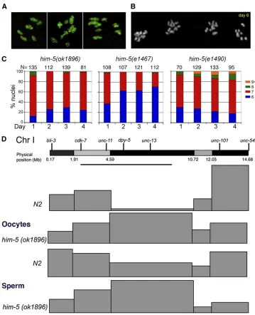
Figure 1 D1086.4 corresponds tohim-5. (A) The genomic locus on cosmid D1086, showing some of the primers (arrowheads) used for analysis of the D1086.4. The primers at the top were used to amplify a 3.65 kB frag-ment used for cosuppression. The other pairs of primers where used tofind the 39and 59of the D1086.4 gene using RT–PCR. The gene structure inferred from sequencing agreed with the structure displayed in WormBase. (B) Locations of the three him-5 mutations used in this study. The deletion ok1896
removes 519 bp including the upstream region, the 59-UTR, all of the first exon, and nearly all of the first intron. Both
e1467ande1490are G-to-A transitions, af-fecting the ATG start codon and the splice acceptor site at the start of exon 4 (or at the end of intron 3), respectively. The putative small isoform identified by WormBase is shown in the dashed lines and stippled boxes; this consists of thefinal 47 amino acids with short 59 -and 39-UTRs. This isoform would not have been detected in our experiments.
Table 1 Hatching rates and male progeny production
Genotype No. eggs % hatchinga No. animals % males
Wild type 3347 98.6 3300 0.06
him-5(e1467) 4572 60 2743 18
him-5(ok1896) 4960 71 3546 37
him-5(e1490) – – 873 35
e1490/ok1896 – – 1868 36
e1467/ok1896 – – 2152 27
Cosupp line 1 – – 3786 30
Cosupp line 2 – – 3102 32
xnd-1(ok709) 3834 60 2288 23
ok709;e1467 1945 62 1209 23
ok709;ok1896 – – 1520 39
mes-2(bn27) – – .4000 0
mes-2;ok1896 – – 3093 24
mes-2/+;ok1896 – – 435 46
Cosupp, cosuppression.
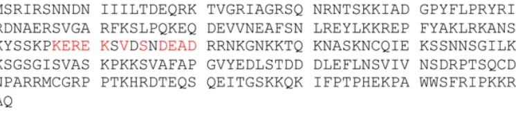
databases found in GenBank. Nonetheless, our attention was drawn to the presence of the sequence KEREKxVxxxDEAD, which is also found in the HSP83 chaperone protein sequence inDrosophila(Figure S1B). This identity is embedded in a re-gion of100 amino acids which, as determined by LALIGN, has an expected similarity value ofE,0.05 betweenHIM-5
and HSP83. The LALIGN program was implemented at the University of Virginia FASTA server. The results are shown in
Figure S1. While the similarity is significant at the 5% level, the importance of these amino acids in HSP83 is not known, and they are only very weakly conserved in other HSP82 and HSP83 proteins.
SNP analysis
him-5(ok1896)was introgressed into the HawaiianCB4856 background by repeated backcrosses. Hawaiian-specific alleles used in these studies were confirmed by PCR geno-typing. Spontaneous males from this stock were crossed to dpy-17(e164); him-5(ok1896) and non-Dpy; him-5 trans-heterozygous progeny were crossed to GFP+ males. GFP+ L4 hermaphrodites were individually plated and genotyped as previously described (Lim et al.2008) using established primer sets for chromosomes X and I (Wagneret al.2010). Chromosome I was also analyzed in sperm by mating the non-Dpy trans-heterozygous males from the above cross to dpy-18hermaphrodites and SNP genotyping the individually plated non-Dpy cross-progeny. Wild-type controls for both egg and sperm were previously reported (Wagner et al. 2010) and are included herein for comparison. Chi square tests were performed to test for significant differences in CO frequency and position between wild type andhim-5. Map units (m.u.) for the whole chromosome were calculated us-ing the formula: m.u. = (number of sus-ingle COs (SCOs) + 2 (number of double COs (DCOs))/sample size · 100. For each interval, the map size was calculated using the formula m.u. = (number of CO in interval/number of total COs on the chromosome)·m.u. for the chromosome.
Immunolocalization
Fixation and treatment of gonads were performed according to established protocols (Chanet al.2003). Briefly, animals were picked onto slides with 2.5ml of M9, rinsed and dis-sected in 2.5ml of 1·sperm salts, andfixed for 5 min in 1% paraformaldehyde with 1% Triton X-100 in a humid cham-ber. A coverslip was added and slides were placed on a metal block on dry ice for 10–20 min. Samples were freeze cracked and placed in 95% ethanol for 1 min. Prehybridization and hybridization were performed with 1· phosphate buffered saline (PBS) with 0.5% Triton X-100. Except where noted, all stainings were performed on 1-day-old adult animals. Antibodies were used at the following concentrations: rabbit anti–HIM-5 (Novus Biologicals, Littleton, CO) 1:5000; guinea pig anti–XND-11:2000 (Wagneret al.2010); guinea pig anti–SYP-11:1000 (Colaiacovoet al.2003); rabbit anti–
RAD-51 1:1000 (Rinaldo et al. 2002); rat anti–HTZ-1
1:2000 (Csankovszki et al. 2009); antinuclear pore
mono-clonal antibody mAb414 (Abcam, Cambridge, MA) 1:2000; guinea pig anti–HTP-31:1000 (Goodyeret al.2008); guinea pig anti–HIM-8 1:500 (Phillips et al. 2005); anti-H2AacK5 (Cell Signaling, Danvers, MA) 1:1000; secondary antibodies were all Alexa-488, Alexa-568, and Alexa-633 (Invitrogen, Carlsbad, CA), all used at 1:1000–1:2000 dilution. The
HIM-5 antibody was made by Strategic Diagnostics (New-ark, DE) as part of the modENCODE project using the100 C-terminal amino acids of the protein. DAPI staining bodies were assessed after Carnoy’s fixation of whole animals. Fluorescentin situhybridization (FISH) was performed us-ing probes to XR and 5S, accordus-ing to established protocol (Phillipset al.2005). Analysis of stained nuclei was carried out as previously described (Colaiacovo et al. 2003). All samples were mounted in Prolong Gold with DAPI and im-aged on a Nikon A1r confocal microscope (Nikon Instru-ments, Melville, NY) with 0.2-mm sections. Stacks were reconstructed and analyzed using Volocity 3D imaging soft-ware (Perkin-Elmer, Waltham, MA).
Irradiation
Rescue of bivalent formation at diakinesis was performed with 20 Gy irradiation as described previously (Wagneret al. 2010). To determine whether irradiation rescued desynapsis and meiotic progression, 1-day-old adults were exposed to 5 Gy using a Nordion Gamma Cell 1000 Irradiator (Ottawa, ON, Canada). Animals were fixed att= 0, 2, 4, 6, 8, and 24 hr postirradiation and stained with anti–SYP-1antibodies and DAPI. Rescue of desynapsis could be seen in a subset of nuclei att= 4 hr, was apparent in almost all nuclei att= 8 hr, and persisted untilt= 24 hr. Rescue of the clustering pheno-types was apparent int= 4 and most obvious int= 6 andt= 8 hr samples.
Analysis of RAD-51 foci
The dynamics of RAD-51focus formation were assessed by dividing germlines into seven sections between the transi-tion zone (leptotene) and the pachytene/diplotene border (Figure S5). The number ofRAD-51foci in each nucleus was determined using 3D reconstruction and image manipula-tion software, as described above.RAD-51foci were plotted as a heat map for each region to rapidly visualize differences in repair dynamics.
To determine whether breaks were made on the X chromosome, wild-type and him-5(ok1896) L1 animals were exposed torad-54 (RNAi) and dissected as L4 larvae as previously described (Wagner et al. 2010). X chromo-somes were distinguished from autochromo-somes on the basis of the lack of anti–HTZ-1 staining (Mets and Meyer 2009). Similar protocols were used to determine the total number of breaks, but germlines were stained with anti–SYP-1and anti–RAD-51.
Analysis of apoptosis
S7574) or acridine orange (20 mg/ml, Molecular Probes A3568) directly ontoC. elegans growth plates. Plates were incubated at room temperature in the dark for 1 hr, and then worms were transferred to fresh plates without staining so-lution to destain for another 1 hr in the dark. Destained worms were mounted in 4 mM Levamisole in EN buffer. Fluorescence microscopy was used to count the number of apoptotic nuclei on a Nikon Eclipse Ti wide-field microso-cope (Nikon Instruments).
Results
The him-5 gene corresponds to D1086.4
him-5 was mapped using morphological markers and se-quence-tagged sites to a region on chromosome V delimited by three cosmids that have 22 predicted protein-coding genes. The predicted gene D1086.4 was considered to be a strong candidate to behim-5on the basis of its expression in thegermline. Three lines of evidence support the conclu-sion thatD1086.4is thehim-5locus. First, transgenic lines containing a 3.65-kb region beginning 2.9 kb upstream of the predicted start codon onD1086.4(Figure 1A) produced
30% male self-progeny on average, indicating that cosup-pression of this locus gives a phenotype that resembles him-5mutants (Table 1). Second, sequencinghim-5mutant alleles revealed mutations within the D1086.4region: the molecular lesions for three him-5 mutations are shown in Figure 1B. These include the two previously characterized alleles,e1467ande1490, as well as a deletion allele created by theC. elegansKnockout Consortium, ok1896. Since the PCR fragment used for cosuppression included most of the neighboring gene,D1086.5, as well as its upstream region, this gene was also sequenced in wild-type and him-5 mutants. No sequence changes in theD1086.5coding region were found in any him-5 alleles. Third, RNA interference (RNAi) using dsRNA against the 59 or 39 regions of the
D1086.4locus, but not againstD1086.5, resulted in the pro-duction of male progeny.
The structure of the gene (Figure 1B) was confirmed using RT–PCR. him-5 has seven exons encoding a protein of 252 amino acids, which is an exceptionally basic protein, with an inferred isoelectric point (pI) of 10.7 (Figure S1A). WormBase annotations suggest the existence of a shorthim-5 isoform transcribed from an internal promoter and encoding only the last 47 amino acids (Figure 1B). We would not have observed this transcript in our RT–PCR experiments and can-not rule out the possibility that a very shortHIM-5 polypep-tide exists. The three him-5 alleles are shown in Figure 1B, the deletion allele ok1896 removes much of the promoter, the translational start and all of exon 1;e1467mutates the start codon; ande1490is a splice site mutation, predicted to reduce the efficiency of splicing. The severity ofe1490 sug-gests it may produce a truncated, dominant-negative protein. All three alleles are within the domains unique to the long him-5isoform and presumably would not affect the structure of the putative shorter isoform. Extensive homology searches
and syntenic alignment failed to identify any extended open reading frame or sequence alignments of.35 amino acids (data not shown). InC. briggsae, in which the alignment for this region is most coextensive withC. elegans, there appears to have been an insertion of a sequence of4.3 kb; part of the region also appears to have been inverted, and other base substitutions have occurred. While the precise molecular rearrangements are difficult to reconstruct, it is clear that no orthologous, full-length gene tohim-5could be identified inC. briggsae,C. remaneii,C. japonica,C. brenneri, or in the more distantly related nematodesBrucei malayiand Pristo-nichus pacificus. Thus, whilehim-5 plays an important role in meiosis in C. elegans, it may not to be evolutionarily conserved.
Crossover defects in him-5 mutants are X biased
The presence of males in the population is a hallmark of meiotic nondisjunction mutants and indicates underlying defects in the transmission of the X chromosome (Hodgkin et al. 1979). Defects in autosomal segregation are instead manifest as an increase in embryonic lethality because monosomy and trisomy of autosomes is incompatible with life (or subvital) (Zetka and Rose 1992). In mutants de-fective for CO formation on all chromosomes, e.g.,spo-11 (Dernburget al.1998), viability of zygotes is,2%, and for strong loss-of-function mutations in other meiotic genes affecting all chromosomes, e.g., him-3(e1256) (Zetka et al.1999) andhim-6(e1423)(Hodgkinet al.1979), em-bryonic lethality can be$80%. In contrast, in the strongest him-5alleles, close to 40% of progeny are male, and up to
30% of eggs do not hatch (Table 1). Although this rate of embryonic lethality is substantial, the majority of these un-hatched eggs can be attributed to X chromosome nondis-juction (Hodgkinet al.1979). Therefore, compared to the high frequency of males, him-5 may have only a minor effect on segregation of the autosomes, as previously sug-gested (Hodgkin et al. 1979; Broverman and Meneely 1994).
100% of the time (Table 2), further confirming a more strin-gent requirement forhim-5activity on the X chromosome.
One unexplained result is that the frequency of autosomal nondisjunction appears to be age dependent (Figure 2, B and C). For the weakest allelehim-5(e1467), the rate of X chro-mosome univalent formation is highest among the first ova and then declines somewhat with maternal age (Figure 2C). For him-5(e1490), the rate of X chromosome univalent for-mation shows no age dependence, but the rate of autosomal univalent formation increases with maternal age (Figure 2C). Similarly for the deletion allele, although not statistically dif-ferent between days 1 and 4, the trend is toward increased numbers of nuclei with autosomal univalents; for example, 12 univalents can be seen in day 4 gonads but in the course of many experiments have never been seen in 1-day-old adults (Figure 2, B and C and data not shown).
To test genetically for the presence of autosomal non-disjunction, we used the assay of Haack and Hodgkin (1991). When him-6males were mated to hermaphrodites that were mutant for him-6 and two unlinked, recessive autosomal markers [for example, unc-24 (IV) and dpy-11 (V)], 14 Unc and 17 Dpy offspring were found among 6079 cross-progeny, a frequency of 0.25–0.3% of excep-tional progeny per chromosome. These excepexcep-tional progeny arise from two different nondisjunction events, one in each parent. The experiment was repeated by mating him-5 (e1490)males to him-6 unc-24; dpy-11 hermaphrodites to test for the presence of nullisomic sperm arising fromhim-5 males. No Unc or Dpy animals were found among 9604 cross-progeny, suggesting that autosomal loss during sper-matogenesis inhim-5 males is extremely low and may not occur at all. To test the effect ofhim-5 on oogenesis,him-6 males were mated to unc-24; dpy-11 him-5(e1490) her-maphrodites. In this case, 12Uncand 10Dpyprogeny were found among 13,074 cross-progeny. The presence of these progeny that have inherited both copies of an autosome in the ova indicates that him-5 mutations result in autosomal nondisjunction during oogenesis. The rate is somewhat lower inhim-5 hermaphrodites than forhim-6 hermaphro-dites (per chromosome, 0.09% of the progeny for him-5 rather than 0.3% forhim-6) but still readily detected above background in which no autosomal nondisjunction has been observed. Similar results were found for other autosomes usingdpy-5I;unc-4 II; him-5 andunc-25III; dpy-11 him-5 hermaphrodites (data not shown). Since these genetic assays require nondisjunction events in both parents as well as the survival of the offspring, it is difficult to compare the rate of autosomal nondisjunction by the genetics and cytol-ogy. Nonetheless, these experiments reveal that autosomal
nondisjunction occurs in him-5 mutants, albeit at a much lower rate than X chromosome nondisjunction.
him-5 alters recombination distribution and frequency
Prior studies of the weak loss-of-function allele, him-5 (e1467), indicated that the genetic map on the X chromo-some was decreased to 35% of its wild-type levels (Hodgkin et al. 1979). On the autosomes, the genetic map was in-creased in some intervals (chromosomes I, II, III, and part of V) and decreased in others (chromosomes IV and part of V). The effect on different genetic intervals suggested the possibility thathim-5would be required not only for crossover frequency, but also for crossover positioning genome-wide. To better understand how the recombination landscape is af-fected by him-5, we performed single nucleotide polymor-phism (SNP) mapping across chromosomes X and I in wild type andhim-5(ok1896). SNP mapping of the X chromosome identified only 7/250 recombinants, yielding a map size of 2.8 m.u. for this chromosome. The paucity of recombinant prog-eny, however, limited our ability to determine whether there was also a change in crossover distribution on the X.
On autosomes, the vast majority of crossovers occur toward the chromosome ends in wild type, leaving a region in the central third of the chromosome with a greatly reduced rate of recombination (Figure 2D and Table S2). Similar regions with reduced recombination appear on each autosome and correspond to gene clusters that contain the majority of active genes (Barnes and Hodgkin 1996). In him-5(ok1896) mutants, the crossover landscape is strikingly different: nearly a third of the crossovers occur within the gene cluster on chromosome I (Figure 2D and Table S2). However, the overall length of the genetic map on chromo-some I is unchanged (52.5 m.u. in wild typevs.50.5 m.u. in him-5), and there is no increase in the number of double crossovers (3 DCO/124 CO in wild type vs.3 DCO/162 CO events in him-5). Therefore, the crossovers that are made within the gene cluster inhim-5(ok1896)must occur at the expense of crossovers on the chromosome arms, as reflected by the change in the map size of each of these intervals. These results also suggest the absence of an interchromo-somal effect which might have been expected if COs formed on the autosomes at the expense of the X. An interchromo-somal effect has been observed for thehim-8mutant that is defective in X chromosome CO formation due to an up-stream defect in X chromosome pairing (Phillips et al. 2005). By contrast, no interchromosomal effect was observed inxnd-1mutants, which functions to promote DSBs on the X. We also analyzed the effect ofhim-5on CO placement in the male germline. The CO landscape of him-5 sperm is
Table 2 Fish analysis of bivalent formation in diakinesis oocytes
Bivalents 6 5 + 2Xa 5 + 2Va 5 + 2Aa 4+2X+2Va 4+2X+2Aa 4 + 4Aa
N2 100 (63) 0 (0) 0 (0) 0 (0) 0 (0) 0 (0) 0 (0)
him-5(ok1896) 24 (20) 65 (54) 0 (0) 1 (1) 6 (5) 5 (4) 0 (0)
Values are percentage of nuclei (number analyzed).
similar to that ofhim-5oocytes, with a substantial number of COs occurring in the middle of the gene cluster at the expense of COs in more telomere-proximal regions (Figure 2D and
Table S3). As in oocytes, the differences in CO position can-not be explained by an increase in crossover formation on this chromosome as the map size of 49.8 m.u. does not differ significantly from wild type, 50.6 m.u. Together the SNP analyses suggest thathim-5+has two different effects on
re-combination:first, it influences recombination distribution on the autosomes such that crossovers in wild type occur more frequently in the gene-sparse outer regions of the chromo-some than in the gene-rich clusters; second, it facilitates crossover formation on the X chromosome.
X chromosome and autosomal nondisjunction in him-5 mutants are rescued by irradiation
The requirement forhim-5+for normal crossover frequency
and distribution suggested the possibility that him-5 may
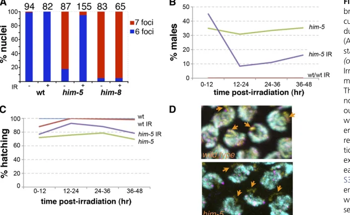
function to regulate the formation of meiotic double strand breaks (DSBs). To determine whetherhim-5affects the ini-tiation of meiotic DSBs, we asked whether DSBs introduced byg-irradiation could rescue the defect in CO formation on the X. This assay has been successfully used to show that a number of meiotic mutants are deficient in the induction of meiotic DSBs (Dernburget al.1998; Reddy and Villeneuve 2004; Wagneret al.2010). As shown in Figure 3,Table S4, andTable S5,g-irradiation (IR) is an effective suppressor of the him-5 mutant phenotypes. In unirradiated him-5 her-maphrodites,.80% of the ova havefive bivalents and two univalents (Figure 3A). Following exposure to a low dose of irradiation, only3% of the ova from ahim-5 hermaphro-dite have univalent chromosomes, and most ova show six normally paired bivalents. The same dose of irradiation has no effect on wild-type bivalent formation. As expected, irradiation also does not rescue the X chromosome pairing defective mutant, him-8 (Figure 3A), since chromosome
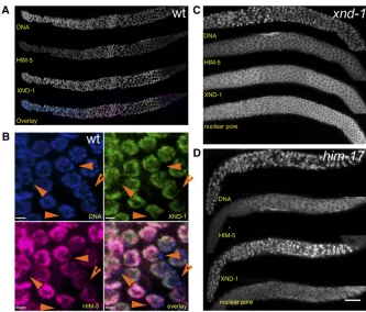
Figure 2 Bivalent formation and recom-bination frequency and position. (A and B) Crossover formation is assessed by the number of DAPI-staining bivalents and univalents at diakinesis. (A) FISH of chromosomes X and V reveals defects in bivalent formation in adult day 1 her-maphrodites. (Left) Six bivalents are present in wild-type and a small fraction ofhim-5(ok1896)nuclei. (Center) Seven foci, five bivalents corresponding to autosomes (V, yellow foci) and two uni-valent X chromosomes (purple foci) are seen most often in him-5. (Right) Rare nuclei show additional univalents (two purple X foci + 2 yellow chromosome V foci), indicating defects in bivalent mation on autosomes. (B) Chiasma for-mation is further compromised in aging
him-5 germlines as shown by the ap-pearance of multiple nuclei with more than nine DAPI staining bodies in adult day 6 germlines. (C) Quantification of DAPI-staining bodies with age inhim-5
alleles. (Day 1 is different from day 4, Mann–Whitney test,e1490, P¼ 0.04;
e1467,P,0.0001) (D) Recombination landscape is altered on autosomes in
pairing is a prerequisite to synapsis and DSB repair off the homologous chromosome in C. elegans. These results indi-cate that him-5 is competent in processing artificially in-duced DSBs into crossovers on the X chromosome.
Time course analysis of the irradiatedhim-5animals con-firmed that theHimphenotype was substantially suppressed from ova that had been exposed to irradiation (Figure 3B). Irradiation also suppressed the low level of embryonic le-thality associated with him-5 (Figure 3C). In some of the irradiated animals, hatching rates reached 100%, indicating that complete suppression is possible (data not shown). Since a portion of the lethality must be a consequence of the defect in bivalent formation on autosomes, the ability of irradiation to rescue the hatching defect suggests that autosomal aneuploidy results from a defect in DSB forma-tion. This result also supports our earlier observation that autosomal aneuploidy is minimal in sperm from him-5 males. Since sperm are produced during L3 and early L4 larval stages and stored in the spermathecae, they have already completed meiosis at the time of irradiation. Sperm from irradiated animals were competent to confer full viability to their offspring. Thus, autosomal nondis-junction is minimal or nonexistent during hermaphrodite spermatogenesis.
HIM-5 is required for DSB formation on the X
Our results with irradiation indicate that the aneuploidy defects that arise in him-5 mutants can be effectively bypassed by artificially inducing DSBs. To observe DSB for-mation more directly, we monitoredRAD-51focus formation in him-5 mutant germlines. RAD-51 is a single-stranded DNA binding protein that is required for strand exchange
during DNA damage repair. Since RAD-51 foci overlap
SPO-11 induced DSBs with .95% confidence (Mets and Meyer 2009),RAD-51localization is an accurate and widely used indicator for DSB formation in meiosis. The number and location of loci that have received a DSB can be assessed in situby looking at RAD-51 foci afterrad-54(RNAi) treat-ment. rad-54(RNAi) prevents repair of the DSBs at a step after the recruitment of RAD-51 to the lesion effectively trapping RAD-51 at the sites of repair (Mets and Meyer 2009). The X chromosome can be unambiguously distin-guished from the autosomes on the basis of differential lo-calization of variant histones and histone post-translational modifications, which serve as reporters of transcriptional status (Kelly et al. 2002). In these studies, as previously shown (Wagner et al. 2010), we used antibodies against
HTZ-1, a H2A.Z homolog (Csankovszkiet al.2009), to neg-atively mark the X chromosome. By this combination of methods, recruitment of RAD-51 to the X chromosome was assessed. In agreement with previous results, we could identify aRAD-51focus on the X chromosome in wild-type worms in almost every nucleus (Figure 3D). In contrast, in him-5(ok1896)mutants close to 90% of the nuclei lacked
RAD-51 foci on the X (Figure 3D). These results further support a role forhim-5+in promoting meiotic DSB
forma-tion on the X chromosome.
An alternative interpretation of these results is thathim-5 is defective in the recruitment ofRAD-51to the break site. We do not favor this interpretation because unlike mutations inrad-51, which lead to fragmented and fused chromosomes at diakinesis, him-5mutants always show well-formed uni-valents and biuni-valents. Nonetheless, we cannot rule out the possibility thathim-5has two roles, one in the formation of
DSBs and a second role, immediately downstream, in early repair events.
him-5 mutants are delayed in progression through the meiotic germline
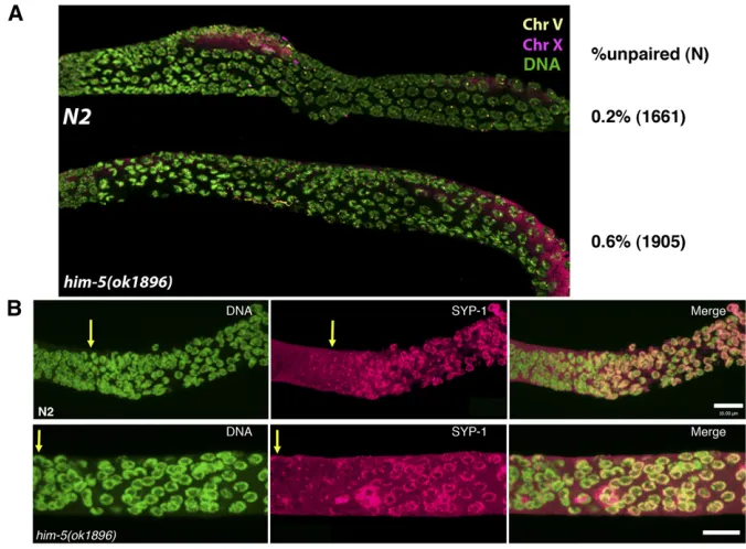
The ability of IR to rescuehim-5but nothim-8(Figure 3A), suggested that chromosome pairing occurs normally inhim-5 mutants. In support of this, FISH probes revealed that chro-mosomes X and V demonstrate pairing with wild-type dynam-ics and efficiency from zygotene through pachytene (Figure 4 and Figure S2). Quantification of pairing using anti–HIM-8
antibodies confirmed that X chromosome pairing levels are indistinguishable between wild type and mutant from the transition zone through pachytene (Figure 4A). In addition, the chromosome axes and the synaptonemal complex assem-bled normally inhim-5mutants (Figures 4B,Figure S3A, and data not shown). Thus, the early events of homolog pairing and synapsis do not depend onhim-5+activity.
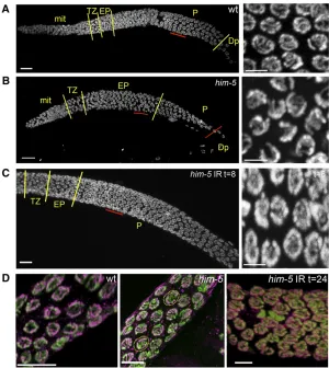
By contrast, the normal progression of meiosis is altered in him-5mutants. Specifically,him-5mutant nuclei appear to be delayed in their progression through the early pachytene stage of meiosis, as assessed by comparing the morphology of DAPI-stained nuclei in wild type and him-5 mutants. In wild type, thegermlineis organized in a spatial and temporal gradient with a stereotypical number of nuclei in each stage of meiosis distinguished by a unique DNA morphology (Fig-ure 5A). Chromosomes in leptotene/zygotene (that is, the transition zone) are tightly clustered at one side of the nu-clear periphery and appear as a bright crescent with DAPI staining. As nuclei enter early pachytene, the chromosomes remain attached to the nuclear periphery but are individually visible and thus less tightly packed. By midpachytene, the chromosomes are fully dispersed around the nuclear periph-ery (Figure 5A, closeup).
In him-5 mutants, the number of nuclei with the clus-tered morphology characteristic of early pachytene is dra-matically increased, and many fewer nuclei are seen with full pachytene morphology (Figure 5B). The persistence of early pachytene nuclei is not associated with a substantial increase in apoptosis, as expected if DSBs were not repaired or if the synapsis checkpoint were activated (Table 3 and
Table S6). To determine whether the delay in progression was instead a consequence of the failure to make a DSB on one or more chromosomes, we asked whether exogenous breaks could promote progression through early pachy-tene. In wild-type germlines, meiotic progression is unaf-fected by 20 Gy of irradiation (not shown). The same dose of irradiation completely suppressed the delay in progres-sion observed in thehim-5mutant (Figure 5C). Therefore, we infer that the failure to make DSBs on one or more chromosomes leads to a delay in progression through early pachytene.
Another cytological feature of him-5 germlines is also noteworthy: the appearance of late pachytene nuclei with desynapsed X chromosomes (Figure 5D). Although full syn-apsis was observed in early pachytene,him-5(ok1896) nu-clei (Figure S3), at slightly later stages of pachytene, a chromosome withoutSYP-1labeling is observed in nearly everyhim-5mutant nucleus, suggesting that a chromosome pair has desynapsed (Figure 5D). Localization of HIM-8
identified the X as the desynapsed chromosome in these nuclei (Figure S3). In addition, rare nuclei with two unla-beled chromosomes are seen, suggesting that autosomes, as well as the X, are susceptible to desynapsis (Figure 5D). We note that desynapsis at this stage does not immediately cause the homologs to separate from each other since sep-arated X chromosomes were not observed until diplotene (Figure 2A and Figure S2). Induction of exogenous DSBs
Figure 4Pairing and synapsis are normal in
him-5 mutants. (A) Wild type and him-5 (ok1896)werefixed and hybridized with FISH probes for chromosomes V (yellow) and X (ma-genta) or anti–HIM-8 antibody (Figure S2) to assess the establishment and maintenance of pairing in zygotene to pachytene nuclei (quan-tification was done using the HIM-8 images in
by IR suppresses desynapsis, as shown by chromosomes in him-5germlines that are fully labeled with anti–SYP-1 anti-bodies at 8–24 hr postirradiation (Figure 5D, right). The maintenance of synapsis after irradiation indicates that desynapsis is a secondary consequence of the failure to make a break on the X and autosomes and that him-5 does not directly function in synaptonemal complex (SC) mainte-nance. These observations are consistent with the results seen in xnd-1 mutants and support the conclusion that DSB formation is coordinated with synapsis and the main-tenance of the synaptonemal complex.
him-5 and xnd-1 appear to function in the same genetic pathway
Many of the features ofhim-5mutants are strikingly similar to those described forxnd-1 (Wagneret al.2010). Specifi -cally both mutants appear to have primary defects in making DSBs on the X chromosome and in CO placement on auto-somes, as well as defects in pachytene progression and SC maintenance. These similarities prompted us to determine
whetherhim-5+might function in the same pathway as
xnd-1+. To assess genetically whether these genes interact to
control crossover formation on the X, we made a double mutant between xnd-1(ok709) and him-5(e1467), both of which independently increase the frequency of males in the population to nearly 20% (Table 1). Thexnd-1(ok709); him-5(e1467) double mutant showed no change in male fre-quency. The lack of suppression or synthetic enhancement between the mutations suggests thathim-5andxnd-1 oper-ate in the same functional pathway.
HIM-5 is enriched on the autosomes
The inferred amino acid sequence ofHIM-5predicts a novel and highly basic protein. To assess the localization ofHIM-5
in worms, antibodies generated to the C-terminus were used for immunolocalization (see Materials and Methods). As shown in Figure 6A,HIM-5protein is found associated with chromosomes from the mitotic region of the germlineuntil late pachytene. No staining is observed outside of the gonad (data not shown). In the germlines of wild-type hermaph-rodites,HIM-5preferentially localizes to most meiotic chro-mosomes but is clearly excluded from one pair. A number of other proteins have been shown to be enriched on auto-somes, including a set that is required for modulating X chromosome gene expression [MES-4(Benderet al.2006),
MRG-1 (Takasakiet al.2007), the DRM complex (Tabuchi et al. 2011), and histone modifications associated with
Figure 5Deficit in DSBs leads to a delay in pachytene progression and to desynapsis. (A–C) DAPI-stained germ-line of wild type (A),him-5(ok1896)(B), or irradiatedhim-5 (ok1896)(C) are shown with yellow lines demarcating the different regions of the germline as indicated by chromo-some morphology (Bar, 10mm). The region underlined in red is shown to the right in the higher magnification pro-jection (Bar, 2mm). Note that him-5 mutants have an extended early pachytene region at the expense of full pachytene (B) and that normal progression is restored after irradiation (C). (D) Desynapsis ensues in the absence of breaks. Full synapsis is seen between all homologs in wild type in late pachytene (left) as shown by the overlap be-tween DNA (green) and anti–SYP-2 (magenta). In him-5 (ok1896), the X chromosome desynapses in almost all nu-clei (center). In occasionalhim-5nuclei, multiple chromo-somes without SYP-2 staining are observed (dashed yellow circles). Irradiation suppresses desynapsis (right) and SYP-2 staining persists between homologs in late pachytene nu-clei (Bar, 5mm).
Table 3 Apoptosis analysis with SYTO 12
No. apoptotic
nuclei/gonad 0 1 2 3 4 5 6 N Mean SD SE mean
N2 1 10 19 14 4 0 0 48 2.208 0.9444 0.136
him-5(ok1896) 2 1 9 11 4 2 2 31 2.903 1.4226 0.256
transcriptional activation (Schaner and Kelly 2006), as well as XND-1 (Wagner et al. 2010)]. To determine whether
HIM-5 is also underrepresented on the X, hermaphrodite germlines were costained with anti–XND-1and anti–HIM-5
antibodies. As shown in Figure 6B,XND-1andHIM-5 coloc-alize to the same subset of chromosomes, indicating that
HIM-5 is also enriched on autosomes. We cannot tell from these images whether there are subnuclear differences be-tween the two proteins in their localization on individual autosomes.HIM-5staining persists in nuclei that are beyond the zone ofXND-1staining. No signal was detected in these regions in the deletion allelehim-5(ok1896), confirming the specificity of the antibody (Figure S4).
The autosomal localization ofHIM-5and the similarities between thexnd-1andhim-5 mutant phenotypes raise the possibility that localization of these proteins could be de-pendent on one another. Previous studies showed that
XND-1 localizes normally in him-5 mutants (Wagner et al. 2010). In contrast, immunolocalization of HIM-5 on auto-somes in the mitotic through midpachytene germlines was significantly reduced in thexnd-1mutant background (Fig-ure 6C).HIM-5 localization was also diminished inhim-17 (e2806) mutants, another meiotic gene with alleles that preferentially affect X chromosome crossover formation (Figure 6D). Thus, the wild-type activities of both him-17 andxnd-1appear to be necessary for correct localization of
HIM-5to the autosomes and/or its expression. The stronger
Him phenotype inhim-5deletion vs.xnd-1 (40%vs. 25%) suggests that either the unlocalizedHIM-5protein inxnd-1 mutants is functional or that reduced quantities of protein are below the level of detection with our antibodies.
Fur-thermore, the dependency ofHIM-5localization onxnd-1, and not vice versa, suggests thathim-5acts downstream ofxnd-1.
him-5 is suppressed by changes in X chromosome architecture
Within the germlines of wild-type worms, the majority of genes on the X chromosome are not expressed, and the X chromosome is replete with histone post-translational mod-ifications associated with heterochromatin and silent chro-matin (Kelly and Fire 1998; Kelly et al.2002). In contrast, modifications associated with transcriptional activation are predominantly found on the autosomes (Schaner and Kelly 2006). We previously showed that xnd-1 could be sup-pressed by mutations inmes-2in which X chromosome gene silencing does not occur, raising the possibility that silencing of the X blocks crossover formation on this chromosome (Wagner et al. 2010). To determine whether the require-ment forhim-5+activity is diminished when the X
chromo-some is desilenced, the frequency of males was assessed in themes-2(bn27); him-5(ok1896)double mutant (Table 1). Loss ofmes-2results in grand-maternal sterility so that the frequency of males can be assessed in the F2 brood. In an otherwise wild-type background, all of the progeny ofmes-2 mothers are hermaphrodites, indicating that loss of mes-2 function does not itself cause nondisjunction. him-5 (ok1896) broods yield 40–45% males, while mes-2; him-5 broods contained 25% males. Therefore, loss of mes-2 function partially suppresses the him-5 X nondisjunction phenotype. These results suggest thatmes-2acts in opposi-tion tohim-5and suggests thathim-5+is sensitive to
chro-matin configurations on the X.
Figure 6HIM-5 is enriched on the autosomes. (A and B) Anti–HIM-5 staining in wild-type germlines. (A) HIM-5 can be seen at low levels in the mitotic zone, accumulates in the transi-tion zone and early pachytene, and decreases mildly upon entry into pachytene and more dra-matically in late pachytene. Costaining with anti–XND-1 reveals differences in the accumu-lation patterns of these two proteins. (B) En-larged region of late pachytene shows the overlap between XND-1 (green), HIM-5 (ma-genta), and DNA (blue) showing their exclusion from the X chromosome (orange arrowheads). Similar enrichment is observed in mitotic and early pachytene nuclei (not shown). Bar, 2
We showed previously that acetylation of histone H2A lysine 5 (H2AacK5) is increased inxnd-1mutants, suggest-ing a role for this mark when crossover frequency is re-duced (Wagner et al. 2010). However, we observed no gross changes in H2AacK5 levels inhim-5(ok1896)mutant germlines (Figure S5). Therefore, despite the ability of mes-2 to suppress nondisjunction in bothxnd-1 and him-5 mutants and the requirement for xnd-1+ function for HIM-5 localization, the two genes appear to function at different steps to control X chromosome crossover formation.
RAD-51 dynamics distinguish him-5 from xnd-1
Further evidence that these genes may have distinct functions comes from analysis of RAD-51accumulation in the mutants. In wild-type nuclei,RAD-51foci are dynamic, increasing during early pachytene, and peaking with ap-proximately sevenRAD-51foci per nucleus (Figure 7 and
Figure S6).RAD-51foci, and thus DSBs (Mets and Meyer 2009), are initiated within the transition inhim-5mutants and wild type; yet, the number ofRAD-51foci per nucleus in thehim-5mutant is lower at each subsequent stage until late pachytene (Figure 7B). Because initiation of DSBs occurs at the same time inhim-5and wild type, the differ-ences inRAD-51focus formation could be explained by an overall decrease in breaks, a change in the overall kinetics of DSB formation or both. We observed a decrease in the total number of DSBs by analysis ofRAD-51foci inrad-54 (RNAi). Consistent with previous reports, we observed that wild-type animals enjoy 14 DSBs (Table S7) (Mets and Meyer 2009). In contrast, in him-5 mutants, we only ob-served10 breaks/nucleus. This does not rule out an ad-ditional role for him-5 in regulating the kinetics of DSB repair, but these results support the hypothesis thathim-5 plays a role in controlling CO dynamics through the regu-lation of meiotic DSB formation.
We note that RAD-51foci also persist longer in him-5 (ok1896)than in wild type, with foci visible in late pachy-tene nuclei and disappearing in diplopachy-tene (Figure 7B and
Figure S6). We attribute the persistence ofRAD-51foci as a consequence of the delay in meiotic progression (de-scribed above and Figure 5), although this has not been tested.
The dynamics of break formation in him-5 are strik-ingly different from that seen in xnd-1 mutants (Figure 7 and Figure S6). In xnd-1 mutants,RAD-51foci appear simultaneously rather than accumulating over time as they do in wild type andhim-5, with the maximum num-ber of foci visible as early as the transition zone/early pachytene nuclei. As nuclei progress into pachytene in xnd-1 mutants, the RAD-51 foci appear to get larger within the extended early pachytene zone and into late pachytene until they disappear upon entry into diplotene (Figure S6). The absence ofRAD-51foci in diplotene indi-cates that both mutants (and wild type) have repaired all damaged DNA.
Discussion
Thehim-5gene inC. eleganswas among the original meiotic mutations found in worms, and him-5 mutants are widely used in strain construction and other types of genetic anal-ysis (Hodgkinet al.1979). Despite its familiarity, the molec-ular identity of the him-5 gene has not been previously demonstrated. We show that him-5 corresponds to
D1086.4, which encodes a novel, small and extremely basic protein. Mutations inhim-5are notable for greatly reduced recombination and elevated nondisjunction of the X chro-mosome (Hodgkin et al. 1979; Broverman and Meneely 1994), whereas effects on the autosomes appear to be comparatively minor. We find that the overall number of crossovers on the autosomes, as reflected in the genetic map units, is not reduced in him-5 mutants compared to wild type (at least for chromosome I), but crossovers are redistributed such that the recombination map and the phys-ical maps are more congruent inhim-5mutants than in wild type. Defects in crossover formation on other autosomes can
Figure 7 Dynamics of RAD-51 accumulation differ betweenxnd-1and
be inferred from the presence of multiple pairs of univalents at diakinesis.
Role of HIM-5 in DSB formation and repair
Possibly the most informative results for understanding the mutant phenotype ofhim-5are that we do not observe RAD-51foci on the X in therad-54(RNAi) and that nearly every aspect of thehim-5 mutant phenotype is suppressed by ra-diation-induced DSBs. These phenotypes include X chromo-some and autochromo-some nondisjunction, premature chromochromo-some desynapsis, and a delay in pachytene progression. Two mod-els could explain the ability of IR to rescue: either (1) him-5+functions upstream of theSPO-11nuclease to potentiate
the X chromosome for recombination or (2)him-5acts im-mediately downstream of DSB formation, in a processing step prior to RAD-51recruitment. The latter model would need to explain how DSBs are ultimately repaired in him-5 mutants since we see no evidence of increased apoptosis (which would occur if there were persistent damage). Addi-tionally, despite passaging for hundreds of generations as homozygotes, no germline transmissible mutations have been identified in these strains, suggesting that the breaks are not repaired by the mutagenic NHEJ pathways nor are they leading to chromosome fusions. For these reasons, we favor a model in whichhim-5acts upstream of break forma-tion, although it may also have a role downstream in break repair.
One of these things is not like the other one: X/autosome differences in crossover control
A number of studies have revealed that the X and autosomes respond differently to loss of certain meiotic gene functions. A subset of mutations, e.g., htp-1, him-3, and cra-1, more severely affect autosomes (Couteauet al.2004; Couteau and Zetka 2005; Smolikov et al. 2008); whereas other muta-tions, e.g., him-17, him-19, xnd-1, and him-5 (Hodgkin et al.1979; Reddy and Villeneuve 2004; Tanget al.2010; Wagneret al.2010), have more severe consequences for the X. The latter class all affect very early events in CO forma-tion, in either DSB formation or processing. The observation that double COs have been documented on the autosomes but never on the X is consistent with this chromosome re-ceiving fewer meiotic breaks, although this has not been directly tested in the nematode system. In the analysis of him-17, Reddy and Villeneuve (2004) proposed a model that they referred to as a“window of opportunity”to explain its different effects on the X and the autosomes. Their hy-pothesis is that the accessibility of the chromosomes to SPO-11 is limited to a specific time period during prophase I. According to their model, the “window”for the X chromo-some closes before the window for the autochromo-somes. Thus, in a mutant likehim-17, in which overall accessibility for DSBs is limited even further, the X chromosome is more sensitive because its window is normally smaller. Since its localization is dependent onhim-17, we can apply this model to explain the effects of him-5 and hypothesize that it is one of the
genes responsible for determining when the X chromosome window is open.
X/autosome differences in DSB formation, however, may alternatively be explained by the gross differences in chromatin states between these chromosomes. Because crossovers occur preferentially in “open” chromatin (Ohta et al.1994; Wu and Lichten 1994; Nicolas 1998), the het-erochromatic-like X chromosome ofC. elegansmight present an inhospitable environment for crossover formation by cre-ating a kinetic barrier to theSPO-11complex. Therefore, we postulate thathim-5may have evolved as part of a mecha-nism to promote crossovers in heterochromatin. Our obser-vation that the HIM phenotype is suppressed by mutations inmes-2andmes-3, components of a histone H3K27 meth-yltransferase complex that is required for silencing X-linked genes, supports the model that the chromatin architecture of the X normally limits CO formation.
Models of HIM-5 function
Because the total number of breaks is reduced in him-5 mutants, we favor models in whichHIM-5directly regulates theSPO-11machinery. The simplest model would have him-5 regulating the expression and/or activity of the SPO-11
machinery. However, while this could account for the in-crease in X and autosomal univalents at diakinesis and resulting males and lethality, as well as the decreased total number ofRAD-51foci observed (9vs.14 inhim-5vs.wild type), it is more difficult to explain how reducing SPO-11
activity could account for the change in CO positioning to go from almost none to nearly half of all COs occurring in the autosomal gene clusters. Simply reducingspo-11expression would be expected to reduce CO formation throughout the genome, not simply divert COs on the autosome to the cen-tral gene cluster.
Instead, we favor the possibility that HIM-5 functions, either directly or indirectly, to target theSPO-11machinery to the chromosome. In other species, SPO11 is known to interact with accessory proteins that regulate its recruitment and function (Keeney 2001). One possibility is thatHIM-5is a functional homolog of one of these components. Another possibility is thathim-5could function to define the recom-bination hotspot, either as a landmark itself or by creating a critical chromatin signature. Alternatively, him-5 could function indirectly to influenceSPO-11recruitment by mod-ifying the closed chromatin milieu on the X (and distal autosome regions). This could occur either by directly recruiting histone-modifying complexes (or chromatin-asso-ciated proteins) to the X (and distal autosomes) or by pre-venting these complexes from accessing the autosomes so that modifications are made on the X. Each of these models posits thatSPO-11is functional but cannot be properly tar-geted to recombination hotspots. We propose that without normalcisortrans-acting localization features, theSPO-11
number of breaks may be reduced inhim-5 as compared to in wild type either because crossover interference is so strong in the worm that breaks are redistributed to the cen-ter of the chromosome effectively inhibiting additional breaks distally to both ends, or because passive recruitment to the DNA is comparatively inefficient.
These models predict that HIM-5 proteins would be found on the X and/or unequally distributed between the gene clusters and distal autosomal regions. Immunofl uores-cence does not provide adequate resolution to determine whether HIM-5 is biased in its autosomal distribution. De-spite our inability to detect HIM-5proteins on the X, there may be subthreshold levels sufficient for function.
Pathways for X chromosome breaks
him-5 phenotypes closely resemble those of xnd-1. Double mutant analyses are consistent with these genes working in the same pathway rather than parallel pathways. This in-terpretation is supported by the observation that xnd-1+
activity is required for properHIM-5localization and places
HIM-5 downstream in the pathway. However, two lines of evidence indicate thatHIM-5andXND-1must have distinct roles in crossover control. First, previous studies have revealed pronounced differences in H2AacK5 in the xnd-1 mutant (Wagneret al.2010), whereas no overt differences in this modification are observed in thehim-5mutant ( Fig-ure S5). Second, the early dynamics ofRAD-51 accumula-tion in xnd-1 and him-5 differ dramatically (Figure 7). In xnd-1 mutants, the majority of programmed DSBs, as assessed byRAD-51foci, appear simultaneously at the onset of break formation. By contrast, inhim-5, fewer breaks are made, but the time course of break formation is more similar to wild type. Therefore, although both XND-1 and HIM-5
affect crossover formation on the X as well as crossover distribution genome-wide, andHIM-5localization is depen-dent on xnd-1+, it appears that these proteins may have
additional, independent roles in regulating DSB formation and repair.
One possible model to explainXND-1andHIM-5suggests that XND-1’s primary function is to negatively regulate
H2AK5 acetylation, allowing heterochromatic regions of the genome to “compete” effectively with the autosomal gene clusters for crossover formation. HIM-5’s association
with chromatin could be reduced in thexnd-1mutant back-ground either because the high levels of H2AacK5 prevent its association with DNA or because XND-1 protein is di-rectly required for its expression and/or localization.
HIM-5 localization also requires functional him-17, a THAP-domain protein required for meiotic DSB break for-mation on all chromosomes. Weak mutations ofhim-17have more severe effects for the X than for autosomes, which we suggest may now be explained by the defect inHIM-5 local-ization. Whether the effects ofhim-17onHIM-5localization are direct or not await further analysis of these proteins, however, the observation that HIM-17 protein localizes to all chromosomes (Reddy and Villeneuve 2004) favors
a model in which him-17indirectly controlsHIM-5protein distribution.
Desynapsis and delayed meiotic progression
In addition to effects on recombination and disjunction, him-5mutants also exhibit a delay in meiotic progression. Since the other mutant phenotypes ofhim-5 can be explained by a role in DSB formation and the delay is suppressed after exposure to irradiation, we suggest that the delay in meiotic progression is also attributed to the defect in DSB formation. Because DSBs would normally be deleterious, the events of meiosis I are closely monitored to ensure that each chromo-some receives a crossover and DNA damage is fully repaired. Checkpoints ensure complete synapsis and the repair of DNA damage. InC. elegans, these checkpoints require the activity of conserved pathways mediated bypch-2andcep-1, respec-tively (Gartneret al.2000; Bhalla and Dernburg 2005). In addition, mechanisms to couple pairing, synapsis, and DSB formation have been elucidated (Couteau and Zetka 2005; Goodyeret al.2008; Smolikovet al.2008), suggesting that fail-safe mechanisms ensure the completion of meiotic processes.
The analyses of him-5 presented here and our previous studies onxnd-1(Wagneret al.2010) suggest that an addi-tional safeguard exists to maintain the synaptonemal com-plex association with chromosomes after formation of a DSB. In both mutants, the synaptonemal complex poly-merizes along every chromosome with normal kinetics and is fully polymerized by the end of the transition zone region. Thus, him-5+ and xnd-1+ do not appear to be needed to
initiate synapsis or SC formation. Moreover, it does not ap-pear that him-5+ and xnd-1+ are simply required for the
maintenance of the SC since irradiation can prevent desy-napsis in the mutant background. Instead, our data are most consistent with a model in which the underlying defect in these mutants, the failure to make a break on the X chro-mosome, apparently leads to subsequent desynapsis of this chromosome. The ability of a DSB to stabilize the synapto-nemal complex does not appear to be limited to the X chro-mosome. Inhim-5mutants, as many as 10% of nuclei have at least one autosome that does not receive a crossover, as revealed by an increase in the number of univalent chromo-somes observed at diakinesis. In a subset of him-5 mutant nuclei, we could observe two chromosomes that failed to label with anti–SYP-1 antibodies (Figure 5D), suggesting that the synaptonemal complex has dissociated from both chromosomes. Since this desynapsis is also rescued by irra-diation, we propose that the formation of a DSB (or a sub-sequent crossover intermediate) must trigger a change in the SC allowing for its stabilization on the homologs.
appears to be independent of the known germline check-points. The inability of him-5 mutants to activate the pch-2–dependent synapsis checkpoint suggests the possibility that this checkpoint is only active during a short window of time in early pachytene. We noted that the most distal him-5 nuclei in which the X chromosomes desynapsed had not yet progressed into full pachytene, on the basis of nu-clear morphology. This suggests that monitoring of break formation and stabilization of the SC occurs during early pachytene. The transition into full pachytene could reflect two inherent changes with regard to the synaptonemal com-plex, both a closing of the window for activation of the synapsis checkpoint and a DSB-induced stabilization of the synaptonemal complex.
It is unclear as yet what controls these transitions, but it could be mediated through communication with the nuclear periphery. The Jantsch laboratory has shown that distinct phosphorylation states of the nuclear membrane protein
SUN-1are required for association of DNA with the nuclear periphery, its movements there, and its ultimate release (Penkneret al.2009). It is tempting to speculate that signals emanating from an early crossover intermediate lead to a specific alteration inSUN-1phospho status and contribute to release from the nuclear periphery. Further analysis of him-5 will help to elucidate the mechanisms that control the formation and monitoring of DSBs and how these are coordinated with meiotic events to ensure the timely and accurate execution of meiotic recombination.
Is break formation sensitive to age?
The nondisjunction phenotype ofhim-5 appears to worsen with age, suggesting a greater requirement forhim-5+
ac-tivity in older germlines. Maternal age effects on nondisjunc-tion are a familiar feature of human meiosis, but the time scale over which these effects occur in humans is measured in decades rather than in hours, as they are in worms, so the underlying mechanism could be quite different. For him-5, the differences among the three alleles suggest that these mutations may be revealing an underlying property of mei-osis in worms, rather than a specific consequence of the effects onhim-5 function. It has been well established that parental age decreases recombination and increases X chro-mosome nondisjunction rates inC. elegans(Rose and Baillie 1979). him-5 joinshim-19 in a class of meiotic genes that increase severity with maternal age. Tanget al. (2010) hy-pothesized that him-19might affect the stability of an un-known maternally supplied factor that becomes depleted over time. One postulate is that him-5mutations have dif-ferent effects on the stability of this factor, which affects autosomal disjunction more than X disjunction. In both him-5 and him-19, the observed defects can be attributed to defects in early meiotic events. We speculate that early events such as DSB formation might be particularly sensitive to aging, because they are influenced by the packaging of chromatin, which has been shown for somatic tissues to be altered during aging (Greer et al.2010).
Acknowledgments
We thank Verena Jantsch for suggesting the cosuppression assay and doing the injections that facilitate P.M.M.’s anal-ysis of these lines. We also thank Dave Baillie for pointing out the similarity to HSP83 and Alex Ensminger and Nelson Lau for their generosity in providing purified RNAs to P.M.M. We thank Monique Zetka, Anne Villeneuve, Adriana la Volpe, Verena Jantsch, Abby Dernburg, and Gyorgyi Csankovski for antibodies and Barbara Meyer and David Mets for technical advice. P.M.M. thanks the many Haverford College students who worked on different aspects ofhim-5. J.L.Y. thanks Cyn-thia Wagner and Mainpal Rana for discussion and reading of the manuscript. This work has been funded by National Sci-ence Foundation grants RUI MCB 0131321 and RUI MCB 061470 to P.M.M. and by National Institutes of Health K01AG031206 and MWRI start-up funds to J.L.Y.
Literature Cited
Barnes, T. M., and J. Hodgkin, 1996 Thetra-3sex determination gene ofCaenorhabditis elegansencodes a member of the calpain regulatory protease family. EMBO J. 15: 4477–4484.
Barnes, T. M., Y. Kohara, A. Coulson, and S. Hekimi, 1995 Meiotic recombination, noncoding DNA and genomic organization in
Caenorhabditis elegans. Genetics 141: 159–179.
Bender, L. B., J. Suh, C. R. Carroll, Y. Fong, I. M. Fingermanet al., 2006 MES-4: an autosome-associated histone methyltransfer-ase that participates in silencing the X chromosomes in theC.
elegansgerm line. Development 133: 3907–3917.
Bhalla, N., and A. F. Dernburg, 2005 A conserved checkpoint monitors meiotic chromosome synapsis in Caenorhabditis ele-gans. Science 310: 1683–1686.
Brenner, S., 1974 The genetics ofCaenorhabditis elegans. Genetics 77: 71–94.
Broverman, S. A., and P. M. Meneely, 1994 Meiotic mutants that cause a polar decrease in recombination on the X chromosome
inCaenorhabditis elegans. Genetics 136: 119–127.
Chan, R. C., A. Chan, M. Jeon, T. F. Wu, D. Pasqualone et al., 2003 Chromosome cohesion is regulated by a clock gene pa-ralogue TIM-1. Nature 423: 1002–1009.
Colaiacovo, M. P., A. J. MacQueen, E. Martinez-Perez, K. McDonald, A. Adamoet al., 2003 Synaptonemal complex assembly inC. elegans
is dispensable for loading strand-exchange proteins but critical for proper completion of recombination. Dev. Cell 5: 463–474. Cole, F., S. Keeney, and M. Jasin, 2010 Evolutionary conservation
of meiotic DSB proteins: more than just Spo11. Genes Dev. 24: 1201–1207.
Couteau, F., and M. Zetka, 2005 HTP-1 coordinates synaptone-mal complex assembly with homolog alignment during meiosis
inC. elegans. Genes Dev. 19: 2744–2756.
Couteau, F., K. Nabeshima, A. Villeneuve, and M. Zetka, 2004 A component ofC. elegansmeiotic chromosome axes at the inter-face of homolog alignment, synapsis, nuclear reorganization, and recombination. Curr. Biol. 14: 585–592.
Csankovszki, G., K. Collette, K. Spahl, J. Carey, M. Snyder et al., 2009 Three distinct condensin complexes control C. elegans chromosome dynamics. Curr. Biol. 19: 9–19.
Dernburg, A. F., K. McDonald, G. Moulder, R. Barstead, M. Dresser
et al., 1998 Meiotic recombination in C. elegans initiates by