YEASTBOOK CELL CYCLE
Morphogenesis and the Cell Cycle
Audrey S. Howell and Daniel J. Lew1
Department of Pharmacology and Cancer Biology, Duke University Medical Center, Durham, North Carolina 27710
ABSTRACTStudies of the processes leading to the construction of a bud and its separation from the mother cell inSaccharomyces cerevisiaehave provided foundational paradigms for the mechanisms of polarity establishment, cytoskeletal organization, and cyto-kinesis. Here we review our current understanding of how these morphogenetic events occur and how they are controlled by the cell-cycle-regulatory cyclin-CDK system. In addition, defects in morphogenesis provide signals that feed back on the cyclin-CDK system, and we review what is known regarding regulation of cell-cycle progression in response to such defects, primarily acting through the kinase Swe1p. The bidirectional communication between morphogenesis and the cell cycle is crucial for successful proliferation, and its study has illuminated many elegant and often unexpected regulatory mechanisms. Despite considerable progress, however, many of the most puzzling mysteries in thisfield remain to be resolved.
TABLE OF CONTENTS
Abstract 51
Introduction 52
Cell-Cycle Control of Morphogenesis 52
Polarity establishment in G1 52
Events leading to bud emergence: 53
Bud-site selection: 53
Polarization of Cdc42p: 53
Symmetry breaking: 55
Polarization of the cytoskeleton and growth: 56
CDK-mediated regulation of polarity establishment: 56
Bud-site selection: 58
Polarization of Cdc42p: 59
Polarization of the cytoskeleton and growth: 59
Apical-Isotropic Switch in G2 60
Reversal of Cln-CDK-promoted polarization: 60
Lipid-mediated GAP activation: 60
Dissociation of GEF-PAK complexes: 61
Breakdown in mother-bud asymmetry 61
Cytokinesis 61
Events leading to cell separation: 61
Actomyosin-ring formation: 61
Continued
Copyright © 2012 by the Genetics Society of America doi: 10.1534/genetics.111.128314
Manuscript received March 2, 2011; accepted for publication June 14, 2011
CONTENTS,continued
Splitting of the septin collar: 62
Cleavage-furrow ingression and primary-septum deposition: 62
Secondary-septum deposition: 63
Cell separation: 63
CDK-mediated regulation of cell separation: 63
Actomyosin-ring formation: 63
Splitting of the septin collar: 64
Cleavage-furrow ingression and primary-septum deposition 64
Secondary-septum deposition: 64
Cell separation: 64
Control of Cdc28p by the Morphogenesis Checkpoint 64
Regulation of Cdc28p tyrosine phosphorylation during the cell cycle 65
Cdc28p phosphorylation in unperturbed cells: 65
Swe1p degradation during the unperturbed cell cycle: 65
Swe1p degradation is coupled to localization at the mother-bud neck: 65
Effect of Swe1p phosphorylation on its activity: 66
Regulation of Mih1p: 66
Regulation of Cdc28p tyrosine phosphorylation in response to stress 67
Swe1p is stabilized upon disruption of the actin cytoskeleton: 67
Parallel regulatory pathways combine with Swe1p stabilization to delay nuclear division: 67
What does the morphogenesis checkpoint monitor? 67
Septin organization: 68
Actin organization: 68
Bud size: 68
Bud emergence: 68
Specific stresses: 69
Sensing morphogenesis defects or stresses 69
Checkpoint kinase Hsl1p: 69
Hsl1p regulation by septins: 69
Hsl1p regulation in response to cell shape: 70
Hsl1p regulation in response to osmotic shock: 70
Perspectives and Open Questions 70
I
T has long been recognized that yeast cell shape is corre-lated with cell-cycle progression: indeed, arrest of prolif-eration with a uniform cell shape formed the basis of the landmarkcdcscreen of Hartwellet al.(1970). It follows that morphogenesis and the cell cycle are somehow coordinated, and numerous subsequent studies have established that the core cell-cycle machinery both regulates morphogenetic events and is in turn regulated by progression of (or defects in) cell morphogenesis. Here we review our imperfect un-derstanding of this bidirectional communication.Cell-Cycle Control of Morphogenesis
Early studies identified four major morphogenetic events of the cell cycle (Figure 1):
1. Polarization of the cytoskeleton and secretion in late G1, leading to bud emergence.
2. The apical-isotropic switch in early G2, a depolarization of growth within the bud leading to uniform bud expansion. 3. A breakdown of mother-bud asymmetry in growth, oc-curring in late mitosis. Before this, all growth is directed
toward the bud; afterward it is evenly directed to both mother and bud.
4. Refocusing of growth toward the neck upon mitotic exit, leading to cytokinesis and cell separation.
Events 1, 2, and 4 were associated with specific changes in the activity of the CDK Cdc28p (Lew and Reed 1993) (Figure 1); event 3 remains mysterious to this day.
Polarity establishment in G1
Bud emergence is dependent on G1 CDK activity and can be induced prematurely by premature CDK activation, indicat-ing that CDK activation is the regulatory trigger for this event (Pringle and Hartwell 1981; Cross 1988; Nash et al.
1988; Richardsonet al.1989). There is considerable genetic redundancy in terms of specific cyclin requirements, but the major drivers for bud emergence appear to be the Cdc28p
cyclins Cln1p and Cln2p, with some assistance from the
must first briefly summarize what is known regarding the molecular underpinnings of this process.
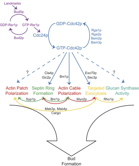
Events leading to bud emergence: A series of seminal studies from John Pringle and colleagues (reviewed in Pringle et al. 1995) identified most of the key regulators of cell polarity in yeast and led to a hierarchical model for polarity establishment in which“bud-site selection” machin-ery recruits the master regulatorCdc42p, which then orients the cytoskeleton for bud growth (Figure 2).
Bud-site selection: At the top of the hierarchy is a set of
“bud-site selection” proteins (reviewed in the YeastBook chapter by Bi and Park, in press). These define a machinery for properly placing and interpreting a set of guidepost or
“landmark”proteins that are inherited by newborn cells at specific positions and influence subsequent bud placement. Many landmarks are integral plasma membrane proteins whose extracellular domains may interact with the cell wall to restrict their mobility, thereby preserving their initial lo-calization (Halmeet al.1996; Roemeret al.1996; Harkins
et al.2001; Kanget al.2004a). The intracellular domains of the landmarks can interact with the GEF for the Ras-related
Rsr1pGTPase (Kanget al.2001, 2004b), and this is thought to result in localized accumulation of GTP-Rsr1pnear the landmark. GTP-Rsr1pcan interact with theCdc42p-directed GEF, Cdc24p (Zheng et al. 1995), as well as with GDP-bound Cdc42p (Kozminski et al. 2003), connecting the bud-site selection landmarks to the next level of the hierarchy.
Polarization of Cdc42p:At the next level (Figure 2) there is a set of“polarity establishment”proteins centered on the conserved Rho-family GTPaseCdc42p. BothCdc42pand its GEFCdc24pare absolutely required for polarized organiza-tion of the cytoskeleton and for bud emergence (Hartwell
et al. 1974; Sloat et al. 1981; Adams and Pringle 1984; Adams et al. 1990). Cdc42p is concentrated in a patch at the presumptive bud site (Zimanet al.1993; Richmanet al.
2002) and then recruits and/or regulates a variety of“ effec-tor”proteins (Table 1) that bind specifically to GTP-Cdc42p
and promote events in the next level of the hierarchy. It is universally assumed that localization of Cdc42p (and, in particular, GTP-Cdc42p) is critical to establish polarity, so
the key question is: How does Cdc42pbecome localized to the presumptive bud site?
In principle, localization of Cdc42pcould occur through interaction with a prelocalized anchoring structure such as a landmark protein. However, Cdc42p does not appear to interact with landmarks, and although localization studies have uncovered many examples of proteins that become Figure 1Morphogenetic events of the cell cycle. The four major morphogenetic events are (1) polarization in late G1, triggered by Cln1,2p-Cdc28p; (2) the apical-isotropic switch in early G2, triggered by Clb1,2p-Cdc28p; (3) breakdown of mother-bud asymmetry in late mitosis (trig-ger unknown); and (4) refocusing of growth toward the neck following mitotic exit, triggered by Clb-Cdc28p in-activation. Actin (red), septin (green), and Cdc42p (blue) localization during the cell cycle is indicated.
localized through their interaction with GTP-Cdc42p, we know of no GTP-Cdc42p interactors that could act as anchors to localize Cdc42p itself. Moreover, polarization can occur at random sites presumed to lack prelocalized anchors (see Symmetry breaking). Thus, it is thought that
Cdc42pcan become clustered at a nascent polarization site and remain clustered, despite diffusion, without needing to be anchored to a stable structure. Fluorescence recovery after photobleaching (FRAP) experiments indicate that po-larized GFP-Cdc42pexchanges in and out of the polarization site very quickly (t1/24–5 s) (Wedlich-Soldneret al.2004; Slaughter et al. 2009), arguing that the cluster of concen-tratedCdc42pis very dynamic. How is such a dynamic clus-ter established and maintained?
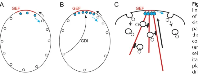
Localized GTP-Rsr1pin the vicinity of a landmark protein could recruit [and perhaps activate (Shimadaet al.2004)] the GEF Cdc24pfrom the cytoplasm, leading to local GTP loading ofCdc42pat the membrane. This would create a lo-cal patch of GTP-Cdc42p in a sea of GDP-Cdc42p at the plasma membrane. However, localized GEF activity would not, in itself, lead to local accumulation of GTP-Cdc42p to a concentration higher than that of the surrounding
GDP-Cdc42p: inward diffusion of GDP-Cdc42p would provide a substrate for the GEF to generate more GTP-Cdc42p, but that would be balanced by outward diffusion of
GTP-Cdc42p, so the overallCdc42pconcentration would not in-crease at the polarization site (Figure 3A). How, then, is the overall concentration ofCdc42pelevated at the presumptive bud site?
Cdc42p undergoes C-terminal prenylation that is critical for membrane association and function (Zimanet al.1991, 1993). Yeast contain a single Rho-GDI homolog,Rdi1p, that can extract prenylatedCdc42pfrom the membrane (Masuda
et al. 1994; Koch et al. 1997; Tcheperegine et al. 2005; Tiedje et al. 2008), and work on the human Cdc42p/GDI interaction suggests that GDI preferentially extracts GDP-bound (as opposed to GTP-GDP-bound)Cdc42pfrom membranes (Johnsonet al.2009). Because the cytoplasmic diffusion of
Cdc42p-GDI complexes is expected to be fast and the yeast cell is small, the GDI could in principle“move”GDP-Cdc42p
between outlying areas and the polarization site much faster than the rate at which GTP-Cdc42p diffuses at the plasma
membrane (Figure 3B). Localized GEF activity would impart directionality to this process by locally converting the GDI-extractable (and therefore mobile) GDP-Cdc42pto the less extractable/mobile GTP-Cdc42p, causing accumulation of GTP-bound Cdc42p at the polarization site (Figure 3B). Mathematical modeling suggests that this mechanism would suffice to concentrateCdc42pat the polarization site (Goryachev and Pokhilko 2008). Moreover, FRAP studies (Slaughteret al.2009) indicate that GFP-Cdc42pexchange in and out of the polarization site is significantly slowed in rdi1Dmutants (t1/220 svs. t1/24–5 s in wild-type cells), supporting an important role for the GDI in concentrating the dynamic pool ofCdc42p.
As rdi1D mutants are viable (Masudaet al. 1994), and still manage to concentrateCdc42p at the polarization site (Slaughteret al.2009; Boulteret al.2010), there must also be a GDI-independent route for concentrating Cdc42p. Some studies reported the presence ofCdc42p in cytoplas-mic fractions even in rdi1D mutant cells (Kochet al.1997; Tiedjeet al.2008), suggesting that there are other mecha-nisms that can extract prenylatedCdc42pfrom membranes. If such (currently undescribed) mechanisms were selective for GDP-Cdc42p, then (like the GDI) they too would pro-moteCdc42pconcentration at the polarization site.
An alternative proposed mechanism for concentrating
Cdc42pinvolves vesicular traffic (Marcoet al.2007; Slaugh-ter et al. 2009). GTP-Cdc42p orients actincables (see be-low), which deliver secretory vesicles. If GTP-Cdc42pwere sufficiently concentrated on such vesicles, then vesicle-me-diated Cdc42p delivery could promote concentration of
Cdc42pat the polarization site. As theCdc42pdiffuses away, endocytosis could remove the Cdc42p from the plasma membrane and deliver it to endosomes, maintaining a dy-namically polarizedCdc42plocalization by vesicle-mediated recycling (Figure 3C). Treatment of cells with Latrunculin A to depolymerizeactinand block vesicle recycling resulted in slightly slower FRAP recovery of GFP-Cdc42pat the polariza-tion site (t1/25–6 svs. t1/24–5 s in untreated cells), which has been interpreted as support for the idea that vesicle recy-cling assistsCdc42ppolarization (Slaughteret al.2009).
It is not known whether vesicles carry sufficientCdc42p
Mathematical modeling indicates that effectiveCdc42p po-larization by a vesicle recycling mechanism would require that theCdc42pbe actively endocytosed and that it diffuse very slowly in the plasma membrane (Laytonet al.2011). At present, there is no evidence for active internalization of
Cdc42p, and current estimates of theCdc42pdiffusion con-stant (Marcoet al.2007) are an order of magnitude higher than the values required for the model to develop robust polarity (Layton et al.2011), so the viability ofCdc42p ve-sicular recycling as a way to concentrateCdc42pat the pre-bud site remains unclear.
Symmetry breaking: The preceding discussion assumed thatCdc42p concentration was triggered by prior localiza-tion of its GEF to a site demarcated by a landmark protein. However, elimination of RSR1 randomizes the location of bud emergence (Bender and Pringle 1989), yet rsr1 cells still pick one and only one (randomly located) bud site with apparently normal timing and efficiency. This process is sometimes called“symmetry breaking.”
Symmetry-breaking behavior suggests that there is a pos-itive feedback loop or amplification mechanism that allows a stochastic fluctuation in polarity factor concentration at some random site to promote accumulation of more polarity factors at that site (Turing 1952). Polarization ofCdc42pin rsr1Dcells does not require polymerizedactinor microtubules (Irazoqui et al. 2003), suggesting that Cdc42p symmetry breaking requires neither the upstream nor the downstream levels of the hierarchy and that the polarity establishment machinery itself contains a positive feedback loop.
A proposed mechanism for the positive feedback is that stochastically arising GTP-Cdc42p can recruit the GEF
Cdc24pto generate more GTP-Cdc42pin its vicinity, thereby growing a cluster of GTP-Cdc42p(Figure 4). This model was derived from the observation that polarization of rsr1D mutants requires the scaffold protein Bem1p (Irazoqui
et al.2003), which appears to function by bringing together the GEF Cdc24p and a p21-activated kinase (PAK)-family kinase (either Cla4p orSte20p) (Kozubowskiet al. 2008). The PAKs are effectors ofCdc42p: they bind to GTP-Cdc42p
and that interaction relieves autoinhibition to activate the kinase (Bagrodia and Cerione 1999). Thus, GTP-Cdc42pat the membrane can (via PAK interaction) recruit a
PAK-Bem1p-GEF complex that can then [via GEF activity, which may be stimulated by Bem1p interaction (Shimada et al.
2004)] convert neighboring GDP-Cdc42p to GTP-Cdc42p. This new GTP-Cdc42pcan then recruit more GEF-containing complexes in a positive feedback loop (Figure 4). Support for this model comes from the striking observation that
Bem1pfunction in symmetry breaking can be bypassed by in-troducing an artificial GEF-PAK fusion protein (Kozubowski
et al.2008).
As discussed above for the Rsr1p-localized GEF, this
Bem1p-mediated positive feedback loop could generate a local cluster of GTP-Cdc42p in a sea of GDP-Cdc42p, but other mechanisms would be needed to concentrate the GTP-Cdc42p
to a level higher than that of the surrounding GDP-Cdc42p. Mathematical modeling suggests that, in combination with the GDI, Bem1p-mediated positive feedback would suffice to ex-plain symmetry-breaking behavior (Goryachev and Pokhilko 2008). However, it is worth noting that the model works only within a limited parameter space, and we do not have suffi -ciently detailed knowledge of the relevant concentrations and rate constants in cells to know whether or not the parameter estimates are realistic.
Like the landmark Rsr1p pathway, the Bem1p positive feedback loop relies on localized GEF activity to concentrate GTP-Cdc42p. Thus, ifCdc42pwere loaded with GTP in some other way (bypassing the GEF), these mechanisms would not be able to concentrateCdc42p. Experimentally, this sit-uation is approximated using theCdc42pG12VorCdc42pQ61L mutants, which bind to GTP upon initial folding and then cannot hydrolyze the GTP, so they remain GTP-bound and bypass the GEF. When endogenousCdc42pwas inactivated by a temperature-sensitive (ts) mutation and replaced by near-endogenous levels of Cdc42pQ61L, the cells failed to polarize (Irazoqui et al. 2003). Thus, GTP hydrolysis by
Cdc42p appears to be essential for polarity establishment, consistent with the idea that localized GTP loading of
Cdc42pby the GEF (which can occur only once the initially bound GTP is hydrolyzed) is needed to concentrate
GTP-Cdc42p at the polarization site.
Unlike near-endogenous levels of Cdc42pQ61L, overex-pression ofCdc42pQ61LorCdc42pG12Vdoes lead to concen-tration of the mutant protein, as well as clustering of cortical
actin patches, at discrete sites (Gulli et al. 2000; Irazoqui
delivery along actin cables (Wedlich-Soldner et al. 2003). On the basis of these findings, a proposed mechanism for symmetry breaking is that vesicle-mediated delivery of
Cdc42pQ61L combined with Cdc42pQ61L-mediated orienta-tion of actincables constitutes a positive feedback loop for concentratingCdc42pQ61L(Wedlich-Soldneret al.2003).
Cdc42pQ61L polarization often produces more than one polarization site and results in cell death by lysis (Gulliet al.
2000; Wedlich-Soldneret al.2003). These features raise the concern that this overexpression system is a pathological manifestation of cells attempting to cope with weak points in the cell wall, rather than an informative mimic of the normal polarization process.
In summary, polarity establishment involves the concen-tration of GTP-Cdc42p at the presumptive bud site on the plasma membrane. In wild-type cells, this is probably initi-ated by localized recruitment of the GEF Cdc24p by
GTP-Rsr1pto a site defined by a previously deposited landmark protein. However, in the absence ofRsr1p,Cdc42p neverthe-less becomes concentrated at an apparently random site. This symmetry breaking is presumably initiated by stochas-tic localfluctuations in polarity protein concentrations and subsequently amplified by positive feedback. A feedback loop involving a complex between Bem1p, Cdc24p, and a PAK that would generate a cluster of GTP-Cdc42p has been proposed (Figure 4). Two mechanisms (one mediated by the GDI Rdi1p and the other by vesicle recycling) that could then allow the concentration of GTP-Cdc42p in the cluster to rise above that of the surrounding GDP-Cdc42p
have also been proposed (Figure 3), but their importance remains uncertain, and the existence of as-yet-uncharacter-ized mechanisms seems likely.
Polarization of the cytoskeleton and growth:Once a polar-ization site with concentrated GTP-Cdc42p is established,
actin cables are oriented toward the site, actin patches [which are sites of endocytosis (Kaksonenet al.2003)] clus-ter around the site, a ring of septinfilaments is assembled around the site, and exocytosis is targeted toward the site (Figure 2). Cell-wall glucan synthesis must also be activated at the polarization site, perhaps via localized activation of the glucan synthase regulatorRho1p(Abeet al.2003). Tar-geted secretion, combined with localized cell-wall synthesis, then promotes bud emergence (Pruyneet al.2004b).
To a significant degree, the downstream events initiated byCdc42pare independent of each other:actinpolarization can occur in the absence of organized septins (Adams and Pringle 1984), and septin rings can form in the absence of polymerized actin(Ayscough et al. 1997). Targeted secre-tion and even bud emergence can occur without septin rings (Hartwell 1971; Haarer and Pringle 1987) or actin cables (Sahin et al.2008; Yamamoto et al. 2010). However, sub-sequent bud growth requiresactincables (Yamamotoet al.
2010), and proper shaping of the bud requires neck-local-ized septins (Gladfelteret al.2005).
In mutants lackingactincables, the small size and ovoid geometry of the unbudded yeast cell may enable bud
emer-gence through chance encounters between secretory vesicles undergoing Brownian motion and theCdc42ppatch, which may promote local fusion via the exocyst (Figure 2). How-ever, once the Cdc42ppatch is separated from the bulk of the cell by a narrow bud neck,actin-mediated transport of vesicles through the neck would be needed to promote effi -cient secretory vesicle fusion at the bud tip. In addition, the septin collar at the neck somehow promotes expansion of the bud base so that it bulges out from the neck.
The ability of downstream events to occur independently suggests thatCdc42pis a master regulator of the microma-naging variety, separately promoting several parallel path-ways required for harmonious bud growth. Supporting this view, specific cdc42 alleles have been isolated that impair targeted exocytosis without overt effects onactinor septins (Adamo et al. 2001), whereas other alleles impair septin organization without overt effects on actin or secretion (Gladfelter et al.2002; Cavistonet al.2003). It is thought that different pathways are carried out by subsets ofCdc42p
effectors (Table 1).Bni1pplays a prominent role in oriented
actin cable assembly (Evangelistaet al. 1997, 2002; Sagot
et al. 2002). The PAKs (Longtineet al. 2000; Weiss et al.
2000; Gladfelteret al.2004; Versele and Thorner 2004) and the Gic1p and Gic2p proteins (Iwase et al. 2006) aid in septin ring assembly. The exocyst components Sec3p and
Exo70p (Zhang et al. 2001; Baek et al. 2010; Wu et al.
2010) and the scaffold proteins Boi1p and Boi2p (Adamo
et al. 2001) promote targeted secretion. However, effectors are not restricted to one pathway (Gladfelter et al. 2001), and the detailed mechanisms by which the effectors operate remain largely unknown.
Although differentCdc42poutputs can occur individually when other outputs are blocked, there are also many inter-connections among these downstream outputs. In some cases, direct mechanistic links have been identified: septin rings recruit the forminBnr1p, which nucleatesactincable formation in mother cells (Pruyneet al.2004a). Septins also recruit the endocytic actin patch initiator protein Syp1p, promoting patch clustering at the mother-bud neck (Qiu
et al. 2008; Stimpson et al.2009). In other cases, the evi-dence is less direct. Actin perturbations can impair septin ring assembly (Kadota et al.2004; Kozubowskiet al.2005; Iwaseet al.2006), perhaps suggesting that some septin-or-ganizing factors are delivered byactincables. And perturba-tions of vesicle traffic can affect actin polarity (Gaoet al.
2003) and the localization of Cdc42p(Wedlich-Soldneret al.
2004; Irazoqui et al. 2005; Zajac et al. 2005; Yamamoto
et al. 2010), although the basis for these effects remains unclear.
CDK-mediated regulation of polarity establishment: The above tour through polarity establishment indicates that CDK-mediated regulation of bud emergence could occur at multiple levels. A variety of fixed-cell synchrony experi-ments indicated that unpolarized cells become polarized
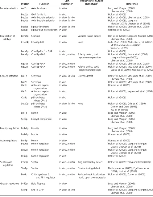
Table 1 CDK substrates with roles in morphogenesis
Protein Function
CDK substratea
Phosphosite mutant
phenotypeb Reference
Bud-site selection Axl2p Axial landmark In vitro Loog and Morgan (2005);
Ubersaxet al.(2003)
Bud2p GAP for Rsr1p In vivo Holtet al.(2009)
Bud3p Axial bud-site selection In vitro,in vivo Holtet al.(2009); Ubersaxet al.(2003) Bud4p Axial bud-site selection In vitro,in vivo Holtet al.(2009); Loog and
Morgan (2005); Ubersaxet al.(2003) Bud8p Bipolar landmark In vitro,in vivo Holtet al.(2009); Ubersaxet al.(2003) Rax2p Bipolar bud-site selection In vitro Ubersaxet al.(2003)
Polarization of Cdc42p
Bem1p Scaffold In vitro Vacuole fusion defects Hanet al.(2005); Loog and Morgan (2005); Ubersaxet al.(2003)
Cdc24p Cdc42p GEF In vitro None Gulliet al.(2000); McCuskeret al.(2007); Moffat and Andrews (2004);
Waiet al.(2009)
Bem2p Cdc42p/Rho1p GAP In vivo Holtet al.(2009)
Bem3p Cdc42p GAP In vivo,in vitro Polarity defect, toxic upon overexpression
Holtet al.(2009); Knauset al.(2007); Loog and Morgan (2005); Ubersaxet al.(2003)
Rga1p Cdc42p GAP In vivo,in vitro Holtet al.(2009); Ubersaxet al.(2003) Rga2p Cdc42p GAP In vivo,in vitro Polarity defect, toxic
upon overexpression
Holtet al.(2009); McCuskeret al.(2007); Sopkoet al.(2007); Ubersaxet al.(2003)
Cdc42p effectors Boi1p Secretion In vitro,in vivo Growth defect Holtet al.(2009); McCuskeret al.(2007); Ubersaxet al.(2003)
Boi2p Secretion In vivo Holtet al.(2009); McCuskeret al.(2007)
Gic1p Actin and septin organization
In vitro Ubersaxet al.(2003)
Gic2p Actin and septin organization
In vivo Holtet al.(2009); Jaquenoudet al.(1998)
Cla4p p21-activated kinase (PAK)
In vivo Holtet al.(2009)
Ste20p p21-activated kinase (PAK)
In vitro,in vivo None Holtet al.(2009); Odaet al.(1999); Oehlen and Cross (1998); Wuet al.(1998)
Bni1p Formin In vitro Loog and Morgan (2005);
Ubersaxet al.(2003)
Sec3p Exocyst component In vitro Loog and Morgan (2005);
Ubersaxet al.(2003)
Polarity regulators Msb1p Polarity In vitro Loog and Morgan (2005);
Ubersaxet al.(2003)
Msb2p Mucin In vitro Ubersaxet al.(2003)
Actin regulators Bnr1p Formin In vitro Ubersaxet al.(2003)
Bud6p Formin regulator In vivo,in vitro Holtet al.(2009); Loog and Morgan (2005); Ubersaxet al.(2003) Spa2p Formin regulator In vivo,in vitro Holtet al.(2009); Loog and Morgan
(2005); Ubersaxet al.(2003)
Pea2p Formin regulator In vivo Holtet al.(2009)
Septins and regulators
Cdc3p Septin In vivo,in vitro Ring disassembly defect Holtet al.(2009); Tang and Reed (2002)
Shs1p Septin In vivo,in vitro Gin4p-binding defect Dephoureet al.(2005); Egelhoferet al. (2008); Holtet al.(2009)
Bni4p Chitin synthase 3 and PP1 regulator
In vivo,in vitro Reduced neck localization, toxic upon overexpression
Holtet al.(2009); Zouet al.(2009)
Growth regulators Dnf2p Lipidflippase In vitro Loog and Morgan (2005);
Ubersaxet al.(2003)
Sac7p Rho1p GAP In vitro,in vivo Holtet al.(2009); Loog and Morgan (2005); Ubersaxet al.(2003)
1987; Ford and Pringle 1991; Kimet al.1991; Lew and Reed 1993; Ziman et al. 1993; Ayscough et al.1997), and this timing was confirmed by live-cell filming (Howell et al.
2009). As G1 CDK activation (a.k.a. START) occurs 15– 20 min before bud emergence (Lew and Reed 1993; Di Talia
et al. 2007), these studies suggested that G1 CDK activity might promote concentration ofCdc42p(and the rest of the polarity establishment machinery) at the presumptive bud site. Below, we summarize the evidence for CDK involve-ment at different steps in polarity establishinvolve-ment and discuss possible mechanisms.
Not all studies on polarity establishment fit easily with the view that CDK triggers polarization. In particular, some studies suggested that the poorly understood proteinSpa2p
could polarize before G1 CDK activation (Snyderet al.1991; Padmashree and Surana 2001). Moreover, in some strain backgrounds, cells arrested without (or with only a little) G1 CDK activity can polarize their growth and produce pro-jections (Madden and Snyder 1992; Lew and Reed 1993). Thesefindings suggest that cytoskeletal polarization is pos-sible without (much) CDK input, although it does not lead to bud emergence.
One way to reconcile the apparently contradictory results on the role of G1 CDK in promoting polarization would be to posit that a small amount of CDK activity suffices to promote polarization of Cdc42p, actin, and secretion, but a larger amount of CDK activity is needed to promote both septin ring assembly and actual bud emergence. Thus, depending on the specific strain and CDK manipulation, CDK inhibition may block all polarization or only septin ring assembly and bud emergence.
Bud-site selection: Before polarity establishment in G1,
Rsr1pis localized all over the plasma membrane (Michelitch and Chant 1996; Park et al.2002), while its GEF Bud5pis
concentrated near the various landmark proteins (Kang
et al.2001; Marstonet al.2001) and its GAPBud2pis delo-calized (Parket al.1999; Marstonet al.2001). This pattern suggests that GTP-Rsr1p would be concentrated near the landmarks. However, the Cdc42p-directed GEF Cdc24p, which directly binds to GTP-Rsr1p(Parket al.1997), does not concentrate at that site in early G1. In diploids,Cdc24p
is diffusely localized in the cytoplasm in early G1, whereas in haploids it is concentrated in the nucleus, due to interac-tion withFar1p, to prepare for potential mating (although if the haploid-specificFAR1is deleted, thenCdc24pis diffusely localized in haploids as well) (Nern and Arkowitz 2000; Shimada et al. 2000). Activation of the G1 CDK promotes
Cdc24p localization to the pre-bud site, even in cdc42 mutants where any feedback pathways would be inoperative (Gulli et al.2000).
Perhaps the simplest way to interpret these observations is that CDK activation promotes GTP loading ofRsr1pby its prelocalized GEF, thereby enabling interaction of the local-ized GTP-Rsr1pwithCdc24p. Interestingly, in late G1, both
Rsr1p and its regulators become concentrated at the po-larization site (Park et al. 1999, 2002; Kang et al. 2001; Marston et al. 2001), consistent with the idea that they are somehow regulated by the CDK. However, that behavior could also reflect regulation of bud-site selection proteins downstream of Cdc42p localization. Bud2p is a putative CDK target (Holt et al. 2009), but the significance of that phosphorylation is untested.
An alternative interpretation of the localization data is that a localized pool of GTP-Rsr1pexists throughout G1, but thatCdc24pcan bind to GTP-Rsr1peffectively only follow-ing CDK activation in late G1, either because some maskfollow-ing factor is removed or because phosphorylation of Cdc24p
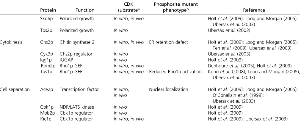
itself or a cofactor enhances its binding affinity for GTP-Table 1 Continued
Protein Function
CDK substratea
Phosphosite mutant
phenotypeb Reference
Skg6p Polarized growth In vitro,in vivo Holtet al.(2009); Loog and Morgan (2005); Ubersaxet al.(2003)
Tos2p Polarized growth In vitro Ubersaxet al.(2003)
Cytokinesis Chs2p Chitin synthase 2 In vitro,in vivo ER retention defect Holtet al.(2009); Loog and Morgan (2005); Tehet al.(2009); Ubersaxet al.(2003)
Cyk3p Chs2p regulator In vitro Ubersaxet al.(2003)
Iqg1p IQGAP In vivo Holtet al.(2009)
Rom2p Rho1p GEF In vitro,in vivo Dephoureet al.(2005); Holtet al.(2009) Tus1p Rho1p GEF In vitro,in vivo Reduced Rho1p activation Konoet al.(2008); Loog and Morgan (2005);
Ubersaxet al.(2003)
Cell separation Ace2p Transcription factor In vitro, in vivo
Nuclear localization Holtet al.(2009); Loog and Morgan (2005); O’Conallainet al.(1999);
Ubersaxet al.(2003)
Cbk1p NDR/LATS kinase In vivo Holtet al.(2009)
Mob2p Cbk1p regulator In vivo Holtet al.(2009)
Kic1p Cbk1p regulator In vitro,in vivo Holtet al.(2009); Ubersaxet al.(2003)
Rsr1p. Cdc24p is a CDK substrate in vitro (Moffat and Andrews 2004; McCusker et al. 2007), but mutation of 6 putative CDK target sites (Gulli et al. 2000) or up to 35 phosphorylation sites mapped by mass spectrometry (Wai
et al.2009) did not appear to affectCdc24plocalization or function. Thus, CDK activation probably promotes Rsr1p
-Cdc24p interaction in vivo, leading toCdc24p localization, but the relevant substrates and underlying mechanism re-main unclear.
Polarization of Cdc42p:Cells arrested in G1 due to lack of the G1 cyclinsCln1p-3p failed to polarizeCdc24p,Cdc42p,
Bem1p, or the effectorsGic2pandBni1p(Gulliet al.2000; Jaquenoud and Peter 2000; Wedlich-Soldner et al. 2004). Induction ofCln2pin the arrested cells led to polarization of all of those factors, even in the absence of polymerizedactin. These findings suggested that G1 CDK activity acts at the level ofCdc42pregulators to promote polarization (Cdc42p
is not itself known to be phosphorylated). Such regulation could involve a change inCdc42p-directed GEF or GAP ac-tivity leading to an increase in GTP-Cdc42p and triggering a localization feedback loop (Figure 4).
As mentioned above, the GEFCdc24pis a CDK substrate
in vitro, but as yet genetic analyses have not uncovered any role for that phosphorylation, so attention has turned to the
Cdc42p-directed GAPs. The yeast genome encodes 11 pro-teins with Rho-GAP domains. Genetic analyses suggested that three of these (Bem3p, Rga1p, and Rga2p) might be
Cdc42p-specific and that their GAP domains catalyze GTP hydrolysis by Cdc42p in vitro (Bender and Pringle 1991; Zheng et al. 1993, 1994; Stevenson et al. 1995; Chen
et al. 1996; Gladfelter et al. 2002; Smith et al. 2002). A fourth Rho-GAP (Bem2p) with genetic links to Cdc42p
was initially thought to be selective for Rho1p (Zheng
et al.1993), but was later shown to act onCdc42pas well, at leastin vitro(Marquitzet al.2002). All of these GAPs are probably CDK substrates (Ubersax et al. 2003; Holt et al.
2009) (Table 1). Biochemical assays suggest that two other Rho-GAPs (Rgd2p and Lrg1p) may act on Cdc42p as well (Roumanieet al.2001).
For Bem3p(Knauset al. 2007) andRga2p(Sopkoet al.
2007), mutation of putative or mapped phosphorylation sites revealed that overexpression of nonphosphorylatable mutants is more toxic to cells than overexpression of the wild-type proteins. Toxicity was associated with accumula-tion of depolarized cells and was abolished by mutaaccumula-tions impairing GAP activity, suggesting that high levels of non-phosphorylatable GAPs can block polarity establishment, perhaps because they are resistant to phosphorylation-mediated inhibition. This suggests the attractive hypothesis that high GAP activity keeps GTP-Cdc42plevels low in early G1 and that CDK activation promotes polarization by phos-phorylating GAPs to reduce total GAP activity (Knaus et al.
2007; Sopkoet al.2007).
As yet, biochemical evidence that phosphorylation inhib-its GAP activity is lacking. Moreover, combined deletion of BEM3andRGA2does not overtly accelerate polarization, so
inhibition of these two GAPs is not sufficient to trigger po-larization. Replacement of BEM3 or RGA2 with nonphos-phorylatable versions expressed at endogenous levels does not overtly delay polarization, but it remains possible that parallel regulation of several GAPs triggers polarization or that combined regulation of both the GEF and the GAPs constitutes redundant pathways to promote polarization.
An alternative to GEF/GAP regulation is that CDK activation regulates the capacity for positive feedback. We do not know whether polarization is accompanied by a rise in GTP-Cdc42plevels within the cells or whether the
GTP-Cdc42pis simply redistributed from a delocalized to a local-ized pool. In principle, enabling a locallocal-ized positive feedback pathway would be sufficient to promote polarization even if GEF and GAP activities were unchanged. For example, CDK could promote assembly of the PAK-Bem1p-GEF complex to enable the positive feedback loop illustrated in Figure 4. Like Cdc24p, Bem1p and the PAKs Ste20p and Cla4p are CDK substrates, but mutation of putative or mapped phos-phorylation sites has thus far failed to reveal any role for those phosphorylations in polarization (Oda et al. 1999; Ubersax et al.2003; Hanet al.2005).
In summary, CDK activity is thought to promote Cdc42p
polarization, and many polarity establishment proteins are probably direct CDK substrates (Enserink and Kolodner 2010), but genetic analysis has thus far failed to demon-strate the significance of those phosphorylations. Either the CDK acts in a complex and highly redundant manner or key substrates remain to be identified.
Polarization of the cytoskeleton and growth: The Rho1p
GTPase is not necessary for polarity establishment but is crucial for cell-wall biosynthesis and bud growth.
GTP-Rho1p is concentrated at the polarization site (Abe et al.
2003) and activates the glucan synthases critical for new cell-wall deposition (Drgonova et al. 1996; Qadota et al.
1996), as well as several other effectors. In a very elegant study, Konoet al.(2008) showed thatRho1pGTP loading is cell-cycle-regulated, peaking at around the time of bud emer-gence. Rho1p activation results fromCln2p-CDK-mediated phosphorylation of the Rho1p-GEF Tus1p (Kono et al.
2008). Phosphorylation-site mutants of Tus1p abolished CDK-mediated accumulation of GTP-Rho1p, but the mutant cells nevertheless survived, implying that sufficient Rho1p
function was still provided. One attractive possibility is that, in the mutant cells, the attempt to engage in polarized growth with insufficient glucan synthesis caused transient cell-wall defects detected by the “cell integrity pathway” (Levin 2005), which led to compensatory activation of the stress-responsiveRho1p-GEFRom2p(Grayet al.1997; Kono
et al. 2008). The Kono et al. (2008) study provides the clearest instance of a downstream event directly regulated by the G1 CDK.
1992; Lew and Reed 1993), although polarization may be delayed relative to wild-type controls. However, such cells do not assemble septin rings and they do not make buds. There is strong genetic evidence that Cln1p and Cln2p in particular are needed to promote proper septin ring assem-bly (Benton et al. 1993; Cvrckova et al. 1995; Gladfelter
et al. 2005) and that some septins are direct CDK targets (Tang and Reed 2002; Egelhofer et al. 2008), although phosphosite mutants of individual septins did not have any obvious effect on septin ring assembly. In contrast to the inconclusivefindings fromS. cerevisiae, analogous work in the related Candida albicans provided strong evidence that CDK-mediated septin phosphorylation directly impacts septin organization and hyphal growth (Sinha et al.2007; Gonzalez-Novoet al.2008). Thus, it seems highly likely that CDKs directly regulate septin assembly as well as indirectly promote septin organization throughCdc42ppolarization.
Is CDK-mediated septin regulation sufficient to explain why cdk-ts cells make projections rather than buds? As septins are dispensable for bud emergence, there may be other targets of the CDK that promote budding itself. However, the difference between projection formation and bud formation is subtle and morphological, and it has been shown that improperly organized septins can lead to the formation of aberrantly shaped“buds”that resemble projec-tions (Gladfelteret al.2005).
In summary, considerable evidence supports the hypoth-esis that G1 CDK triggers polarization of Cdc42pand other polarity establishment proteins. Additional evidence suggests further links between the CDK and downstream events, in-cludingRho1pactivation and septin organization. However, despite the identification of numerous CDK substrates with roles in polarity establishment (Table 1), we are not yet in a position to state that any given set of phosphorylations can explain any specific step in polarity establishment.
Apical-Isotropic Switch in G2
Following bud emergence, most growth and new cell-wall deposition is targeted to the tip of the bud, but at some point this“apical”growth mode switches to a uniform or“ isotro-pic” mode of growth (Farkas et al. 1974; Lew and Reed 1993). After the apical-isotropic switch, growth is still di-rected toward the bud (and the mother cell does not grow significantly), but it is now distributed diffusely within the bud. The proteins that were highly polarized in late G1 (Cdc42p, etc.) remain polarized during apical growth but become distributed around much of the bud cortex after the switch. The apical-isotropic switch is dependent on G2 CDK activity (primarilyClb2p, assisted byClb1p) and can be induced prematurely byClb1porClb2poverexpression, sug-gesting that Clb1p,2p-CDK activation is the regulatory trig-ger for this event (Lew and Reed 1993). Compared to polarity establishment, much less research has gone into understanding the basis for this depolarizing switch, but at least three interesting ideas have been put forward for how it might be triggered.
Reversal of Cln-CDK-promoted polarization: Clb1p,2 p-CDK activity represses the transcription of a set of promoters that includes those forCLN1andCLN2(Amonet al.1993). Thus, if polarity-promoting G1-CDK substrates need to be continuously phosphorylated and cannot be phosphorylated by G2-CDK, then the apical-isotropic switch could simply reflect the reversal of G1-CDK-targeted phosphorylations following G1 cyclin repression. In support of this idea, over-expression of CLN1orCLN2from theGAL1promoter leads to prolonged apical growth in otherwise wild-type cells (Lew and Reed 1993). However, it is possible that the overex-pressed G1 cyclins compete with endogenousClb2pfor ac-cess to the CDK and that the continued apical growth stems from absence of sufficient G2 CDK, rather than from the presence of sufficient G1 CDK. Consistent with that possibil-ity, an intriguing study reported that the continued apical growth of cells overexpressingCLN1was dependent on the G2-CDK inhibitor Swe1p (Ahn et al. 2001). Moreover, in-activation of temperature-sensitivecdc28alleles in G2 leads to a return to apical growth (Lew and Reed 1993), which is difficult to explain if G1-Cdc28pactivity is continuously re-quired to promote such growth (especially as the samecdc28 alleles effectively block G1-CDK-induced budding). Thus, on balance it appears that G2 CDK activity does more than simply inactivate G1 CDK.
Lipid-mediated GAP activation: The lipid composition of many eukaryotic plasma membranes is highly asymmetric, with phosphatidylserine (PS) and phosphatidylethanolamine (PE) enriched in the inner leaflet and phosphatidylcholine and sphingolipids enriched in the outer leaflet. Using a probe for PE in the outer leaflet, Saitoet al.(2007) found that the probe was readily detectable at the polarization site during apical growth, but not detectable during isotropic growth. Moreover, lipid“flippases”thought to translocate PS and PE from the outer to the inner leaflet also displayed a polarized localization during apical growth, and mutations in the genes encoding theflippases led to persistent external PE staining, polarizedCdc42p, and continued apical growth at low tem-peratures, resulting in elongated buds (Saito et al. 2007). These findings suggested that lipid flipping at the bud tip might trigger the apical-isotropic switch.
Cells in which the apical-isotropic switch is impaired would be expected to display elongated buds, so this phenotype was consistent with the idea that lipid flipping might be important for triggering the apical-isotropic switch. However, a large majority of elongated-bud mutants turn out to affect the timing ofClb2p-CDK activation (generally via effects on a septin-dependent Swe1p-regulatory path-way, as discussed below) (Barral et al. 1999; Edgington
et al.1999; Longtineet al.2000; Thomaset al.2003), rather than affecting the apical-isotropic switch per se. Saitoet al.
active Clb2p-CDK, as in some cases partial CDK inhibition blocks the apical-isotropic switch without blocking nuclear division (Lew and Reed 1993).
If Clb2p-CDK does trigger the apical-isotropic switch by activating lipid flippases, then how would flipping lipids affect polarized growth? Using in vitro GAP assays, Saito
et al. (2007) showed that Rga1p and Rga2p GAP activity could be stimulated by PS or PE. They suggested that
Clb2p-CDK-stimulated flipping of PS and PE to the inner leaflet at the bud tip would activate these GAPs to clear the local GTP-Cdc42p, terminating polar growth. This is an intriguing hypothesis worthy of further investigation. But it cannot be the whole story because flippase mutants exhibit only a delayed apical-isotropic switch at low temperatures.
Dissociation of GEF-PAK complexes: As discussed above (Figure 4), the GEF Cdc24p can form complexes with
Bem1p and a PAK, and such complexes are important for polarity establishment. In these complexes the Cdc24p
becomes heavily phosphorylated by the PAK (Gulli et al.
2000; Boseet al. 2001). On the basis of a variety of obser-vations, Gulliet al.(2000) suggested thatCdc24p phosphor-ylation might cause it to dissociate fromBem1p, terminating polarized growth. This hypothesis does not address why the inhibitory effects ofCdc24pphosphorylation would be man-ifested only in G2 or how this pathway might be regulated by the G2 CDK. In addition, later studies found that phos-phorylated Cdc24p could still bind to Bem1p (Bose et al.
2001) and that neither fusion ofBem1ptoCdc24p(to pre-vent their separation) (Kozubowski et al.2008) nor muta-tion of 35 mapped phosphorylamuta-tion sites onCdc24p(which greatly reducedCdc24pphosphorylation) (Waiet al.2009) affected the apical-isotropic switch. However, the idea that GEF inhibition may be involved in triggering depolarization in G2 remains attractive, and although fusion of Bem1pto
Cdc24phad no effect, fusion ofCla4ptoCdc24pdid lead to the development of elongated buds (Kozubowski et al.
2008). Thus, it remains possible that the G2 CDK somehow disrupts the Cdc24p-Bem1p-Cla4p complex to trigger the apical-isotropic switch. As with the lipid flippase pathway above, this pathway (if it exists) can be only part of the story, as only 11% of cells containing the Cdc24p-Cla4p fusion exhibited elongated buds (Kozubowskiet al.2008).
In summary, it seems likely that the apical-isotropic switch is actively triggered by the G2 CDK and is not a passive consequence of diminished G1 CDK activity. De-polarization may involve regulated lipid translocation and GAP activation, disassembly of a GEF-containing complex, or both, leading to diminished GTP-Cdc42plevels. However, both of these hypotheses remain tentative, and other mech-anisms may well be important.
Breakdown in mother-bud asymmetry
Even after the apical-isotropic switch, growth remains re-stricted to the bud for most of G2/M. This mother-bud asymmetry requires polymerized actin, myosin V (Karpova
et al. 2000), and an intact septin collar at the mother-bud neck (Barral et al. 2000). The asymmetry is most easily visualized by looking at the distribution of cortical actin
patches, which are abundant in the bud and almost absent in the mother (Adams and Pringle 1984; Amberg 1998).
Actin patches represent sites of endocytosis at a late stage where the plasma membrane is in the process of invaginat-ing (Kaksonen et al. 2006). Markers of an earlier step of endocytosis (Ede1por clathrin) are not as highly asymmet-ric (Newpheret al.2005; Stimpsonet al.2009), and it was recently suggested that endocytic patches wait until theyfill up with cargo before they initiateactinpolymerization and invagination (Layton et al. 2011). In buds, where directed secretion delivers many proteins (e.g., v-SNAREs) to the plasma membrane that subsequently become endocytic cargo, the clathrin patchesfill with cargo rapidly and convert to actin patches; in mothers, where there is little secretion, the clathrin patches must wait much longer to collect suffi -cient cargo, so conversion to actin patches is rare (Layton
et al.2011). In this way, theactinpatch distribution reflects the polarization of secretion.
For a brief time prior to cytokinesis, the actin-patch distribution becomes symmetric between mother and bud, presumably reflecting a breakdown in the mother-bud asym-metry of secretion described above. Cell-cycle arrest by DNA checkpoints or the spindle assembly checkpoint results in the accumulation of cells withactinpatches distributed be-tween mother and bud (Jacobset al.1988). Similarly, cells expressing nondegradable mitotic cyclins arrest with sym-metrically distributed actinpatches (Lew and Reed 1993). However, these treatments do not accelerate the switch to symmetric actinpatches (Lew and Reed 1993), suggesting that the switch is not simply a response to some threshold level of CDK activity. Thus, the breakdown in mother-bud asymmetry is not clearly linked to a change in CDK activity, and the regulatory trigger for this morphogenetic event remains enigmatic.
Cytokinesis
In S. cerevisiae, cytokinesis occurs at the mother-bud neck (see the YeastBook chapter by Bi and Park, in press). Below wefirst briefly summarize the series of events leading to cell separation and then discuss what is known regarding how these events are regulated by the cell cycle.
Events leading to cell separation: Cytokinesis involves the assembly and constriction of an actomyosin ring, which guides deposition of a chitinous primary septum, which is followed shortly by deposition of a glucan- and mannan-rich secondary septum on either side. The actual separation of mother and daughter involves the action of chitinase, which degrades the primary septum, as well as some glucanases. These processes are summarized in Figure 5.
Actomyosin-ring formation:The actomyosin ring contains
Localization of these proteins to the mother-bud neck re-lies on the septins, which form a collar tethering various proteins to that site (Epp and Chant 1997; Bi et al. 1998; Lippincott and Li 1998).
Myo1pis a classical two-headed non-muscle myosin with a long coiled-coil tail that has a pronounced kink region in which there are two independent“targeting domains”(Fang
et al.2010). One of these binds to the septin-binding protein
Bni5p and targets Myo1p to the neck from late G1 until anaphase. Thisfirst targeting mechanism is largely dispens-able for actomyosin-ring formation and may reflect earlier roles for Myo1p. The second targeting domain promotes neck localization in anaphase/telophase and largely suffices for actomyosin-ring formation and constriction (Fanget al.
2010).
Iqg1p contains a calponin-homology domain that inter-acts with F-actin, and several light-chain-binding IQ motifs (Epp and Chant 1997; Lippincott and Li 1998; Shannon and Li 1999).Iqg1ptargeting to the neck requires the light chain
Mlc1p(Boyneet al.2000; Shannon and Li 2000; Luoet al.
2004).Iqg1pis synthesized during G2/M, becomes localized to the neck in anaphase, and is targeted for degradation by the anaphase-promoting complex (APC) ubiquitin ligase fol-lowing cytokinesis to promote orderly disassembly of the constricted actomyosin ring (Ko et al. 2007; Tully et al.
2009).
Actin recruitment to the ring requires both Myo1p and
Iqg1p, as well as one or the other of the forminsBni1pand
Bnr1p (Biet al.1998; Lippincott and Li 1998; Vallenet al.
2000; Tolliday et al. 2002). It is thought that Rho1p-GTP activates the formins to produce the neck-ring actin fi la-ments at this stage and thatRho1p and its GEFTus1pare also targeted to the neck in anaphase (Tollidayet al.2002; Yoshidaet al.2006, 2009).
Given the precedents from other systems, it was expected that the actomyosin ring would consist of actin filaments
aligned and cross-linked by bipolar myosin filaments via interactions betweenactinand the myosin motor domains. Remarkably, however, theMyo1pmotor domain is dispens-able for actomyosin-ring formation, and even (largely) for its constriction (Lordet al.2005; Fanget al.2010). Thus, it appears that the Myo1p tail (which is not thought to bind
actin) promotesactinrecruitment indirectly, presumably by affectingIqg1pinteraction withactin(Fanget al.2010).
Splitting of the septin collar: Upon bud emergence, the initial septin ring spreads to form an hourglass-shaped collar at the neck, which persists until mitotic exit and then abruptly splits into two discrete rings (Kimet al.1991; Lip-pincottet al.2001). Ring splitting involves dramatic changes in septin organization and dynamics (Cavistonet al.2003; Dobbelaere et al. 2003; Vrabioiu and Mitchison 2006). It seems likely that ring splitting is necessary for the invagina-tion of the cleavage furrow, but this has not been directly tested as no mutations are known that specifically block the process.
Cleavage-furrow ingression and primary-septum deposition:
Coincident with or immediately after septin-ring splitting, the actomyosin ring constricts and the cleavage furrow ingresses, centripetally depositing a primary septum com-posed of chitin in its wake (Figure 5).
The primary septum is deposited by chitin synthase 2 (Chs2p), an integral membrane protein that polymerizes chitin from the precursor UDP-N-acetyl-glucosamine and extrudes it through the plasma membrane.Chs2pis synthe-sized in G2/M and accumulates in the endoplasmic reticu-lum until mitotic exit, when it rapidly traverses the secretory pathway and is delivered to a ring of plasma membrane at the bud neck (Chuang and Schekman 1996; Zhang
et al. 2006). Targeting of Chs2p depends on the septins, and in mutant cells where septins assemble in aberrant patches away from the neck, Chs2p is targeted to those patches and synthesizes chitin ectopically (Roh et al.
2002). Following primary-septum deposition, Chs2p is re-moved from the neck by endocytosis and transferred to the vacuole for degradation (Chuang and Schekman 1996). Several proteins colocalize with Chs2p during primary-septum formation, including Hof1p, Cyk3p, and Inn1p
(Lippincott and Li 1998; Korinek et al. 2000; Vallen et al.
2000; Sanchez-Diaz et al. 2008; Nishihama et al. 2009). These proteins interact with one another, and Inn1p and
Cyk3p appear to activate Chs2p (Jendretzki et al. 2009; Nishihamaet al.2009; Meitingeret al.2010).
The actomyosin ring constricts together with the cleavage furrow as the primary septum forms. Cells lacking the myosin motor domain constrict the ring a little more slowly (Lordet al.2005; Fanget al.2010), suggesting that myosin-mediated contractility normally contributes modestly to this process. Consistent with contractile activity, in cells that can-not form a primary septum (e.g.,chs2orinn1mutants) the actomyosin ring appears to pull itself off the membrane and collapse to a dot on one side or disassemble asymmetrically (Verplank and Li 2005; Nishihama et al. 2009). However, cleavage (although a bit slower) is largely normal in cells lacking the myosin motor domain, suggesting that the pri-mary force for constriction derives from centripetal deposi-tion of the rigid septum.
Mutant cells with impaired actomyosin rings often display misoriented, wavy, or branched primary septa, supporting the hypothesis that the main role of the actomyosin ring is to guide the primary septum so that it precisely bisects the neck (Fang et al. 2010; R. Nishihama and J. R. Pringle, personal communication). Interestingly, mutations that impair different aspects of actomyosin-ring formation have effects of quite different severity on the over-all process of cytokinesis: lack of anactinring leads to mild defects, lack of myosin to more severe defects, and lack of
Iqg1pto a complete block in cytokinesis (although this can be overcome by extra Cyk3p or Inn1p) (Shannon and Li 1999; Nishihama et al.2009; Fang et al.2010). Thus, sig-nificant primary-septum guidance can be provided byIqg1p
andMyo1pin the absence of anactinring.
Secondary-septum deposition: Immediately after primary-septum completion, cells deposit secondary septa on each side of the chitin plate. The secondary septum is similar in composition to the bulk of the yeast cell wall and contains glucans (polymers of glucose) and mannan (a heteroge-neous set of heavily glycosylated cell-wall proteins bearing abundant mannose sugars) (planned YeastBook chapter by Orlean and Strahl). As for cell-wall deposition during bud growth, secondary-septum deposition is thought to involve directed secretion andRho1p-mediated activation of glucan synthases. Actincables are oriented toward the neck, and
actinpatches cluster at the neck during this process.Cdc42p
and many other polarity-establishment proteins are also concentrated at the neck during this process, but almost all temperature-sensitive cdc24 and cdc42 alleles complete cytokinesis and cell separation and arrest as unbudded cells in the next cell cycle at restrictive temperature (Adamset al.
1990; Adamo et al. 2001; D. J. Lew, unpublished results), suggesting thatCdc42pandCdc24pare completely dispens-able for cytokinesis. The mechanisms responsible for redi-recting actin and vesicle traffic to the neck remain mysterious.
Secondary-septum formation normally begins only when the primary septum is complete, but can proceed in the absence of an actomyosin ring or a primary septum. In such cells, secondary-septum deposition is quite exuberant,filling the neck with large amounts of disorganized cell-wall material that can trap pockets of cytoplasm (Schmidt et al.
2002; Rancati et al. 2008; Nishihama et al. 2009). These observations suggest that the primary septum may initially restrict deposition of the secondary septum and subse-quently guide that process to the correct location.
Cell separation:Upon completion of primary- and second-ary-septum formation, mother and daughter cells are con-nected by a trilaminar cell wall. Daughter cells then synthesize and secrete a chitinase, Cts1p, to degrade the primary septum (Kuranda and Robbins 1991) (see chapter by Weiss, in press). At least three glucanases,Dse2p,Dse4p, andEgt2p, are also made by daughters at this time (Colman-Lerneret al.2001), presumably to enable degradation of the outer cell wall that attaches mother and daughter (Figure 5), allowing cell separation.
CDK-mediated regulation of cell separation: During mi-totic exit, APC-mediated degradation of cyclins inactivates the CDK. This process involves a signaling pathway called the mitotic-exit network (MEN), which is activated when the anaphase spindle elongates through the mother-bud neck and results in the release of the phosphatase Cdc14p from the nucleolus (Yeong et al. 2002). Cdc14p contributes to CDK inactivation and dephosphorylates many CDK substrates (Stegmeier and Amon 2004). When CDK inactivation is pre-vented using MEN pathway mutants or nondegradable cyclin mutants, the actomyosin ring forms but all other aspects of cytokinesis are blocked (Lew and Reed 1993; Corbett et al.
2006; Yoshidaet al.2006).
Interestingly, the terminal MEN kinase Mob1p-Dbf2p
relocates to the mother-bud neck during cytokinesis, and this is apparently triggered by CDK inactivation (Frenz
et al. 2000; Xu et al. 2000; Luca et al. 2001; Hwa Lim
et al. 2003). Thus, individual cytokinetic events could be triggered by MEN activity itself, instead of being triggered by the ensuing CDK inactivation. As MEN activity is needed for CDK inactivation and CDK inactivation promotes MEN component localization, it is not a straightforward process to tease apart which of these processes is the specific trigger for a given event. Thus, the most incisive findings come from experiments in which strains are manipulated so that CDK inactivation is uncoupled from MEN activity. Below, we dis-cuss what is known regarding the regulation of the specific events leading to cell separation.
Iqg1pabundance because overexpression ofIqg1pleads to premature neck localization ofIqg1p(Epp and Chant 1997). Interestingly, premature Iqg1p localization is often accom-panied by premature actinring formation (Epp and Chant 1997), suggesting thatIqg1psuffices for some level ofactin -ring assembly.
Another pathway important for actin-ring formation is mediated by the Polo-family kinase Cdc5p (Yoshida et al.
2006). LikeIqg1p,Cdc5paccumulates in G2/M due to reg-ulated transcription and is degraded following mitotic exit by the APC (Shirayama et al.1998).Cdc5pphosphorylates theRho1pGEFsTus1pandRom2p(after priming phosphor-ylations at CDK target sites), and mutations that reduce
Cdc5p-mediated phosphorylation impair actin-ring forma-tion, whereas phosphomimetic mutations at some Cdc5p
target sites onTus1pcan partially bypass theactin-ring de-fect incdc5mutants (Yoshidaet al.2006). Phosphorylation of Tus1p appears to promote its localization to the neck, where it assists in Rho1p recruitment and GTP loading.
Rho1pGTP loading spikes at around the time of cytokinesis (Kono et al.2008), and theRho1p-GTP is thought to pro-mote actin-ring formation by stimulating formin-mediated
actinpolymerization at the neck (Tollidayet al.2002; Yoshida
et al.2006).
Splitting of the septin collar:Splitting of the septin collar is blocked by inactivation of the upstream MEN pathway regulator Tem1p (a GTPase), even when other mutations allow Cdc14p release, CDK inactivation, and mitotic exit (Lippincott et al. 2001). Inactivation of the downstream MEN pathway kinaseDbf2palso blocks splitting of the sep-tin collar, but in this context CDK inactivation can trigger septin splitting (Meitingeret al.2010). Thus, it appears that a combination of CDK inactivation and MEN components upstream ofDbf2ptriggers this event, although the mecha-nism remains unknown.
Cleavage-furrow ingression and primary-septum deposition
Traffic of Chs2p from the ER to the plasma membrane requires CDK inactivation and can be triggered by CDK in-activation even in the absence of MEN activity (in cdc15 mutants) (Zhang et al. 2006). However, neck targeting of
Chs2pfollowing release from the ER is not as robust in MEN pathway mutants (Meitinger et al. 2010). Chs2p is a CDK substrate (Loog and Morgan 2005; Holt et al. 2009), and phosphomimetic mutations in consensus CDK target sites blockChs2pER exit, whereas nonphosphorylatable mutants permit Chs2pER exit regardless of CDK status (Teh et al.
2009). Thus, CDK-mediated Chs2p phosphorylation blocks
Chs2pexit from the ER, and CDK inactivation relieves that block, allowingChs2pdelivery to the neck.
Despite some Chs2p localization, inactivation of MEN components impairs furrow ingression even when CDK in-activation is triggered (Lippincott et al. 2001; Luca et al.
2001; Meitingeret al.2010). Localization of theChs2p acti-vatorsInn1pandCyk3pto the neck is MEN-regulated, and
Inn1p [as well as its binding partner Hof1p (Vallenet al.
2000; Blondelet al.2005; Corbettet al.2006)] undergoes
MEN-dependent phosphorylation (Nishihama et al. 2009). However, it is unclear which MEN components are respon-sible for regulating furrow ingression, and the functional significance of MEN-stimulated phosphorylations of Chs2p
regulators has not yet been tested. In summary, it seems likely that MEN-mediated phosphorylations of Chs2p regu-lators (and perhaps of Chs2p itself) trigger furrow ingres-sion, once CDK inactivation has enabledChs2pexit from the ER and delivery to the neck.
Secondary-septum deposition: Redirection of theactin cy-toskeleton (Lew and Reed 1993) and secretory pathway (Verplank and Li 2005) to the neck requires CDK inactiva-tion. CDK inactivation can apparently trigger relocation of the exocyst componentSec3pto the neck even when MEN activity is blocked (Verplank and Li 2005), suggesting that MEN pathway activity impacts this process primarily by aid-ing in CDK inactivation. How CDK inactivation promotes redirection of actin and secretion to the neck remains unknown.
Cell separation: Synthesis of chitinase and glucanases is directed by a daughter-specific transcription program that is initiated by concentration of the transcription factorAce2p
into the bud-localized nucleus immediately after nuclear di-vision. Asymmetric Ace2p distribution is controlled by the kinase Cbk1p, which itself is regulated by the “RAM” net-work, and the phosphatase Cdc14p, activated by the MEN (Weisset al.2002; Nelsonet al.2003; Braceet al.2011) (see the YeastBook chapter by Weiss, in press).
In summary, many aspects of cytokinesis are triggered by CDK inactivation or MEN pathway activity, but, although candidate CDK and MEN substrates exist, the detailed mechanisms have not yet been elucidated.
Control of Cdc28p by the Morphogenesis Checkpoint
Successful progression through the cell cycle requires that certain events be executed in a specific order. For example, chromosomal DNA must be replicated before the chromo-somes can be segregated, and chromochromo-somes must be segregated before the cell divides. In the normal course of events, these processes are triggered in the proper order by the sequential activation and inactivation of cyclin-CDK complexes. However, stochastic or environmental factors can occasionally derail a key process, potentially throwing off the correct order of events. Checkpoint controls are surveillance pathways that can detect such problems and restore order by delaying subsequent cell-cycle progression (Hartwell and Weinert 1989).
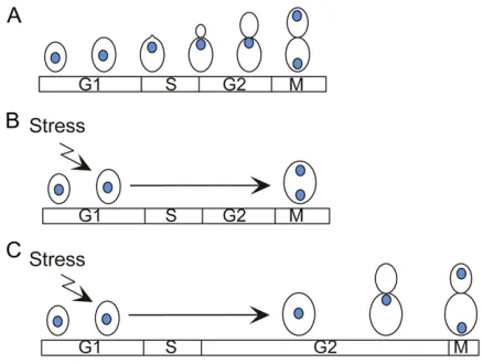
In budding yeast, the morphogenesis checkpoint delays nuclear division until a bud has been formed (reviewed in Lew 2003; Keaton and Lew 2006) (Figure 6). The existence of this checkpoint was first suggested by the observation that environmental stresses, genetic manipulations, or drug treatments that delayed bud formation also caused a delay in nuclear division (Lew and Reed 1995; McMillan et al.
the CDK-inhibitory kinaseSwe1p(Siaet al.1996).Swe1pis homologous to Wee1-family kinases in other organisms and phosphorylates tyrosine 19 ofCdc28p(Booheret al.1993). Below we summarize what has been learned regarding
Swe1paction and its regulation during the unperturbed cell cycle and then address the question of what processes are monitored by the checkpoint and how that sensing takes place.
Regulation of Cdc28p tyrosine phosphorylation during the cell cycle
Cdc28p phosphorylation in unperturbed cells: Given the precedent fromSchizosaccharomyces pombe, where Cdc2 ty-rosine 15 phosphorylation inhibits the mitotic CDK and enforces a long G2 delay in every cycle, it was quite a sur-prise when early studies indicated that CDK tyrosine phos-phorylation had no discernible effect on the S. cerevisiae
cell cycle, even in the face of treatments that triggered arrest via the DNA replication or spindle assembly checkpoints (Amon et al. 1992; Sorger and Murray 1992). Some (Lim
et al. 1996; Harvey and Kellogg 2003; Rahal and Amon 2008), but not all (McNulty and Lew 2005), subsequent studies found that Swe1p did have a small effect on the timing of spindle assembly. Why is the effect of Swe1p
so minor?
Cdc28p tyrosine phosphorylation occurs only in G2, al-thoughSwe1pis synthesized during late G1 as part of a large set of periodically expressed genes (Limet al.1996; Siaet al.
1996). However, at that time, the predominant G1 CDK (Cln1-3p-Cdc28p) complexes are not recognized bySwe1p
(Booher et al. 1993). Later Clb-CDK complexes are all
Swe1psubstrates, but the S-phaseClb5p-Cdc28pcomplexes are poorer substrates than the M-phaseClb2p-Cdc28p com-plexes and are initially protected from phosphorylation by binding of the CDK inhibitor Sic1p (Keaton et al. 2007). Even onceSic1pis degraded,Cdc28ptyrosine phosphoryla-tion does not accumulate because S-phase CDK complexes are excellent substrates of the Cdc25-related phosphatase
Mih1p (Keaton et al. 2007), which is present throughout the cell cycle (Keaton et al. 2008; Pal et al. 2008). These features account for the lack ofCdc28pphosphorylation in S phase even thoughSwe1pis abundant at that time.
In G2, cells no longer makeSwe1pand begin to degrade it (Sia et al.1998), so Swe1p abundance decreases as the mitoticClb2p-Cdc28pcomplexes [which are excellentSwe1p
substrates (Keatonet al.2007)] accumulate. The combination of Swe1pdegradation andMih1p-mediated dephosphoryla-tion of Cdc28pexplains why Swe1pdoes not greatly delay the cell cycle.
Swe1p degradation during the unperturbed cell cycle:
Swe1p degradation is cell-cycle-regulated in unstressed cells. In early G1, any residual Swe1p left over from the previous cycle is degraded slowly [t1/290 min (Siaet al. 1998)], probably via ubiquitination by the APC (Thornton and Toczyski 2003). In G2/M, Swe1p is degraded more
rapidly (t1/2 14 min) in a manner that requires both
Clb1p,2p-Cdc28p (Sia et al. 1998) and the Polo-family ki-nase Cdc5p(Sakchaisriet al. 2004). Both of these kinases phosphorylateSwe1p at multiple sites, and mutation of 18
Cdc28ptarget sites (Harveyet al.2005) or up to 20Cdc5p
target sites (Sakchaisri et al. 2004) significantly retards
Swe1p degradation. Phosphorylation by Cdc28p primes
Swe1p for subsequent phosphorylation by Cdc5p (Asano
et al.2005). The ubiquitin ligase responsible forSwe1p deg-radation was initially identified as SCFMet30 (Kaiser et al.
1998), although subsequent studies indicated that Met30p
was not required forSwe1pdegradation in a strain lacking
Met4p (a transcription factor also targeted by SCFMet30)
(McMillan et al. 2002). This finding indicates that Swe1p
can be degraded by other pathways, but it remains possible that in wild-type cells SCFMet30 is a major contributor. In
mammalian cells, Wee1 degradation involves sequential Wee1 phosphorylation by cyclin B-CDK1 and by the Polo-family kinase Plk1, and these phosphorylations generate a phosphodegron recognized by the SCFMet30 homolog
SCFbTrCP(Watanabeet al.2004, 2005). Thus, it is attractive to speculate (although it has yet to be proved) that the multisite phosphorylation ofSwe1pin yeast similarly creates phosphodegrons recognized by SCFMet30or another
ubiqui-tin ligase.
targeting requires interaction ofSwe1pwithHsl7p, which is also concentrated at the neck (McMillanet al.1999; Shule-witz et al. 1999; Longtine et al. 2000). Hsl7p is a protein methyltransferase, although that activity appears to be dis-pensable for Swe1p regulation (Theesfeld et al. 2003).
Hsl7pitself is targeted to the neck by interaction withHsl1p, a neck-localized protein kinase (Barralet al.1999; Shulewitz
et al. 1999; Longtine et al. 2000). Small mutations that abrogate the direct interactions between Hsl1p and Hsl7p
(Cidet al.2001) orHsl7pandSwe1p(McMillanet al.2002) preventSwe1pneck targeting and also blockSwe1p degra-dation, suggesting that neck localization is critical forSwe1p
degradation.
Multisite phosphorylation of Swe1p is rapidly reversed upon Cdc28p inhibition (Harvey et al. 2005). Thus, there appear to be very active (although currently uncharacter-ized) Swe1p-directed phosphatases that would presumably antagonize Swe1p degradation. Like Swe1p, the Clb2p
-Cdc28p complex (Baillyet al.2003) and the Cdc5pkinase (Sakchaisriet al.2004) are also concentrated at the mother-bud neck. It is attractive to speculate that neck localization serves to co-concentrateSwe1pwith the kinases that target it for degradation, thereby overcoming the barrier provided bySwe1p-directed phosphatases. This hypothesis remains to be rigorously tested.
Effect of Swe1p phosphorylation on its activity:In addition to slowingSwe1pdegradation, mutation of 18 CDK consen-sus target sites onSwe1p generated a protein with signifi -cantly reduced CDK-inhibitory activity (Harveyet al.2005). The simplest interpretation of this result is thatCdc28p -me-diated Swe1p phosphorylation activates Swe1p to inhibit Clb-Cdc28p. This would constitute a negative feedback loop whereby Cdc28p promotes its own inhibition. AsCdc28p -mediated Swe1p phosphorylation also targets Swe1p for degradation (a double-negative feedback loop with the same consequence as a positive feedback loop in Cdc28p
activation), the combined feedbacks would create a rather confusing scenario.
In the well-studied Xenopus egg extract system, it is clear that CDK-mediated Wee1 phosphorylation inhibits
Wee1 (rather than activates it) (Dunphy 1994). Analysis of Wee1 phosphorylation-site mutants indicated that multi-site phosphorylation targeted two inhibitory multi-sites and at least three “decoy”sites (Kim et al.2005; Kim and Ferrell 2007). Phosphorylation of the decoy sites, which were pref-erentially targeted by the CDK, did not affect Wee1 activity. Rather, the decoys delayed phosphorylation of the inhibitory sites. Thesefindings suggested that, when there is little CDK activity, Wee1 undergoes repeated phosphorylation and de-phosphorylation at decoy sites and that the inhibitory sites are phosphorylated only when there is high CDK activity. This arrangement is thought to introduce ultrasensitivity to Wee1 regulation by the CDK (Kim and Ferrell 2007). Conceivably, Swe1p phosphorylation may involve a large number of decoy sites; in that case, mutational removal of
the decoys may enhance the targeting of less-preferred in-hibitory sites, resulting in less activeSwe1p(as observed for the 18-site mutant).
Distinguishing between the different hypotheses on the role ofSwe1pphosphorylation may not be trivial:Swe1pis phosphorylated at many nonconsensus sites (Harvey et al.
2005), and phosphosite mutants carry the risk of altering
Swe1pactivity for reasons unrelated to phosphorylation. In the case of Cdc5p-targeted Swe1p phosphorylation, the nonphosphorylatable mutants enhance the potency of
Swe1p, as expected for mutants that increaseSwe1p abun-dance (Sakchaisriet al.2004). Because there is not a great correlation between the abundance and potency of mutants affecting different clusters of target sites, it may be that some phosphorylations act to inhibitSwe1pwhereas others target Swe1p for degradation. Analysis of a mathematical model for the morphogenesis checkpoint (Ciliberto et al.
2003) suggested that for a robust checkpoint it would be useful to inhibitSwe1p(a rapid event) prior to its degrada-tion (a slower event). The possibility that a subset ofSwe1p
phosphorylations (catalyzed by either Cdc28p or Cdc5p) inhibits Swe1pactivity merits further investigation.
In addition to the kinases discussed above, the PAKCla4p
can phosphorylate Swe1p in vitro(Sakchaisriet al. 2004). Because of overlapping site specificity, it has been difficult to discern the role of that phosphorylationin vivo.