Prognostic significance of immune cells in non-small cell lung
cancer: meta-analysis
Ross A. Soo
1,2,5, Zhaojin Chen
3, Rebecca Siew Yan Teng
4, Hon-Lyn Tan
1, Barry
Iacopetta
5, Bee Choo Tai
3,6and Richie Soong
2,71Department of Haematology-Oncology, National University Health System, Singapore 2Cancer Science Institute of Singapore, National University of Singapore, Singapore 3Investigational Medicine Unit, National University Health System, Singapore 4Yong Loo Lin School of Medicine, National University of Singapore, Singapore 5School of Surgery, The University of Western Australia, Perth, Australia
6Saw Swee Hock School of Public Health, National University of Singapore, Singapore 7Department of Pathology, National University Health System, Singapore
Correspondence to: Ross A. Soo, email: ross_soo@nuhs.edu.sg
Keywords: non-small cell lung cancer; immune cells; dendritic cells; tumor associated macrophages; mast cells Received: December 29, 2017 Accepted: March 06, 2018 Published: May 15, 2018
Copyright: Soo et al. This is an open-access article distributed under the terms of the Creative Commons Attribution License 3.0
(CC BY 3.0), which permits unrestricted use, distribution, and reproduction in any medium, provided the original author and source are credited.
ABSTRACT
Background: Tumor-associated immune cells are prognostic in non-small cell lung cancer (NSCLC) but findings have been conflicting.
Objectives: To determine the prognostic role of immune cells according to localization in NSCLC patients.
Methods: A systematic literature review and meta-analysis was performed on dendritic cell (DC), tumor associated macrophages (TAM), mast cells (MC), natural killer (NK) cells, T and B cells and tumor CTLA-4 and PD-L1 studies.
Results: We analysed 96 articles (n= 21,752 patients). Improved outcomes were seen with increased tumor DCs (overall survival (OS) hazard ratio (HR) 0.55; 95% confidence interval (CI) 0.44–0.68), NK cells (OS HR 0.45; 0.31–0.65), TAMs (OS HR 0.33; 0.17–0.62), M1 TAMs (OS HR 0.10; 0.05–0.21), CD3+ T cells (disease specific survival (DSS) HR 0.64; 0.48–0.86), CD8+ T cells (OS HR 0.78; 0.66–0.93), B cells (OS HR 0.65; 0.42–0.99) and with increased stroma DC (DSS HR 0.62; 0.47–0.83), NK cells (DSS HR 0.51; 0.32–0.82), M1 TAMs (OS HR 0.63; 0.42–0.94), CD4+ T cells (OS HR 0.45; 0.21–0.94), CD8+ T cells (OS HR 0.77; 0.69–0.86) and B cells (OS HR 0.74;0.56–0.99). Poor outcomes were seen with stromal M2 TAMs (OS HR 1.44; 1.06–1.96) and Tregs (relapse free survival (RFS) HR 1.80; 1.34–2.43). Tumor PD-L1 was associated with worse OS (1.40; 1.20–1.69), RFS (1.67) and DFS (1.24).
Conclusion: Tumor and stroma DC, NK cells, M1 TAMs, CD8+ T cells and B cells were associated with improved prognosis and tumor PD-L1, stromal M2 TAMs and Treg cells had poorer prognosis. Higher quality studies are required for confirmation.
www.oncotarget.com
Oncotarget, 2018, Vol. 9, (No. 37), pp: 24801-24820
INTRODUCTION
Lung cancer is one of the most common malignancies
globally, accounting for 1.5 million cases annually. It is also
the leading cause of cancer deaths globally, causing 1.3
million deaths annually [1]. The tumor microenvironment
has a major role in influencing cancer development [2], of
which immune cells are considered to contribute to tumor
destruction, as well as tumor development by promoting
growth and invasion [3, 4].
In recent times, a major advance in the treatment
of non-small cell lung cancer (NSCLC) has been the use
Meta-Analysis
of immunotherapy, such as immune checkpoint inhibitors
targeting cytotoxic T lymphocyte antigen-4 (CTLA-4),
programmed death receptor-1 (PD-1) and programmed
death receptor ligand-1 (PD-L1) [5]. In the advanced stage
NSCLC setting, many PD-1/PD-L1 inhibitors have been
approved for use [6], although results from trials in the
resected tumor setting have been less encouraging [7].
The potential of the immune system to contribute
functionally to both tumor elimination and promotion, and
the observed significant effects of its modulation through
immunotherapy, have supported that the immune system
can be a significant determinant of the outcomes of NSCLC
patients. As such, numerous studies have investigated the
prognostic and predictive significance of many different
cell types of the immune system over the years [4]. The
different immune cell types have included mast cells,
dendritic cells, natural killer cells, macrophages, neutrophils
of the innate immune system, T and B lymphocytes of the
adaptive immune system, as well as CTLA-4 and
PD-L1-expressing cells targeted by immunotherapy. In many cases,
specific subtypes of immune cells, such as M1 and M2
macrophages, and CD3+, CD4+, CD8+, and regulatory T
cells have been examined. Moreover, assessment according
to localization of the immune cells in tumor parenchyma
or stroma has also been performed. This is based on the
observed varied presence of these cells in the tissue
compartments, and associated functional implications.
Findings from such reports have been numerous
and varied according to immune cell type, outcome
endpoint, tissue localization, study quality, as well as
results. This study was undertaken with the goal of
consolidating knowledge on the prognostic significance
of the many immune cell types in NSCLC, and according
to investigated co-factors.
METHODS
Search strategy
Meta-analysis was conducted according to the
guidelines of the Systematic Reviews and Meta-Analyses
(PRISMA) [8]. Relevant articles were identified through
a systematic search of PUBMED using the MeSH terms:
“Immune cell type” AND “lung neoplasm” limited to
“Human”, “English”. MeSH terms for the immune cell
types were “mast cells”, “macrophage”, “dendritic cell”,
“NK cell”, “regulatory T cell”, “CD3 T-lymphocyte”, “CD4
T-lymphocyte”, “CD8 T-lymphocyte”, “B cell”,
“CTLA-4 antigen” and “Antigen, CD27“CTLA-4” (PD-L1). Articles
published up to 7 July 2017 were included in our search.
Inclusion and exclusion criteria
The inclusion criteria for articles were those that
reported on samples from patients with primary lung
tumors with NSCLC, having no systemic treatment or
radiation therapy prior to sample collection, and sufficient
prognostic information to determine pooled Hazard
Ratios (HR). Where HRs were not reported, included
studies had to have sufficient information to extrapolate
HR. The exclusion criteria included studies on blood
or other body fluids or pre-clinical models, studies on
the optimization of immunohistochemistry (IHC) or
quantitative immunoflurorescence (QIF) methods, or
using non-IHC/QIF based methods to detect immune
cells, as well as letters and case reports. References cited
in retrieved articles were checked for additional relevant
articles. Data from other reviews and meta-analyses were
not included, but articles identified through references
cited were reviewed. Irrelevant and/or duplicate studies
were removed by manual curation. Study eligibility was
assessed independently by two authors (RAS and ZC).
Data extraction
Two investigators (RAS and ZC) independently
extracted the data. The following details were extracted
from each study: first author, publication year, PMID,
country of origin of the study population, immune cell
studied, phenotype, markers used to define immune cell
type, localization of immune cells (defined as “tumor”,
“stroma” or, if the localization was unspecified, “general”
compartment), sample size, number of events, tumor
stage, treatment setting, and histology (adenocarcinoma,
squamous cell carcinoma or mixed). For study
methodology, data was collected on the assay used, tissue
sample used (full tissue sections or tissue microarray),
antibodies used, scoring method and thresholds used to
define expression. Survival outcomes annotated included
disease-free survival (DFS), relapse-free survival (RFS),
disease-specific survival (DSS) and overall survival (OS).
Assessment of study quality and risk of bias
RAS and HLT independently assessed study quality
according to the criteria developed by McShane 2005 and
Hayes [9, 10] for tumor marker prognostic studies. In brief,
the criteria assessed seven domains including: inclusion
and exclusion criteria, prospective or retrospective study
design, patient and tumor characteristics, method or assay
description, outcome measures defined, patient follow
up and number of patients lost to follow-up or otherwise
unavailable for analysis.
Statistical analysis
The prognostic effect of an immune cell was
quantified by HR, defined as the relative hazard of death
or disease progression of patients with high or positive
immune cell levels against those with low or negative
immune cell levels. Where HRs were not reported, they
were estimated using hazard ratio, odds ratio, or the
ratio of median survival, as proposed by Parmar [11].
Stratified analysis was conducted according to localization
(tumor, stroma, general), or phenotype of immune cells.
A meta-analysis was performed when there were at least
two studies in a stratum. Therefore, a single study with
a reported HR in an analytic stratum was not analyzed.
For studies with considerable heterogeneity, studies were
modelled for random-effects, according to the methods
of DerSimonian and Laird [12]. Otherwise, a fixed-effect
model was used. Heterogeneity was considered to be low,
moderate, and high for I
2values of 25–50%, 50–75%,
and >75%, respectively [13]. Results for each immune
cell type were displayed using a forest plot. A funnel
plot was constructed to visualize small-study effects and
possible publication bias for a stratum with five or more
studies. To test for small-study effect, the Egger’s test
was subsequently performed when there were at least 10
studies in a stratum. Median survival times were derived
from the Kaplan-Meier survival curves using DigitizeIt
2.2. All analyses were performed using StataSE14
(StataCorp LP, College Station, Texas) by assuming a
two-sided statistical test with 5% significance level.
RESULTS
A systematic search of PubMed and referenced
articles resulted in 3,291 records, from which 96 individual
studies, assessing 21,752 patients, were eligible for
meta-analyses (Supplementary Tables 1–2, Supplementary
Figure 1). The majority of studies were from East Asia (61,
64%) and mixed NSCLC histology (60, 63%). IHC was
used in 92 (96%) of studies, and full tissue sections were
used in 67 (70%). The average study quality score for all
studies was 4.7.
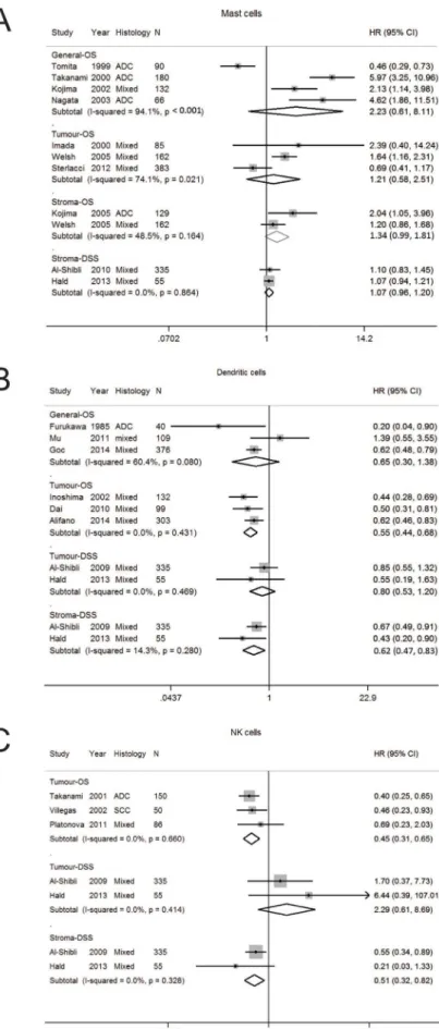
Mast cells
Mast cells (MC) play a key role in allergic diseases
but are also involved in immune responses. Depending on
the type of solid tumor, mast cells can enhance adaptive
immunity but also play a key role in tumor angiogenesis,
tumor invasion, and immune suppression [14]. Ten
studies were analysed [15–24]. (Supplementary Table 1
and 2, Supplementary Figure 1A). The number of studies
analysed per stratum ranged from two to three (Table 1).
Early studies reported MC counts without consideration
of tumor localisation (Supplementary Table 2). Three out
of four studies reported increased MC was associated
with a worse OS, however the associations did not reach
statistical significance in pooled analysis (HR 2.23; 95%
CI 0.61–8.11) (Table 1, Figure 1A). Later studies assessed
outcomes according to localisation, and reported MC
were not significantly associated with OS in the tumor
(HR 1.21; 0.58–2.51) or stroma (HR1.34; 0.99–1.81). A
high degree of heterogeneity was seen in studies on OS
according to general (I
294.1%, p < 0.001) and tumor
(I
274.1%, p = 0.021) localisation.
Dendritic cells
Dendritic cells (DC) are the most potent antigen
presenting cells and regulate the immune system to respond
to foreign antigens while avoiding autoimmunity and
therefore are important in cancer, generating both immunity
and tolerance [25]. Eight studies (were suitable for analysis
(Supplementary Tables 1, 2, Supplementary Figure 1B).
The average quality score was 4.5 (Supplementary Table 1)
[24, 26–32]. On pooled analysis, the HR for OS for general
DC was 0.65 (0.30–1.38) (Table 1, Figure 1B). Inoshima
et al. first reported high DCs in the tumor compartment
was associated with longer OS [27]. In pooled analysis,
increased tumor DC was prognostic for OS (HR 0.55;
0.44–0.68) but not for DSS (HR 0.80; 0.53–1.20). In
contrast, stromal DC was significantly associated with DSS
(HR 0.62; 0.47–0.83). Study heterogeneity was generally
low in the studies examined for OS in tumor and DSS in the
tumor and stroma. Funnel plot analysis was not performed
as there was an inadequate number of publications per
stratum.
Natural killer (NK) cells
Natural killer (NK) cells are the major effector cells
of the innate immune system, and have an important role
in the immune response against cancer [33]. Only five
studies were suitable for pooled analysis (Supplementary
Tables 1, 2 Supplementary Figure 1C) [24, 28, 34–36].
Pooled analysis revealed increased tumor NK cells were
associated with an improved OS (HR 0.45; 0.31–0.65) but
not DSS (HR 2.29; 0.62–8.69), whereas stromal NK cells
were associated with better DSS (HR 0.51; 0.32–0.82)
(Table 1, Figure 1C). Study heterogeneity was low. As
the number of publications per stratum was only 2 or 3,
further studies of adequate sample size should be pursued.
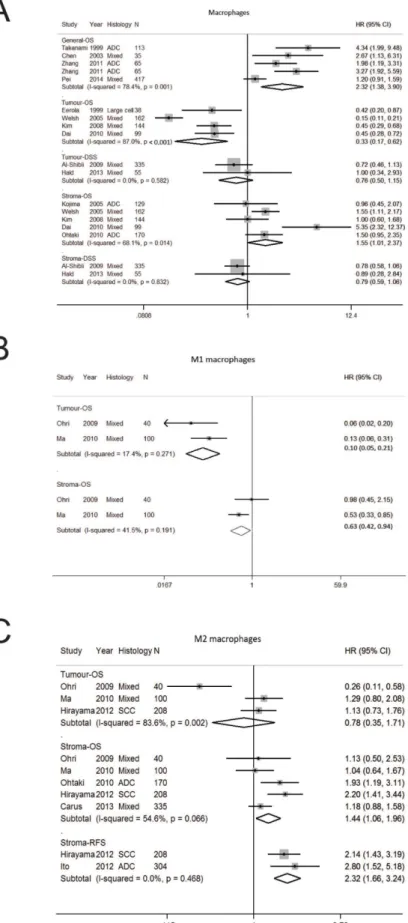
Tumor associated macrophages
Tumor associated macrophages (TAMs) are part of
the innate immune system and have an essential function
against foreign pathogens [37]. Eighteen studies were
analysed (Supplementary Tables 1, 2, Supplementary
Figure 1D) [20, 21, 24, 28, 29, 38–50]. The number of
studies per stratum was 2–5 (Table 1). Initial studies
reported on tumor-associated macrophages (TAM) in
the general compartment [39, 40] and subsequently the
importance of tumor localization was first recognised
by Eeroloa et al. who found increased tumoral TAM
was associated with an improved OS and DFS [38]. The
significance of TAM localization was extended further in
a pivotal paper that reported increased tumor TAM had an
improved OS whereas stromal TAMs had worse OS [21].
In our pooled analysis, increased TAMs in general
had worse OS (HR 2.32; 1.38–3.90) (Table 1, Figure 2A).
When analysed according to localization, increased
TAMs in the tumor compartment was associated with a
better OS (HR 0.33; 0.17–0.62) whereas stromal TAM
was associated with poorer OS (HR 1.55; 1.01–2.37). In
terms of DSS, TAMs in the tumor (HR 0.76; 0.50–1.15)
and stromal (HR 0.79; 0.59–1.06) compartments was not
significant (Figure 2A). A high degree of heterogeneity
was seen in studies on OS according to general (I
278.4%, p = 0.001) and tumor (I
287.0%, p < 0.001)
localisation. Funnel plot analysis suggest publication bias
on macrophages in general compartment whereas no bias
was seen for stroma macrophages (Supplementary Figure
2A and 2B).
Distinct macrophage phenotypes have been
described including M1 macrophages that induce host
defense, antitumor immunity and inflammatory responses
and M2 macrophages reduces inflammation, suppress
antitumor immunity and promote angiogenesis [37]. Given
the presence of different macrophage phenotypes, we
determined the prognostic effect of M1 and M2 macrophages
(Table 1, Figure 2B, 2C) and found M1 macrophages was
associated with improved OS in the tumor (HR 0.10;
0.05–0.19) stromal M1 and stroma (HR 0.63; 0.42–0.94).
Whilst tumor M2 macrophages was not significant for OS,
stroma M2 macrophages was associated with a worse OS
(HR 1.44; 1.06–1.96) and RFS (HR 2.32; 1.66–3.24).
Neutrophils
Neutrophils, a key effector immune cell, has a
complex role in tumorigenesis [51]. After screening,
four full text papers were reviewed [49, 52–54] but no
Table 1: Summary of hazard ratios, sample sizes and average quality scores from pooled analyses for cell types
according to cellular localization
General Tumor Stroma
DSS OS DSS OS DSS OS Mast cells NA 2.23 [0.61–8.11] n468(4) Q4.25 NA 1.21 [0.58–2.51] n630(3) Q5.3 1.07 [0.96–1.20] n390(2) Q5 1.34 [0.99–1.81] (n291(2) Q6.5 Dendritic cells NA 0.65 [0.30–1.38] n525(3) Q4 0.80 [0.53–1.20] n390(2) Q5.5 0.55 [0.44–0.68] n534(3) Q4.67 0.62 [0.47–0.83] n390(2) Q2 NA Natural Killer cells NA NA 2.29 [0.61–8.69] n390(2) Q5.5 0.45 [0.31–0.65] n374(3) Q2.67 0.51 [0.32–0.82] n390(2) Q5 NA Macrophages NA 2.32 [1.38–3.90] n695(5) Q4.2 0.76 [0.50–1.15] n350(2) Q5 0.33 [0.17–0.62] n443(4) Q5 0.79 [0.59–1.06] n350(2) Q5 1.55 [1.01–2.37] n704(5) Q5.4 Macrophages, M1 NA NA NA 0.10 [0.05–0.21] n140(2) Q4.5 NA 0.63 [0.42–0.94] n140(2) Q4.5 Macrophages, M2 NA NA NA 0.78 [0.35–1.71] n348(3) Q4.67 2.32 [1.66–3.24] n512(2) Q57 1.44 [1.06–1.96] n853(5) Q5 T cells, CD3+ NA 0.72 [0.53–0.97] n848(4) Q4.5 0.64 [0.48–0.86] n350(2) Q52,3 0.88 [0.74–1.05] n420(3) Q3 1.13 [0.38–3.35] n350(2) Q5 0.86 [0.62–1.18] n325(2) Q3 T cells, CD4+ NA NA 0.86 [0.61–1.21] n350(2) Q5.54 0.74 [0.48–1.15] n678(4) Q4.5 0.23 [0.06–0.95] n350(2) Q5.5 0.45 [0.21–0.94] n358(3) Q5.67 T cells, CD8+ 0.70 [0.48–1.02] n554(3) Q4.341 0.80 [0.56–1.15] n1348(6) Q4.5 0.69 [0.50–0.96] n350(2) Q5.55,6 0.78 [0.66–0.93] n2844(9) Q4.55 0.47 [0.36–0.63] n1187(3) Q5.679 0.77 [0.69–0.86] n2157(8) Q5 T cells, regulatory NA 1.42 [0.78–2.60] n146(2) Q3.5 1.43 [0.69–2.94] n578(2) Q47 1.00 [0.75–1.34] n578(2) Q4 1.80 [1.34–2.43] n678(3) Q47,10 1.43 [0.69–2.94] n750(5) Q4.4 B cells NA NA NA 0.65 [0.42–0.99] n590(2) Q4.5 NA 0.74 [0.56–0.99] n644(2) Q4.5 PD–L1 NA NA n2245(10) Q4.81.67 [1.22–2.29] 7,8 1.40 [1.20–1.69] n8970(35) Q5 NA NA Results are expressed as pooled HR [95% confidence limit], number of patients (number of studies), Q (average quality score).
DFS = disease-free survival, DSS = disease-specific survival, NA = not analyzable, OS = overall survival, RFS = relapse-free survival. 1DFS, no DSS available. 2RFS HR = 0.73 [0.40–1.32]. 3DFS HR = 1.01 [0.54–1.88]. 4RFS HR = 1.11 [0.73–1.68]. 5DFS HR = 0.58 [0.32–1.04]. 6RFS HR = 1.22 [0.82–1.83]. 7RFS, no DSS available. 8DFS HR = 1.24 [1.01–1.52]. 9DFS HR = 1.69 [1.11–2.55]. 10DFS HR = 1.24 [1.01–1.52].
Figure 1:
Forest plot of studies assessing (A) Mast cells, (B) dendritic cells, (C) Natural killer cells and survival in patients with non-small cell lung cancer. Adenocarcinoma, ADC; confidence interval, CI; disease specific survival, DSS; hazard ratio, HR; overall survival, OS; programmed cell death-ligand 1, PD-L1; progression free survival, PFS; squamous cell carcinoma, SCC.Figure 2:
Forest plot of studies assessing (A) Macrophages (B) Macrophages M1 (C) Macrophages M2 and survival in patients with non-small cell lung cancer (NSCLC) stratified according to localisation (in general, tumor or stroma compartment). Adenocarcinoma, ADC; confidence interval, CI; disease specific survival, DSS; hazard ratio, HR; overall survival, OS; programmed cell death-ligand 1, PD-L1; progression free survival, PFS; relapse free survival, RFS; squamous cell carcinoma, SCC.studies were selected for pooled analysis. One study was
excluded as neo-adjuvant chemotherapy was administered
in 9% of patients [54] and three other studies were in a
single stratum [49, 52, 53] (Supplementary Table 1,
Supplementary Figure 1E). In the first study by Carus et al,
increased neutrophils in the tumor and stroma was not
associated with RFS or OS [49] whereas in the second
study, increased tumor associated neutrophils (TAN) was
associated with a poorer DFS [52]. In the third study, high
intratumoral TANs was a positive prognosticator for DSS
in SCC NSCLC whereas TAN was associated with worse
DSS [53].
Tumor-associated neutrophils have a dual function
characterized by the N1 and N2 phenotype in a
context-dependent process. N1 neutrophils have an anti-tumor
phenotype through its interaction with T cells whereas
the N2 phenotype promotes tumor growth [55]. Future
studies examining the prognostic role of tumor-associated
neutrophils in NSCLC should take into account the
distribution of N1 and N2 phenotypes within the tumor
microenvironment.
T cells, CD3 positive
Twelve studies were analysed (Supplementary
Tables 1, 2, Supplementary Figure 1F) [21, 24, 52, 56–64].
Elevated CD3+ T cells in general compartment was
associated with improved OS (HR 0.72; 0.53–0.97)
(Table 1, Figure 3A). When analysed according to
localisation, increased tumor CD3+ T cells was associated
with longer DSS (HR 0.64; 0.48–0.86) and suggestive
for better OS (HR 0.88; 0.74–1.05) but not for RFS (HR
0.73; 0.40–1.32) (Table 1). Stromal CD3+ T cells was not
associated with survival outcomes and may be suggestive
of poorer DSS (HR 1.13) or DFS (HR 1.2), although
results were not statistically significant. A high degree of
heterogeneity was seen in studies on OS in the general
compartment (I
270.6%, p = 0.002). Funnel plot suggest
potential publication bias with smaller studies with
favourable OS in general compartment being reported
(Supplementary Figure 2C).
T cells, CD4 positive
Eleven studies were analysed (Supplementary
Tables 1, 2, Supplementary Figure 1G) [23, 24, 60, 64–71].
Study quality was generally good with an average score of
5.1 (Supplementary Table 1). CD4+ T cells in the general
or tumor compartment had no influence on OS, RFS or
DSS (Table 1, Figure 3B). In contrast, CD4+ T cells in
the stroma compartment was associated with better OS
(HR 0.45; 0.21–0.94) and DSS (HR 0.23; 0.06–0.95).
Significant heterogeneity was seen in studies in the
stromal compartment for OS (I
277.0%, p = 0.013) and
DSS (I
277.4%, p = 0.035).
T cells, CD8 positive
Twenty-three studies underwent pooled analysis
(Supplementary Table 1, Supplementary Figure 1H) [23, 24,
29, 31, 32, 60–64, 66–78]. Although CD8+ T cells in the
general compartment was not associated with OS (HR 0.80;
0.56–1.15) or DFS (HR 0.70; 0.48–1.02), when analysed
according to tumor or stroma compartment, the prognostic
value of CD8+ T cells could be appreciated (Table 1,
Figure 3C). CD8+ T cells in the tumor compartment was
associated with better prognosis in terms of OS (HR 0.78;
0.66–0.93) and DSS (HR 0.69; 0.50–0.96) but not for
DFS (HR 0.58; 0.32–1.04) or RFS (HR 1.22; 0.82–1.83).
Similarly, stromal CD8+ T cells conferred an improved
OS (HR 0.77; 0.69–0.86), DFS (HR 0.73; 0.58–0.93) and
DSS (HR 0.47; 0.36–0.63). Heterogeneity was low for most
analytic strata. Funnel plot suggest generally no publication
bias (Supplementary Figure 2D–2F).
T cells, regulatory
Regulatory T cells (Tregs) are a subpopulation
of CD4+ CD25+ T lymphocytes that inhibit anti-tumor
immunity by promoting immune tolerance through
direct suppressive functions on T cells or by secreting
immunosuppressive cytokines such as IL-10 and TGF-b
[79]. Tregs are purported to express and functionally
depend on the transcription factor forkhead box protein
P3 (FoxP3). As such many studies commonly use FoxP3
as a single marker for Tregs. Eleven studies (n=1977
patients) were reviewed (Supplementary Table 1 and 2,
Supplementary Figure 1I). [23, 59, 60, 70, 71, 77, 80–84].
General Tregs infiltration was not associated with OS (HR
1.42; 0.78–2.60) (Table 1, Figure 3D) whereas tumor Tregs
was not associated with OS (HR 1.00; 0.75–1.34) or RFS
(HR 1.53; 0.64–3.67). Stromal Tregs was not associated
with OS (HR1.43; 0.69–2.94), but however was associated
with worse RFS (HR 1.80; 1.34–2.43) and DFS (HR 1.69;
1.11–2.55). Most analytic strata showed low heterogeneity.
studies of tumoral Treg and RFS (I
280.0%, p = 0.025) and
stromal OS (I
287.1%, p < 0.001). Possible publication bias
was not observed in studies of stromal Treg at the extreme
HR for OS (Supplementary Figure 2G).
B cells
Apart from its role in humoral immune responses,
B cells have a pro- or anti-tumorigenic function [85].
After screening, four studies were analysed for OS
(Supplementary Table 1 and 2, Supplementary Figure 1J)
[38, 61, 86, 87]. Several studies were excluded as they
had no prognostic information, insufficient information to
impute HR or were the only study in an analytic stratum
[21, 52, 64, 68, 88, 89]. Results of pooled analysis found
B cells in the tumor and stroma was associated with an
improved OS with a HR for 0.65 (0.42–0.99) and 0.74
(0.56–0.99), respectively (Table 1, Figure 4). High
heterogeneity was not seen for studies of tumor and stroma
B cells and OS.
Cytotoxic T lymphocyte antigen-4
CTLA-4 is not only expressed on T cells but is also
found on NSCLC tumors. Three studies were assessed in
full (Supplementary Table 1, Supplementary Figure 1K).
Two studies had different endpoints: OS [90] and DSS
(91) therefore pooled analysis was not performed. Tumor
CTLA-4 overexpression was not associated with OS [90]
or DSS [91]. In the third study gene expression arrays was
used and CTLA-4 overexpression was associated worse
OS [92].
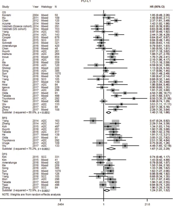
Programmed death ligand-1
The prognostic impact of PD-L1 was reported in 38
studies with 10,034 patients (Supplementary Tables 1, 2,
Supplementary Figure 1L) [30, 70, 71, 75–77, 93–124].
Two additional studies were excluded [125, 126] as
there was insufficient information to calculate the HR.
Our meta-analysis found tumor PD-L1 over expression
was associated with worse OS (HR 1.40; 1.20–1.69),
RFS (HR 1.67; 1.22–2.29) and DFS (1.24; 1.01–1.52)
(Table 1, Figure 5). Heterogeneity was high in the
studies for OS (I
280.8 %, p < 0.001) and RFS (I
275.2 %,
p < 0.001), and moderate for DFS (I
272.9 %, p < 0.001).
Publication bias was not observed and Egger’s test for
small-study effects was not significant for studies on
Figure 3:
Forest plot of studies assessing (A) CD3+ T cells, (B) CD4+ T cells, (C) CD8+ T cells, (D) FOXP3+ regulatory T cells (Treg) and survival in patients with non-small cell lung cancer (NSCLC) stratified according to localisation (in general, tumor or stroma compartment). Adenocarcinoma, ADC; confidence interval, CI; Hazard ratio, disease specific survival, DSS; hazard ratio; HR; overall survival, OS; programmed cell death-ligand 1, PD-L1; progression free survival, PFS; relapse free survival, RFS; squamous cell carcinoma, SCC.OS (p = 0.132), RFS (p = 0.663) and DFS (p = 0.656)
(Supplementary Figure 2H–2J).
Potential factors for heterogeneity
We performed sensitivity analysis and subgroup
analyses for studies of each immune cell to identify
potential factors responsible for the heterogeneity.
Sensitivity analysis was performed by excluding
studies with quality score of three or less. After
excluding low quality studies, there were an inadequate
number of studies of M1 macrophages for
meta-analysis. Mast cells, NK cells, M2 and macrophages,
CD3 lymphocytes, FOXP3+ T cells and PD-L1
meta-analysis were re-analysed and prognosis were affected
for two immune cell types: OS for macrophages in
general was no longer associated with poor prognosis
(Supplementary Figure 3C) whereas stromal regulatory
T cells was now associated with poorer prognosis
(Supplementary Figure 3F). Prognostic patterns were
unchanged for the other immune cells (Supplementary
Figure 3). Studies of dendritic cells, CD4 and CD8 T
lymphocytes were not re-analysed as all studies had
quality score of at least 4.
Subgroup analyses were performed for immune
cells with relatively large number of studies (≥7
studies). For this analysis, only studies of PD-L1 and
CD8 satisfied this threshold. The subgroups analysed
included ethnicity, publication year, sample size,
sample type, and threshold for positive score. For CD8
cells, we found ethnicity, publication year, sample size
and cut-off point were confounders for the association
between tumour location (tumor, stroma) and OS
(Supplementary Tables 4 and 5) but not for general
location (Supplementary Table 3). For PD-L1 and
OS, there was no confounding effect for the following
variables: geographic location, publication year, sample
size and histology (Supplementary Table 6). Ethnicity,
histology and publication year may be a potential
confounder for the association between PD-L1 and
DFS (Supplementary Table 7) whereas histology may
be a potential confounder for the association between
PD-L1 and RFS (Supplementary Table 8). The role
of histology as a confounder is limited by the small
number of studies (two studies with mixed histology).
DISCUSSION
The immune system has been implicated to have
a dual role in tumorigenesis, both suppressing tumor
growth through the elimination of cancer cells and also
promoting tumor growth through supply of growth and
survival factors. With the rapid progress being achieved
in tumor immunology and the development of cancer
immunotherapy approaches, an understanding of the
role of immune cells in the tumor microenvironment in
NSCLC may enhance these advances in immunotherapy
drug development.
Figure 4: Forest plot of studies assessing B cells and overall survival (OS) in patients with non-small cell lung cancer
(NSCLC) according to localisation in tumor or stroma compartment.
Confidence interval, CI; hazard ratio, HR.The role of tumor-infiltrating immune cells in
NSCLC is complex and its prognostic value has been
studied with variable and often conflicting results. Whilst
previous meta-analyses have examined the prognostic
effect of specific immune cells or immune markers such
as T cells [127, 128], PD-L1 [129–135] in NSCLC, the
current study to our knowledge is the first meta-analysis
on the prognostic impact of NK cells, DC, MC, and
macrophages. DC, NK cells, M1 macrophages, CD3+
and CD8+ T cells were found to be associated with a
favourable prognosis, whereas M2 macrophages and Tregs
in the stroma were associated with a worse prognosis.
These results are consistent with the role of these immune
cells in anti- and pro-tumor immunity, and support the
pursuit of immunotherapy as a potential therapeutic
modality in NSCLC [136–140].
We also confirmed localisation influenced prognosis
(Table 1). DCs, NK cells, M1 macrophages and CD8+
T cells in the tumor and stroma was associated with
improved prognosis. TAMs localised in the tumor had a
Figure 5: Forest plot of studies assessing tumor PD-L1 expression and survival outcomes in patients with non-small
cell lung cancer (NSCLC).
Adenocarcinoma, ADC; confidence interval, CI; Hazard ratio, disease free survival DFS; hazard ratio, HR; overall survival, OS; programmed cell death-ligand 1, PD-L1; progression free survival, PFS; relapse free survival, RFS; squamous cell carcinoma, SCC.better prognosis, whereas stromal TAMs were associated
with a worse prognosis. Tumor M1 macrophages were
associated with a better prognosis, whereas stromal M2
macrophages were associated with poorer OS, consistent
with our understanding of the function of M1 and M2
macrophages [37]. These findings highlight the prognostic
importance of both the immune cell phenotype (M1 or
M2) as well as immune cell localisation in the tumor
microenvironment, further emphasising the importance
of a full understanding of the complexity of the cellular
interactions within the tumor microenvironment.
The prognostic effect of immune cells has been
reported in other tumors. Increased mast cells are
associated with poorer prognosis in colorectal cancer
(CRC) [141], malignant melanoma [142], pancreatic
adenocarcinoma [143] and improved prognosis in
malignant mesothelioma [144], ovarian cancer [145],
and breast cancer [146]. Increased NK cells have been
reported to be associated with an improved prognosis in
gastric carcinoma [147, 148], CRC [149], and laryngeal
cancer [150]. High CD3+ TILs have been associated with
an improved OS in NSCLC [128], gastric [151], breast
[152] and hepatocellular carcinoma (HCC) [153]. CD8+
T cells infiltration have been associated with a favourable
prognosis in breast [152], ovarian [154], gastric [151],
CRC [155] and HCC [153]. High FoxP3+ Tregs was
associated with worse OS in cervical, renal cell carcinoma
(RCC), melanoma, HCC, gastric and breast cancers and
an improved OS in CRC, head and neck cancer (HNC),
and oesophageal cancer whereas the DFS rate was lower
in lung cancer [156]. B cell infiltration into the tumor
stroma has been reported to be associated with different
outcomes with an improved survival seen in breast [157],
but reports for melanoma, prostate, HCC, ovarian and
HNC [158] have been inconsistent. High intratumoral
neutrophil density was associated with poorer OS for HCC
and intrahepatic cholangiocarcinoma, HNC, NSCLC and
RCC [159]. In the same meta-analysis, the HR for OS
with NSCLC was borderline, with 95% CI of 1.0–1.35
and included one study where pre-operative chemotherapy
was given [54].
A tremendous degree of heterogeneity was observed
in terms of sample size (ranging from 38 to 1290 patients),
geographical location of the patient population (East
Asian/ non-East Asian); stage (I, I–III, I–IV), histology
(adenocarcinoma or squamous cell only, or mixed
histology), study methodology (sections or TMAs,
antibodies used, scoring cutoffs), and survival endpoints
(OS, DSS, DFS, RFS) (Table 1, Supplementary Tables 1,
2). The vast majority of studies examined mixed NSCLC
histology with only a small number of studies focused
adenocarcinoma or squamous cell histology only. The
number of studies with East Asian varied depending on
the immune cell studied. Immune cell studies published
frequently from East Asia countries included PD-L1
(79%), TAMs (66.7%), regulatory T cells (64%), and MC
(60%) whereas non-East Asian studies included CD3+ T
cells (83%), NK cells (75%), B cells (75%) and CD8+
T cells (61%) (Supplementary Table 1). Such differences
may account for prognostic differences observed between
some studies. Subgroup analyses showed ethnicity,
publication year, sample size and cut-off point were
confounders for OS in CD8 (Supplementary Tables 4
and 5). The studies analysed were generally moderate in
quality with an average of 3.8 for NK cell studies to 5.1 for
CD4+ T cells studies (Supplementary Table 1). Therefore,
higher quality studies are needed to validate the results.
The choice of antibodies used may be important as
different subpopulations of immune cells exist with regard
to its maturation, differentiation and state of activation.
This is exemplified by the various antibody clones used to
detect DCs: S100 in earlier studies [26, 27], subsequently
CD1a [24, 28, 30] or CD83 [29] and more in recent studies
CD208 [32, 58]. The choice of antibodies used may be
important as different subpopulations of dendritic cells
exist in regard to its maturation, differentiation and state of
activation. Mature DC has T cell co-stimulatory molecules
that induce immune reactions, whereas inactivated DCs
lack such T cell stimulating ability. The use of S100 IHC
Ab is controversial as its expression in DC is not specific
[160]. CD83 and CD208 are markers expressed in mature
DCs; whereas CD1a is expressed in immature DCs [161].
As a result, the prognostic effect of DCs in the general
compartment may be affected the maturation of DC: two
studies using mature DC marker were associated with
favorable OS [26, 32] whereas the study of immature DC
(CD1a) was associated with worse OS [30]. Similarly,
in a study examining the role of DC maturation status
in patients with breast cancer, CD83 expression was
prognostic for overall and relapse free survival whereas
CD1a, a marker of immature DC, was not [162]. Future
prognostic studies should be conducted with mature DC
markers localized to the tumor center.
The number of studies analyzed according to the
same tissue localization of immune cell and clinical
outcome was small, further limiting our ability to draw
firm conclusions (Table 1). Further studies are also
required to define the prognostic role of neutrophils,
CTLA-4 expression in tumor cells and PD-L1 expression
in immune cells.
Tumor infiltrating lymphocytes (TILs) as a whole
has been reported recently in a meta-analysis to be
associated with an improved PFS [127]. However, as TILs
are a heterogeneous population comprising of different T
cell subsets, we elected to focus on the individual subsets
of TILs separately due to their different functions in
tumor microenvironment [3, 4] and thus did not analyse
for the prognostic effect of tumor infiltrating lymphocytes
as a group.
Our findings should be interpreted within the
limitations of a meta-analysis as the data was confounded
by factors such as absence of individual patient data,
variation in study quality, HRs calculated based on the data
extracted from the survival curves, differences in tissue
processing, IHC staining protocols, definition of
region-of-interest, scoring methodology, differences in thresholds
for positivity and prognostic end points. The use of
different protocols, antibodies, and scoring systems creates
complexity in the interpretation of studies and applicability
in clinical practice. This is especially pertinent for the
detection of PD-L1, where meta-analyses studies have
shown PD-L1 expression to be associated with improved
outcomes in patients with NSCLC treated with immune
checkpoint inhibitors [163–165]. Given the role of
PD-L1 as a prognostic biomarker and, more importantly, as a
predictive marker for treatment selection, further efforts
are clearly required to standardise the detection of
PD-L1 expression and also to determine factors of variability
between IHC assays [166, 167]. Apart from to PD-L1,
international efforts are also underway to standardise the
assessment of tumor-infiltrating lymphocytes in NSCLC
as well as other solid tumors [168, 169].
Future studies should examine the role of immune
cells as a new prognostic factor in staging. Similar to
developments were made in CRC [170, 171], while
approaches to integrate tumor-infiltrating lymphocytes
into NSCLC staging are being pursued [172]. In addition
to incorporating tumor-infiltrating lymphocytes, further
prospective studies using immune cell panels/
multi-parametric IHC are also desirable to determine the most
promising combination of immune cells as a prognostic
marker in NSCLC. Recent studies of molecular tumor
profiling with immune cell phenotyping [173, 174] has
improved our understanding of the complex relationship
between tumor and the tumor microenvironment and may
lead to improvements in therapeutic outcomes in NSCLC.
CONCLUSIONS
Our findings suggest DC, NK cells, M1
macrophages, CD8+ T cells, and B cells in the tumor
and stroma are associated with an improved prognosis
and stromal M2 macrophages, regulatory T cells and
PD-L1 overexpression are associated with poorer
prognosis in NSCLC. Future research should focus
on the standardisation of immune cell detection, use of
multi-immune cell panels as a prognostic biomarker, and
incorporating immune cells into prognostic models.
ACKNOWLEDGMENTS
This study was supported by the National Research
Foundation Singapore and the Singapore Ministry of
Education under its Research Centres of Excellence
initiative.
CONFLICTS OF INTEREST
The authors declare no conflicts of interest.
REFERENCES
1. Torre LA, Bray F, Siegel RL, Ferlay J, Lortet-Tieulent J, Jemal A. Global cancer statistics, 2012. CA Cancer J Clin. 2015; 65:87–108.
2. Hanahan D, Weinberg RA. Hallmarks of cancer: the next generation. Cell. 2011; 144:646–74.
3. Hanahan D, Coussens LM. Accessories to the crime: functions of cells recruited to the tumor microenvironment. Cancer Cell. 2012; 21:309–22.
4. Fridman WH, Pagès F, Sautès-Fridman C, Galon J. The immune contexture in human tumours: impact on clinical outcome. Nat Rev Cancer. 2012; 12:298–306.
5. Sundar R, Soong R, Cho BC, Brahmer JR, Soo RA. Immunotherapy in the treatment of non-small cell lung cancer. Lung Cancer. 2014; 85:101–09.
6. Soo RA, Stone EC, Cummings KM, Jett JR, Field JK, Groen HJ, Mulshine JL, Yatabe Y, Bubendorf L, Dacic S, Rami-Porta R, Detterbeck FC, Lim E, et al. Scientific Advances in Thoracic Oncology 2016. J Thorac Oncol. 2017; 12:1183–209.
7. Pakkala S, Ramalingam SS. Adjuvant therapy for nonsmall cell lung cancer: recent advances and future perspectives. Curr Opin Oncol. 2016; 28:150–58.
8. Moher D, Liberati A, Tetzlaff J, Altman DG, and PRISMA Group. Preferred reporting items for systematic reviews and meta-analyses: the PRISMA statement. PLoS Med. 2009; 6:e1000097.
9. McShane LM, Altman DG, Sauerbrei W, Taube SE, Gion M, Clark GM, and Statistics Subcommittee of the NCI-EORTC Working Group on Cancer Diagnostics. REporting recommendations for tumour MARKer prognostic studies (REMARK). Br J Cancer. 2005; 93:387–91.
10. Hayes DF, Bast RC, Desch CE, Fritsche H Jr, Kemeny NE, Jessup JM, Locker GY, Macdonald JS, Mennel RG, Norton L, Ravdin P, Taube S, Winn RJ. Tumor marker utility grading system: a framework to evaluate clinical utility of tumor markers. J Natl Cancer Inst. 1996; 88:1456–66. 11. Parmar MK, Torri V, Stewart L. Extracting summary statistics
to perform meta-analyses of the published literature for survival endpoints. Stat Med. 1998; 17:2815–34.
12. DerSimonian R, Laird N. Meta-analysis in clinical trials. Control Clin Trials. 1986; 7:177–88.
13. Higgins JP, Thompson SG, Deeks JJ, Altman DG. Measuring inconsistency in meta-analyses. BMJ. 2003; 327:557–60.
14. Dalton DK, Noelle RJ. The roles of mast cells in anticancer immunity. Cancer Immunol Immunother. 2012; 61:1511–20.
15. Tomita M, Matsuzaki Y, Onitsuka T. Correlation between mast cells and survival rates in patients with pulmonary adenocarcinoma. Lung Cancer. 1999; 26:103–08.
16. Takanami I, Takeuchi K, Naruke M. Mast cell density is associated with angiogenesis and poor prognosis in pulmonary adenocarcinoma. Cancer. 2000; 88:2686–92. 17. Imada A, Shijubo N, Kojima H, Abe S. Mast cells correlate
with angiogenesis and poor outcome in stage I lung adenocarcinoma. Eur Respir J. 2000; 15:1087–93.
18. Kojima H, Shijubo N, Abe S. Thymidine phosphorylase and vascular endothelial growth factor in patients with Stage I lung adenocarcinoma. Cancer. 2002; 94:1083–93.
19. Nagata M, Shijubo N, Walls AF, Ichimiya S, Abe S, Sato N. Chymase-positive mast cells in small sized adenocarcinoma of the lung. Virchows Arch. 2003; 443:565–73.
20. Kojima H, Shijubo N, Yamada G, Ichimiya S, Abe S, Satoh M, Sato N. Clinical significance of vascular endothelial growth factor-C and vascular endothelial growth factor receptor 3 in patients with T1 lung adenocarcinoma. Cancer. 2005; 104:1668–77.
21. Welsh TJ, Green RH, Richardson D, Waller DA, O’Byrne KJ, Bradding P. Macrophage and mast-cell invasion of tumor cell islets confers a marked survival advantage in non-small-cell lung cancer. J Clin Oncol. 2005; 23:8959–67.
22. Al-Shibli K, Al-Saad S, Andersen S, Donnem T, Bremnes RM, Busund LT. The prognostic value of intraepithelial and stromal CD3-, CD117- and CD138-positive cells in non-small cell lung carcinoma. APMIS. 2010; 118:371–82.
23. Sterlacci W, Wolf D, Savic S, Hilbe W, Schmid T, Jamnig H, Fiegl M, Tzankov A. High transforming growth factor β expression represents an important prognostic parameter for surgically resected non-small cell lung cancer. Hum Pathol. 2012; 43:339–49.
24. Hald SM, Bremnes RM, Al-Shibli K, Al-Saad S, Andersen S, Stenvold H, Busund LT, Donnem T. CD4/ CD8 co-expression shows independent prognostic impact in resected non-small cell lung cancer patients treated with adjuvant radiotherapy. Lung Cancer. 2013; 80:209–15. 25. Mellman I. Dendritic cells: master regulators of the immune
response. Cancer Immunol Res. 2013; 1:145–49.
26. Furukawa T, Watanabe S, Kodama T, Sato Y, Shimosato Y, Suemasu K. T-zone histiocytes in adenocarcinoma of the lung in relation to postoperative prognosis. Cancer. 1985; 56:2651–56.
27. Inoshima N, Nakanishi Y, Minami T, Izumi M, Takayama K, Yoshino I, Hara N. The influence of dendritic cell infiltration and vascular endothelial growth factor expression on the prognosis of non-small cell lung cancer. Clin Cancer Res. 2002; 8:3480–86.
28. Al-Shibli K, Al-Saad S, Donnem T, Persson M, Bremnes RM, Busund LT. The prognostic value of intraepithelial and stromal innate immune system cells
in non-small cell lung carcinoma. Histopathology. 2009; 55:301–12.
29. Dai F, Liu L, Che G, Yu N, Pu Q, Zhang S, Ma J, Ma L, You Z. The number and microlocalization of tumor-associated immune cells are tumor-associated with patient’s survival time in non-small cell lung cancer. BMC Cancer. 2010; 10:220.
30. Mu CY, Huang JA, Chen Y, Chen C, Zhang XG. High expression of PD-L1 in lung cancer may contribute to poor prognosis and tumor cells immune escape through suppressing tumor infiltrating dendritic cells maturation. Med Oncol. 2011; 28:682–88.
31. Alifano M, Mansuet-Lupo A, Lococo F, Roche N, Bobbio A, Canny E, Schussler O, Dermine H, Régnard JF, Burroni B, Goc J, Biton J, Ouakrim H, et al. Systemic inflammation, nutritional status and tumor immune microenvironment determine outcome of resected non-small cell lung cancer. PLoS One. 2014; 9:e106914.
32. Goc J, Germain C, Vo-Bourgais TK, Lupo A, Klein C, Knockaert S, de Chaisemartin L, Ouakrim H, Becht E, Alifano M, Validire P, Remark R, Hammond SA, et al. Dendritic cells in tumor-associated tertiary lymphoid structures signal a Th1 cytotoxic immune contexture and license the positive prognostic value of infiltrating CD8+ T cells. Cancer Res. 2014; 74:705–15.
33. Vivier E, Tomasello E, Baratin M, Walzer T, Ugolini S. Functions of natural killer cells. Nat Immunol. 2008; 9:503–10.
34. Takanami I, Takeuchi K, Giga M. The prognostic value of natural killer cell infiltration in resected pulmonary adenocarcinoma. J Thorac Cardiovasc Surg. 2001; 121:1058–63.
35. Villegas FR, Coca S, Villarrubia VG, Jiménez R, Chillón MJ, Jareño J, Zuil M, Callol L. Prognostic significance of tumor infiltrating natural killer cells subset CD57 in patients with squamous cell lung cancer. Lung Cancer. 2002; 35:23–28.
36. Platonova S, Cherfils-Vicini J, Damotte D, Crozet L, Vieillard V, Validire P, André P, Dieu-Nosjean MC, Alifano M, Régnard JF, Fridman WH, Sautès-Fridman C, Cremer I. Profound coordinated alterations of intratumoral NK cell phenotype and function in lung carcinoma. Cancer Res. 2011; 71:5412–22.
37. Mantovani A, Marchesi F, Malesci A, Laghi L, Allavena P. Tumour-associated macrophages as treatment targets in oncology. Nat Rev Clin Oncol. 2017; 14:399–416.
38. Eerola AK, Soini Y, Pääkkö P. Tumour infiltrating lymphocytes in relation to tumour angiogenesis, apoptosis and prognosis in patients with large cell lung carcinoma. Lung Cancer. 1999; 26:73–83.
39. Takanami I, Takeuchi K, Kodaira S. Tumor-associated macrophage infiltration in pulmonary adenocarcinoma: association with angiogenesis and poor prognosis. Oncology. 1999; 57:138–42.
40. Chen JJ, Yao PL, Yuan A, Hong TM, Shun CT, Kuo ML, Lee YC, Yang PC. Up-regulation of tumor interleukin-8 expression by infiltrating macrophages: its correlation with tumor angiogenesis and patient survival in non-small cell lung cancer. Clin Cancer Res. 2003; 9:729–37.
41. Kim DW, Min HS, Lee KH, Kim YJ, Oh DY, Jeon YK, Lee SH, Im SA, Chung DH, Kim YT, Kim TY, Bang YJ, Sung SW, et al. High tumour islet macrophage infiltration correlates with improved patient survival but not with EGFR mutations, gene copy number or protein expression in resected non-small cell lung cancer. Br J Cancer. 2008; 98:1118–24.
42. Ohri CM, Shikotra A, Green RH, Waller DA, Bradding P. Macrophages within NSCLC tumour islets are predominantly of a cytotoxic M1 phenotype associated with extended survival. Eur Respir J. 2009; 33:118–26.
43. Ohtaki Y, Ishii G, Nagai K, Ashimine S, Kuwata T, Hishida T, Nishimura M, Yoshida J, Takeyoshi I, Ochiai A. Stromal macrophage expressing CD204 is associated with tumor aggressiveness in lung adenocarcinoma. J Thorac Oncol. 2010; 5:1507–15.
44. Ma J, Liu L, Che G, Yu N, Dai F, You Z. The M1 form of tumor-associated macrophages in non-small cell lung cancer is positively associated with survival time. BMC Cancer. 2010; 10:112.
45. Zhang BC, Gao J, Wang J, Rao ZG, Wang BC, Gao JF. Tumor-associated macrophages infiltration is associated with peritumoral lymphangiogenesis and poor prognosis in lung adenocarcinoma. Med Oncol. 2011; 28:1447–52. 46. Zhang B, Yao G, Zhang Y, Gao J, Yang B, Rao Z,
Gao J. M2-polarized tumor-associated macrophages are associated with poor prognoses resulting from accelerated lymphangiogenesis in lung adenocarcinoma. Clinics (São Paulo). 2011; 66:1879–86.
47. Hirayama S, Ishii G, Nagai K, Ono S, Kojima M, Yamauchi C, Aokage K, Hishida T, Yoshida J, Suzuki K, Ochiai A. Prognostic impact of CD204-positive macrophages in lung squamous cell carcinoma: possible contribution of Cd204-positive macrophages to the tumor-promoting microenvironment. J Thorac Oncol. 2012; 7:1790–97. 48. Ito M, Ishii G, Nagai K, Maeda R, Nakano Y, Ochiai A.
Prognostic impact of cancer-associated stromal cells in patients with stage I lung adenocarcinoma. Chest. 2012; 142:151–58.
49. Carus A, Ladekarl M, Hager H, Pilegaard H, Nielsen PS, Donskov F. Tumor-associated neutrophils and macrophages in non-small cell lung cancer: no immediate impact on patient outcome. Lung Cancer. 2013; 81:130–37.
50. Pei BX, Sun BS, Zhang ZF, Wang AL, Ren P. Interstitial associated macrophages combined with tumor-derived colony-stimulating factor-1 and interleukin-6, a novel prognostic biomarker in non-small cell lung cancer. J Thorac Cardiovasc Surg. 2014; 148:1208–1216.e2.
51. Treffers LW, Hiemstra IH, Kuijpers TW, van den Berg TK, Matlung HL. Neutrophils in cancer. Immunol Rev. 2016; 273:312–28.
52. Kurebayashi Y, Emoto K, Hayashi Y, Kamiyama I, Ohtsuka T, Asamura H, Sakamoto M. Comprehensive Immune Profiling of Lung Adenocarcinomas Reveals Four Immunosubtypes with Plasma Cell Subtype a Negative Indicator. Cancer Immunol Res. 2016; 4:234–47.
53. Rakaee M, Busund LT, Paulsen EE, Richardsen E, Al-Saad S, Andersen S, Donnem T, Bremnes RM, Kilvaer TK. Prognostic effect of intratumoral neutrophils across histological subtypes of non-small cell lung cancer. Oncotarget. 2016; 7:72184–96. https://doi.org/10.18632/ oncotarget.12360.
54. Ilie M, Hofman V, Ortholan C, Bonnetaud C, Coëlle C, Mouroux J, Hofman P. Predictive clinical outcome of the intratumoral CD66b-positive neutrophil-to-CD8-positive T-cell ratio in patients with resectable nonsmall cell lung cancer. Cancer. 2012; 118:1726–37.
55. Fridlender ZG, Sun J, Kim S, Kapoor V, Cheng G, Ling L, Worthen GS, Albelda SM. Polarization of tumor-associated neutrophil phenotype by TGF-beta: “N1” versus “N2” TAN. Cancer Cell. 2009; 16:183–94.
56. Johnson SK, Kerr KM, Chapman AD, Kennedy MM, King G, Cockburn JS, Jeffrey RR. Immune cell infiltrates and prognosis in primary carcinoma of the lung. Lung Cancer. 2000; 27:27–35.
57. Petersen RP, Campa MJ, Sperlazza J, Conlon D, Joshi MB, Harpole DH Jr, Patz EF Jr. Tumor infiltrating Foxp3+ regulatory T-cells are associated with recurrence in pathologic stage I NSCLC patients. Cancer. 2006; 107:2866–72.
58. Dieu-Nosjean MC, Antoine M, Danel C, Heudes D, Wislez M, Poulot V, Rabbe N, Laurans L, Tartour E, de Chaisemartin L, Lebecque S, Fridman WH, Cadranel J. Long-term survival for patients with non-small-cell lung cancer with intratumoral lymphoid structures. J Clin Oncol. 2008; 26:4410–17.
59. Kayser G, Schulte-Uentrop L, Sienel W, Werner M, Fisch P, Passlick B, Zur Hausen A, Stremmel C. Stromal CD4/CD25 positive T-cells are a strong and independent prognostic factor in non-small cell lung cancer patients, especially with adenocarcinomas. Lung Cancer. 2012; 76:445–51.
60. Suzuki K, Kadota K, Sima CS, Nitadori J, Rusch VW, Travis WD, Sadelain M, Adusumilli PS. Clinical impact of immune microenvironment in stage I lung adenocarcinoma: tumor interleukin-12 receptor β2 (IL-12Rβ2), IL-7R, and stromal FoxP3/CD3 ratio are independent predictors of recurrence. J Clin Oncol. 2013; 31:490–98.
61. Schalper KA, Brown J, Carvajal-Hausdorf D, McLaughlin J, Velcheti V, Syrigos KN, Herbst RS, Rimm DL. Objective measurement and clinical significance of TILs in non-small cell lung cancer. J Natl Cancer Inst. 2015; 107:dju435.
62. Tian C, Lu S, Fan Q, Zhang W, Jiao S, Zhao X, Wu Z, Sun L, Wang L. Prognostic significance of tumor-infiltrating CD8+ or CD3+ T lymphocytes and interleukin-2 expression
in radically resected non-small cell lung cancer. Chin Med J (Engl). 2015; 128:105–10.
63. Djenidi F, Adam J, Goubar A, Durgeau A, Meurice G, de Montpréville V, Validire P, Besse B, Mami-Chouaib F. CD8+CD103+ infiltrating lymphocytes are tumor-specific tissue-resident memory T cells and a prognostic factor for survival in lung cancer patients. J Immunol. 2015; 194:3475–86.
64. Hernández-Prieto S, Romera A, Ferrer M, Subiza JL, López-Asenjo JA, Jarabo JR, Gómez AM, Molina EM, Puente J, González-Larriba JL, Hernando F, Pérez-Villamil B, Díaz-Rubio E, Sanz-Ortega J. A 50-gene signature is a novel scoring system for tumor-infiltrating immune cells with strong correlation with clinical outcome of stage I/II non-small cell lung cancer. Clin Transl Oncol. 2015; 17:330–38. 65. Wakabayashi O, Yamazaki K, Oizumi S, Hommura F,
Kinoshita I, Ogura S, Dosaka-Akita H, Nishimura M. CD4+ T cells in cancer stroma, not CD8+ T cells in cancer cell nests, are associated with favorable prognosis in human non-small cell lung cancers. Cancer Sci. 2003; 94:1003–09. 66. Hiraoka K, Miyamoto M, Cho Y, Suzuoki M, Oshikiri T,
Nakakubo Y, Itoh T, Ohbuchi T, Kondo S, Katoh H. Concurrent infiltration by CD8+ T cells and CD4+ T cells is a favourable prognostic factor in non-small-cell lung carcinoma. Br J Cancer. 2006; 94:275–80.
67. Yoshida N, Abe H, Ohkuri T, Wakita D, Sato M, Noguchi D, Miyamoto M, Morikawa T, Kondo S, Ikeda H, Nishimura T. Expression of the MAGE-A4 and NY-ESO-1 cancer-testis antigens and T cell infiltration in non-small cell lung carcinoma and their prognostic significance. Int J Oncol. 2006; 28:1089–98.
68. Al-Shibli KI, Donnem T, Al-Saad S, Persson M, Bremnes RM, Busund LT. Prognostic effect of epithelial and stromal lymphocyte infiltration in non-small cell lung cancer. Clin Cancer Res. 2008; 14:5220–27.
69. Donnem T, Al-Shibli K, Andersen S, Al-Saad S, Busund LT, Bremnes RM. Combination of low vascular endothelial growth factor A (VEGF-A)/VEGF receptor 2 expression and high lymphocyte infiltration is a strong and independent favorable prognostic factor in patients with nonsmall cell lung cancer. Cancer. 2010; 116:4318–25.
70. Yang CY, Lin MW, Chang YL, Wu CT, Yang PC. Programmed cell death-ligand 1 expression is associated with a favourable immune microenvironment and better overall survival in stage I pulmonary squamous cell carcinoma. Eur J Cancer. 2016; 57:91–103.
71. Ohue Y, Kurose K, Nozawa R, Isobe M, Nishio Y, Tanaka T, Doki Y, Hori T, Fukuoka J, Oka M, Nakayama E. Survival of Lung Adenocarcinoma Patients Predicted from Expression of PD-L1, Galectin-9, and XAGE1 (GAGED2a) on Tumor Cells and Tumor-Infiltrating T Cells. Cancer Immunol Res. 2016; 4:1049–60.
72. Ruffini E, Asioli S, Filosso PL, Lyberis P, Bruna MC, Macrì L, Daniele L, Oliaro A. Clinical significance of tumor-infiltrating lymphocytes in lung neoplasms. Ann Thorac Surg. 2009; 87:365–71.
73. Miotto D, Lo Cascio N, Stendardo M, Querzoli P, Pedriali M, De Rosa E, Fabbri LM, Mapp CE, Boschetto P. CD8+ T cells expressing IL-10 are associated with a favourable prognosis in lung cancer. Lung Cancer. 2010; 69:355–60. 74. Donnem T, Hald SM, Paulsen EE, Richardsen E, Al-Saad S,
Kilvaer TK, Brustugun OT, Helland A, Lund-Iversen M, Poehl M, Olsen KE, Ditzel HJ, Hansen O, et al. Stromal CD8+ T-cell Density—A Promising Supplement to TNM Staging in Non-Small Cell Lung Cancer. Clin Cancer Res. 2015; 21:2635–43.
75. Kim MY, Koh J, Kim S, Go H, Jeon YK, Chung DH. Clinicopathological analysis of PD-L1 and PD-L2 expression in pulmonary squamous cell carcinoma: comparison with tumor-infiltrating T cells and the status of oncogenic drivers. Lung Cancer. 2015; 88:24–33.
76. Ameratunga M, Asadi K, Lin X, Walkiewicz M, Murone C, Knight S, Mitchell P, Boutros P, John T. PD-L1 and Tumor Infiltrating Lymphocytes as Prognostic Markers in Resected NSCLC. PLoS One. 2016; 11:e0153954.
77. Teng F, Meng X, Wang X, Yuan J, Liu S, Mu D, Zhu H, Kong L, Yu J. Expressions of CD8+TILs, PD-L1 and Foxp3+TILs in stage I NSCLC guiding adjuvant chemotherapy decisions. Oncotarget. 2016; 7:64318–29. https://doi.org/10.18632/oncotarget.11793.
78. Ye SL, Li XY, Zhao K, Feng T. High expression of CD8 predicts favorable prognosis in patients with lung adenocarcinoma: A cohort study. Medicine (Baltimore). 2017; 96:e6472.
79. Zou W. Regulatory T cells, tumour immunity and immunotherapy. Nat Rev Immunol. 2006; 6:295–307. 80. Shimizu K, Nakata M, Hirami Y, Yukawa T, Maeda A,
Tanemoto K. Tumor-infiltrating Foxp3+ regulatory T cells are correlated with cyclooxygenase-2 expression and are associated with recurrence in resected non-small cell lung cancer. J Thorac Oncol. 2010; 5:585–90.
81. Dimitrakopoulos FI, Papadaki H, Antonacopoulou AG, Kottorou A, Gotsis AD, Scopa C, Kalofonos HP, Mouzaki A. Association of FOXP3 expression with non-small cell lung cancer. Anticancer Res. 2011; 31:1677–83. 82. Tao H, Mimura Y, Aoe K, Kobayashi S, Yamamoto H,
Matsuda E, Okabe K, Matsumoto T, Sugi K, Ueoka H. Prognostic potential of FOXP3 expression in non-small cell lung cancer cells combined with tumor-infiltrating regulatory T cells. Lung Cancer. 2012; 75:95–101.
83. Zhang GQ, Han F, Fang XZ, Ma XM. CD4+, IL17 and Foxp3 expression in different pTNM stages of operable non-small cell lung cancer and effects on disease prognosis. Asian Pac J Cancer Prev. 2012; 13:3955–60.
84. Kinoshita T, Ishii G, Hiraoka N, Hirayama S, Yamauchi C, Aokage K, Hishida T, Yoshida J, Nagai K, Ochiai A.
Forkhead box P3 regulatory T cells coexisting with cancer associated fibroblasts are correlated with a poor outcome in lung adenocarcinoma. Cancer Sci. 2013; 104:409–15. 85. Tsou P, Katayama H, Ostrin EJ, Hanash SM. The Emerging
Role of B Cells in Tumor Immunity. Cancer Res. 2016; 76:5597–601.
86. Fujimoto M, Yoshizawa A, Sumiyoshi S, Sonobe M, Kobayashi M, Koyanagi I, Aini W, Tsuruyama T, Date H, Haga H. Stromal plasma cells expressing immunoglobulin G4 subclass in non-small cell lung cancer. Hum Pathol. 2013; 44:1569–76.
87. Lohr M, Edlund K, Botling J, Hammad S, Hellwig B, Othman A, Berglund A, Lambe M, Holmberg L, Ekman S, Bergqvist M, Pontén F, Cadenas C, et al. The prognostic relevance of tumour-infiltrating plasma cells and immunoglobulin kappa C indicates an important role of the humoral immune response in non-small cell lung cancer. Cancer Lett. 2013; 333:222–28.
88. Pelletier MP, Edwardes MD, Michel RP, Halwani F, Morin JE. Prognostic markers in resectable non-small cell lung cancer: a multivariate analysis. Can J Surg. 2001; 44:180–88.
89. Germain C, Gnjatic S, Tamzalit F, Knockaert S, Remark R, Goc J, Lepelley A, Becht E, Katsahian S, Bizouard G, Validire P, Damotte D, Alifano M, et al. Presence of B cells in tertiary lymphoid structures is associated with a protective immunity in patients with lung cancer. Am J Respir Crit Care Med. 2014; 189:832–44.
90. Salvi S, Fontana V, Boccardo S, Merlo DF, Margallo E, Laurent S, Morabito A, Rijavec E, Dal Bello MG, Mora M, Ratto GB, Grossi F, Truini M, Pistillo MP. Evaluation of CTLA-4 expression and relevance as a novel prognostic factor in patients with non-small cell lung cancer. Cancer Immunol Immunother. 2012; 61:1463–72.
91. Paulsen EE, Kilvaer TK, Rakaee M, Richardsen E, Hald SM, Andersen S, Busund LT, Bremnes RM, Donnem T. CTLA-4 expression in the non-small cell lung cancer patient tumor microenvironment: diverging prognostic impact in primary tumors and lymph node metastases. Cancer Immunol Immunother. 2017; 66:1449–61.
92. Deng L, Gyorffy B, Na F, Chen B, Lan J, Xue J, Zhou L, Lu Y. Association of PDCD1 and CTLA-4 Gene Expression with Clinicopathological Factors and Survival in Non-Small-Cell Lung Cancer: Results from a Large and Pooled Microarray Database. J Thorac Oncol. 2015; 10:1020–26. 93. Konishi J, Yamazaki K, Azuma M, Kinoshita I,
Dosaka-Akita H, Nishimura M. B7-H1 expression on non-small cell lung cancer cells and its relationship with tumor-infiltrating lymphocytes and their PD-1 expression. Clin Cancer Res. 2004; 10:5094–100.
94. Chen YB, Mu CY, Huang JA. Clinical significance of programmed death-1 ligand-1 expression in patients with non-small cell lung cancer: a 5-year-follow-up study. Tumori. 2012; 98:751–55.
95. Azuma K, Ota K, Kawahara A, Hattori S, Iwama E, Harada T, Matsumoto K, Takayama K, Takamori S, Kage M, Hoshino T, Nakanishi Y, Okamoto I. Association of PD-L1 overexpression with activating EGFR mutations in surgically resected nonsmall-cell lung cancer. Ann Oncol. 2014; 25:1935–40.
96. Velcheti V, Schalper KA, Carvajal DE, Anagnostou VK, Syrigos KN, Sznol M, Herbst RS, Gettinger SN, Chen L, Rimm DL. Programmed death ligand-1 expression in non-small cell lung cancer. Lab Invest. 2014; 94:107–16. 97. Yang CY, Lin MW, Chang YL, Wu CT, Yang PC.
Programmed cell death-ligand 1 expression in surgically resected stage I pulmonary adenocarcinoma and its correlation with driver mutations and clinical outcomes. Eur J Cancer. 2014; 50:1361–69.
98. Zhang Y, Wang L, Li Y, Pan Y, Wang R, Hu H, Li H, Luo X, Ye T, Sun Y, Chen H. Protein expression of programmed death 1 ligand 1 and ligand 2 independently predict poor prognosis in surgically resected lung adenocarcinoma. OncoTargets Ther. 2014; 7:567–73.
99. Koh J, Go H, Keam B, Kim MY, Nam SJ, Kim TM, Lee SH, Min HS, Kim YT, Kim DW, Jeon YK, Chung DH. Clinicopathologic analysis of programmed cell death-1 and programmed cell death-ligand 1 and 2 expressions in pulmonary adenocarcinoma: comparison with histology and driver oncogenic alteration status. Mod Pathol. 2015; 28:1154–66.
100. Kim S, Kim MY, Koh J, Go H, Lee DS, Jeon YK, Chung DH. Programmed death-1 ligand 1 and 2 are highly expressed in pleomorphic carcinomas of the lung: comparison of sarcomatous and carcinomatous areas. Eur J Cancer. 2015; 51:2698–707.
101. Cooper WA, Tran T, Vilain RE, Madore J, Selinger CI, Kohonen-Corish M, Yip P, Yu B, O’Toole SA, McCaughan BC, Yearley JH, Horvath LG, Kao S, et al. PD-L1 expression is a favorable prognostic factor in early stage non-small cell carcinoma. Lung Cancer. 2015; 89:181–88.
102. Schmidt LH, Kümmel A, Görlich D, Mohr M, Bröckling S, Mikesch JH, Grünewald I, Marra A, Schultheis AM, Wardelmann E, Müller-Tidow C, Spieker T, Schliemann C, et al. PD-1 and PD-L1 Expression in NSCLC Indicate a Favorable Prognosis in Defined Subgroups. PLoS One. 2015; 10:e0136023.
103. Tsao MS, Le Teuff G, Shepherd FA, Landais C, Hainaut P, Filipits M, Pirker R, Le Chevalier T, Graziano S, Kratze R, Soria JC, Pignon JP, Seymour L, Brambilla E. PD-L1 protein expression assessed by immunohistochemistry is neither prognostic nor predictive of benefit from adjuvant chemotherapy in resected non-small cell lung cancer. Ann Oncol. 2017; 28:882–89.
104. Mori S, Motoi N, Ninomiya H, Matsuura Y, Nakao M, Mun M, Okumura S, Nishio M, Morikawa T, Ishikawa Y. High expression of programmed cell death 1 ligand 1 in lung adenocarcinoma is a poor prognostic factor particularly