Mechanisms of Antibiotic Resistance
I. PROBLEM OF RESISTANCE
A. Resistance varies with setting, e.g. hospital vs. community B. Resistance varies with geographical location
C. Multiple antibiotic resistance - Staph. aureus, Strep. pneumoniae, tuberculosis D. Infectious resistance - infection may be in abcess or at intracellular location
II. MOLECULAR GENETICS OF ANTIBIOTIC RESISTANCE
Bacteria are rapidly growing organisms. A typical infection that causes symptoms will contain 109 to 1012 bacteria. Based on normal genetic variability, this population of bacteria will have a wide
variability of response to an individual antibiotic. In the face of antibiotic pressure, genetic mutations leading to resistance are a necessary and likely outcome for the bacteria i.e. "survival of the fittest".
A. Chromosome-mediated resistance - Spontaneous mutations
1. Frequency of spontaneous mutations is 107 -109. Low frequency is unrelated to presence of antibiotic.
2. Not a major reason for massive sudden emergence of drug resistance 3. Mutations rarely lead to complete resistance
4. If mutation is stable, selection pressure will rapidly increase the numbers of drug resistant mutants.
B. Plasmid-Mediated Resistance - "Conjugation"
1. Importance from a clinical standpoint
a. Occurs in many different species, especially Gram – rods.
50% of GI tract inhabitants can transfer resistant plasmids (R-factors). b. Plasmids frequently mediate resistance to multiple drugs
c. Plasmids have a high rate of transfer from one cell to another. 2. Mechanism of plasmid-mediated resistance
a. Definitions
Plasmid – non-essential, extrachromosomal, self-replicating element composed of circular, double-stranded DNA.
Episome - DNA that can integrate into chromosomal DNA R-factor – a plasmid that encodes for antimicrobial resistance
b. R-factors (resistance factors)
i. Transfer occurs during mating to drug sensitive recipient bacteria ii. Replicate independently. Cell may contain multiple copies. iii. Can be transferred to bacteria of other species and genera
iv. Two sizes. Large plasmids (MW = 10 million) are conjugative R-factors that contain extra DNA for conjugation process. Small R-factors only contain resistance genes.
3. Transposon-mediated resistance - "Transduction" & "Transformation" a. Definition
Transposon - resistance genes that are transferred within or between large pieces of either chromosomal DNA or plasmids.
b. Transduction
i. phage-mediated transfer of resistance genes. A phage is a virus that infects bacteria. During lysogeny (bursting of cell releasing many copies of phage), phage which pick up resistance genes that can infect antimicrobial sensitive cells. ii. Clinically important, especially for Gram + bacteria e.g. Staph.
c. Transformation
i. Uptake of resistance transposon by a sensitive bacterium after lysis of a resistant bacteria.
III. SPECIFIC MECHANISMS OF RESISTANCE
A. Inactivation of drug by enzymes - usually plasmid-mediated 1. Penicillins & Cephalosporins - b-lactamases (penicillinases) 2. Aminoglycosides - phosphorylation, adenylation, or acetylation 3. Chloramphenicol – acetylation
4. Macrolides - erythromycin esterase
B. Alteration of membrane permeability
1. Common mechanism in Gm – bacteria. Change in porins or transport proteins. 2. Examples - tetracyclines, b-lactams, aminoglycosides, quinolones
C. Efflux pumps – Active transport pump to remove antimicrobial agent
1. Active transport or efflux pumps tetracyclines, quinolones, and macrolides out of bacteria
D. Alteration of intracellular target site
1. Macrolides - methylation of 23S ribosomal RNA, blocking erythromycin binding 2. Aminoglycosides - altered protein in 30S ribosome
E. Alteration of intracellular target enzyme
1. Trimethoprim - production of dihyrofolate reductase with high Km
2. b-lactams - alteration in pencillin binding proteins with less affinity or decreased production of PBPs.
3. Rifampin - altered DNA dependent RNA polymerase 4. Quinolones - modified DNA gyrase and topoisomerase IV
F. Overproduction of target enzyme
1. Sulfonamides - increased levels of dihydropteroate synthetase 2. Trimethoprim - increased levels of DHFR
G. Auxotrophs that bypass blocked step
1. Sulfonamides – Resistant auxotrophs can utilize exogenous folic acid. 2. Trimethoprim - loss of thymidylate synthetase. Reistant bacteria take up thymidine and produce thymidylate via salvage pathways.
H. Absence of autolytic enzymes
1. b-lactams activate murein hydrolase (autolysin), an enzyme that breaks down the peptidoglycan. Tolerant cells lack this enzyme.
IV. CONTROL OF RESISTANCE A. Give the optimal antibiotic
1. Is it necessary ?
2. Is the pathogen sensitive ?
3. Will the drug get to the site of infection ?
4. Are therapeutic concentrations achieved at the site of infection ? 5. Is toxicity acceptable (risk vs. benefit)
6. Is the therapy cost effective ?
B. Administer high antibiotic loading doses for antibiotics that display concentration-
dependent killing. e.g a one-time dose for STDs or single dose daily therapy for aminoglycosides.
C. Stress good patient compliance - directly observed therapy for STDs & tuberculosis D. Simultaneous therapy with unrelated antibiotics - Synergy & decreased chance of resistance.
E. Use antibiotics only when necessary -strict formulary review & use criteria
F. Place absolute limits on use of certain antibiotics e.g. quinolones ?, amikacin, linezolid G. Reduce antibiotic exposure - animal feeds, self-limiting infections, third-world use H. Randomly rotate use of different antibiotics - limits continual exposure in institution I. Use inhibitors of inactivating enzymes ?
The Pathogens - "Know Thine Enemy"
Table 5. Major Bacterial Pathogens
Type of Organism
Genus
Disease
Readily Gram stained
Gram positive cocci Staphylococcus
aureus Staph. epidermidis Streptococcus pyogenes
Strep. pneumoniae Enterococcus
Skin and tissue abcesses, osteomyelitis, pneumonia enterocolitis, toxic shock endocarditis, prosthetic inf. cellulitis, pharyngitis
Pneumonia, meningitis, otitis media, sinusitis
endocarditis, UTIs Gram negative coccobacilli Haemophilus influenzae
Neisseria, Moraxella catarrhalis,
otitis media, sinusitis, bronchitis, meningitis gonorrhea, meningitis otitis media, bronchitis, sinusitis
Gram positive rods Corynebacterium
Listeria, Bacillus Clostridium Actinomyces Nocardia diphtheria meningitis
anthrax, food poisoning tetanus, gas gangrene, botulism
actinomycosis, pneumonia pneumonia, meningitis Gram negative rods - facultative aerobes
Enteric tract organisms
Pathogenic inside & outside GI tract Escherichia
Salmonella UTIs, diarrhea, pneumoniaenterocolitis, typhoid fever Pathogenic primarily inside GI tract Shigella
Vibrio Campylobacter Helicobacter Aeromonas enterocolitis cholera enterocolitis gastric ulcers enterocolitis, wound infections
Pathogenic outside GI tract Klebsiella-Enterobacter-Citrobacter-Serratia group, Proteus-Providencia-Morganella group, Pseudomonas- Acinetobacter-Xanthomonas group pneumonia, UTIs UTIs pneumonia, UTIs
Gram negative rods - anaerobes Bacteroides peritonitis Respiratory tract organisms Haemophilus
Legionella, Bordetella
otitis media, pneumonia pneumonia
pertussis Organisms from animal sources Brucella
Franciscella Pasteurella Yersinia brucellosis tularemia cellulitis bubonic plague UTIs = urinary tract infections
Type of Organism
Genus
Disease
Not Readily Gram Stained
Non-obligate intracellular parasites Mycobacterium Mycoplasma Treponema Leptospira Borrelia tuberculosis, leprosy pneumonia syphilis leptospirosis Lyme disease Obligate intracellular parasites Chlamydia
Rickettsia urethritis, PID, pneumoniaRocky Mt. spotted fever, typhus, Q fever
PID = pelvic inflammatory disease
Table 6.
Top Notifiable Bacterial Diseases in U.S. 2000
Infectious Disease Number of cases per yearChlamydia 642,588
Gonorrhea 335,098
Lyme disease 13,309
Tuberculosis 12,942
Pertussis 6,755
Syphilis, primary &
secondary 5,894 E. coli O157:H7 4,410 Cryptosporidiosis 2,573 Meningococcal infections 2,035 Malaria 1,288 Legionellosis 1,249 Haemophilus infections (invasive disease) 982 Listeriosis 662
Note: The numbers provided above are those reported to State Departments of Health or the CDC. Data was collected through the 52th week (Dec. 30th 2000). It is likely that these numbers are
significantly under-reported. More common bacterial diseases such as streptococcal pharyngitis are not required to be reported to the CDC. In 2000, NETSS reported 36,762 cases of Salmonellosis and 20,721 cases of Shigellosis. Nationwide reporting for Chlamydia began in 1996.
For Strep. pneumoniae, the number of infections per year in the U.S. has been estimated as follows:
Meningitis 3,000
Bacteremia 50,000
Pneumonia 500,000
Otitis media 7,000,000 Reference: J. Inf. Dis. 116:1346-53 (1992).
Chlamydia trachomatis is the most prevalent sexually transmitted disease with 4 million new cases per year in the U.S.
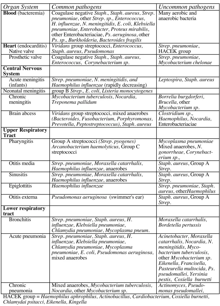
Table 7. Bacteria Associated with Human Disease
Organ System
Common pathogens
Uncommon pathogens
Blood (bacteremia) Coagulase negative Staph., Staph. aureus, Strep. pneumoniae, other Strep. sp., Enterococcus, H. influenzae, N. meningitidis, E. coli, Klebsiella pneumoniae, Enterobacter, Proteus mirabilis, other Enterobacteriaciae, Ps. aeruginosa, other
Ps. sp., Burkholderia, Bacteroides fragilis
Many aerobic and anaerobic bacteria
Heart (endocarditis)
Native valve ViridansStaph. aureus, Pseudomonas group streptococci, Enterococcus, Strep. pneumoniaeHACEK group , Prosthetic valve Coagulase negative Staph., Staph. aureus,
Enterococcus, Corynebacterium sp. Strep. pneumoniae,Mycobacterium chelonae
Central Nervous System
Acute meningitis
(infants) Strep. pneumoniae, N. meningitidis, andHaemophilus influenzae (rapidly decreasing) Leptospira, Staph. aureus Neonatal meningitis group B Strep., E. coli, Listeria monocytogenes
Chronic
meningitis Mycobacterium tuberculosis, Nocardia,Treponema pallidum Borrelia burgdorferi,Brucella, other
Mycobacterium sp.
Brain abcess Viridans group streptococci, mixed anaerobes
(Bacteroides, Fusobacterium, Porphyromonas, Prevotella, Peptostreptococcus), Staph. aureus
Clostridium sp.,
Haemophilus, Nocardia,
Enterobacteriaciae Upper Respiratory
Tract
Pharyngitis Group A streptococci (Strep. pyogenes) Arcanobacterium haemolyticus, Group C streptococci
Mycoplasma pneumoniae
Mixed anaerobes, N. gonorrheae, Corynebact-erium sp.,
Otitis media Strep. pneumoniae, Moraxella catarrhalis,
Haemophilus influenzae, anaerobes Staph. aureusStrep. , Group A
Sinusitis Strep. pneumoniae, Moraxella catarrhalis,
Haemophilus influenzae, anaerobes Staph. aureusStrep. , Group A
Epiglottitis Haemophilus influenzae Strep. pneumoniae, Staph.
aureus, otherHaemophilus
Otitis externa Pseudomonas aeruginosa (swimmer's ear) Staph. aureus, Group A
Strep.
Lower respiratory tract
Bronchitis Strep. pneumoniae, Staph. aureus, H. influenzae, Klebsiella pneumoniae,
Chlamydia pneumoniae, Mycoplasma pneum.
Moraxella catarrhalis, Bordetella pertussis
Acute pneumonia Strep. pneumoniae, Staph. aureus, H. influenzae, Klebsiella pneumoniae, Chlamydia pneumoniae, Mycoplasma
pneumoniae, E. coli, Pseudomonas aeruginosa,
mixed anaerobes Acinetobacter, Moraxella catarrhalis, Nocardia, N. meningitidis, Myco-bacterium tuberculosis, other Mycobacterium sp. Eikenella, Francisella, Pasteurella multocida, Ps. pseudomallei, Yersinia pestis, Coxiella burnetti
Chronic
pneumonia Mixed anaerobes, Nocardia, other Mycobacterium sp.Mycobacterium tuberculosis, Actinomyces, Pseudo-monas pseudomallei,
Organ System
Common pathogens
Uncommon pathogens
Intra-abdominalSpontaneous
peritonitis E. coli, Klebsiella pneumoniae, Strep.pneumoniae, Enterococcus Staph. aureus, gonorrheae, Chlamydiaanaerobes, N. trachomatis, M.
tuberculosis
Secondary
peritonitis E. coli, Bacteroides fragilis,anaerobes, Enterococcus, Ps. aeruginosa other enteric Staph. aureus, N.gonorrheae, M. tuberculosis
Dialysis-associated peritonitis Coagulase negative Streptococcus sp., Corynebacterium sp.Staph., Staph. aureus, E. coli, Klebsiella,Enterobacter, Proteus, Ps.
Intraabdominal
abcess Bacteroides fragilisEnterococcus sp. group, E. coli, Klebsiella, Enterobacter,Proteus, Ps., Staph. aureus
Urinary Tract
Cystitis (bladder) E. coli, Proteus mirabilis, Klebsiella, Enterobacter, Staph. saprophyticus, Pseudomonas, Enterococcus
Staph. aureus, Ureaplasma urealyticum,
Corynebacteri-um
ureolyticus, Clostridum sp., Bacteroides fragilis
Pyelonephritis E. coli, Proteus mirabilis, Klebsiella, Staph.
aureus Enterococcus,Corynebacterium ureolyticus
Prostatitis E. coli, Klebsiella, Enterobacter, Proteus
mirabilis, Enterococcus Neisseria gonorrheae
Genital
Urethritis Neisseria gonorrheae, Chlamydia trachomatis Ureaplasma urealyticum, Mycoplasma genitalum
Cervicitis Neisseria gonorrheae, Chlamydia trachomatis Actinomyces,
Mycobacterium tuberculosis
Bacterial vaginosis (vaginitis)
Synergistic infection with anaerobes
(Mobiluncus, Bacteroides sp., Peptostrepto-coccus) and possibly Gardnerella vaginalis
Genital ulcers Treponema pallidum, Haemophilus ducreyi, Chlamydia trachomatis (LGV)
Bone and Joint
Septic arthritis Staph. aureus, N. gonorrheae, Streptococcus sp., Haemophilus influenzae, Borellia
burgdorferi
Brucella, Nocardia, Mycobacterium sp. Osteomyelitis Staph. aureus, Enterobacteriaciae M. tuberculosis, other
mycobacterial sp, anaerobes
Prosthesis-associated infection Staph. aureus, Streptococcus sp.coagulase negative Staph., Peptostreptococcusmisc. aerobic gm - bacilli Skin & Soft tissue
Impetigo Staph. aureus, Group A Streptococcus,
Furuncles &
Carbuncles (boils) Staph. aureus
Erysipelas Group A Streptococcus
Cellulitis Group A Streptococcus, Staph. aureus, Haemophilus influenzae
Necrotizing cellulitis & fasciitis
Group A Strep., Clostridium perfingens, B. fragilis, Peptostreptococcus, other Gm – anaerobes, Enterobacteriaciae, Ps. aeruginosa
Eye
Conjuctivitis Strep. pneumoniae, Staph. aureus, coagulase neg. Staph., H. influenzae & aegyptius, N. gonorrheae, Chlamydia trachomatis
Table 7 adapted from Murray, Kobayashi, Pfaller, & Rosenthal, Medical Microbiology, 2nd ed.,
Mosby-Year Book, New York, 1994
IDENTIFICATION AND CLASSIFICATION OF BACTERIA
Bacteria are classified, identified, and described by their staining characteristics, morphology, spatial relationships, and biochemical testing characteristics e.g Staphyloccocus aureus can be described in the following way:
staining characteristics morphology
ÿ ÿ
Staph. aureus: Gram-positive, Coagulase-positive cocci in clusters ↑ ↑
biochemical test spatial relationship
A. Microscopic Identification 1. Staining characteristics
a. Gram's stain - 4 step procedure
i. apply crystal violet to stain cells blue.
ii. Iodine solution (mordant) forms crystal violet-iodine complex with cell wall (all cells blue).
iii. Wash with acetone or alcohol. Decolorizes gram-negative bacteria. Gram positive bacteria remain purple or blue.
iv. Counterstain with safranin, a red dye that stains gram negatives. b. Acid fast stain - useful for identification of Mycobacteria and Nocardia
i. These organisms aren't stained with safranin in Gram stain. ii. Counter stain with carbolfuchsin which binds to mycolic acid in the cell wall of these organisms.
2. Morphology
a. cocci (spheres) - e.g Staphyloccocus, Streptococcus, Neisseria
b. bacilli (rods) - e.g. Enterobacteriaciae (E. coli, Proteus), Pseudomonas
i. spiral shaped rods - Treponema, Borellia
ii. Curved-shape rods - Vibrio
iii. Filamentous rods - Lactobacillus, Propionobacterium
3. Spatial Relationships
a. Chains - Streptococcus pyogenes, Group B Strep., Enterococcus
b. Pairs - Streptocococcus pneumoniae (Gm +), Neisseria (Gm –) c. Clusters (groups) - Staphylococcus aureus & epidermidis
Diplococci Strep. pneumoniae Cocci in Clusters Staph. aureus Bacilli or rods E. coli Cocci in chains Strep. pyogenes
B. Biochemical Testing (and Morphology)
C. Sensitivity and Selectivity Testing 1. Definitions
MIC = Minimal Inhibitory Concentration. The lowest concentration of antimicrobial agent that will visually inhibit bacterial growth following an overnight incubation (18h) at 35°C of an initial inoculum of 104-105 colony forming units (CFU) per ml. Usually done in microtiter plates.
MBC = Minimal Bactericidal Concentration. The lowest concentration that kills 99.9% of the bacteria in culture. Requires reculture of MIC incubations that show no growth.
SIT = Serum Inhibitory Titer - Serum from a patient receiving animicrobial therapy is serially diluted & a standard inoculum of bacteria is added to each dilution. The SIT is the greatest dilution that prevents bacterial growth e.g. 1:8 or 1:16 is considered adequate. SBT = Serum Bactericidal Titer - The SBT is the greatest dilution of serum that will kill 99.9% of the bacteria in the inoculum after re-plating.
Bactericidal = MBC/MIC is ≤4. Indicates that the antibiotic is capable of killing at a concentration near the MIC. Bactericidal antibiotics are aminoglycosides, quinolones, vancomycin, and ß-lactams.
Bacteriostatic = MBC/MIC is between 4 and 32. These antibiotics inhibit growth but require good immune function to eliminate the infecting organism. Examples are chloramphenicol, macrolides, and tetracyclines.
Tolerant = MBC/MIC >32. The bacteria is considered to tolerant to the effects of the antibiotic. Enterococcus sp. are often tolerant to aminoglycosides.
2. Sensitivity Testing - Testing of the sensitivity of a patient isolate (a pure culture of the organism isolated from a patient that is responsible for the infection) to a variety of antimicrobial agents.
a. Kirby-Bauer Disk Diffusion Method
1 4 5 3 6 7 1 = Resistant 2 = Sensitive 3 = Resistant 4 = Resistant 5 = Sensitive 6 = Resistant 7 = Intermediate 2
Paper disks impregnated with various antibiotics are placed on Mueller-Hinton agar plates. The agar is commonly seeded with a set number of CFUs or an even lawn of bacteria is spread on the surface of the plate. The organism is allowed to grow (18 hrs) until the surface of the plate contains contiguous colonies. Antibiotics that inhibit bacterial growth with have a clear zone of inhibition surrounding the plate. Bacteria are usually placed into three groups (Resistant, Intermediate, or Sensitive) depending upon the diameter of the clear zone. When well standardized, the zone diameter can be related to the MIC.
b.) E-Test
A plastic strip impregnated with antibiotic is placed into agar seeded with an even lawn of bacteria. Diffusion into the media provides a continuous concentration gradient that yields a quantitative measurement of the MIC value.