Nijmegen
The following full text is a publisher's version.
For additional information about this publication click this link.
http://hdl.handle.net/2066/138677
Please be advised that this information was generated on 2017-12-05 and may be subject to
change.
DNA Sequences Proximal to Human Mitochondrial DNA
Deletion Breakpoints Prevalent in Human Disease Form
G-quadruplexes, a Class of DNA Structures Inefficiently
Unwound by the Mitochondrial Replicative Twinkle Helicase
*
Received for publication, March 19, 2014, and in revised form, August 18, 2014Published, JBC Papers in Press, September 5, 2014, DOI 10.1074/jbc.M114.567073 Sanjay Kumar Bharti‡, Joshua A. Sommers‡, Jun Zhou§¶1, Daniel L. Kaplan储, Johannes N. Spelbrink**‡‡2, Jean-Louis Mergny§¶3, and Robert M. Brosh, Jr.‡4
From the‡Laboratory of Molecular Gerontology, NIA, National Institutes of Health, NIH Biomedical Research Center, Baltimore, Maryland 21224, the§ARNA Laboratory, University of Bordeaux, F-33000 Bordeaux, France,¶INSERM U869, Institut Europe´en de Chimie et Biologie (IECB), F-33600 Pessac, France, the储Department of Biomedical Sciences, Florida State University College of Medicine, Tallahassee, Florida 32312, the**FinMIT Centre of Excellence, BioMediTech and Tampere University Hospital, Pirkanmaa Hospital District, University of Tampere, FI-33014 Tampere, Finland, and the‡‡Department of Pediatrics, Nijmegan Centre for Mitochondrial Disorders, Radboud University Medical Centre, Geert Grooteplein 10, P. O. Box 9101, 6500 HB Nijmegen, The Netherlands
Background:Mitochondrial DNA deletions are prominent in human genetic disorders and cancer.
Results:Predicted mitochondrial G-quadruplex-forming sequences map in close proximity to known deletion breakpoints and form G-quadruplexesin vitro.
Conclusion:The mitochondrial replicative helicase Twinkle inefficiently unwinds intra- and intermolecular G-quadruplexes. Significance:Mitochondrial G-quadruplexes are likely to cause genome instability by perturbing replication machinery.
Mitochondrial DNA deletions are prominent in human genetic disorders, cancer, and aging. It is thought that stalling of the mitochondrial replication machinery during DNA synthesis is a prominent source of mitochondrial genome instability; how-ever, the precise molecular determinants of defective mitochon-drial replication are not well understood. In this work, we per-formed a computational analysis of the human mitochondrial genome using the “Pattern Finder” G-quadruplex (G4) predic-tor algorithm to assess whether G4-forming sequences reside in close proximity (within 20 base pairs) to known mitochondrial DNA deletion breakpoints. We then used this information to map G4P sequences with deletions characteristic of representa-tive mitochondrial genetic disorders and also those identified in various cancers and aging. Circular dichroism and UV spectral analysis demonstrated that mitochondrial G-rich sequences near deletion breakpoints prevalent in human disease form G-quadruplex DNA structures. A biochemical analysis of puri-fied recombinant human Twinkle protein (gene product of c10orf2) showed that the mitochondrial replicative helicase inefficiently unwinds well characterized intermolecular and intramolecular G-quadruplex DNA substrates, as well as a
uni-molecular G4 substrate derived from a mitochondrial sequence that nests a deletion breakpoint described in human renal cell carcinoma. Although G4 has been implicated in the initiation of mitochondrial DNA replication, our current findings suggest that mitochondrial G-quadruplexes are also likely to be a source of instability for the mitochondrial genome by perturbing the normal progression of the mitochondrial replication machin-ery, including DNA unwinding by Twinkle helicase.
The human mitochondrial genome consists of a 16,569-bp circular DNA molecule that encodes essential subunits of mito-chondrial complexes I, II, III, IV, and V in oxidative phosphor-ylation, a process responsible for the generation of the majority of the cellular ATP pool (1). Therefore, energy production is intimately linked to an intact and correctly coding mitochon-drial DNA (mtDNA) sequence. Because of the utmost impor-tance of the mitochondrion, it naturally follows that the accu-mulation of mitochondrial DNA mutations leading to mitochondrial dysfunction are implicated in human genetic disorders (2), neurodegeneration (3), cancer (4), and aging (5). Understanding the molecular basis for mitochondrial DNA mutations has attracted considerable interest in the clinical and basic science fields.
mtDNA deletions are prominent in genetic disorders such as Pearson marrow-pancreas syndrome (PMPS),5 Kearns-Sayre syndrome (KSS), and progressive external ophthalmoplegia
*This research was supported, in whole or in part, by the National Institutes
of Health, NIA, Intramural Research Program.
1Recipient of a European Union Marie Curie International Incoming
Fellow-ship (IIF).
2Supported by the Academy of Finland (Centre of Excellence funding) and
the Netherlands Organization for Scientific Research (NWO), VICI Grant 865.10.004.
3Supported by Fondation ARC, the Aquitaine Regional Council, and the
Agence Nationale de la Recherche (ANR Grants Quarpdiem and Oligoswitch).
4To whom correspondence should be addressed: Laboratory of Molecular
Gerontology, National Institutes of Health, NIA, NIH Biomedical Research Center, 251 Bayview Blvd., Baltimore, MD 21224. Tel.: 410-558-8578; Fax: 410-558-8162; E-mail: [email protected].
5The abbreviations used are: PMPS, Pearson marrow-pancreas syndrome;
CSBII, conserved sequence block II; FANCJ, Fanconi anemia complementa-tion group J; G4, G-quadruplex; G4P, potential G4-forming (sequence); H, heavy; L, light; HRCC, human renal cell carcinoma; KSS, Kearns-Sayre syn-drome; PEO, progressive external ophthalmoplegia; PNA, peptide-nucleic acid;
ssDNA, single-stranded DNA; ATP␥S, adenosine 5⬘-O-(thiotriphosphate).
at RADBOUD UNIVERSITEIT NIJMEGEN on March 6, 2017
http://www.jbc.org/
(PEO) as well as various cancers and age-related diseases (6). mtDNA deletions are often located in regions of the genome flanked by tandem repeat sequences. It is proposed that at least one mechanism likely to underlie the mitochondrial genomic instability that leads to deletions is the stalling of the mitochon-drial DNA synthesis machinery during replication. Mitochon-drial replication stalling can lead to replication slippage, aber-rant DNA structures, and double strand breaks, which can in turn promote mtDNA deletions. Although this model is attrac-tive, the molecular determinants of mitochondrial replication stalling are not well understood.
The mitochondrial genome consists of a heavy (H) strand, which is rich in guanine bases, and a complementary light (L) strand, which is rich in cytosine bases. Because of its chemical nature, the guanine-rich (G-rich) strand is prone to form G-quadruplex (G4) DNA, an alternative DNA structure char-acterized by planar stacks of four guanines interacting by non-conventional Hoogsteen hydrogen bonds. G4 DNA is stabilized by a monovalent cation (typically Na⫹or K⫹) that resides in the central barrel of the G-tetrad. There has been growing interest in nuclear G-quadruplex DNA metabolism in recent years with increasing evidence that G4 existsin vivo(7, 8) (for review see Refs. 9 –11). However, it has been only very recently that researchers have begun to focus on the mitochondrial genome and its propensity to form G-quadruplexes or other alternate DNA structures. The Zakian laboratory (12) performed the first analysis of G4 DNA motifs in yeast mitochondria, which indi-cates a 10-fold greater density of G4 motifs in mitochondrial
versusnuclear genomes ofSaccharomyces cerevisiae. Damaset al.(13) conducted a computational analysis of human mtDNA deletions and correlated these with non-B DNA conformations including hairpin, cruciform, and cloverleaf-like elements. Their findings suggest that non-B form conformations are likely to contribute to the formation of mitochondrial deletions. In 2013, Oliveiraet al.(14) described their findings from meta-analysis of human mtDNA sequence leading them to propose that noncanonical DNA structures, including helix-distorting intrinsically curved regions and long G-quadruplex elements, are likely to drive mtDNA rearrangements.
In the current study, we employed “Pattern Finder,” a G4 predictor algorithm, to map potential G4-forming (G4P) sequences throughout the human mitochondrial genome and chart them to known mitochondrial deletion breakpoints. We utilized this information to correlate G4P sequences with dele-tions characteristic of representative mitochondrial genetic disorders and also those identified in various cancers and aging. We performed physical analysis of representative predicted G4-forming sequences residing adjacent to clinically relevant mitochondrial deletions to substantiate that they do indeed form G-quadruplexes. Finally, we tested G-quadruplex DNA substrates with various topologies including a unimolecular G4 derived from human mitochondrial sequence for unwinding by the human mitochondrial DNA helicase Twinkle. Our results provide insight into the hypothesis that mitochondrial G-qua-druplexes are likely to be a source of instability for the mito-chondrial genome by perturbing the normal progression of the mitochondrial replication machinery.
EXPERIMENTAL PROCEDURES
Mitochondrial G-quadruplex Motif Prediction and Correlation with Mitochondrial Deletions—Mitochondrial G4P sequences were searched using Pattern Finder software to predict G4 in a user-defined quadruplex pattern for a given input sequence (15). Human mtDNA sequences used for analysis were taken from accession number NC_012920 (formerly, AC_000021). Mitochondrial L-strand (C-rich) and H-strand (G-rich) DNA was subjected to Pattern Finder analysis to assign G4 motifs with a stem size of 2–5 nucleotides (nt) and loop size of 1–7 nt. The mitochondrial deletion breakpoints are reported based on L-strand numbering. The mitochondrial deletion data were obtained from MitoMap and MitoBreak.
Circular Dichroism Spectroscopy, UV Melting Curves, and Thermal Difference Spectra—Oligonucleotides used to prepare G4 DNA substrates for biophysical analysis were purchased from Eurogentec (Seraing, Belgium) without further purifica-tion. The oligonucleotides derived from human mitochondrial sequence were: KSS, 5⬘ -GGGGAGGGGTGTTTAAGGGGTT-GGCTAGGG-3⬘; human renal cell carcinoma (HRCC), 5⬘ -GGG-GGTTGGGTATGGGGAGGGGGG-3⬘; and PMPS, 5⬘ -GGGAC-GCGGGCGGGGGATATAGGG-3⬘. Strand concentrations were determined by UV absorbance using the extinction coefficients provided by the manufacturer. All of the chemicals used were pur-chased from Sigma unless otherwise stated. The samples were heated to 95 °C for 5 min, cooled slowly to room temperature, and then stored at 4 °C before use unless otherwise stated (the concen-tration of each strand was 5M, and the buffer was prepared with
10 mMcacodylic acid buffered to pH 7.0 with LiOH containing 100
mMK⫹or Na⫹).
Circular dichroism (CD) spectra were recorded on a Jasco J-815 equipped with a Peltier temperature control accessory (Jasco, Hachioji, Japan). Each CD spectrum was obtained by averaging five scans at a speed of 100 nm/min, and the collec-tion temperature was set at 20 °C. UV absorbance measure-ments were performed on an Uvikon XS (Secomam) equipped with a circulating water bath. Temperature was measured with an inert glass sensor immersed into a quartz cell. UV absor-bance was monitored at 295 nm with a temperature gradient of 0.5 °C/min. For each sample, the thermal difference spectrum was obtained by subtracting the absorbance spectrum at lower temperature from the one at higher temperature as described previously (16).
Recombinant Helicase Proteins—Recombinant human Twin-kle protein (gene product ofc10orf2) with a C-terminal hexa-histidine tag was expressed in bacterial cells and purified as described previously (17). Recombinant human Fanconi ane-mia complementation group J (FANCJ) protein with a C-termi-nal FLAG tag was expressed in insect cells and purified as described previously (18). DnaB was purified fromEscherichia colias described previously (19).
DNA Helicase Assays—Gel-purified oligonucleotides used to prepare G4 DNA substrates for helicase assays were purchased from Lofstrand Labs Ltd. (Gaithersburg, MD) (Table 1). The four-stranded (tetramolecular) parallel TP-G4 substrate (20), two-stranded (bimolecular) anti-parallel OX-1-G2⬘ DNA substrate (20), four-stranded TelR2-G4 (20), four-stranded
at RADBOUD UNIVERSITEIT NIJMEGEN on March 6, 2017
http://www.jbc.org/
TABLE 1
DNA substrates used in this study
at RADBOUD UNIVERSITEIT NIJMEGEN on March 6, 2017
http://www.jbc.org/
CEB1-G4 (21, 22), and unimolecular poly(A) Zic1-G4 substrate with 5⬘single-stranded DNA (ssDNA) tail lengths of 20, 30, or 40 nt were prepared from gel-purified oligonucleotides as described previously (23). The 19-bp forked duplex DNA sub-strate was prepared by annealing DC26 and TSTEM25 as described earlier (24).
Standard helicase reaction mixtures (20 l) containing 5 fmol of the indicated TP-G4 or OX-1-G2⬘DNA substrate or the forked duplex DNA substrate (0.25 nMDNA substrate
concen-tration) and the indicated concentration of FANCJ (20) or DnaB hexamer (25) were as described previously unless stated otherwise. For the Twinkle hexamer, the reaction conditions for the forked duplex were as described previously (26) except for the inclusion of 25 mMKCl and the additional presence
throughout the incubation period of a 100-fold excess of oligo-nucleotide of the same sequence as the labeled strand in the partial duplex substrate to serve as a displaced strand trap (17). Forked duplex helicase reactions were terminated as described previously and resolved on nondenaturing 12% polyacrylamide (19:1 acrylamide-bisacrylamide) gels (24). Twinkle reaction conditions for the intermolecular G4 substrates (TP-G4, OX-1-G2⬘, TelR2-G4, and CEB1-G4) were the same as for the forked duplex except there was no additional oligonucleotide present during the reaction. For the unimolecular poly(A) Zic1-G4 or HRCC-G4 DNA substrate (5 fmol), helicase reac-tions (20l) were performed in a reaction buffer containing a
20-fold excess of peptide-nucleic acid complementary oligonu-cleotide (23). G4 helicase reactions were terminated by the addition of 20 l of stop buffer (74% glycerol, 0.01% xylene cyanol, 0.01% bromphenol blue, 10 mm KCl, and 20 mm EDTA). Reaction products for intermolecular and unimolecu-lar G4 DNA substrates were resolved on 10 and 15% nondena-turing polyacrylamide (19:1 acrylamide-bisacrylamide) gels, respectively. Gels were scanned by PhosphorImager and quan-titated as described previously (24).
RESULTS
Bioinformatic Analysis of Potential G-quadruplex-forming Sequences in the Human Mitochondrial Genome and Their Locations with Respect to Deletion Breakpoints—The human mitochondrial genome contains G-rich sequences predicted to form G-quadruplex DNA (Fig. 1); G4P sequences can be found in both the L-strand (top) and H-strand (bottom). However, a greater number of G4P sequences are present in the H-strand, which is also the G-rich strand. G4P sequences reside within the coding sequences of mitochondrial genes and also the tRNA genes. G4P sequences can also be found to a lesser extent in noncoding regions of the mitochondrial genome.
We compared the G4P sequences in human mitochondrial DNA with other mammalian species to determine the degree of conservation. The number of G4 motifs in the mitochondrial genomes of mouse, rat, and monkey was markedly less than
FIGURE 1.Graphical representation of predicted G4 motifs and deletion breakpoints in the human mitochondrial genome coding and noncoding
sequences.Mitochondrial genes, tRNA, and G-rich sequences predicted to form G4 are shown in the L-strand (top) and the H-strand (bottom). The 5⬘and 3⬘
deletion breakpoints, collected from MitoMap and MitoBreak, residing within or adjacent to predicted G4-forming sequences, are shown. The number of guanines (2–5 G) and loop size (1–7 nt) were employed for search criteria using QuadBase Pattern Finder. The predicted G4-forming sequences with
param-etersⱖG3 and loop size 1–7 nt are also shown bybold dots.RNR1, 12 S ribosomal RNA;RNR2, 16 S ribosomal RNA;ND1, NADH dehydrogenase 1;ND2, NADH
dehydrogenase 2;COX1, cytochromecoxidase I;COXII, cytochromecoxidase II;ATP8, ATP synthase 8;ATP6, ATP synthase 6;COXIII, cytochromecoxidase III;
ND3, NADH dehydrogenase 3;ND4L, NADH 4L dehydrogenase;ND4, NADH dehydrogenase 4;ND5, NADH dehydrogenase 5;ND6, NADH dehydrogenase 6;
CYTB, cytochromeb.
at RADBOUD UNIVERSITEIT NIJMEGEN on March 6, 2017
http://www.jbc.org/
found in human (Table 2). The number of conserved G4P sequences identified by the Pattern Finder parameters between these species and humans was greatest in monkey followed by rat and then mouse.
The human mitochondrial genome deletions (collected from MitoMap and MitoBreak) that reside adjacent to the G4P sequences derived from our analysis are shown in Fig. 1. We identified both 5⬘and 3⬘deletion breakpoints that reside in the G4P sequence or adjacent (within 20 bp) to the G4P sequence. The mitochondrial genome deletion breakpoints were mapped proximal to G4P sequences located within the coding regions of
mitochondrial genes and tRNA genes, as well as noncoding regions to a lesser extent.
To ascertain the relative abundance of deletion breakpoints in predicted mitochondrial G4-forming sequences, we com-pared the distribution and density of unique mitochondrial deletion breakpoints in G4P sequences, tRNA genes, and the D-loop region (Table 3). The total number of mitochondrial 5⬘ or 3⬘deletion breakpoints was greater in G4P as compared with tRNA or the D-loop region. We next determined the deletion breakpoint density (deletions/kb) for each region of mitochon-drial DNA. The 5⬘mitochondrial deletion breakpoint density was⬃1.4- and 7.6-fold greater in G4P as compared with tRNA genes and the D-loop, respectively. The 3⬘deletion breakpoint density of mitochondrial G4P was 1.4- greater and 0.8-fold less than that of tRNA genes and D-loop, respectively (Table 3).
Association of Potential G-quadruplex-forming Sequences with Mitochondrial DNA Deletion Breakpoints Prevalent in Human Disease—Our computational analysis revealed that G4P sequences reside proximal to mtDNA deletions found in a number of genetic diseases (Table 4). Among the genetic disor-ders associated with mitochondrial deletions near G4P sequences is progressive external ophthalmoplegia. PEO is characterized by the adult onset of muscular deficiencies, including those of the external eye and skeletal system (27). G4P sequences were also located adjacent to mitochondrial deletion TABLE 2
Evolutionary conservation of G4 motifs in mitochondrial genomes of human and selected mammals
Organisma Mitochondrial genome Sequence similarity Total G4 motifsb Conserved G4 motifsc bp % Human 16,569 100 270 270 Monkey 16,564 78 148 81 Rat 16,313 67 108 46 Mouse 16,299 66 59 34 a
Accession numbers: human (NC_012920 AC_000021); monkey (NC_005943); rat (NC_001665); mouse (NC_005089).
b
Total G4P motifs obtained based on stem size of 2–5 nt and loop size of 1–7 nt through Pattern Finder analysis.
c
Number of predicted G4 motifs that are common in the indicated mammal and human.
TABLE 3
Distribution and density of 5ⴕand 3ⴕdeletion breakpoints
The number of unique deletion breakpoints⫽620 5⬘deletion breakpoints and 497 3⬘deletion breakpoints (13).
Deletion breakpoint
G4Pa tRNAb D-loop (AT-rich)c
Deletion breakpoints Deletion breakpoints Deletion breakpoints Total no.d %e Density (deletions/kb)f Total no.d %e Density (deletions/kb)f Total no.d %e Density (deletions/kb)f
5⬘deletion breakpoints 194 31 38 57 9 28 6 1 5 3⬘deletion breakpoints 125 25 25 36 7 18 37 7 31 aNumber of deletion breakpoints in G4P excluding those overlapping in tRNA and D-loop region.
bNumber of deletion breakpoints in tRNA excluding those overlapping in G4P or D-loop region. cNumber of deletion breakpoints in D-loop excluding those overlapping in G4P or tRNA. dTotal number of unique deletion breakpoints in the case of G4P, tRNA, and D-loop.
eNumber of deletion breakpoints in percentage (%) with respect to either 620 5⬘deletions or 497 3⬘deletions for G4P, tRNA, and D-loop. fDeletion breakpoint density is based on 5071, 2017, and 1180 bp, respectively, for G4P, tRNA, and D-loop.
TABLE 4
Selected disease-associated mitochondrial deletion breakpoints in proximity to predicted G4-forming sequences
Deletion
breakpointa G4P neighboring sequenceb Genetic disease References
5⬘10190 (10187) tata2tcccccgcccgcgtcccttt (10210) Pearson marrow-pancreas syndrome
(29, 84) 3⬘13753 (13751) tt2tcccccgcatcccccttccaaacaacaatccccctcta (13790)
5⬘12113 (12081) tatcccccattctcctcctatccctcaaccccg2ac (12115)
5⬘12103 (12078) cctatcccccattctcctcctatcc2ctc (12106) Kearns-Sayre syndrome, mitochondrial encephalomyopathy
(85–87) 3⬘14414 (14412) cc2tcaacccctgacccccatgcc (14435)
5⬘4398 (4374) ccgtgccacctatcacaccccatcc2ta (4400) Diabetes mellitus (88, 89) 3⬘14822 (14806) cctccccaccccatcc2aacatctcc (14830)
5⬘7974 (7942) ctcctacatacttcccccattattcctagaacc2agg (7977) Kearns-Sayre syndrome, mitochondrial encephalomyopathy, lactic acidosis, stroke-like episodes syndrome
(90, 91) 3⬘15496 (15495) t2aggcgacccagacaattataccctagccaaccccttaaacacccctccccacatc (15550)
5⬘6325 (6325) c2ctccgtagacctaaccatcttctcct (6351) Mitochondrial myopathy (92) 3⬘13989 (13971) ccaaaacctgcccctact2cctcctagacctaacc (14004)
5⬘6329 (6311) ctcccaccctggagcctc2cgtagacc (6336) Chronic progressive external
ophthalmoplegia (93) 3⬘13994 (13970) gccaaaacctgcccctactcctcc2tagacctaacc (14004)
5⬘6330 (6311) ctcccaccctggagcctccg2tagacctaacc (6341) Kearns-Sayre syndrome, Pearson marrow-pancreas syndrome
(87) 3⬘13994 (13970) gccaaaacctgcccctactcctcc2tagacctaacc (14004)
5⬘7491 (7365) accccccaaagctggtttcaagccaac2cccatggcctcc (7503) Kearns-Sayre syndrome, progressive external ophthalmoplegia (85) 3⬘11004 (10995) caagccaac2gccacttatccagtgaacc (11022)
a
Deletion breakpoints reside within the G4P sequence orⱕ20 nt away from G4P. The breakpoint is indicated by a down arrow.
b
L-strand sequence is shown (5⬘to 3⬘). The predicted G4-forming sequence (G4P) is underlined.
at RADBOUD UNIVERSITEIT NIJMEGEN on March 6, 2017
http://www.jbc.org/
breakpoints associated with KSS, a clinical subgroup of mito-chondrial encephalomyopathies associated with PEO and skel-etal muscle defects (28).
Our analysis revealed that a G4P sequence is located in a breakpoint region of a mitochondrial DNA deletion associated with PMPS, a fatal disease of early infancy affecting the hema-topoietic system and pancreas exocrine system (29) (Table 4). The molecular features of PMPS are attributed to a large-scale deficiency in oxidative phosphorylation. The site-specific mito-chondrial deletions associated with PMPS and other mitochon-drial disorders (KSS and PEO) are characterized by direct repeats of 8 –13 bp present in the normal mitochondrial genome, which flank the deletion breakpoints (29 –31). It has been proposed that homologous recombination may be responsible for the direct repeat signature pattern characteris-tic of PMPS and other sporadic mtDNA deletion disorders (32). The common 4977-bp deletion flanked by a 13-bp repeat in the normal mtDNA sequence frequently identified in sporadic PEO and other mitochondrial disorders (33) is characterized by a 5⬘breakpoint at mtDNA position 8468 bp, which resides in a predicted G4-forming sequence according to the Pattern Finder algorithm. Unusual secondary DNA structures such as G-quadruplexes may be prone to form in the mitochondrial genome at loci such as the 5⬘ breakpoint of the common
4977-bp deletion and interfere with normal mitochondrial DNA metabolic processes such as replication or repair, leading to double strand breaks.
Association of Potential G-quadruplex-forming Sequences with Mitochondrial DNA Deletion Breakpoints Found in Cancer—We identified a number of cases in which G4P sequences flanked mtDNA deletion breakpoints reportedly associated with cancer (Table 5). G4P sequences flanking mtDNA deletions in thyroid tumors were prominent. G4P-as-sociated mitochondrial genome instability may be relevant to previous observations that a subset of thyroid tumors display elevated mtDNA deletions (34). A relatively uncommon and small 264-bp deletion in the mitochondrial coding sequence for the first subunit (ND1) of NADH:ubiquinone oxidoreductase of the electron transport chain was reported in a HRCC (35). This deletion is flanked by G4P sequences according to our computational analysis (Table 5). Biophysical studies of the G4P flanking sequence confirmed its ability to form G4 (see Fig. 2Aand “Biophysical Analysis of Selected G4P Mitochondrial Sequences” below). A 6-bp direct repeat flanked the deleted region (35), suggesting that replication slippage due to mtDNA synthesis stalling at G4 DNA may play a role.
Using the G4P algorithm, we determined that a G4P sequence flanked a 7079-bp mtDNA deletion site reported to
FIGURE 2.Circular dichroism and UV spectral analyses of G-rich human mitochondrial DNA sequences in the proximity of deletion breakpoints
prevalent in human renal cell carcinoma, Pearson marrow-pancreas syndrome, and Kearns-Sayre syndrome.Oligonucleotide sequences used for
biophysical analysis are listed under “Experimental Procedures.”A, CD spectra for 5Msamples as indicated, in 10 mMlithium cacodylate buffer (pH 7.0)
containing 100 mMNa⫹or K⫹. The spectra were collected at 20 °C.B, thermal difference spectra (TDS) for 5Msamples as indicated, in 10 mMcacodylate buffer
(pH 7.0) containing 100 mMNa⫹or K⫹.
TABLE 5
Selected cancer-associated mitochondrial deletion breakpoints in proximity to predicted G4-forming sequences Deletion
breakpointa G4P neighboring sequenceb Cancer References
5⬘3323 (3310) cccatggccaacct2cctactcc (3331) Human renal cell carcinoma (35)
3⬘3588 (3566) cccccctccccatacccaaccc2cctggtcaaccct (3600)
5⬘8290 (8252) cccgtatttaccctatagcaccccctctaccccctctag2agcccactgt (8300) Thyroid tumor (51) 3⬘16275 (16250) ccaaagccacccctcacccactagg2ataccaacaaacctacccaccc (16296)
5⬘8281 (8262) ccctatagc accccctctac2cccctctag agccc (8295)
3⬘16371 (16351) atcccttctc gtccccatgg2atgacccccc (16380)
3⬘13383 (13364) ccgggtccatcatccacaa2ccttaaca (13390)
3⬘16280 (16250) ccaaagccacccctcacccactaggatacc2aacaaacctacccaccc (16296)
5⬘8261 (8252) cccgtattta2ccctatagcaccccctctaccccctctagagccc (8295) Colon adrenocarcinoma (94) 5⬘15591 (15568) cctattcgcctacacaattctccg2atccgtccc (15660) Lung cancer (52)
5⬘8992 (8983) ccaatagccc2tggccgtacgcctaaccg (9010) Hepatic tumor (36)
5⬘306 (295) ccaccaaacccc2ccctcccccgcttctggcc (325) Hepatocellular carcoma (95)
3⬘16280 (16250) ccaaagccacccctcacccactaggatacc2aacaaacctacccaccc (16296) Thyroid tumor (96) aDeletion breakpoints reside within G4P sequence orⱕ20 nt away from G4P. The breakpoint is indicated by a down arrow.
bL-strand sequence is shown (5⬘to 3⬘). The predicted G4-forming sequence (G4P) is underlined.
at RADBOUD UNIVERSITEIT NIJMEGEN on March 6, 2017
http://www.jbc.org/
exist in cirrhotic liver that surrounds hepatic tumors (36) (Table 5). This mtDNA deletion was not characterized by direct repeat flanking sequences, suggesting that a mechanism other than replication slippage may be involved. Interestingly, the 7079-bp mtDNA deletion was detected only in the cirrhotic liver surrounding the hepatic tumor but not in the tumor itself, suggesting that mitochondrial dysfunction contributes to liver cirrhosis, which in turn serves as a risk factor for liver cancer. Next generation sequencing of mtDNA from tumor-derived tissues should provide a more comprehensive analysis of the mtDNA mutation spectra, which may be informative for understanding the molecular mechanism underlying the origin of the mtDNA deletions associated with cancer and the poten-tial role of G4P sequences.
Association of Potential G-quadruplex-forming Sequences with Mitochondrial DNA Deletion Breakpoints Observed in Aging—Using the computational analysis to map G4P sequences flanking mitochondrial deletion breakpoints, we sought to determine whether G4P sequences might play a role in the mitochondrial genome instability prevalent in aged tissues of the human body based on a survey of the literature. A compar-ison of the mitochondrial G4P sequences with reported mtDNA deletions associated with aging suggest this to be the case (Table 6). We will briefly discuss several examples to illus-trate the potential importance of G4-forming sequences in mitochondrial DNA deletions found in aging tissues. An anal-ysis of photo-aged tumor-free skin of nonmelanoma skin can-cer patients reveals an increase in mitochondrial DNA dele-tions that correlates with patient age (37). Most of the identified mtDNA deletions contained repeat sequences, suggesting a replication slippage model. This would be consistent with the proposal that mtDNA photoadducts interfere with normal mtDNA replication; however, G4 DNA may also contribute to replication stalling and mitochondrial genomic instability. Understanding the potential role of G-quadruplexes in replica-tion slippage or other aspects of replicareplica-tion mismanagement is an important area of study that deserves greater attention. Aberrant DNA structures such as G4 DNA, in addition to bulky
adducts imposed by chronic UV exposure, may contribute to the accumulation of mtDNA deletions in skin during chrono-logical aging.
A decline in mitochondrial function is prevalent in age-re-lated disorders characterized by neurodegeneration (6). Kray-tsberget al.(38) found that aged human substantia nigra con-tain a great abundance of mitochondrial DNA deletions, some of which we determined to be in close proximity to G4P sequences (Table 6). It was observed that mtDNA deletions were enriched in cytochromecoxidase-deficient neurons, sug-gesting that the mitochondrial genome instability was respon-sible for impaired cellular respiration and functional impair-ment of the aged neurons of the substantia nigra (38). Further work in this area to explore the potential role of G-quadru-plexes in brain mtDNA deletions may be informative for age-related disorders such as Parkinson disease, which is character-ized by primary neurodegeneration in the substantia nigra (39).
Biophysical Analysis of Selected G4P Mitochondrial Sequences—
To determine whether representative mitochondrial G4P sequences residing adjacent to known deletion breakpoints form G-quadruplex structures, the classical spectroscopic methods of CD and UV light absorption were used. The oligo-nucleotides selected for physical analysis corresponded to G4P sequences flanking mitochondrial DNA deletion breakpoints detected in PMPS, KSS, and HRCC. CD spectral analysis dem-onstrated that HRCC and PMPS showed a positive peak around 265 nm and negative peak at 240 nm in the presence of 100 mM
K⫹(Fig. 2A), suggesting that parallel G-quadruplex structures were formed (40). However, PMPS and KSS exhibited positive and negative peaks around 295 and 260 nm, respectively, in the presence of 100 mMNa⫹, which is in agreement with the for-mation of nonparallel G-quadruplexes (40). Interestingly, both HRCC in Na⫹ and KSS in K⫹ displayed two positive peaks around 295 and 260 nm. These spectra are similar to those reported previously for the G-rich hTERT promoter, implying the coexistence of two different G-quadruplex conformations (41). Furthermore, the thermal difference spectra of all three sequences showed a negative peak around 295 nm and two TABLE 6
Selected age-associated mitochondrial deletion breakpoints in proximity to predicted G4-forming sequences
Deletion
breakpointa G4P neighboring sequenceb Aging features References
5⬘540 (535) ccccata2ccccgaaccaaccaaaccccaaagacacccccc (573) Heart tissue (97) 3⬘13764 (13751) tttcccccgc atc2ccccttcc ( 13771)
5⬘5433 (5421) gaacatacaaaa2cccaccccattcctcccc (5450)
3⬘13023 (13010) cccaattaggtct2ccacccctgactcccctcagcc (13044)
5⬘1491 (1482) cacaccgcccgtc2accctcctc (1500) Skin tissue (37) 3⬘5206 (5191) cacccttaattccat2ccaccctcctctccc (5220)
5⬘6329 (6311) ctcccaccctggagcctcc2gtagacctaac catcttctcc (6350) Skeletal muscle (98) 3⬘13994 (13971) ccaaaacctgcccctactcctcc2tagacctaacc (14004)
5⬘6437 (6420) ccccctgccataacccaa2taccaaacgcccc (6450) Substantia nigra neurons (99) 3⬘14077 (14043) ccaaatct ccacctccat catcacctcaacccaa2aaag (14080)
5⬘6501 (6463) ccgtcctaatcacagcagcctatctctcc2cagtcctagc (6511) Substantia nigra neurons (38) 3⬘13802 (13781) tccccctctacctaaaactca2cagccctcg (13810)
3⬘16284 (16250) ccaaagccacccctcacccactaggataccaaca2aacctacccaccc (16296) Cochlear tissue (100) 5⬘7409 (7396) gccccccaccctac2cacacattcga (7420) Substantia nigra neurons of aged
individual with Parkinson’s disease
(101)
3⬘5447 (5333) cccaccccattcct2ccccacactcatcgcccttaccac gctactccta cctatctcccc (5491) Skeletal muscle (102)
a
Deletion breakpoints reside within G4P sequence orⱕ20 nt away from G4P. The breakpoint is indicated by a down arrow.
b
L-strand sequence is shown (5⬘to 3⬘). The predicted G4-forming sequence (G4P) is underlined.
at RADBOUD UNIVERSITEIT NIJMEGEN on March 6, 2017
http://www.jbc.org/
positive peaks around 275 and 243 nm (Fig. 2B), which is a typical feature of G-quadruplex DNA (42). In addition, UV melting profiles of all these sequences exhibited a hypochromic transition at 295 nm, which is in agreement with the formation of G-quadruplex structures (42) (Fig. 3). Interestingly, Tm depends on the nature of the cation (compareleft-and right-hand panelsin Fig. 3), which is strong evidence for G-quadru-plex formation (42).
Assessment of Twinkle Helicase to Unwind G-quadruplex DNA Substrates—Based on our computational analysis and biophysical studies, it is highly probable that human mitochon-drial DNA forms G4 structures under physiological conditions. This led us to consider how the mitochondrial replication machinery might tolerate G-quadruplexes. The gene product of
c10orf2, also known as Twinkle, is a DNA helicase required for the replication of human mitochondrial DNA (43). To acquire a better understanding of the potential role of Twinkle helicase in mitochondrial G-quadruplex DNA metabolism, we purified recombinant human Twinkle protein for biochemical assays
with G4 DNA substrates. A representative Coomassie-stained polyacrylamide gel showing the purified recombinant Twinkle protein appears in Fig. 4A. Purified recombinant Twinkle tein unwound a forked duplex (19 bp) DNA substrate in a pro-tein concentration-dependent manner (Fig. 4,BandC). DNA unwinding by Twinkle was ATP-dependent, as demonstrated by its poor activity in the absence of ATP or the presence of ATP␥S (Fig. 4D).
We began tests for putative Twinkle G4 unwinding activity by assessing its ability to unwind previously characterized inter-molecular G4 substrates with two distinct topologies: a four-stranded (tetramolecular) parallel TP-G4 substrate and a two-stranded (bimolecular) anti-parallel OX-1-G2⬘substrate (Fig. 5). We monitored unwinding of the TP-G4 substrate with a 21-nt 5⬘ssDNA tail (TP-G4 –21) as a function of Twinkle heli-case concentration (Fig. 5A). A low but detectable level of activ-ity was observed, which reached 9% of the substrate unwound at 25 nMTwinkle hexamer concentration. In contrast, FANCJ
unwound nearly all of the TP-G4 –21 substrate at 3 nMprotein
FIGURE 3.UV melting profiles of G-rich human mitochondrial DNA sequences (A–F).UV melting profiles at 295 nm of G4-prone sequences (5M) strand
concentrations of G4-prone sequences in the presence of 100 mMNa⫹or K⫹, as indicated in the respective panels.Arrowsindicate direction of temperature
changes.
at RADBOUD UNIVERSITEIT NIJMEGEN on March 6, 2017
http://www.jbc.org/
monomer concentration (Fig. 5B). Twinkle (12.5 nMhexamer)
inefficiently unwound the TP-G4 substrate with a 40-nt 5⬘ ssDNA tail TP-G4 (TP-G4 – 40) (Fig. 5C,lane 2), whereas the TP-G4 – 40 substrate was unwound efficiently by FANCJ (Fig. 5C, lane 4), as reported previously (20). We tested whether Twinkle would unwind the bimolecular OX-1-G2⬘substrate with a 20-nt 5⬘ssDNA tail (Fig. 5D). Twinkle showed little to no activity throughout the protein titration (Fig. 5D,lanes 2–7). In contrast, FANCJ (3 nMmonomer) efficiently unwound
OX-1-G2⬘(Fig. 5D,lane 10), as reported previously (20). Quantitative analyses of Twinkle helicase activity on the TP-G4 or OX-1-G2⬘ substrates are shown in Fig. 5E. In contrast to the low unwind-ing activity on the G4 substrates, under identical reaction con-ditions Twinkle (12.5 nMhexamer) unwound to near
comple-tion the 20-bp forked duplex DNA substrate (Fig. 5F).
To further investigate whether Twinkle was able to unwind G4 DNA with distinct sequence and topology, we tested its activity on two previously well characterized G4 substrates, one formed by a human telomeric sequence designated TelR2-G4 –20 (20) and the other formed by a human CEB1 minisatel-lite G-rich sequence designated CEB1-G4 –20 (21, 22). As reported previously (20), FANCJ efficiently unwound to near completion the TelR2-G4 –20 substrate (Fig. 6A). We detected modest Twinkle helicase activity on the TelR2-G4 –20 sub-strate, reaching a plateau of⬃22% of the substrate unwound even at the greatest Twinkle concentration tested, 125 nM
hex-amer (Fig. 6B). Twinkle-catalyzed unwinding of the
TelR2-G4 –20 substrate was dependent on hydrolysable ATP, as evi-dent by the absence of helicase activity when the reactions were conducted in the presence of ATP␥S (Fig. 6C). A quantitative assessment of Twinkle helicase activity on TelR2-G4 –20 is shown in Fig. 6D. The G4 ligand telomestatin, shown previously to inhibit FANCJ unwinding of the TelR2-G4 –20 substrate (20), also inhibited the modest DNA unwinding catalyzed by Twinkle in the presence of ATP (Fig. 6E) but did not affect Twinkle unwinding of a forked duplex DNA substrate (Fig. 6F). Next, we tested whether Twinkle helicase would unwind a G4 substrate formed by the CEB1 minisatellite G-rich sequence with a 20-nt 5⬘single-stranded tail, designated CEB1-G4 –20. This G4 substrate was reported previously to be unwound by the Pif1 helicase (21, 22). FANCJ (3 nMmonomer) unwound the
CEB1-G4 –20 substrate to near completion (Fig. 6G). In con-trast, we detected little to no activity of Twinkle on this sub-strate up to 140 nMhexamer concentration (Fig. 6H). Based on
these results, we concluded that Twinkle did not unwind the CEB1-G4 substrate under the conditions in which it efficiently unwound a simple forked duplex substrate.
The relatively weak efficiency of Twinkle in unwinding the intermolecular G4 substrates raised the question of whether other helicases that share sequence homology with Twinkle are able to unwind G4 DNA. Therefore, we tested theE. coli repli-cative DnaB helicase, a member of superfamily 4 to which Twinkle belongs (44) (Fig. 7A). DnaB, like Twinkle, is a 5⬘-to-3⬘ hexameric DNA helicase (45). The results from helicase protein
FIGURE 4.Purified recombinant Twinkle protein and its helicase activity on a forked duplex DNA substrate.A, Coomassie-stained denaturing
SDS-polyacrylamide gel showing the purity of recombinant Twinkle protein (500 ng).kD(kilodalton), molecular mass protein standards.B, representative native
polyacrylamide gel image showing helicase products from reaction mixtures containing the indicated concentration of purified recombinant Twinkle protein (hexamer) incubated with 19-bp forked duplex DNA substrate (0.25 nM) for 30 min at 30 °C as described under “Experimental Procedures.” Products were
resolved on native 12% polyacrylamide gels, and a representative phosphorimage of a typical gel is shown.Triangle, heat-denatured DNA substrate control.
Asterisk, denotes 5⬘-32P end label.C, quantitative analysis of data from Twinkle helicase reactions with forked duplex DNA substrate is shown. Data represent
the mean of at least three independent experiments with S.D. indicated byerror bars.D, Twinkle-catalyzed DNA unwinding is dependent on ATP. Twinkle (12.5
nM) was incubated with the 19-bp fork duplex substrate (0.25 nM) and the indicated nucleotide for 30 min at 30 °C and subsequently analyzed as described under “Experimental Procedures.”
at RADBOUD UNIVERSITEIT NIJMEGEN on March 6, 2017
http://www.jbc.org/
titrations demonstrated that DnaB unwound the OX-1-G2⬘ bimolecular G4 substrate in a protein concentration-depen-dent manner (Fig. 7B) similar to that observed for the forked duplex substrate (Fig. 7C), albeit with a slightly reduced effi-ciency (Fig. 7E). In contrast, DnaB failed to unwind the four-stranded parallel TP-G4 substrate (Fig. 7, D and E). DnaB unwound the OX-1-G2⬘substrate in the presence of ATP, as evidenced by the product of the helicase reaction, which comi-grated with the unannealed control radiolabeled oligonucleo-tide (Fig. 7,BandF). DnaB failed to unwind the G4 substrate in the absence of ATP or in the presence of ATP␥S (Fig. 7F), indi-cating that DnaB-catalyzed G4 unwinding requires the hydro-lysis of nucleoside triphosphate. Based on these results, we con-cluded that, unlike the sequence-related Twinkle helicase, DnaB is able to unwind the two-stranded anti-parallel G2⬘ substrate.
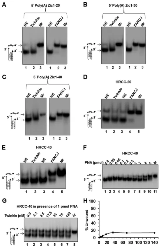
We next assessed the ability of Twinkle helicase to unwind an entropically favored unimolecular G4 DNA substrate (poly(A) Zic1) that was shown previously to be unwound by FANCJ heli-case (46). Twinkle was tested on a series of poly(A) Zic1-uni-molecular G4 DNA substrates with increasing 5⬘ single-stranded DNA tail lengths of 20, 30, and 40 nt and was found to be completely inactive for unwinding (Fig. 8,A–C). In contrast, FANCJ efficiently unwound all three poly(A) Zic1 unimolecu-lar G4 DNA substrates (Fig. 8,A–C). We also assessed the
activ-ity of Twinkle on a unimolecular G4 DNA substrate derived from a human mitochondrial sequence located in the vicinity of a deletion breakpoint observed in HRCC (Fig. 8,DandE). As described above, we determined by biophysical analysis that the HRCC G4P sequence incubated in the presence of potassium formed a G-quadruplex structure that conformed to the char-acteristic G4 parameters detected by CD and UV spectroscopy (Figs. 2 and 3). Twinkle failed to unwind the unimolecular HRCC G4 substrate flanked by a 20-nt 5⬘ssDNA tail (HRCC-20) (Fig. 8D) or a 40-nt 5⬘ssDNA tail (HRCC-40) (Fig. 8E). Moreover, FANCJ was able to efficiently unwind the HRCC-20 and HRCC-40 unimolecular G4 DNA substrates (Fig. 8, D
andE).
In these helicase assays with a unimolecular G4 substrate, a peptide-nucleic acid (PNA) trap was used to capture the unwound radiolabeled complementary strand. The low but detectable unwinding by Twinkle of certain intermolecular G4 substrates raised the possibility that Twinkle might also unwind an intramolecular G4 substrate; however, due to rapid refolding the unfolded G4 failed to be captured. To investigate this pos-sibility, we set out to test Twinkle unwinding of unimolecular G4 under conditions that would favor trapping of the unfolded G4 structure. Initially, we performed a titration of complemen-tary PNA into Twinkle-free reaction mixtures containing the HRCC-40 G4 DNA substrate (5 fmol) and monitored G4
FIGURE 5.Twinkle inefficiently unwinds intermolecular G4 DNA substrates TP-G4 and OX-1-G2ⴕ.Helicase reactions (20l) were performed at 30 °C for 30
min under standard helicase reaction conditions as described under “Experimental Procedures.”A, Twinkle hexamer titration in reaction mixtures containing
5 fmol of tetramolecular TP-G4 with a 21-nt 5⬘ssDNA tail (TP-G4 –21).B, FANCJ (3 nMmonomer) efficiently unwinds 5⬘ssDNA tail (TP-G4 –21).C, Twinkle
hexamer (12.5 nM) or FANCJ monomer (3 nM) was incubated with 5 fmol of TP-G4 with a 40-nt 5⬘ssDNA tail (TP-G4 – 40).D, Twinkle hexamer titration on
bimolecular G4 DNA (OX-1-G2⬘) with a 20-nt 5⬘ssDNA tail. As a control, FANCJ (3 nMmonomer) unwound OX-1-G2⬘substrate.E, quantitative analysis of data
from Twinkle helicase reactions with TP-G4 –21 and OX-1-G2⬘as shown inAandD, respectively. Data represent the mean of at least three independent
experiments with S.D. indicated byerror bars.F, Twinkle hexamer (12.5 nM) or FANCJ monomer (3 nM) unwound 20 bp forked duplex with 20 nt 5⬘and 3⬘ssDNA
tails. Products were resolved on native 10% polyacrylamide gels, and representative phosphorimages of typical gels are shown.NE, no enzyme control;open
triangle, heat-denatured DNA substrate control or ssDNA used as a marker.
at RADBOUD UNIVERSITEIT NIJMEGEN on March 6, 2017
http://www.jbc.org/
unfolding. As shown in Fig. 8F, a greater quantity of spontane-ously unfolded HRCC-40 G4 substrate was captured by increasing the amount of PNA in the reaction mixture. We then tested for unwinding of the HRCC-40 G4 substrate by increas-ing the concentrations of Twinkle in the presence of a fixed level of PNA (1 pmol), which trapped⬃30% of the unfolded G4 structure in the absence of helicase. Under these conditions, Twinkle unwound a maximal 13% of the HRCC-40 G4 DNA substrate over background (Fig. 8,GandH). We performed a
similar set of Twinkle helicase assays with the poly(A) Zic1 G4 substrate, which included the corresponding PNA (1 pmol) in the reaction mixture, but did not detect any Twinkle-catalyzed unfolded G4 product over background (data not shown). These results suggest that Twinkle cannot uniformly unwind all uni-molecular G4 substrates, even in the presence of excess com-plementary PNA to capture the unfolded G4.
DISCUSSION
The source of mitochondrial DNA deletions and rearrange-ments is debated (47). A prominent hypothesis for a number of mitochondrion-based genetic disorders is that a significant fraction of mtDNA deletions are attributed to defective repli-cation of the organelle’s genome. Given the interest in and growing evidence that G-quadruplexes existin vivoand can be sources of genomic instability, we sought to investigate their potential abundance in mitochondria by performing a meta-analysis of predicted G-quadruplex-forming human mitochon-drial sequences. Using a G4 algorithm and information from mitochondrial deletion databases, we mapped predicted G-quadruplexes in the human mitochondrial genome to known mitochondrial deletion breakpoints and correlated this infor-mation with mitochondrial deletion breakpoints prevalent in human disease, cancer, and aging. As we used a conservative upper limit of 7 nt for all G4 loops, the number of G4-prone motifs found in mitochondrial human genome might be even larger (stable G4 may be formed with loops consisting of 8 or more nucleotides). A systematic bioinformatics and biophysi-cal study of this genome using more relaxed parameters may yield interesting new correlations. Using biophysical approaches, we substantiated that selected predicted G4-form-ing human mitochondrial sequences indeed form G-quadru-plexesin vitro. These results suggest that mitochondrial G-qua-druplexes may be prevalent at sites of double strand breaks, which impose a source of mitochondrial genome instability observed in certain genetic mitochondrial disorders, cancer, and aging.
Mitochondrial G4 DNA and Genetic Disease—Mutations in several nuclear genes are associated with the mitochondrial DNA instability of PEO patients, including genes encoding the mitochondrial DNA polymerase ␥and its accessory subunit, Twinkle/C10orf2 mitochondrial helicase, the RRM2B ribonu-cleotide reductase subunit, and the adenine nuribonu-cleotide translo-cator (48). Interestingly, a recent study by Roos et al. (49) described a sibling pair with compound heterozygous POLG1 mutations that displayed PEO, cognitive impairment, and mitochondrial myopathy, characterized by multiple mitochon-drial deletions. A subnormal level of pol␥in vitroresulted in reduced initiation of lagging strand synthesis, leading the authors to suggest uncoupled leading and lagging strand mito-chondrial DNA synthesis and the prolonged presence of repli-cation intermediates in which the H-strand is present in a non-double-stranded DNA conformation. It is plausible that delayed maturation of the G-rich template strand may contrib-ute to prevalent mitochondrial genome deletions in the major arc between OHand OL and that this is exacerbated in this particular case of pol␥deficiency but perhaps also in other POLG1-associated multiple deletion syndromes. It is
notewor-FIGURE 6.Twinkle shows modest activity for unwinding a G4 substrate
formed by human telomeric G-rich sequence but is inactive on G4
formed by CEB1 minisatellite G-rich sequence.Helicase reactions (20l)
were performed at 30 °C for 30 min with the indicated helicase and DNA substrate (5 fmol) under standard helicase reaction conditions as described
under “Experimental Procedures.”A, FANCJ (3 nMmonomer) was incubated
with TelR2-G4 –20 substrate.B, the indicated concentration of Twinkle
hex-amer was incubated with TelR2-G4 –20 substrate.C, Twinkle hexamer was
incubated with TelR2-G4 –20 substrate as inB, except 4 mMATP␥S was
sub-stituted for ATP.D, quantitative analysis of data from Twinkle helicase
reac-tions with TelR2-G4 –20 substrate in the presence of ATP or ATP␥S is shown.
Data represent the mean of at least three independent experiments with S.D.
indicated byerror bars.EandF, the indicated concentrations of the G4 ligand
telomestatin (TMS) was included in reaction mixtures containing 6.12 nM
Twinkle hexamer and the TelR2-G4 –20 DNA substrate (E) or a 19-bp forked
duplex (F).G, FANCJ (3 nM) was incubated with CEB1-G4 –20 substrate.H, the
indicated concentrations of Twinkle hexamer were incubated with
CEB1-G4 –20 substrate.NE, no enzyme control or ssDNA used as a marker.
at RADBOUD UNIVERSITEIT NIJMEGEN on March 6, 2017
http://www.jbc.org/
thy that the density of deletion breakpoints within 20 bp of predicted G-quadruplexes in the major arc between OHand OL (33 deletions/kb) is considerably greater than the G4P-associ-ated deletion breakpoints in the minor arc (5 deletions/kb), suggesting an alternative explanation for the prominence of mtDNA deletions in the major arc region.
Defects in nuclear genes encoding mitochondrial DNA rep-lication proteins are responsible for mitochondrial multiple deletion syndromes. For example, the mitochondrial disorder Alpers syndrome is a progressive neurodevelopmental mito-chondrial DNA depletion disease caused by mutations in polymerase␥(48). The prominent role of the mitochondrial DNA polymerase␥ in mitochondrial DNA maintenance and human disease is attested to by the fact that⬃200 disease-causing mutations in POLG1 are reportedly responsible for clinical diseases characterized by mitochondrial depletion and/or deletions. Mutations in the mitochondrial DNA heli-case Twinkle (c10orf2) are also prominently associated with instability of mitochondrial DNA in patients with PEO or encephalopathy (48). Wanrooijet al.(50) examined the conse-quences of POLG or Twinkle defects for the spectrum of mul-tiple mtDNA deletions characteristic of PEO patients. Using
the G4 Pattern Finder algorithm, we determined that of these reported deletion breakpoints, approximately one-half reside within 20 bp of predicted G4-forming sequences (Table 7). The frequency of the indicated 5⬘or 3⬘deletion breakpoint residing withinⱕ20 bp of the G4P sequence was calculated based on the number of specified deletion breakpoints per total number of mitochondrial deletions reported for the respective PEO patient. The most frequent deletion breakpoints residing in proximity to predicted G4 DNA were commonly found in more than one PEO patient. For example, 5⬘deletion breakpoints, all occurring in the residue 7397–7401 window, were found in patient 1 (POLG), patient 2 (POLG), patient 3 (POLG), and patient 7 (Twinkle). Another common 5⬘deletion breakpoint found in the residue 7397–7401 window appeared in patient 1 (POLG), patient 2 (POLG), patient 3 (POLG), and patient 7 (Twinkle). 3⬘deletion breakpoints residing in close proximity to the G4P sequence were found only in patient 1 (POLG).
A comparison of G4P-associated deletion breakpoint per-centages with percentage values for tRNA genes or D-loop for the PEO patients with POLG or Twinkle defects, from the study by Wanrooijet al.(50), suggests that 5⬘breakage in the vicinity of G4 motifs is associated with mtDNA instability (Table 8). All
FIGURE 7.DnaB unwinds anti-parallel bimolecular G4 DNA substrate.A, conserved helicase motifs inHomo sapiensTwinkle andE. coliDnaB proteins.B–D,
DnaB was tested for helicase activity on various DNA substrates. The indicated concentration of DnaB hexamer was incubated at 37 °C for 15 min with 5 fmol
of the OX-1-G2⬘bimolecular G4 DNA substrate (B), a 19-bp forked duplex DNA substrate (C), or TP-G4 –21 four-stranded parallel DNA substrate (D) under
standard helicase conditions as described under “Experimental Procedures.”Lane 8,open triangle, heat-denatured DNA substrate control (C) or ssDNA as a
marker control (B,D, andF).E, quantitative analysis of helicase activity on all DNA substrates is shown with S.D. indicated byerror bars.Filled squares,19-bp
forked duplex;filled diamonds, OX-1-G2⬘;filled triangles, TP-G4 –21.F, DnaB unwinding of OX-1-G2⬘is ATP-dependent. Reactions were performed as described
under “Experimental Procedures” in the presence or absence of ATP or the poorly hydrolyzable ATP analog ATP␥S. Products were resolved on native 10%
polyacrylamide gels, and representative phosphorimages of typical gels are shown.
at RADBOUD UNIVERSITEIT NIJMEGEN on March 6, 2017
http://www.jbc.org/
FIGURE 8.Twinkle inefficiently unwinds unimolecular G4 DNA substrates.Helicase reactions (20l) were performed at 30 °C for 30 min under standard helicase conditions in the presence of complementary PNA-oligonucleotide as described under “Experimental Procedures.” In contrast to helicase experiments with intermolecular G4 substrates, a PNA-conjugated complementary trap oligonucleotide was added to the reaction mixture to prevent refolding of the
intramolecular G4 after helicase unwinding.A–C, Twinkle hexamer (12.5 nM) or FANCJ (0.5 nM) was incubated with 5 fmol of 5⬘poly(A) Zic1 unimolecular G4
DNA substrate with increasing 5⬘ssDNA tail lengths of 20 (A), 30 (B), or 40 nt (C).DandE, Twinkle (12.5 nM) or FANCJ (0.5 nM) was tested for unwinding of a
unimolecular G4 DNA substrate derived from human mitochondrial DNA sequence flanked by a 20- or 40-nt ssDNA tail in HRCC-20 (D) and HRCC-40 (E),
respectively. Helicase reaction products were resolved on native 15% polyacrylamide gels, and representative phosphorimages of typical gels are shown.F,
increasing amounts of complementary PNA (HRCC-PNA) were included in reaction mixtures to trap transiently unfolded HRCC-40 G4 substrate.G, Twinkle
shows modest helicase activity on intramolecular G4 substrate HRCC-40 (5 fmol) in reaction mixtures containing an excess of HRCC-PNA (1 pmol) to capture
unfolded G4.H, a quantitative analysis of helicase data from experiments as shown inGis shown. Data represent the mean of at least three independent
experiments with S.D. indicated byerror bars.
at RADBOUD UNIVERSITEIT NIJMEGEN on March 6, 2017
http://www.jbc.org/
TABLE 7
Mitochondrial deletion breakpoints in proximity to predicted G4-forming sequences from PEO patients with mutation in POLG or Twinkle
aThe mitochondrial deletion breakpoints from PEO patients were collected from the supplemental data of Ref. 50.
bDeletion breakpoints mapped to within 20 bp of the predicted G4-forming sequences (shown) as determined by Pattern Finder analysis. cRatio of number of 5⬘or 3⬘deletion breakpoints per total number of mitochondrial deletions for respective PEO patient as reported in Ref. 50. dL-strand sequence is shown (5⬘to 3⬘).
eThe predicted G4-forming sequence (G4P) is underlined.
at RADBOUD UNIVERSITEIT NIJMEGEN on March 6, 2017
http://www.jbc.org/
patients analyzed showed a greatly reduced percentage of 5⬘ deletion breakpoints in the tRNA genes or D-loop region com-pared with the G4P sequences. With the exception of patient 1, 3⬘deletion breakpoints were not found in the G4P sequences, suggesting preferential breakage at the 5⬘end of G4 motifs. For patient 1, there was a similar percentage of 3⬘deletion break-points in the G4P sequence and the tRNA genes. Although the G4P algorithm predicts G4 motifs with a loop size of 1–7 nt, a greater loop size limit predicts that G4 structures may form at 3⬘common deletion breakpoints (residues 16034 –16035 and 16071–16075) found in the D-loop. Further analysis of mito-chondrion-based genetic diseases should deepen our under-standing of the relationship of mtDNA deletions to sequences that form G-quadruplexes or other alternative DNA structures and their clinical importance.
Mitochondrial G4 DNA and Cancer—Mitochondrial dys-function is prevalent in tumor cells, and mitochondrial genomic instability is well documented in a wide spectrum of human cancers (4). The prevalence of mtDNA deletions in a variety of human malignancies has led researchers to explore the potential usefulness of cataloging mitochondrial deletions for personalized medicine in cancer diagnosis and treatment. The association of potential G-quadruplex-forming sequences with mtDNA deletion breakpoints associated with cancer raises the possibility that G4 DNA itself may serve as a pre-dictive marker for cancer associated with abnormal mtDNA metabolism.
Rogounovitchet al.(51) observed a strong correlation between the number of mitochondrial deletions and the mtDNA content in radiation-associated human thyroid tumors, suggesting a useful biomarker for discerning the molecular differences between spo-radic and radiation-induced tumors of the thyroid. However, the relationship between mtDNA and radiation-associated human thyroid diseases may be specific to random large-scale deletions flanked by 2–7 bp of microhomology versus the common 5000-bp deletion flanked by 13-bp direct repeats. A careful investigation of the potential role of G4P sequences or other alternative DNA structures contributing to the signature mtDNA deletions commonly found in thyroid tumors and other cancers is warranted.
In a 2012 study of a southwestern Chinese population, Zheng
et al.(52) detected a defined 822-bp deletion in mtDNA desig-nated as a cigarette smoking-associated risk factor for lung can-cer. Our computational analysis predicts a G4P sequence adja-cent to the 822-bp mtDNA deletion (Table 5). Further studies will be necessary to determine the putative role of the G4P flanking sequence in the origin of the mitochondrial genome
instability and whether the characteristic small mtDNA dele-tion is associated with other cigarette smoking-associated dis-eases. The use of G4P sequences as biomarkers for various can-cer types and risk factors may become more widely applicable in the future as our knowledge of G-quadruplex DNA metabo-lism in both the mitochondrial and nuclear genomes advances.
Mitochondrial G4 DNA and Aging—The mitochondrial the-ory of aging postulates that the accumulation of somatic mito-chondrial mutations during the life span of an individual results in mitochondrial dysfunction, which contributes to the under-lying pathological basis for age-related phenotypes of cells, tis-sues, and organs (53). An analysis of genetically inherited mtDNA disorders strongly suggests that defects in mtDNA replication make significant contributions to the decline of mitochondrial function, contributing to age-associated pheno-types (48). Moreover, mouse models that show an accumula-tion of mtDNA mutaaccumula-tions recapitulate age-related phenotypes (54). Considerable evidence over the past 2 decades supports the general conclusion that mtDNA deletions accumulate with aging in various tissues. One recent model postulates that rep-lication errors incurred in stem cells during development become clonally expanded, ultimately leading to mitochondrial dysfunction (5). Understanding the molecular-genetic basis for the accumulation of mtDNA mutations remains an active area of study. Two prominent models for the accumulation of mtDNA deletions with aging are: 1) replication slippage over repeated sequences (55) and 2) homologous recombination repair of double strand breaks (32). G-quadruplex DNA, which perturbs the progression of the mtDNA replication machinery, may contribute to either mechanism by causing stalling during DNA synthesis and/or double strand break formation during processing of replication intermediates. In addition, experi-mental evidence from Barros et al. (56) suggests a model in which recombination at the site of a double strand break may be followed by replication slippage of a G4-nested region during DNA synthesis, resulting in duplications.
Mitochondrial Deletions Associated with Potential G-quad-ruplex-forming Sequences as Polymorphic Anthropological Markers—There has been considerable interest in tracing the distant ancestry of an individual or population group by analyz-ing genetic markers (57). Different human populations with distinct mitochondrial genetic markers can be analyzed through generations to dissect their lineage and recreate past migration patterns. An excellent example of this strategy was provided by an analysis of an indigenous Alaskan population known as the Aleuts whose mtDNA variation from other native Americans suggested that multiple migrations occurred during TABLE 8
Distribution of mitochondrial deletion breakpoints in POLG or Twinkle patients
Deletion breakpoint
Patient 1 PolG (64)a Patient 2 PolG (18)a Patient 3 PolG (17)a
Patients 5 and 6
PolG (32)a Patient 7 Twinkle (11)a
G4Pb tRNAc D-loopd G4Pb tRNAc D-loopd G4Pb tRNAc D-loopd G4Pb tRNAc D-loopd G4Pb tRNAc D-loopd % % % % % 5⬘deletion breakpoint 31 5 0 72 0 0 47 0 0 59 0 0 64 9 0 3⬘deletion breakpoint 16 14 13 0 0 28 0 0 53 0 0 41 0 0 38 a
The number of deletion breakpoints in the specified patients is indicated in parentheses.
b
The percentage of deletion breakpoints in G4P, excluding those overlapping with tRNA or D-loop region.
c
The percentage of deletion breakpoints in tRNA excluding those overlapping with G4P or D-loop region.
d
The percentage of deletion breakpoints in D-loop excluding those overlapping with G4P or tRNA.
at RADBOUD UNIVERSITEIT NIJMEGEN on March 6, 2017
http://www.jbc.org/
the habitation of the New World (58). From the G4P computa-tional analysis and a survey of the literature, we identified a reported 9-bp mitochondrial deletion used as a polymorphic anthropological marker for native North American groups (59) as residing within 20 bp of a G4P sequence (5⬘ -GGGGGTAGA-GGGGGTGCTATAGGGTAAATACGGG-3⬘H-strand). The deletion has its highest frequency in the American Southwest and is absent in the Arctic and Subarctic regions (59). The distribution of the G4P-associated mitochondrial deletion marker has provided some insight into the migration patterns of East Asians to the New World. Genographic projects such as this will benefit from further genetic analysis of indigenous populations by using novel genetic markers as important tools. Understanding the role of G-quadruplex metabolism and other molecular mechanisms that affect chromosomal stability and mutations, thereby influen-cing the appearance of mitochondrial genetic polymorphisms, will help in deciphering the origin of polymorphic markers. These efforts will in turn be useful for anthropological studies such as the history of human migration patterns and other fields such as forensic science (60).
Implications of Twinkle G4 Studies for Mitochondrial DNA Replication—Because the Twinkle helicase is required for human mtDNA replication, we assessed whether Twinkle was able to unwind various uni-, bi-, and tetramolecular G4 DNA substrates. We found that the helicase is not efficient in unwinding the various topological forms of G4 DNA tested under the same conditions in which it efficiently unwound con-ventional forked duplex substrates. Therefore, it is reasonable to suggest that G4 DNA structures are likely to persist in the mitochondrial genome and may contribute to double strand breaks in regions nearby G4, ultimately leading to the mito-chondrial deletions prevalent in human disease. Although this study does not definitively support the proposed model for a role of G4 DNA in mitochondrial genome instability, it sheds light on a novel aspect of mtDNA metabolism and provides a reasonable platform for its further study in biological systems. An important biochemical observation made in this study is that Twinkle helicase poorly unwound all of the G-quadruplex substrates tested. This is paradoxical given that the human mitochondrial genome is very G4-prone. It was recently dem-onstrated that close coupling of T7 DNA polymerase with gp4 helicase during leading strand synthesis results in a synergistic interaction in which DNA synthesis drives fork unwinding (61). If Twinkle functions in a similar manner with the mitochon-drial replicative polymerase ␥, then the helicase might more readily melt G-quadruplex structures that impede mitochon-drial DNA replication. Furthermore, auxiliary mitochonmitochon-drial helicases are likely to assist in resolving G4 structures that impede mtDNA replication and other processes such as mtDNA repair. The most likely candidate for the role of G4 resolution in mitochondria is PIF1 helicase. Studies from the Nicolas and Zakian laboratories have implicated yeast PIF in G4 metabolism (21, 22, 62– 66). Although the recombinant nuclear form of human Pif1 has been shown to unwind G4 DNA sub-stratesin vitro(67), a definitive role for human PIF1 in mito-chondrial G4 metabolism remains to be determined. Other DNA helicases with substantial residence time in mitochondria include the SUV3 helicase (68, 69) and DNA2 helicase-nuclease
(70, 71), the latter of which has been shown to process G-quad-ruplex DNAin vitro(72, 73). Mutations in DNA2 were identi-fied recently as associated with progressive myopathy in a mul-tiple deletion syndrome (74).
In addition to helicases, a number of other G4 DNA-binding proteins are known to exist that may play a role in mitochon-drial G4 metabolism (11). The demonstration that nuclear topoisomerase I interacts with G4 DNA structures (75) raises the possibility that the mitochondrion-specific topoisomerase I (76) plays a role in G-quadruplex metabolism as well. G4-bind-ing and G4-metabolizG4-bind-ing proteins may affect not only mtDNA replication during the elongation phase but also replication ini-tiation in which G-quadruplexes have recently been implicated. Mitochondrial transcription creates primers in a CG-rich ele-ment, generally termed conserved sequence block II (CSBII), required for the initiation of leading strand DNA replication. G-quadruplexes formed by RNA result in transcription termi-nation and dictate primer formation for DNA synthesis initia-tion (77). More recently, Wanrooijet al.(78) reported that the mitochondrial RNA primer required for the leading strand ori-gin of mtDNA replication forms a hybrid G-quadruplex between RNA and DNA in CSBII and that this quadruplex for-mation plays a critical role in determining the rate of DNA synthesis initiation in human mitochondria by influencing the architecture and persistence of an RNA-DNA hybrid (R-loop) residing at the leading strand origin of DNA replication. Because CSBII-like sequences are widespread and found in both yeast and mammals (79) (but not, for example, in Drosoph-ila), G-quadruplexes are now believed to play an important and often evolutionarily conserved role in mitochondrial replica-tion initiareplica-tion. Current models of mtDNA replicareplica-tion place the role of Twinkle helicase to unwind duplex DNA downstream of nascent DNA primer synthesized by polymerase␥(80). Thus, our experimental data suggest that Twinkle may be specialized to leave intact the RNA primer-associated G-quadruplex and focus its work on facilitating polymerase␥strand displacement DNA synthesis extension with the help of the mitochondrial ssDNA-binding protein.
Future Directions—The recent development of G4-specific probes (G4-directed antibodies (7, 8, 81) and ligands (82)) has made it more realistic to study the abundance and relevance of G4 mitochondrial structuresin vivo. There has been a growing interest in nuclear G4 DNA metabolism to further the under-standing of the biological roles of G4 structure, and it may also be a potential target for anticancer agents and therapies. This now applies to mitochondrial G4 metabolism as well. Recently, a discovery was made that guanine-rich oligonucleotides, which may be useful anti-cancer drugs, can be introduced into lung cancer cells where they persist as a topologically distinct class of G4 structures in the mitochondria (83). As these authors indicate, a more refined understanding of the cellular tracking and localization of guanine-rich oligonucleotides may provide greater insight into design optimization for cancer therapies (83). From a broader perspective, G-quadruplexes that form in the human mitochondrial genome are likely to have major consequences for mtDNA replication, transcrip-tion, and repair, impacting health and longevity.
at RADBOUD UNIVERSITEIT NIJMEGEN on March 6, 2017
http://www.jbc.org/